Mindfulness Practice and Increases in Gray Matter Density, Gray Matter Volume, and Cortical Thickness: A Scoping Review



Highlights
- Mindfulness practice, increasingly integrated into both clinical and wellness settings, has been associated with structural changes in brain regions tied to cognitive control, interoception, self-referential processing, attention, memory, and emotional regulation.
- Evidence from eight of the nine studies included in this review indicates that the practice is associated with increases in gray matter density, gray matter volume, and cortical thickness.
- Mindfulness practice appears to hold promise as a practical, easy to access, low-cost tool for promoting brain health through associated structural change.
- Given that mindfulness has the potential to mitigate cognitive decline and buffer against psychological hardship related to stress, anxiety, or depression, further high-quality research is certainly warranted.
Abstract
1. Introduction
2. Materials and Methods
2.1. Inclusion and Exclusion Criteria
2.2. Neuroimaging Analysis Techniques
3. Results
Patterns of Structural Brain Change Across Studies
4. Discussion
Limitations
5. Conclusions
Supplementary Materials
Author Contributions
Funding
Institutional Review Board Statement
Informed Consent Statement
Data Availability Statement
Conflicts of Interest
Abbreviations
| ACC | Anterior cingulate cortex |
| CT | Cortical thickness |
| GMD | Gray matter density |
| GMV | Gray matter volume |
| FAM | Focused Attention Meditation |
| IBMT | Integrative body–mind training |
| MAP | Mindfulness Awareness Program |
| MBI | Mindfulness-based interventions |
| MBSR | Mindfulness-based stress reduction |
| MCI | Mild cognitive impairment |
| PCC | Posterior cingulate cortex |
| PFC | Prefrontal cortex |
| PRISMA-ScR | Preferred Reporting Items for Systematic Reviews and Meta-Analyses extension for Scoping Reviews |
| ROI | Region-of-interest |
| RCT | Randomized controlled trial |
| SBM | Surface-based morphometry |
| VBM | Voxel-based morphometry |
References
- Calderone, A.; Latella, D.; Impellizzeri, F.; Tibollo, L.; Picerno, P.; Coratella, G.; Nardo, G.D.; Quartarone, A. Neurobiological changes induced by mindfulness and meditation: A systematic review. Biomedicines 2024, 12, 2613. [Google Scholar] [CrossRef] [Scilit] [PubMed]
- Epstein, R. Mindful practice. In Behavioral Medicine: A Guide for Clinical Practice, 5th ed.; Feldman, M.D., Christensen, J.F., Satterfield, J.M., Laponis, R., Eds.; McGraw-Hill Education: Columbus, OH, USA, 2019; Available online: https://accessmedicine-mhmedical-com.georgefox.idm.oclc.org/content.aspx?bookid=2747§ionid=230248882 (accessed on 1 December 2025).
- Hemond, C.C.; Deshpande, M.; Berrios-Morales, I.; Gaitan, A.M.; Bajaj, S.; Toor, G.; Glanz, B.I.; Bakshi, R.; Healy, B.C.; Chitnis, T. A single-arm, open-label pilot study of neuroimaging, behavioral, and peripheral inflammatory correlates of mindfulness-based stress reduction in multiple sclerosis. Sci. Rep. 2024, 14, 14044. [Google Scholar] [CrossRef] [Scilit] [PubMed]
- Hölzel, B.K.; Carmody, J.; Evans, K.C.; Hoge, E.A.; Dusek, J.A.; Morgan, L.; Pitman, R.K.; Lazar, S.W. Stress reduction correlates with structural changes in the amygdala. Soc. Cogn. Affect. Neurosci. 2010, 5, 11–17. [Google Scholar] [CrossRef] [Scilit] [PubMed]
- Keng, S.L.; Smoski, M.J.; Robins, C.J. Effects of mindfulness on psychological health: A review of empirical studies. Clin. Psychol. Rev. 2011, 31, 1041–1056. [Google Scholar] [CrossRef] [Scilit] [PubMed]
- Hölzel, B.K.; Carmody, J.; Vangel, M.; Congleton, C.; Yerramsetti, S.M.; Gard, T.; Lazar, S.W. Mindfulness practice leads to increases in regional brain gray matter density. Psychiatry Res. 2011, 191, 36–43. [Google Scholar] [CrossRef] [Scilit] [PubMed]
- Kral, T.R.A.; Davis, K.; Korponay, C.; Hirshberg, M.J.; Hoel, R.; Tello, L.Y.; Goldman, R.I.; Rosenkranz, M.A.; Lutz, A.; Davidson, R.J. Absence of structural brain changes from mindfulness-based stress reduction: Two combined randomized controlled trials. Sci. Adv. 2022, 8, eabk3316. [Google Scholar] [CrossRef] [Scilit] [PubMed]
- Kurth, F.; Luders, E.; Wu, B.; Black, D.S. Brain Gray Matter Changes Associated with Mindfulness Meditation in Older Adults: An Exploratory Pilot Study using Voxel-based Morphometry. Neurobiology 2014, 1, 23–26. [Google Scholar]
- Kurth, F.; Strohmaier, S.; Luders, E. Reduced Age-Related Gray Matter Loss in the Orbitofrontal Cortex in Long-Term Meditators. Brain Sci. 2023, 13, 1677. [Google Scholar] [CrossRef] [Scilit] [PubMed]
- Lenhart, L.; Steiger, R.; Waibel, M.; Schocke, M.; Hotter, A. Cortical reorganization processes in meditation naïve participants induced by 7 weeks focused attention meditation training. Behav. Brain Res. 2020, 395, 112828. [Google Scholar] [CrossRef] [Scilit] [PubMed]
- Santarnecchi, E.; D’Arista, S.; Egiziano, E.; Gardi, C.; Rigon, R.; Stefanini, C.; Reda, M.; Rossi, A.; Mondillo, S.; Rossi, S. Correction: Interaction between neuroanatomical and psychological changes after mindfulness-based training. PLoS ONE 2015, 10, e0129754. [Google Scholar] [CrossRef] [Scilit]
- Tang, R.; Friston, K.J.; Tang, Y.Y. Brief Mindfulness Meditation Induces Gray Matter Changes in a Brain Hub. Neural Plast. 2020, 2020, 8830005. [Google Scholar] [CrossRef] [Scilit] [PubMed]
- Valk, S.L.; Bernhardt, B.C.; Trautwein, F.M.; Böckler, A.; Kanske, P.; Guizard, N.; Collins, D.L.; Singer, T. Structural plasticity of the social brain: Differential change after socio-affective and cognitive mental training. Sci. Adv. 2017, 3, e1700489. [Google Scholar] [CrossRef] [Scilit] [PubMed]
- Watson, T.; Walker, O.; Cann, R.; Varghese, A.K. The benefits of mindfulness in mental healthcare professionals. F1000Research 2021, 10, 1085. [Google Scholar] [CrossRef] [Scilit] [PubMed]
- Tang, Y.; Ma, Y.; Wang, J.; Fan, Y.; Feng, S.; Lu, Q.; Yu, Q.; Sui, D.; Rothbart, M.K.; Fan, M.; et al. Short-term meditation training improves attention and self-regulation. Proc. Natl. Acad. Sci. USA 2007, 104, 17152–17156. [Google Scholar] [PubMed]
- Yu, J.; Rawtaer, I.; Feng, L.; Fam, J.; Kumar, A.P.; Cheah, I.K.; Lim, J.X.W.; Tan, Y.; Kua, E.H.; Mahendran, R. Mindfulness intervention for mild cognitive impairment led to attention-related improvements and neuroplastic changes: Results from a 9-month randomized control trial. J. Psychiatr. Res. 2021, 135, 203–211. [Google Scholar] [CrossRef] [Scilit] [PubMed]
- Zu, H.; Song, Y.; Xu, M.; Wang, X.; Li, X.; Liu, J. The brain structure correlates of individual differences in trait mindfulness: A voxel-based morphometry study. Neuroscience 2014, 272, 21–28. [Google Scholar] [CrossRef] [Scilit] [PubMed]
- Page, M.J.; McKenzie, J.E.; Bossuyt, P.M.; Boutron, I.; Hoffmann, T.C.; Mulrow, C.D.; Shamseer, L.; Tetzlaff, J.M.; Akl, E.A.; Brennan, S.E.; et al. The PRISMA 2020 statement: An updated guideline for reporting systematic reviews. BMJ 2021, 372, n71. [Google Scholar] [CrossRef] [Scilit] [PubMed]


| Keywords | Identified | Determined as Potentially Relevant | Included in Scoping Review |
|---|---|---|---|
| Mindfulness | 33,893 | - | - |
| Mindfulness practice | 8455 | - | - |
| Mindfulness practice AND brain | 852 | - | - |
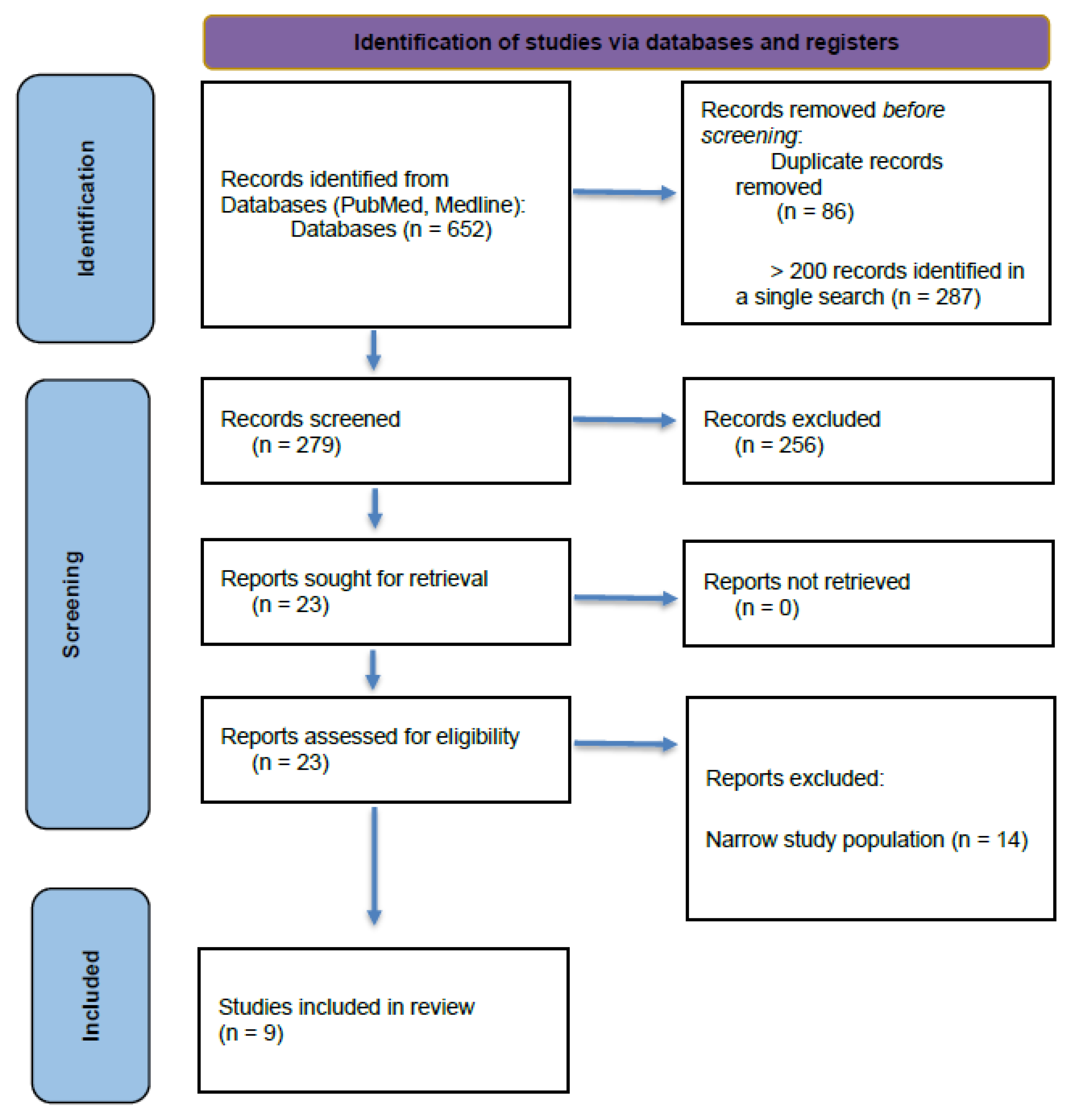
| Mindfulness practice AND brain changes | 287 | - | - |
| Mindfulness practice AND structural brain changes | 44 | 11 | 3 |
| Mindfulness AND brain AND structure changes | 177 | 12 | 5 |
| Mindfulness OR meditation AND brain AND structure changes AND gray matter | 37 | 5 | 3 |
| Mindfulness OR meditation AND brain AND structure changes AND cortical thickness | 23 | 10 | 5 |
| Mindfulness AND brain AND structure changes AND gray matter | 25 | 6 | 5 |
| Mindfulness practice AND gray matter | 36 | 13 | 4 |
| Mindfulness practice AND gray matter density | 4 | 2 | 1 |
| Mindfulness practice AND gray matter volume | 19 | 7 | 1 |
| Author/Year | Study Design | Subjects | Geographical Location | Intervention | MRI Modality | Primary Outcomes | Secondary/Behavioral Outcomes |
|---|---|---|---|---|---|---|---|
| Kral et al. [7] (2022) | 2 pooled RCTs | 218 healthy adults | Madison, Wisconsin United States of America | Duration: 8 weeks, follow-up immediately post intervention Intervention: MBSR Comparison/Control: Active Health Enhancement Program (HEP) and waitlist(WL) Outcomes: GMV, GMD, CT changes Outcome Measures: - Structural Changes: SBM for CT, VBM for GMD/GMV - Behavioral Changes: none | VBM, SBM, CT, ROI | No MBSR-specific structural changes vs. controls There were no significant group differences for change in brain structure for MBSR compared to HEP or WL for any of the ROIs (p > 0.10) ROI-specific: (insula, hippocampus, amygdala, and caudate) = no significant group differences for change in GMV No significant within-group changes in brain structure across time (p > 0.05) | None |
| Santarnecchi et al. [11] (2014) | RCT | 48 healthy adults | Siena, Italy | Duration: 8 weeks, follow-up immediately post intervention Intervention: MBSR (2.5 h weekly, daily home practice, and a retreat) Comparison/Control: Passive control Outcomes: CT changes, behavioral changes (anxiety, worry, alexithymia, depression, mindful attention) Outcome Measures: - Structural Changes: Voxel-Based Cortical Thickness (VBCT) toolbox for Statistical Parametric Mapping (SPM) software - Behavioral Changes: State–Trait Anxiety Inventory (STAI), Beck Depression Inventory II (BDI II), Mindful Attention Awareness Scale (MAAS), Penn State Worry Questionnaire (PSWQ), Toronto Alexithymia Scale (TAS-20) | The VBCT (VBM) toolbox for SPM was used to calculate GM CT | Increase in CT in the right anterior insula and bilateral somatosensory cortices VBCT model (F(1,25) = 5.456; p = 0.008; n2 = 0.467), indicating an increase in CT greater in the MBSR than in the control group Specifically located in the right insula (F(1,25) = 12.134; p = 0.001) and somatosensory cortex (F(1,25) = 10.316; p = 0.002) Change in CT in the control group were not significant for either insula (F(1,25) = −1.12; p = 0.44) and somatosensory cortex (F(1,25) = 0.78; p = 0.65) No clusters of GMV reached the statistical significance threshold for main or interaction effects | Decrease in anxiety, worry, and alexithymia. Insula CT correlated with alexithymia reduction Anxiety: - Mean decrease of ~6.2 points on the STAI Alexithymia decrease: p < 0.004 State anxiety decrease: p < 0.031 Depression level decrease: p < 0.046 MBSR trainees showed a significant pre–post decrease in alexithymia (t(22) = 3.142; p < 0.004), worry (t(22) = 2.665; p < 0.012), state anxiety (t(22) = 2.259; p < 0.031) and depression levels (t(22) = 2.086; p < 0.046), while no differences were detected by the MAAS (t(22) = 0.879; p < 0.418). No differences were found for participants in the control group (p > 0.05 corrected) The rate of changes in CT were correlated with the changes in psychological indexes of interest(Brain/Psy), revealing a significant negative correlation for alexithymia level and insula cluster thickness values (r = −0.712; p < 0.01) in MBSR subjects after the training |
| Tang et al. [12] (2020) | RCT | 40 healthy adults | Dalian, China | Duration: 5 days, with 10 h total, immediate follow-up Intervention: 10 h of Integrative body–mind Training (IBMT) Comparison/Control: Relaxation training (RT) Outcomes: GMV changes, mood changes Outcome Measures: - Structural Changes: GMV changes in the PCC, isthmus of cingulate (ISC), retrosplenial cortex (RSC) - Behavioral Changes: Adult Temperament Questionnaire (ATQ) | VBM, ROI | Increase in GMV in the ventral PCC after brief training (5 days, total 10 h) Significant effect on the right ventral PCC/ISC volume F(1,36) = 5.08, p = 0.03 IBMT group had a significantly higher % change (M = 0.88, SD = 3:50) relative to the RT group (M = −2.66, SD = 5.47). A large effect size, 0.124 (partial eta-squared), was detected No significant difference was detected in RSC volume between the IBMT and RT group (p > 0.05) | Decrease in sadness, as defined by lower level of unpleasant affect, mood, and energy-related to object or person loss, disappointment, and exposure to suffering Sadness was the only one of the four temperamental traits that showed a significantly negative correlation with the right ventral PCC/ISC change (r = −0.535, p < 0.018) |
| Valk et al. [13] (2017) | RCT | Randomized training cohorts, 445 healthy adults | Leipzig, Germany | Duration: 9 months, 3 × 3-month-long modules, with follow-ups at the end of each module Intervention: Modules on presence (mindfulness/attention), affect(compassion/emotion), perspective (theory of mind (ToM)/metacognition) Comparison/Control: Each module served as active control for others, retest cohort Outcomes: CT changes, behavioral changes (domain-specific) Outcome Measures: - Structural Changes: CT, whole brain - Behavioral Changes: EmpaToM task (compassion, ToM), attention test (cued flanker). Compassion was quantified as a mean of compassion ratings across all experimental conditions | CT | Module-specific CT increases - Presence → right PFC and ACC increases FWE < 0.001) - Affect/compassion → Increases in the right insula extending to the temporal pole, with findings consistent across all cohorts undergoing affect training [TC1, T1 → T2 (r = 0.37); TC2, T2 → T3 (r = 0.28); TC3, T0 → T1 (r = 0.31) - Affect/perspective → Bilateral occipital regions extending to inferior temporal cortices (FWE < 0.05 (left) and <0.001 [right]) Increases in thickness in right lateral and medial frontal regions (FWE < 0.001), together with focal decreases in the right lingual gyrus (FWE < 0.025) | Domain-specific behavioral increases tracked: increased attention, affect presence, compassion, ToM |
| Yu et al. [16] (2021) | RCT | 54 older adults w/MCI | Singapore, Singapore | Duration: 9 months, with follow-ups at 3 months and 9 months Intervention: Mindfulness Awareness Program (MAP), weekly sessions × 3 months then monthly sessions × 6 months Comparison: Health Education Program (HEP) Outcomes: CT changes, cognition changes (working memory span, divided attention) Outcome Measures: - Structural Changes: CT of whole brain and specific ROIs - Behavioral Changes: Cognitive assessments, digit span backward and block design tests from the Wechsler Adult Intelligence Scale (WAIS-III), delayed recall and recognition tests from the Rey Auditory Verbal Learning Test(RAVLT), Color Trails Test(CTT), and semantic fluency test | CT, ROI | - Increases in CT in right frontal pole (at 9 months) - Transient increase in CT in the left inferior temporal gyrus (at 3 months) - Decreases in CT in the left ACC The MANOVAs on the CT revealed a significant time * group effect between baseline and ninth months (F(8,12) = 3.03; p = 0.041; η2partial = 0.67), but not between baseline and third month (F(8,29) = 2.01; p = 0.080; η2partial = 0.36) | Improved working memory span and divided attention |
| Lenhart et al. [10] (2020) | Longitudinal pre–post | 27 healthy adults | Innsbruck, Austria | Duration: 7.3 week average, follow-up immediately after intervention Intervention: Focused attention meditation (FAM) (14 guided session over 7 weeks) + daily home practice Comparison/Control: None (within subject) Outcomes: GM changes, WM microstructure, anxiety, quality of life (QOL) Outcome Measures: - Structural Changes: VBM for GM and DWI for FA - Behavioral Changes: Short-form health survey (SF-36), German version of the State–Trait Anxiety Inventory (STADI) | VBM | Within-group comparison of the 27 meditation participants showed increased GM volumes at follow-up compared to the baseline time-point in clusters of the following: The anterior insula (p = 0.001) The inferior frontal gyrus (p = 0.001) The caudate nucleus with adjacent regions of the putamen (p = 0.002) The superior frontal gyrus (p = 0.003) In each case, bi-hemispheric and middle spreading to the superior temporal gyrus (p < 0.001) and the cerebellum (p = 0.004) on the right side were observed Significant GM decreases were revealed in the inferior parietal lobule as well as the superior and middle temporal gyrus (p < 0.001) and the inferior frontal gyrus (p = 0.001) on both sides, as well as in the mPFC (p < 0.001) and the parahippocampal gyrus spreading to the fusiform gyrus (p = 0.001) on the right side Additionally, GM decreases were found in the PCC at height thresholds set to p < 0.01 (p < 0.001) No significant sex differences were found at height thresholds set to p < 0.01 | Decrease in state anxiety associated with MCC/PCC and mPFC changes Longitudinal analysis of the questionnaires revealed significant improvements for excitement(p = 0.012), concern (p = 0.012) and anxiety (p = 0.004) as well as psychological well-being (p = 0.004) Improved scores of the STADI questionnaire |
| Kurth et al. [8] (2014) | Single-group pilot | 6 healthy older adults | Los Angeles, California, United States of America | Duration: 6 weeks, follow-up immediately after intervention Intervention: Mindfulness Awareness Practices (MAPs), 6 weekly 2 h session Comparison/Control: None (within subject) Outcomes: GM changes Outcome Measures: - Structural Changes: VBM - Behavioral Changes: none | VBM | Significant increases in GM in right precuneus. Decreases in GM in left PFC, right hippocampus, right thalamus, and right parietal cortex VBM analysis revealed one cluster indicating significant GM increase in the right precuneus (247 voxels, cluster maximum [x;y;z]: 6;−64;19, p = 2.4 × 10⁻¹⁵) and decreases in the left PFC (408 voxels, x;y;z: −39;50;−8, p = 1.0 × 10⁻¹⁰), right hippocampus (136 voxels, x;y;z: 24;−36;3, p = 1.3 × 10⁻⁷), right thalamus (199 voxels, x;y;z:3;−21;12, p = 1.7 × 10⁻⁵), and right parietal cortex (311 voxels, x;y;z: 9;−45;58, p = 1.8 × 10⁻¹⁵) | n/a |
| Kurth et al. [9] (2023) | Cross-sectional matched | 100 healthy adults: 50 long-term meditators and 50 control participants | Berlin, Germany | Duration: ~20-year practice; cross-sectional analysis Intervention: Long-term meditation practice Comparison/Control: Matched non-meditators Outcomes: Orbitofrontal cortex (OFC) overall tissue loss rate (healthy aging) Outcome Measures: - Structural Changes: ROI morphometry (OFC Fo1-Fo7) - Behavioral Changes: none | ROI | Reduced age-related OFC atrophy in meditators; multiple Fo subregions significant(OFC is made up of 7 (Fo1–Fo7) defined subregions) Significant group-by-age interaction for the left and right OFC, as well as for left and right Fo2, Fo3, Fo4, and Fo7. In addition, significant group-by-age interactions were observed for left Fo5 and right Fo6. Effect sizes (calculated as Cohen’s d) ranged between d = 0.374 and d = 0.806, suggesting a range from small to large effects. | Supports neuroprotection and healthy aging hypothesis |
| Hölzel et al. [6] (2011) | Controlled longitudinal | 33 healthy adults: 16 in MBSR and 17 in WL | Boston, Massachusetts United States of America | Duration: 8 weeks, follow-up immediately post intervention Intervention: MBSR daily, ~27 min Comparison/Control: Waitlist Outcomes: GMD changes, mindfulness facets (behavioral attributes) Outcome Measures: - Structural Changes: VBM of whole brain and in ROI of hippocampus - Behavioral Changes: Five Facet Mindfulness Questionnaire (FFMQ) | VBM, ROI | Increases in GMD in left hippocampus Increases in GMD in PCC, TPJ, cerebellum (whole brain) ROI: MBSR group identified a small cluster in the left hippocampus with increased gray matter concentration (peak voxel MNI coordinates x,y,z: −36,−34,−8; t(15) = 6.89; voxel level p = 0.014, corrected for multiple comparisons with FWE correction; cluster size k = 30 Whole brain: Cluster located in four regions (PCC; left temporo-parietal junction (TPJ); and two clusters in the cerebellum) Group × time interactions were significant for all 4 regions, indicating that increases in GM concentration were significantly greater in the MBSR than in the control group: PCC (F(1,29) = 50.124; p = 0.001), TPJ (F(1,29) = 11.456; p = 0.002), cerebellar vermis/brainstem (F(1,29) = 11.292; p = 0.002), lateral cerebellum (F(1,29) = 9.806; p = 0.004) | MBSR participants significantly increased their mindfulness facet scores (acting with awareness, observing, non-judgmental attitude) Confirmed significant group/time interactions for three of the five mindfulness subscales: - Acting with awareness: F(1,26) = 16.87, p < 0.001 - Observing: F(1,26) = 7.09, p = 0.013 - Non-judging: F(1,26) = 4.61, p = 0.041 - Describing: F(1,26) = 1.95, p = 0.175 - Non-reactivity: F(1,26) = 2.79, p = 0.107 Significant gains in the MBSR group: - Acting with awareness: t(13) = 3.665, p = 0.003 - Observing: t(13) = 4.218, p = 0.001 - Non-judging: t(13) = 3.580, p = 0.003 |
Disclaimer/Publisher’s Note: The statements, opinions and data contained in all publications are solely those of the individual author(s) and contributor(s) and not of MDPI and/or the editor(s). MDPI and/or the editor(s) disclaim responsibility for any injury to people or property resulting from any ideas, methods, instructions or products referred to in the content. |
© 2026 by the authors. Licensee MDPI, Basel, Switzerland. This article is an open access article distributed under the terms and conditions of the Creative Commons Attribution (CC BY) license.
Share and Cite
Rafter, C.; McCarthy, E.; Stilp, C.; Brumitt, J. Mindfulness Practice and Increases in Gray Matter Density, Gray Matter Volume, and Cortical Thickness: A Scoping Review. Brain Sci. 2026, 16, 483. https://doi.org/10.3390/brainsci16050483
Rafter C, McCarthy E, Stilp C, Brumitt J. Mindfulness Practice and Increases in Gray Matter Density, Gray Matter Volume, and Cortical Thickness: A Scoping Review. Brain Sciences. 2026; 16(5):483. https://doi.org/10.3390/brainsci16050483
Chicago/Turabian StyleRafter, Colin, Erika McCarthy, Curt Stilp, and Jason Brumitt. 2026. "Mindfulness Practice and Increases in Gray Matter Density, Gray Matter Volume, and Cortical Thickness: A Scoping Review" Brain Sciences 16, no. 5: 483. https://doi.org/10.3390/brainsci16050483
APA StyleRafter, C., McCarthy, E., Stilp, C., & Brumitt, J. (2026). Mindfulness Practice and Increases in Gray Matter Density, Gray Matter Volume, and Cortical Thickness: A Scoping Review. Brain Sciences, 16(5), 483. https://doi.org/10.3390/brainsci16050483