1. Introduction
Cancer-related cognitive impairment (CRCI) is often framed primarily as a consequence of chemotherapy, as reflected in the popular term “chemobrain.” However, cognitive difficulties can be present before systemic treatment begins, indicating that chemotherapy is not required for cognitive change and that later symptoms cannot automatically be attributed to treatment toxicity alone [
1,
2]. Pretreatment cognitive vulnerability is likely multifactorial, involving cancer-related biological processes together with psychological distress, fatigue, sleep disruption, pain, comorbid conditions, and other clinical factors [
2,
3].
Chemotherapy may then add a second exposure. Post-treatment studies have documented cognitive changes and alterations in brain structure and functional connectivity, although findings vary according to cancer type, treatment regimen, timing of assessment, and the methods used to define impairment [
4,
5].
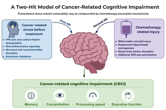
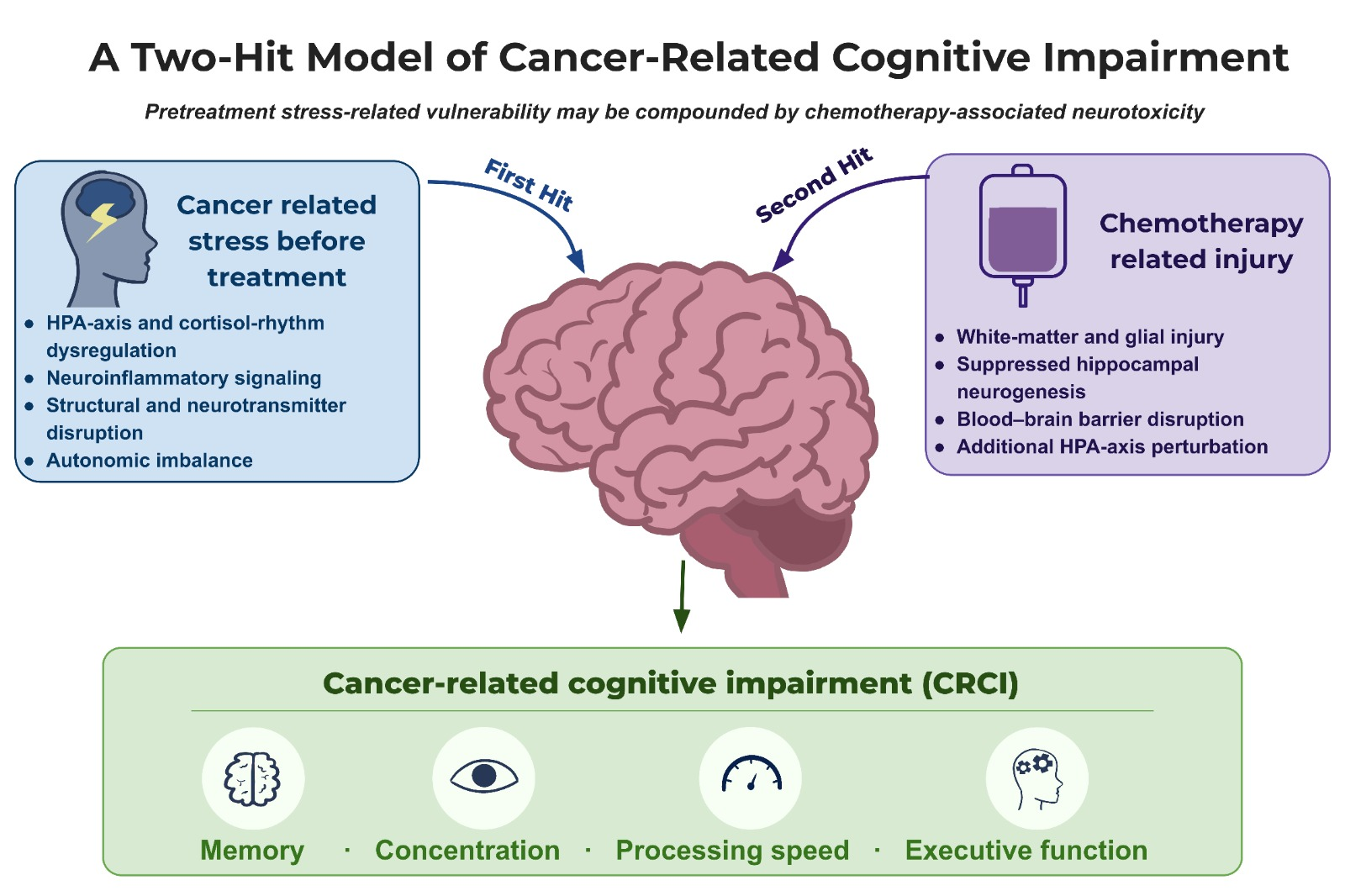
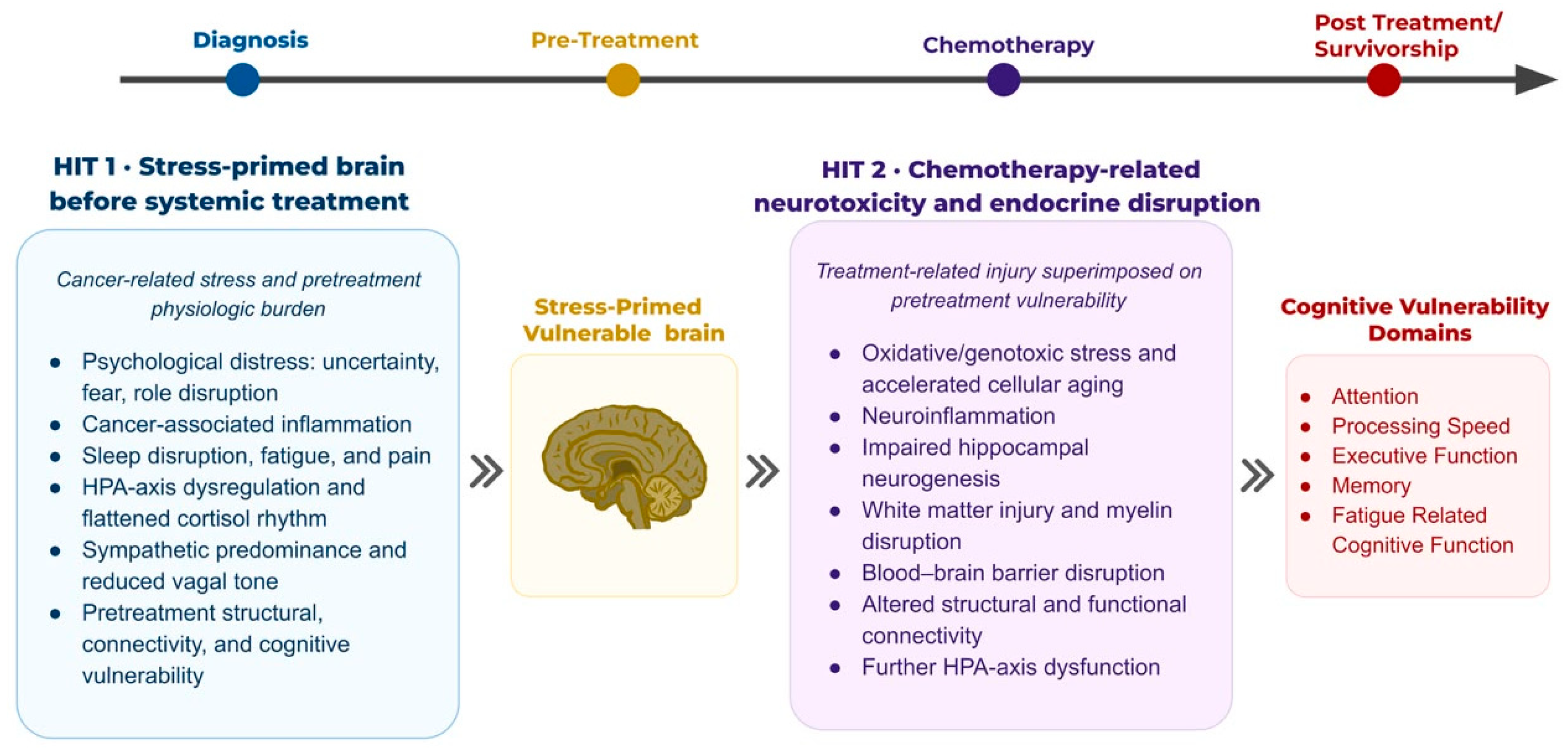
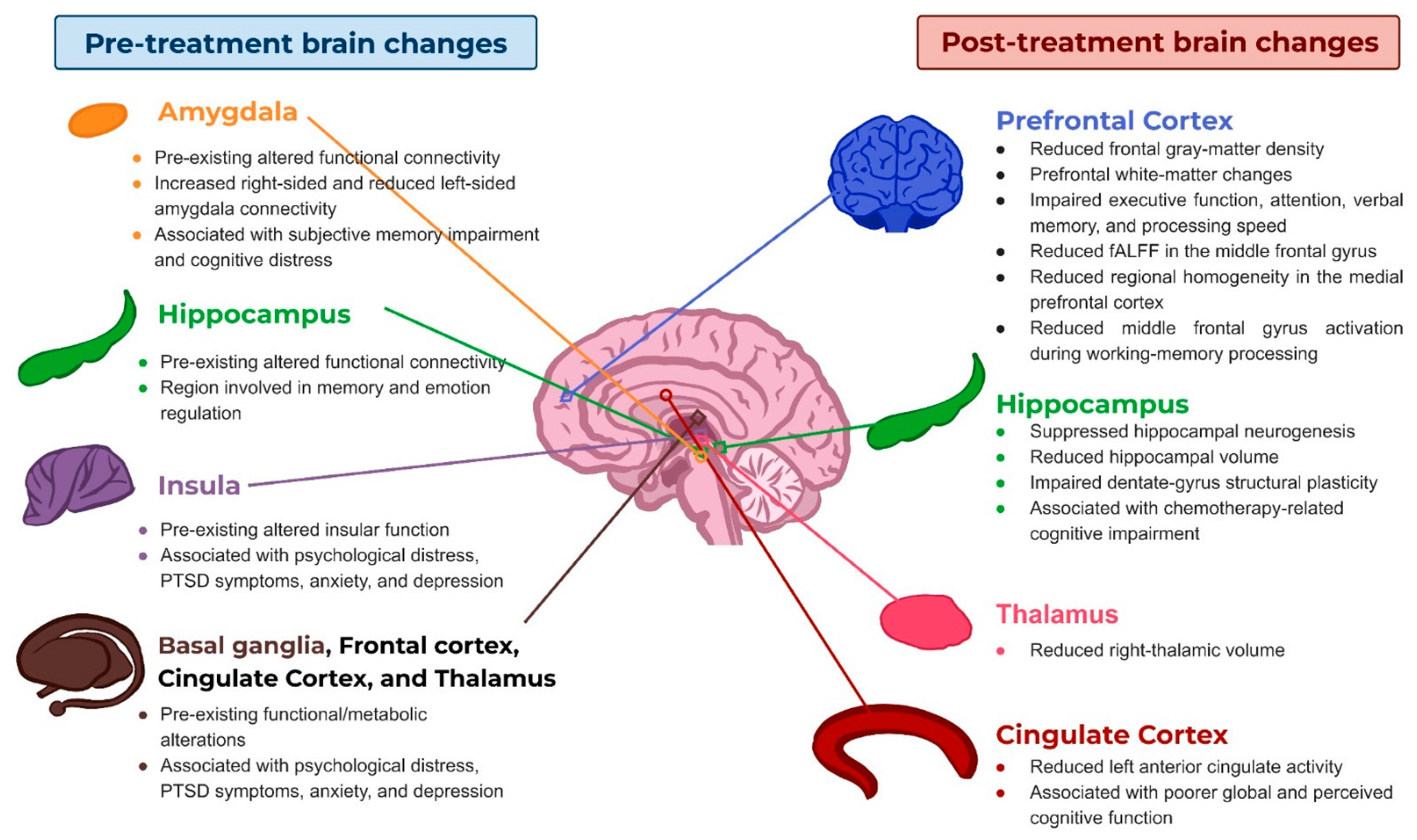
This review therefore adopts a two-hit framework. In the first hit, interacting cancer-, host-, and stress-related factors may create or amplify neural vulnerability before treatment. In the second, chemotherapy may add neurotoxic injury and further cognitive burden (
Figure 1). This framework is intended as an organizing model rather than a proven causal sequence. The review focuses on adults with non-central nervous system cancers; pediatric populations and patients with primary brain tumors fall outside its scope.
2. Methodology
A literature search was performed using PubMed/MEDLINE and electronic resources accessed through the Midwestern University Library for peer-reviewed English-language articles published up to 25 August 2026. No lower publication-date restriction was applied. Search terms included combinations of keywords related to cancer-related cognitive impairment, chemotherapy-associated cognitive impairment, chronic and psychological stress, hypothalamic–pituitary–adrenal axis function, cortisol and glucocorticoid signaling, neuroinflammation, neuroimaging, autonomic function, and interventions relevant to cognitive vulnerability. Articles were selected based on their relevance to the topic, methodological quality, and contribution to mechanistic, clinical, or translational understanding. The review focused on adults with non-central nervous system cancers, and both clinical and preclinical studies were considered. Pediatric populations, primary central nervous system tumors, editorials, conference abstracts, and studies lacking sufficient methodological detail were excluded unless they provided highly relevant or seminal mechanistic or conceptual insights. Additional relevant articles were identified through manual screening of reference lists. Evidence was synthesized qualitatively to examine pretreatment cognitive vulnerability, chemotherapy-associated neurotoxicity, potential interactions with stress and HPA-axis dysregulation, and candidate interventions. Given the narrative nature of the review, no formal risk-of-bias assessment or quantitative synthesis was performed.
3. Epidemiology of Cancer, Psychological Stress, and Cognitive Burden
3.1. Global Cancer Incidence and Survivorship
The global cancer burden is large and continues to grow. In 2022, there were nearly 20 million new cases and 9.7 million deaths worldwide, and demographic pressures, particularly population growth and aging, are projected to increase annual incidence to approximately 35 million by 2050 [
6]. Improvements in cancer detection and treatment have simultaneously increased the population living after a cancer diagnosis. Approximately 18.6 million cancer survivors were living in the United States as of 1 January 2025, a figure expected to surpass 22 million by 2035 [
7]. As more patients survive longer, the cognitive, psychological, and functional consequences of cancer and its treatment have become increasingly important.
Cancer-related cognitive impairment (CRCI) can affect memory, attention, concentration, processing speed, and executive function. Its reported prevalence varies according to the population studied, timing of assessment, and method used to define impairment. A systematic review of longitudinal breast cancer studies found cognitive impairment in approximately 25% of patients before therapy, 24% after therapy, and 21% at follow-up extending to one year [
1] (
Table 1). A separate systematic review and meta-analysis found prevalence estimates of 21–34% using neuropsychological test batteries, compared with 44% by self-report and 16% using brief cognitive screening instruments [
4]. These findings demonstrate that cognitive impairment can precede treatment and that prevalence estimates depend substantially on assessment methodology.
3.2. Psychological Stress in Cancer Populations
Psychological distress often emerges around the time of diagnosis, before systemic treatment begins. Patients may face uncertainty about prognosis, difficult treatment decisions, disruptions in work and family roles, financial strain, and fears related to pain, disability, recurrence, or death [
3]. A 2024 meta-analysis of 84 studies estimated the overall prevalence of anxiety symptoms or disorders among patients with cancer at 23%, with a higher prevalence among women than men—31% compared with 20% [
8]. Considerable heterogeneity accompanied these estimates because studies differed in cancer populations and methods of assessing anxiety.
Individual pretreatment cohorts may report substantially higher levels of psychological distress. Among patients with newly diagnosed locally advanced breast cancer assessed before neoadjuvant chemotherapy, anxiety was present in 47.85%, depression in 59.62%, and overall psychological distress in 65.07% [
9]. These estimates are specific to this population and should not be generalized to all patients with cancer. Nevertheless, they demonstrate that considerable psychological burden may already be present before systemic treatment. Psychological symptoms may also persist into survivorship; a recent meta-analysis estimated anxiety in 24.4% and depression in 23.7% of cancer survivors [
10].
The relationship between stress and cancer is complex and bidirectional. Cancer and its treatment can generate psychological and physiological stress, while chronic stress may influence inflammatory, neuroendocrine, autonomic, and immune pathways relevant to both systemic and neural function [
3]. Current epidemiologic evidence, however, does not support describing psychological stress as an established independent cause of cancer. In the context of CRCI, the more relevant question is whether cancer-related psychological and physiological stress contributes to cognitive vulnerability before treatment.
3.3. Separating Cancer-Related from Chemotherapy-Related Brain Effects
Evidence of cognitive impairment before systemic therapy indicates that post-treatment deficits cannot be attributed solely to chemotherapy [
1,
2]. Potential contributors include psychological distress, cancer-associated physiological changes, fatigue, sleep disruption, pain, comorbid conditions, and other disease-related factors [
2].
Studies enrolling patients only after chemotherapy has begun cannot determine how much cognitive difficulty was already present and how much developed or worsened during treatment. At the same time, post-treatment studies demonstrate cognitive changes and alterations in structural and functional brain networks associated with chemotherapy exposure [
4,
5]. Within the proposed two-hit framework, cancer-related stress and physiological disruption may therefore establish an initial state of neural vulnerability, after which chemotherapy adds a second neurotoxic exposure. This remains a working hypothesis rather than a demonstrated causal sequence. Prospective studies beginning at diagnosis and following patients before, during, and after treatment will be necessary to distinguish pretreatment vulnerability from treatment-related change.
4. Neurobiological Pathways Linking Chronic Stress to Brain Dysfunction
4.1. HPA Axis and Circadian Cortisol Dysregulation
The hypothalamic–pituitary–adrenal (HPA) axis provides the principal neuroendocrine route by which psychological and physical stress are translated into a systemic physiological response. A stressor stimulates hypothalamic corticotropin-releasing hormone (CRH), which drives pituitary adrenocorticotropic hormone (ACTH) release; arginine vasopressin (AVP) acts alongside CRH as a co-regulatory factor and potentiates this response, particularly during chronic or repeated stress adaptation. ACTH then stimulates cortisol secretion from the adrenal cortex. Under acute conditions, the sequence is adaptive, mobilizing energy, modulating immune activity, enhancing arousal, and supporting the behavioral response to challenge [
11,
12,
13,
14].
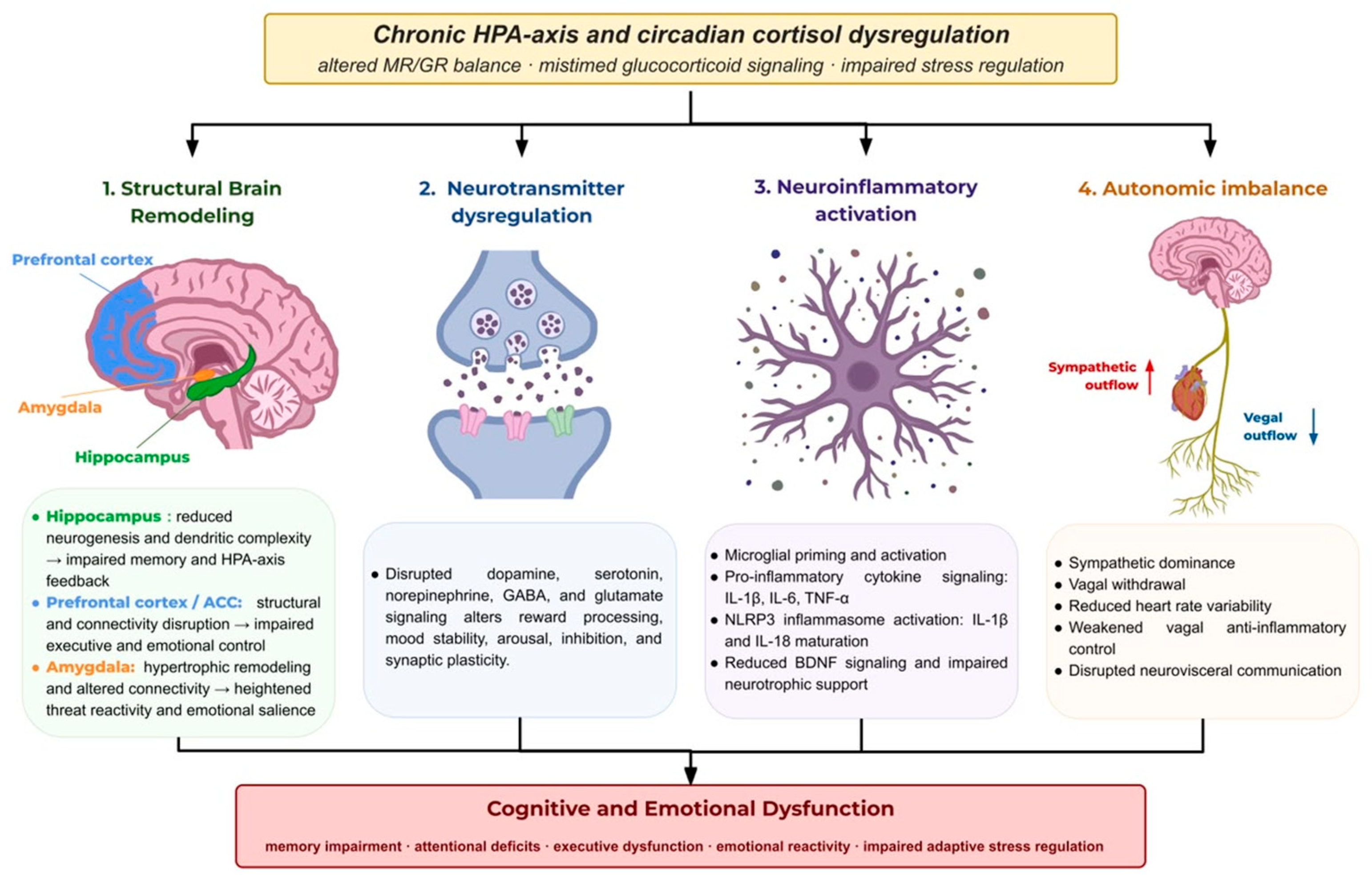
The problem emerges when glucocorticoid signaling no longer remains confined to that adaptive response. Cortisol acts throughout the brain through mineralocorticoid receptors (MRs) and glucocorticoid receptors (GRs), which differ in affinity, distribution, and function. MRs are preferentially occupied under basal conditions and support appraisal, emotional regulation, and cognitive stability, whereas GRs become increasingly engaged during stress and coordinate adaptation, energy mobilization, and negative-feedback termination of the stress response. Normal cognitive and emotional regulation therefore depends in part on the balance between the two systems. Repeated or prolonged stress can shift that balance toward maladaptive glucocorticoid signaling, weakening HPA-axis feedback regulation and increasing allostatic load [
11,
12,
15,
16] (
Figure 2).
Cortisol exposure is defined not only by how much hormone is present but also by when it is present. Under physiological conditions, concentrations rise before awakening, peak shortly after waking, and decline progressively toward evening, with ultradian pulses superimposed across the day. This rhythmic organization coordinates glucocorticoid receptor activation and synchronizes metabolic, immune, autonomic, and neural processes with the sleep–wake cycle [
11,
12,
16,
17].
Chronic HPA-axis dysregulation disturbs that organization, often producing a blunted morning peak, a flatter diurnal cortisol slope, and mistimed glucocorticoid signaling. These changes should not be reduced to a simple question of whether cortisol is “high” or “low.” Absolute cortisol concentration, circadian rhythmicity, and stress responsiveness are distinct dimensions of HPA-axis function, and each may become dysregulated in a different way [
11,
17,
18]. Disrupted rhythmicity has been associated with adverse physical and mental health outcomes, including immune and inflammatory dysfunction, fatigue, depression, cancer-related disease outcomes, and mortality [
17]. HPA-axis dysregulation may therefore interact with several processes examined below, providing a plausible route through which prolonged psychological stress may alter brain structure, neurotransmitter signaling, neuroinflammatory activity, and autonomic balance.
4.2. Structural Brain Alterations Associated with Chronic Glucocorticoid Exposure
Chronic glucocorticoid exposure does not remodel the brain uniformly. Regions involved in cognition, emotional regulation, and HPA-axis feedback are particularly vulnerable because of their corticosteroid receptor density, reciprocal connectivity, and role in coordinating the adaptive stress response. The resulting pattern is therefore one of region-specific remodeling rather than generalized neurodegeneration. The hippocampus, prefrontal cortex, amygdala, and anterior cingulate cortex are among the most consistently affected structures, but they do not respond in the same direction [
19,
20,
21].
The hippocampus is particularly susceptible because it supports memory and HPA-axis feedback and contains a high density of corticosteroid receptors [
19,
20,
21]. Sustained cortisol exposure suppresses adult hippocampal neurogenesis, promotes dendritic retraction of cornu ammonis 3 (CA3) pyramidal neurons, reduces spine density, and is associated with hippocampal volume loss in animal models and human neuroimaging studies [
22,
23,
24,
25,
26]. These changes impair memory and cognitive flexibility and may weaken hippocampal inhibition of the HPA axis [
19,
20].
The prefrontal cortex also shows stress-related retraction. Chronic stress or glucocorticoid exposure induces dendritic remodeling and spine loss in the medial prefrontal cortex, while human studies link chronic stress and cortisol exposure with reduced cortical thickness and gray-matter volume [
26,
27,
28,
29,
30]. The anterior cingulate cortex shows related changes in gray matter, dendritic architecture, and functional connectivity [
20,
21,
31,
32]. These alterations are consistent with poorer executive function, attentional control, and emotional regulation [
19,
20].
The amygdala follows a different pattern. Chronic stress promotes dendritic arborization, spine formation, and increased synaptic connectivity in the basolateral amygdala [
33,
34,
35], accompanied by greater anxiety, fear conditioning, and emotional reactivity [
19,
20]. Memory and control circuits may therefore retract as threat-responsive circuitry becomes more reactive. Stress also alters the neurotransmitter systems through which these regions communicate.
The anterior cingulate cortex adds another component to this stress-sensitive network. Together with interconnected dorsolateral prefrontal regions, the anterior cingulate cortex (ACC) contributes to cognitive control, conflict monitoring, emotional regulation, pain processing, and the integration of cognitive and affective information [
19,
20,
21]. Chronic stress and stress-related psychopathology have been associated with changes in ACC gray matter, dendritic architecture, and functional connectivity [
20,
21,
31,
32]. These alterations track impaired emotional regulation, attentional control, and cognitive performance and may contribute to the executive dysfunction observed under prolonged glucocorticoid dysregulation [
19,
20].
The structural signature of chronic stress is therefore distributed but internally coherent: hippocampal and prefrontal systems supporting memory and cognitive control retract, amygdaloid circuits supporting threat responsiveness expand, and cingulate networks involved in integrating cognition and emotion are altered. These anatomical changes provide one substrate for chronic-stress dysfunction, but structure alone does not account for the resulting cognitive and behavioral phenotype. Stress also changes the chemical signaling through which these networks operate.
4.3. Neurotransmitter Dysregulation in Chronic Stress
Structural remodeling is accompanied by changes in neurotransmitter systems that regulate motivation, mood, attention, learning, and synaptic plasticity [
20,
36]. Chronic stress blunts dopaminergic reward and motivation circuits, contributing to anhedonia, reduced cognitive flexibility, and more habitual behavior [
11,
37], and can alter serotonergic tone through inflammation, impaired neurogenesis, and tryptophan shunting toward kynurenine. This contributes to the depressive symptoms, emotional dysregulation, and memory impairment associated with prolonged glucocorticoid exposure [
20,
21].
Noradrenergic signaling links cortisol more directly to arousal and emotionally salient memory. Norepinephrine regulates vigilance, attention, arousal, and stress-related memory, and cortisol interacts with noradrenergic signaling in amygdala- and hippocampus-dependent circuits during stress [
11,
38]. That interaction strengthens emotional salience and fear-related memory processing. When stress persists, the same relationship may contribute to hypervigilance, impaired attentional control, and heightened emotional reactivity [
11].
The balance between γ-aminobutyric acid (GABA) and glutamate determines another level of vulnerability. GABA is the principal inhibitory neurotransmitter in the central nervous system, and chronic stress disrupts GABAergic signaling particularly within the medial prefrontal cortex, where remodeling of inhibitory interneurons and reductions in markers of GABAergic function have been observed [
20,
39]. The resulting loss of inhibitory regulation can disturb excitation–inhibition balance and increase vulnerability to anxiety, emotional dysregulation, and stress-related psychopathology [
20,
40].
Glutamate provides the excitatory counterpart. As the principal excitatory neurotransmitter of the central nervous system, it is essential for synaptic plasticity, learning, and memory. Chronic glucocorticoid exposure and repeated stress disrupt glutamate release, receptor trafficking, and synaptic plasticity in the hippocampus and prefrontal cortex [
36,
41,
42,
43]. These changes suppress long-term potentiation, facilitate long-term depression, and promote dendritic remodeling, impairing the synaptic adaptations required for learning and memory [
36,
44,
45]. Glutamatergic dysfunction therefore represents one of the principal mechanisms connecting chronic stress to cognitive impairment and stress-related neuropsychiatric dysfunction [
36].
Together, these changes convert the structural vulnerability described above into altered network function. Dopaminergic and serotonergic disruption affects motivation and mood, cortisol–norepinephrine interactions bias attention toward emotionally salient information, impaired GABAergic inhibition weakens regulatory control, and altered glutamatergic plasticity compromises learning and memory. Chronic stress therefore affects not only the architecture of stress-sensitive brain regions but also the chemical signaling required for those regions to function adaptively.
4.4. Neuroinflammation: Cytokines, Microglia, and the Stress-Brain Interface
The stress response extends beyond neurons. Chronic glucocorticoid exposure recruits neuroimmune pathways, placing microglia and inflammatory cytokine signaling alongside structural remodeling and neurotransmitter dysregulation as mechanisms of stress-related brain dysfunction [
46,
47].
Microglia bridge neuroendocrine disturbance and neuroinflammation. Prolonged exposure to stress hormones can shift these normally homeostatic immune cells toward a primed phenotype with altered morphology, immune function, and responsiveness to later inflammatory stimuli [
46,
47]. Activated microglia amplify inflammatory signaling through mediators including interleukin-1β (IL-1β), interleukin-6 (IL-6), and tumor necrosis factor-α (TNF-α), which can impair synaptic plasticity, reduce neurogenesis, alter neurotransmitter signaling, and disrupt neuronal communication in the hippocampus and prefrontal cortex [
46,
47,
48].
The NLR family pyrin domain containing 3 (NLRP3) inflammasome supplies a more specific pathway. Its activation promotes caspase-1 activity and maturation of IL-1β and IL-18; in chronic stress models, this pathway increases local neuroinflammation, impairs hippocampal neurogenesis, and contributes to synaptic dysfunction and cognitive decline [
48,
49].
The relationship is bidirectional. Glucocorticoid dysregulation can promote microglial priming and inflammatory signaling, while inflammatory mediators can further disrupt HPA-axis regulation through limbic and hypothalamic circuits [
46,
47,
48,
49]. Reduced brain-derived neurotrophic factor (BDNF) signaling may add to this vulnerability by limiting neuronal survival, synaptic plasticity, and hippocampal neurogenesis [
20,
21,
48]. The neuroinflammatory state associated with chronic stress is therefore not an isolated immune phenomenon. Sustained microglial activation, excessive cytokine signaling, and impaired neurotrophic support reinforce the same structural and synaptic vulnerabilities already produced by glucocorticoid dysregulation.
4.5. Autonomic Nervous System: Sympathetic Dominance and Parasympathetic Withdrawal
Chronic stress also alters brain–body regulation through the autonomic nervous system. Acute sympathetic activation supports vigilance and energy mobilization, while parasympathetic activity promotes recovery after the stressor resolves. Repeated stress shifts this balance toward sustained sympathetic drive and reduced vagal activity [
50,
51].
Sympathetic activation becomes maladaptive when adrenergic signaling persists beyond the acute threat. Sustained signaling maintains physiological hyperarousal and can promote inflammatory activity through effects on immune cells and peripheral tissues. In this setting, sympathetic overactivation becomes another chronic amplifier of neuroendocrine and neuroinflammatory dysregulation rather than a transient adaptive response [
50,
51].
At the same time, parasympathetic withdrawal removes an important counter-regulatory pathway. The vagus nerve is the principal parasympathetic conduit linking the brain to peripheral organs and plays a central role in limiting excessive inflammatory responses. Through the inflammatory reflex and its efferent cholinergic arm, vagal signaling suppresses production of pro-inflammatory cytokines including TNF-α, IL-1β, and IL-18 [
52,
53,
54]. Reduced vagal tone therefore weakens an anti-inflammatory brake and allows inflammatory signaling to persist, further disrupting neuroendocrine and neural homeostasis [
52].
Heart rate variability (HRV) provides a non-invasive readout of this autonomic balance. Defined as beat-to-beat variation in heart rate, HRV is strongly influenced by vagal activity [
51,
55]. Higher HRV generally reflects greater parasympathetic regulatory capacity and greater flexibility in responding to physiological and psychological demands [
56]. Chronic stress, by contrast, is commonly associated with reduced HRV, diminished vagal modulation, increased autonomic rigidity, and impaired recovery from stress [
51,
57].
The same imbalance has consequences for brain function. Central autonomic networks spanning the prefrontal cortex, anterior cingulate cortex, insula, amygdala, hypothalamus, and brainstem integrate internal bodily signals, emotional salience, cognitive appraisal, and physiological responses to stress [
56,
57]. Persistent sympathetic activation and reduced vagal tone may therefore impair emotional regulation, pain processing, attentional control, and cognitive performance, linking peripheral autonomic imbalance to the cortical and limbic dysfunction described above [
56,
57].
5. Pre-Chemotherapy Brain Changes: Pretreatment Neural Vulnerability at Diagnosis
5.1. Structural and Connectivity Alterations Before Treatment Begins
The mechanisms described in
Section 4 become clinically relevant only if corresponding brain abnormalities are detectable before chemotherapy begins. Neuroimaging evidence suggests that they can be. Even before cytotoxic treatment, patients with cancer may show structural and functional alterations across networks involved in cognition and emotional regulation. A comprehensive neuroimaging review linked the psychological stress and emotional burden surrounding a cancer diagnosis with functional and metabolic changes involving the prefrontal cortex, thalamus, cingulate cortex, hypothalamus, basal ganglia, and insula, regions also associated with anxiety, depression, posttraumatic stress symptoms, and psychological distress [
58].
Resting-state imaging extends this evidence to limbic networks. D’Imperio et al. (2021) [
59] examined women with breast cancer before chemotherapy using resting-state functional magnetic resonance imaging and identified altered connectivity involving the amygdala and hippocampus, structures central to emotion regulation and memory. Among patients with greater subjective memory complaints, resting-state connectivity was increased in the right amygdala and hippocampus and reduced in the left amygdala [
58,
59]. The finding places measurable neural alteration before the first cytotoxic exposure rather than exclusively after it.
5.2. Pre-Existing Cognitive Impairment Before Treatment
The pretreatment phenotype is not limited to neuroimaging. Treatment-naïve patients also demonstrate measurable cognitive abnormalities before receiving systemic therapy. Compared with healthy controls, treatment-naïve adolescent and young-adult cancer patients exhibited elevated baseline inflammatory markers, including IL-2, IL-4, IL-6, IL-8, IL-10, and IFN-γ, together with lower brain-derived neurotrophic factor (BDNF) levels [
2]. These findings identify inflammatory and neurobiological disturbances present before treatment that potentially contribute to baseline cognitive vulnerability [
60,
61].
Pretreatment cognitive abnormalities vary by cancer population. Patients with Hodgkin lymphoma showed deficits in verbal learning and memory, processing/psychomotor speed, and abstraction/executive functions relative to matched controls [
61], whereas pretreatment non-small-cell lung cancer (NSCLC) cohort found objective impairment in 36% of patients, with performance related to cognitive reserve and psychological factors [
62]. Their presence before systemic treatment reinforces the distinction made in
Section 3: cancer-related cognitive impairment cannot be treated as a phenomenon that begins only after chemotherapy.
That timing has practical consequences for interpretation. Without a cognitive baseline at diagnosis, disease-related cognitive changes and diagnosis-associated psychological distress can later be attributed to chemotherapy even when part of the impairment preceded treatment. Pretreatment assessment is therefore important for distinguishing subsequent treatment-associated change from cognitive abnormalities already present at diagnosis.
5.3. The Nocebo Effect: Anticipatory Cognition as a Potential Contributor
Pretreatment vulnerability can also be shaped by what patients expect treatment to do. The nocebo effect refers to adverse outcomes attributable to negative expectations and contextual or learning processes beyond the pharmacological effects of treatment. Its clinical consequences include greater symptom burden, poorer treatment adherence, and reduced therapeutic efficacy. Susceptibility reflects negative expectations, prior treatment experiences and conditioning, social observation, patient–clinician communication, contextual cues, negative mood and cognitive appraisal, personality characteristics, and the misattribution of pre-existing symptoms to treatment [
63,
64].
The effect is not confined to subjective symptom reporting. Experimental nocebo paradigms indicate that negative expectations can recruit physiological stress responses. In nocebo-hyperalgesia experiments, negative verbal suggestion has been associated with increased ACTH and cortisol, while Johansen et al. also observed greater cortisol elevation under nocebo conditions [
65,
66]. Cholecystokinin signaling has also been implicated in nocebo hyperalgesia, as cholecystokinin (CCK)-receptor antagonism with proglumide blocked experimentally induced nocebo-related pain amplification [
67]. Anticipatory cognition can therefore recruit physiological stress pathways before or during treatment and add to the symptom burden surrounding chemotherapy exposure.
6. Chemotherapy-Associated Brain Changes: Evidence Across Cancer Populations
6.1. Mechanism and Structural Changes Associated with Chemotherapy Neurotoxicity
If
Section 4 and
Section 5 define the biological and clinical basis of the first hit, chemotherapy provides a second source of injury through mechanisms that act directly on neural, glial, and vascular systems (
Figure 3). One target is the proliferative cell population required for hippocampal plasticity. Chemotherapeutic agents can injure neural stem cells and markedly suppress adult hippocampal neurogenesis within the subgranular zone [
68]. Loss of this dentate-gyrus plasticity has been implicated in chemotherapy-associated cognitive impairment [
69], while neuroimaging studies in treatment-exposed patients demonstrate that breast-cancer survivors showed smaller total hippocampal volume and greater inward hippocampal deformation, particularly in the right hippocampus [
68,
69,
70].
White matter represents a second point of vulnerability. Chemotherapy injures oligodendrocyte progenitor cells, the population responsible for maintaining and repairing myelin [
69]. Chen et al. [
71] identified longitudinal post-chemotherapy changes in white-matter integrity involving the corpus callosum and middle frontal gyrus, with neuropsychological performance associated with abnormalities in the corpus callosum, middle frontal gyrus, posterior limb of the internal capsule, and superior longitudinal fasciculus [
69,
71].
Chemotherapy can promote molecular features of accelerated aging through DNA damage and cellular senescence. Sanoff et al. [
72] demonstrated sustained increases in the p16 inhibitor of cyclin-dependent kinase 4A (p16INK4a), alternative reading frame (ARF), and senescence-associated cytokines after chemotherapy but did not detect chemotherapy-associated telomere shortening. Broader reviews indicate that telomere attrition may occur with particular cancer treatments and treatment modalities, suggesting that the combination of persistent DNA lesions and telomeric degradation can arrest the cell cycle and drive vulnerable nervous-system components toward cellular senescence or programmed apoptosis, providing a substrate for longer-term cognitive and neurological decline [
73].
The blood–brain barrier provides another route to the second hit. In an in vitro rat brain endothelial model, Branca et al. showed that oxaliplatin increased intracellular reactive oxygen species (ROS) and endoplasmic reticulum (ER) stress and altered the organization of zonula occludens-1 (ZO-1) and F-actin, producing a transient reduction in endothelial barrier tightness [
74]. Once barrier integrity is weakened, inflammatory cytokines, systemic chemotherapeutics, and ROS can enter the central nervous system more readily, where they promote neuroinflammation, microglial and astrocyte activation, synaptic dysfunction, and neuronal cell death. These processes are associated clinically with deficits in executive function, processing speed, attention, working memory, and learning [
73,
74,
75].
Glial injury extends beyond actively dividing progenitor populations. Non-mitotic cells such as astrocytes also develop altered cellular physiology after chemotherapy, contributing to a secondary neuroinflammatory environment and reducing the metabolic support available to surrounding neural tissue [
69].
At the macroscopic level, these cellular processes are accompanied by measurable structural change. Longitudinal morphometric imaging demonstrated gray-matter reductions in bilateral frontal, temporal, and cerebellar regions and the right thalamus at an assessment performed approximately one month after chemotherapy completion [
69]. The second hit is therefore visible across scales, from progenitor and glial injury to white-matter disruption, barrier compromise, and regional gray-matter loss.
6.2. Breast Cancer
Breast cancer provides some of the most developed clinical evidence for these treatment-associated brain changes. Prospective neuroimaging studies identify deterioration of white-matter microstructure after chemotherapy, including reduced fractional anisotropy and altered diffusion metrics within the corpus callosum, superior longitudinal fasciculus, inferior longitudinal fasciculus, corticospinal tract, and prefrontal white matter [
76,
77]. These abnormalities are associated with declines in attention, verbal memory, processing speed, and executive function, linking disruption of large-scale structural connectivity with impaired cognitive performance after treatment.
Gray-matter findings point in the same direction. Longitudinal MRI studies demonstrate reductions in frontal gray-matter density after chemotherapy accompanied by increased executive dysfunction [
78]. Multimodal studies add evidence of increased serum neurofilament light chain, consistent with axonal injury, together with persistent abnormalities in white-matter organization and objective cognitive decline [
76]. Across these modalities, the imaging and biomarker findings converge on structural injury involving both gray- and white-matter networks after chemotherapy [
76,
77,
78,
79].
The neuroendocrine system is altered alongside these structural effects. Cancer patients frequently exhibit a flattened diurnal cortisol trajectory rather than the normally steep decline in cortisol across the day, and this pattern has been associated with earlier mortality and persistent cancer-related fatigue [
80,
81]. Cortisol dysregulation has also been associated with impaired cognitive performance, suggesting that HPA-axis dysfunction may contribute to the cognitive burden observed during cancer treatment [
80,
81]. Morning salivary cortisol offers a non-invasive way to assess this system, but collection timing, adherence, sleep patterns, and day-to-day variability limit its interpretation as a standalone biomarker [
81]. Its value is therefore greater when considered alongside behavioral, psychological, and neurobiological measures in identifying patients with stress-related vulnerability and in guiding approaches intended to mitigate cancer-related cognitive decline [
80,
81,
82].
6.3. Lung Cancer
In lung cancer, pretreatment abnormalities may exceed post-treatment change. Simó et al. found more widespread white-matter damage and worse verbal memory in chemotherapy-naïve patients than in treated patients [
83]. A later SCLC cohort found cognitive impairment in about one-third of patients at diagnosis and uncommon de novo impairment after chemotherapy [
84]. Increased BBB leakage has also tracked poorer cognition in advanced disease [
85]. Post-treatment imaging nevertheless shows focal gray- and white-matter abnormalities in paralimbic, frontal, and temporal regions [
83]. The available evidence supports substantial disease-related vulnerability before chemotherapy, with additional treatment-associated change in some patients, while longitudinal evidence indicates that new chemotherapy-associated cognitive impairment is not universal.
6.4. Gastrointestinal Cancer
Functional imaging extends the second-hit evidence to colorectal cancer. Resting-state functional magnetic resonance imaging after systemic chemotherapy has identified altered intrinsic brain activity, particularly in anterior cingulate and frontal regions, together with poorer cognitive performance. Among patients treated with capecitabine- and oxaliplatin-based regimens, fractional amplitude of low-frequency fluctuation was reduced in the left anterior cingulate and middle frontal gyri. Anterior-cingulate fALFF was positively associated with global cognitive and perceived cognitive function, suggesting vulnerability of these regions to chemotherapy-related neurotoxicity [
86].
6.5. Gynecological/Ovarian Cancer
Ovarian cancer adds converging structural and functional imaging evidence after platinum and taxane-based chemotherapy. Structural MRI in these patients showed reduced frontal and parietal gray-matter volume and reduced middle frontal and inferior parietal activation during N-back working memory, although neuropsychological performance did not differ from controls [
87]. Resting-state imaging found reduced regional homogeneity in the middle frontal gyrus, medial prefrontal cortex, and superior parietal lobule [
88]. Both studies were small, cross-sectional investigations from the same research group, so they do not provide independent replication.
6.6. Prostate Cancer
The prostate-specific literature on cognitive effects of cytotoxic chemotherapy is comparatively limited. A prospective multicenter cohort of older men with metastatic castration-resistant prostate cancer found that most patients maintained stable cognition or showed slight improvement; decline of at least 1.5 SD occurred in 0–6.5% depending on the measure, including 3 of 46 patients receiving chemotherapy [
89]. Substantial deterioration was therefore not consistently observed, although the small prostate-specific evidence base limits broader conclusions [
89].
6.7. Patients Receiving Adjuvant Treatment
Prospective studies of adjuvant chemotherapy are particularly informative because pretreatment assessment allows subsequent change to be measured against an established baseline. Adjuvant chemotherapy, given after surgery to eliminate residual malignant cells and reduce recurrence risk, is followed by measurable cognitive change in a subset of patients. Longitudinal studies identify declines after treatment in attention, learning, and processing speed, with some of these deficits persisting at longer-term follow-up [
90].
Meta-analytic evidence is consistent with those findings. Across studies of adjuvant chemotherapy, treatment exposure was significantly associated with cognitive impairment, although the magnitude of impairment was generally small and varied by study design and cognitive domain [
91]. The evidence therefore supports chemotherapy as a potential contributor to cognitive dysfunction while reinforcing the importance of pretreatment measurement of cognitive impairment when attributing subsequent changes to chemotherapy [
90,
91].
7. The Bi-Directional HPA Axis-Chemotherapy Relationship
The preceding sections treated stress-related brain injury and chemotherapy neurotoxicity as parallel exposures, but the evidence suggests potential interaction. Preclinical studies show that endogenous cortisol acting through the glucocorticoid receptor (GR) opposes the cytotoxic action of several chemotherapies at the same time as it damages stress-sensitive brain regions, while clinical studies indicate that cytotoxic treatment can perturb the HPA axis. These findings support a possible bidirectional relationship, although its contribution to CRCI in patients remains unproven.
7.1. Glucocorticoid Signaling and Chemotherapy Response
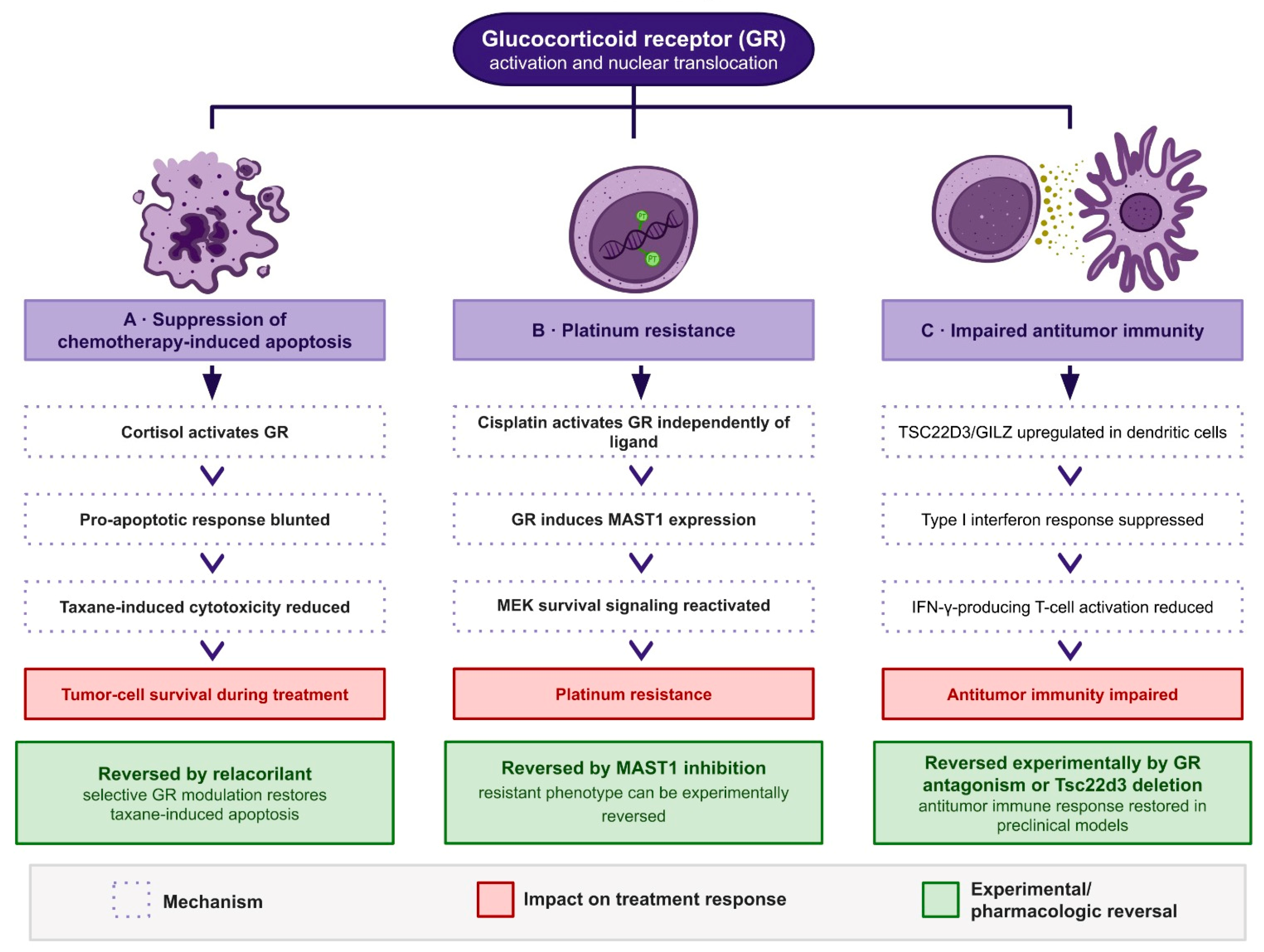
Preclinical studies indicate that glucocorticoid signaling can reduce the effects of microtubule-targeting chemotherapy. In the MIA PaCa-2 pancreatic line, paclitaxel-driven apoptosis is blunted by cortisol and restored by the selective GR modulator relacorilant; in OVCAR5 ovarian cells, relacorilant improved the efficacy of paclitaxel and the potency of platinum agents [
92]. A combination screen in the same work found that microtubule-targeted agents consistently benefited from the addition of relacorilant, and the effect held in xenograft models, including MIA PaCa-2, HeLa, and a cholangiocarcinoma patient-derived xenograft, where tumor-cell apoptosis rose when relacorilant was added to paclitaxel. The pairing has since been tested clinically, where relacorilant with nab-paclitaxel produced durable disease control in taxane-resistant solid tumors [
93].
Platinum agents subvert the same receptor by a different route. Rather than being restrained by circulating cortisol, cisplatin activates the GR directly: Pan et al. [
94] reported that the drug binds cysteine-622 of the receptor and recruits it to the nucleus, where the activated receptor induces microtubule-associated serine/threonine kinase 1 (MAST1) and reactivates mitogen-activated protein kinase kinase (MEK) signaling to produce platinum resistance. Glucocorticoid exposure with dexamethasone reproduced the resistant phenotype, and MAST1 inhibition reversed it. Catecholamines released under sympathetic activation act together with glucocorticoids to support tumor-cell survival and blunt both chemotherapy and immunotherapy [
95], so the stress hormones that accompany a cancer diagnosis converge on drug resistance from more than one receptor system.
7.2. The TSC22D3 Neuro-Immune Pathway
A more recent line of work connects psychological state to the immune arm of treatment response. In mouse models, Yang et al. [
96] showed that chronic stress raised plasma corticosterone, which upregulated the glucocorticoid-inducible factor TSC22D3 (GILZ) in dendritic cells. TSC22D3 blocked type I interferon responses and the activation of interferon-γ-producing T cells, and enforced expression of the gene in dendritic cells was sufficient to abolish the therapeutic control of tumors. Administration of a glucocorticoid receptor antagonist (mifepristone), or dendritic-cell-specific deletion of Tsc22d3, reversed this effect; in patients, plasma cortisol, leukocyte TSC22D3 expression, and negative mood were closely correlated. The pathway supplies a defined molecular link from stress physiology to failure of antitumor immunity, and raises the possibility that lowering glucocorticoid tone could recover immunotherapy sensitivity, a mechanistic rather than purely palliative role for stress reduction (
Figure 4).
7.3. Chemotherapy Directly Disrupts the HPA Axis
Chemotherapy can also alter HPA-axis function. In a systematic review of seventeen studies, Kanter et al. [
97] found altered HPA function in sixteen; twelve reported increased baseline or hyperactive cortisol responses and four a decreased or blunted response, with fatigue, memory complaints, and disturbed sleep among the clinical correlates. Superimposed on this chronic picture is an acute pharmacological effect. Morrow et al. sampled serum cortisol hourly before and after treatment in twenty-three chemotherapy-naïve women with ovarian cancer and observed a significant fall in cortisol immediately after cisplatin or carboplatin infusion that did not occur on a separate day when the same procedures were performed without chemotherapy (
p < 0.003) [
98]. Because the drop appeared only in the presence of the cytotoxic drug, they attributed it to a direct effect rather than to circadian or psychological factors. Whether platinum suppresses adrenal steroidogenesis or pituitary ACTH output has not been resolved, so the mechanism of this acute perturbation remains open.
7.4. Perceived Stress and Cognitive Burden During Treatment
Patient-level evidence ties these mechanisms to the clinic. Ding et al. [
82] recruited 450 patients receiving chemotherapy for breast cancer and tested four candidate routes from perceived stress to cognitive function within a single structural model: psychological resilience, social support, morning salivary cortisol, and the neutrophil-to-lymphocyte ratio. Perceived stress was inversely associated with cognition (direct effect −0.410,
p < 0.001) [
82]. Resilience and morning cortisol each carried a significant indirect effect, giving a total indirect effect of −0.231 (95% CI −0.364 to −0.102) [
82], while paths through social support and the neutrophil-to-lymphocyte ratio did not hold and were dropped from the final model.
Higher perceived stress predicted lower morning cortisol (
p = 0.0020), and lower morning cortisol predicted poorer cognition (
p = 0.034) [
82]. Blunting of the morning value, rather than the elevated output that a simple reading of stress physiology might anticipate, is the signature described in
Section 4.1 as the leading edge of a flattened diurnal rhythm, so the finding converges with the argument that disrupted rhythmicity rather than absolute concentration is what carries consequences. The inference is limited by the cross-sectional design and by the small cortisol-mediated indirect effect compared with resilience (−0.023 vs. −0.208) [
82]. Cortisol may contribute to the stress–cognition pathway, but its temporal position requires longitudinal sampling.
7.5. A Potential Bidirectional Feedback Loop
A flatter diurnal cortisol slope provides one link across the proposed cycle. In the meta-analysis by Adam et al. [
17], flatter slopes were most strongly associated with immune and inflammatory burden (r = 0.288) and were also associated with cancer diagnosis and progression (r = 0.231) and mortality (hazard ratio 1.63 after removal of two extreme outliers). Chemotherapy can further perturb HPA function [
98,
99], while glucocorticoid signaling may suppress chemotherapy-induced apoptosis, promote platinum resistance, and impair antitumor immunity [
92,
94,
96]. Patient-level data also place cortisol within the path from perceived stress to cognition, although its indirect effect was modest [
82]. Together, these findings place cortisol at the intersection of neurocognitive and oncological processes, but do not establish a self-reinforcing causal cycle in patients. Circadian dysregulation is therefore best viewed as a candidate integrative marker rather than a demonstrated driver of CRCI.
8. Candidate Interventions Targeting Pretreatment Vulnerability
8.1. Rationale
If stress-related cortisol dysregulation precedes chemotherapy and compounds its neurotoxicity, then reducing that load becomes a candidate modifier of treatment response, not only a matter of comfort. Two features of the evidence reviewed above shape how such interventions should be designed. The stress-related circadian dysregulation framework [
17] emphasizes restoring a normally steep diurnal decline rather than to lower total cortisol. The TSC22D3 pathway [
96] provides a second rationale, as reducing glucocorticoid drive, whether behaviorally or pharmacologically, may recover a component of antitumor immunity that stress had suppressed. These approaches remain mechanistically motivated, but evidence that they prevent CRCI is currently limited.
8.2. Psychoeducation and Nocebo Mitigation
Because anticipatory expectation engages the stress-response pathways set out in
Section 5.3, patient expectation is itself a modifiable point of intervention [
63]. In a randomized trial of 100 patients beginning first-line chemotherapy for gastrointestinal cancer, those given a brief structured explanation of the nocebo effect reported lower adverse-event intensity at twelve weeks than an attention-matched control group (mean difference 4.04, 95% CI 0.72–7.36, Cohen’s d = 0.480) [
64], an effect carried mainly by non-specific side effects. A review of forty-three reviews identified nine categories of nocebo risk factors, several of which, including information framing and patient–clinician communication, are open to modification during routine care [
99]. Consent conversations and the wording used to prepare patients for treatment are therefore a low-cost lever on symptom burden.
8.3. Autonomic Restoration
Because chemotherapy may worsen the sympathetic dominance and vagal withdrawal described in
Section 4.5, interventions that restore parasympathetic tone may address a specific mechanism rather than general wellbeing. Heart-rate-variability biofeedback, a breathing-based method for raising HRV as an index of emotional and cognitive self-regulation, was associated with improvements in sleep, pain, fatigue, depression, and anxiety across the studies gathered in a scoping review by Spada et al. [
100]. Yoga has been tested directly against chemotherapy’s autonomic cost: in an eighteen-week randomized trial of 68 breast cancer patients receiving anthracycline-based treatment, groups were comparable at baseline, but after chemotherapy, the usual-care group showed higher resting heart rate and lower vagal indices, including the root mean square of successive differences (RMSSD), percentage of successive normal-to-normal intervals differing by more than 50 ms (pNN50), and high-frequency power, with a shift toward sympathetic predominance (higher low-frequency power and LF/HF ratio), while the yoga group was protected from this deterioration [
101]. At the level of structured exercise, a meta-analysis of six trials in 272 patients and survivors found that training improved low-frequency power (SMD 0.97), high-frequency power (SMD 1.49), and the low-to-high-frequency ratio (mean difference −0.32) [
102] relative to controls, with resistance and endurance formats most effective.
8.4. Psychological and Pharmacological Approaches
Cognitive behavioral therapy directed at anticipatory anxiety targets the same expectation circuitry that drives nocebo responses; by reducing the anxiety and neuroticism that Grosso et al. [
99] identify as risk factors, reappraisal-based approaches should lower both distress and its autonomic and endocrine sequelae. On the pharmacological side, GR antagonism has a dual rationale: relacorilant restores chemotherapy-induced apoptosis and augments taxane efficacy [
92,
93], and, because it dampens glucocorticoid signaling, it may in principle reduce cortisol-driven strain on stress-sensitive neurons, though this neuroprotective extension is inferred from mechanism and has not been tested clinically. Beta-adrenergic blockade addresses the sympathetic arm: a week of preoperative propranolol reduced pro-metastatic gene expression in breast tumors [
103], although the clinical benefit of beta-blockade appears to depend on tumor type and context and should not be generalized. Anti-inflammatory adjuncts follow from the finding that elevated cytokines and reduced BDNF accompany cognitive deficits before treatment begins [
2] and from the strong association between a flat cortisol slope and immune burden [
17], but they remain a mechanistic proposal pending trial evidence. Chronotherapy, timing cytotoxic delivery to circadian phase, reduced toxicity and raised the objective response rate for oxaliplatin-based regimens in metastatic colorectal cancer [
104], though a later phase III trial found the survival advantage confined to men [
105]; the approach is promising, but its efficacy data are mixed.
8.5. Sleep and Circadian Restoration
Sleep disruption both flattens the diurnal cortisol rhythm and worsens as that rhythm degrades, which makes sleep one of the more accessible routes to a steeper slope. In 77 cancer survivors with clinical fatigue, a four-week home light-therapy intervention re-steepened the diurnal cortisol slope (B = −0.02, Cohen’s d = 0.57) and raised total cortisol output [
106]. The change occurred under both bright-white and dim-red light and did not statistically account for the improvement in fatigue, so light exposure is best regarded as a plausible means of circadian repair rather than a demonstrated mediator of symptom relief.
8.6. Clinical Integration
The through-line for practice is timing. Psychological state and cognition should be assessed at diagnosis rather than after treatment begins so that stress-related deficits present before the first infusion are not later misattributed to chemotherapy. Taken together, these measures point toward survivorship care organized across oncology, endocrinology, and psychology, in which the cortisol slope, autonomic tone, and cognitive trajectory are followed as related signals rather than in isolation.
9. Conclusions and Future Directions
Cancer-related cognitive impairment may begin before chemotherapy and persist through treatment. This pattern supports a two-hit framework in which multifactorial pretreatment vulnerability precedes additional chemotherapy-associated neurotoxicity [
1,
2,
4,
5]. However, evidence for the proposed sequence remains largely indirect and associative; much of the clinical literature is weighted toward breast cancer, and HPA-axis dysregulation should be viewed as one interacting pathway among tumor biology, inflammation, psychological distress, fatigue, sleep disruption, and pain.
Future research should use prospective, longitudinal designs beginning at diagnosis and continuing before, during, and after treatment. Establishing pretreatment cognitive and psychological baselines will be important for distinguishing pre-existing vulnerability from chemotherapy-related change. Studies integrating cognitive testing with measures of psychological distress, sleep, neuroendocrine function, inflammation, autonomic function, and neuroimaging may help clarify how these factors interact and identify patients at greatest risk. Recognizing that cognitive vulnerability may begin before treatment could ultimately support earlier assessment and more targeted strategies to preserve cognitive function throughout cancer care.
Author Contributions
Conceptualization, L.A., Y.E.K. and N.N.K.; writing—original draft preparation, L.A., Y.E.K., T.G. and T.A.; writing—review and editing, L.A. and Y.E.K.; visualization, L.A.; supervision, N.N.K.; project administration, N.N.K. All authors have read and agreed to the published version of the manuscript.
Funding
This research received no external funding.
Data Availability Statement
No new data were created or analyzed in this study. Data sharing is not applicable to this article.
Acknowledgments
The authors would like to acknowledge Rahaf Atmeh for creating the original illustrations used in this manuscript.
Conflicts of Interest
The authors declare no conflicts of interest.
Abbreviations
The following abbreviations are used in this manuscript:
| ACC | Anterior cingulate cortex |
| ACTH | Adrenocorticotropic hormone |
| ARF | Alternative reading frame tumor suppressor protein |
| AVP | Arginine vasopressin |
| BBB | Blood–brain barrier |
| BDNF | Brain-derived neurotrophic factor |
| CA3 | Cornu ammonis 3 |
| CI | Confidence interval |
| CRCI | Cancer-related cognitive impairment |
| CRH | Corticotropin-releasing hormone |
| DNA | Deoxyribonucleic acid |
| ER | Endoplasmic reticulum |
| fALFF | Fractional amplitude of low-frequency fluctuations |
| fMRI | Functional magnetic resonance imaging |
| GABA | γ-Aminobutyric acid |
| GILZ | Glucocorticoid-induced leucine zipper |
| GR | Glucocorticoid receptor |
| HPA | Hypothalamic–pituitary–adrenal |
| HRV | Heart rate variability |
| IFN-γ | Interferon-γ |
| IL | Interleukin |
| IL-1β | Interleukin-1β |
| IL-2 | Interleukin-2 |
| IL-4 | Interleukin-4 |
| IL-6 | Interleukin-6 |
| IL-8 | Interleukin-8 |
| IL-10 | Interleukin-10 |
| IL-18 | Interleukin-18 |
| LF/HF | Low-frequency/high-frequency ratio |
| MAST1 | Microtubule-associated serine/threonine kinase 1 |
| MEK | Mitogen-activated protein kinase kinase |
| MR | Mineralocorticoid receptor |
| MRI | Magnetic resonance imaging |
| NIRS | Near-infrared spectroscopy |
| NLRP3 | NLR family pyrin domain containing 3 |
| NSCLC | Non-small-cell lung cancer |
| p16INK4a | p16 inhibitor of cyclin-dependent kinase 4A |
| pNN50 | Percentage of successive normal-to-normal intervals differing by more than 50 ms |
| PTSD | Post-traumatic stress disorder |
| RMSSD | Root mean square of successive differences |
| ROS | Reactive oxygen species |
| SCLC | Small-cell lung cancer |
| SMD | Standardized mean difference |
| TMS | Transcranial magnetic stimulation |
| TMS–NIRS | Transcranial magnetic stimulation–near-infrared spectroscopy |
| TNF-α | Tumor necrosis factor-α |
| TSC22D3 | TSC22 domain family member 3 |
| ZO-1 | Zonula occludens-1 |
References
- Dijkshoorn, A.B.C.; van Stralen, H.E.; Sloots, M.; Schagen, S.B.; Visser-Meily, J.M.A.; Schepers, V.P.M. Prevalence of cognitive impairment and change in patients with breast cancer: A systematic review of longitudinal studies. Psycho-Oncology 2021, 30, 635–648. [Google Scholar] [CrossRef] [Scilit] [PubMed]
- Chan, A.; Cheng, I.; Wang, C.; Tan, C.J.; Toh, Y.L.; Ng, D.Q.; Koh, Y.Q.; Zhou, H.; Foo, K.M.; Chan, R.J.; et al. Cognitive impairment in adolescent and young adult cancer patients: Pre-treatment findings of a longitudinal study. Cancer Med. 2023, 12, 4821–4831. [Google Scholar] [CrossRef] [Scilit] [PubMed]
- Talukdar, J.; Megha; Choudhary, H.; Bhatnagar, S.; Pandit, A.; Mishra, A.K.; Karmakar, S.; Sharan, P. The interplay of chronic stress and cancer: Pathophysiology and implications for integrated care. Cancer Rep. 2025, 8, e70143. [Google Scholar] [CrossRef] [Scilit] [PubMed]
- Whittaker, A.L.; George, R.P.; O’Malley, L. Prevalence of cognitive impairment following chemotherapy treatment for breast cancer: A systematic review and meta-analysis. Sci. Rep. 2022, 12, 2135. [Google Scholar] [CrossRef] [Scilit] [PubMed]
- Leskinen, S.; Alsalek, S.; Galvez, R.; Ononogbu-Uche, F.C.; Shah, H.A.; Vojnic, M.; D’Amico, R.S. Chemotherapy-related cognitive impairment and changes in neural network dynamics: A systematic review. Neurology 2025, 104, e210130. [Google Scholar] [CrossRef] [Scilit] [PubMed]
- Bray, F.; Laversanne, M.; Sung, H.; Ferlay, J.; Siegel, R.L.; Soerjomataram, I.; Jemal, A. Global cancer statistics 2022: GLOBOCAN estimates of incidence and mortality worldwide for 36 cancers in 185 countries. CA Cancer J. Clin. 2024, 74, 229–263. [Google Scholar] [CrossRef] [Scilit] [PubMed]
- Wagle, N.S.; Nogueira, L.; Devasia, T.P.; Mariotto, A.B.; Yabroff, K.R.; Islami, F.; Jemal, A.; Alteri, R.; Ganz, P.A.; Siegel, R.L. Cancer treatment and survivorship statistics, 2025. CA Cancer J. Clin. 2025, 75, 308–340, Correction in CA Cancer J. Clin. 2025, 75, 683. [Google Scholar] [CrossRef] [Scilit] [PubMed]
- Amiri, S. The prevalence of anxiety symptoms/disorders in cancer patients: A meta-analysis. Front. Psychiatry 2024, 15, 1422540. [Google Scholar] [CrossRef] [Scilit] [PubMed]
- Omari, M.; Amaadour, L.; Zarrouq, B.; Al-Sheikh, Y.A.; El Asri, A.; Kriya, S.; Nadi, S.; Benbrahim, Z.; Mellas, N.; El Rhazi, K.; et al. Evaluation of psychological distress is essential for patients with locally advanced breast cancer prior to neoadjuvant chemotherapy: Baseline findings from cohort study. BMC Women’s Health 2023, 23, 445. [Google Scholar] [CrossRef] [Scilit] [PubMed]
- Ge, M.-W.; Sheng, J.; Shen, L.-T.; Hu, F.-H.; Jia, Y.-J.; Ur-Rehman, A.; Li, W.; Lan, J.-Z.; Liu, P.; Chen, H.-L. Global prevalence of mental health problems among cancer survivors: A meta-analysis from 31 countries. Psycho-Oncology 2025, 34, e70077. [Google Scholar] [CrossRef] [Scilit] [PubMed]
- de Kloet, E.R.; Joëls, M.; Sarabdjitsingh, R.A. Coping with the multifaceted and multifunctional role of cortisol in the brain. Curr. Res. Physiol. 2024, 8, 100137. [Google Scholar]
- Herman, J.P.; McKlveen, J.M.; Ghosal, S.; Kopp, B.; Wulsin, A.; Makinson, R.; Scheimann, J.; Myers, B. Regulation of the hypothalamic–pituitary–adrenocortical stress response. Compr. Physiol. 2016, 6, 603–621. [Google Scholar] [CrossRef] [Scilit]
- Gillies, G.E.; Linton, E.A.; Lowry, P.J. Corticotropin releasing activity of the new CRF is potentiated several times by vasopressin. Nature 1982, 299, 355–357. [Google Scholar] [CrossRef] [Scilit] [PubMed]
- Aguilera, G.; Rabadan-Diehl, C. Vasopressinergic regulation of the hypothalamic-pituitary-adrenal axis: Implications for stress adaptation. Regul. Pept. 2000, 96, 23–29. [Google Scholar] [CrossRef] [Scilit] [PubMed]
- McEwen, B.S.; Wingfield, J.C. What is in a name? Integrating homeostasis, allostasis and stress. Horm. Behav. 2010, 57, 105–111. [Google Scholar] [CrossRef] [Scilit] [PubMed]
- De Alcubierre, D.; Ferrari, D.; Mauro, G.; Isidori, A.M.; Tomlinson, J.W.; Pofi, R. Glucocorticoids and cognitive function: A walkthrough in endogenous and exogenous alterations. J. Endocrinol. Investig. 2023, 46, 1961–1982. [Google Scholar] [CrossRef] [Scilit] [PubMed]
- Adam, E.K.; Quinn, M.E.; Tavernier, R.; McQuillan, M.T.; Dahlke, K.A.; Gilbert, K.E. Diurnal cortisol slopes and mental and physical health outcomes: A systematic review and meta-analysis. Psychoneuroendocrinology 2017, 83, 25–41. [Google Scholar] [CrossRef] [Scilit]
- Pruessner, J.C.; Kirschbaum, C.; Meinlschmid, G.; Hellhammer, D.H. Two formulas for computation of the area under the curve represent measures of total hormone concentration versus time-dependent change. Psychoneuroendocrinology 2003, 28, 916–931. [Google Scholar] [CrossRef] [Scilit] [PubMed]
- Lupien, S.J.; Juster, R.P.; Raymond, C.; Marin, M.F. The effects of chronic stress on the human brain: From neurotoxicity, to vulnerability, to opportunity. Front. Neuroendocrinol. 2018, 49, 91–105. [Google Scholar] [CrossRef] [Scilit] [PubMed]
- Lucassen, P.J.; Pruessner, J.; Sousa, N.; Almeida, O.F.X.; Van Dam, A.M.; Rajkowska, G.; Swaab, D.F.; Czéh, B. Neuropathology of stress. Acta Neuropathol. 2014, 127, 109–135. [Google Scholar] [CrossRef] [Scilit] [PubMed]
- Cardoner, N.; Andero, R.; Cano, M.; Marin-Blasco, I.; Porta-Casteràs, D.; Serra-Blasco, M.; Via, E.; Vicent-Gil, M.; Portella, M.J. Impact of stress on brain morphology: Insights into structural biomarkers of stress-related disorders. Curr. Neuropharmacol. 2024, 22, 935–962. [Google Scholar] [CrossRef] [Scilit] [PubMed]
- Watanabe, Y.; Gould, E.; McEwen, B.S. Stress induces atrophy of apical dendrites of hippocampal CA3 pyramidal neurons. Brain Res. 1992, 588, 341–345. [Google Scholar] [CrossRef] [Scilit] [PubMed]
- Magariños, A.M.; McEwen, B.S. Stress-induced atrophy of apical dendrites of hippocampal CA3c neurons: Involvement of glucocorticoid secretion and excitatory amino acid receptors. Neuroscience 1995, 69, 89–98. [Google Scholar] [CrossRef] [Scilit] [PubMed]
- Magariños, A.M.; McEwen, B.S.; Flügge, G.; Fuchs, E. Chronic psychosocial stress causes apical dendritic atrophy of hippocampal CA3 pyramidal neurons in subordinate tree shrews. J. Neurosci. 1996, 16, 3534–3540. [Google Scholar] [CrossRef] [Scilit] [PubMed]
- Lupien, S.J.; de Leon, M.; de Santi, S.; Convit, A.; Tarshish, C.; Nair, N.P.V.; Thakur, M.; McEwen, B.S.; Hauger, R.L.; Meaney, M.J. Cortisol levels during human aging predict hippocampal atrophy and memory deficits. Nat. Neurosci. 1998, 1, 69–73, Correction in Nat. Neurosci. 1998, 1, 329. [Google Scholar] [CrossRef] [Scilit]
- Gianaros, P.J.; Jennings, J.R.; Sheu, L.K.; Greer, P.J.; Kuller, L.H.; Matthews, K.A. Prospective reports of chronic life stress predict decreased grey matter volume in the hippocampus. NeuroImage 2007, 35, 795–803. [Google Scholar] [CrossRef] [Scilit] [PubMed]
- Wellman, C.L. Dendritic reorganization in pyramidal neurons in medial prefrontal cortex after chronic corticosterone administration. J. Neurobiol. 2001, 49, 245–253. [Google Scholar] [CrossRef] [Scilit] [PubMed]
- Cook, S.C.; Wellman, C.L. Chronic stress alters dendritic morphology in rat medial prefrontal cortex. J. Neurobiol. 2004, 60, 236–248. [Google Scholar] [CrossRef] [Scilit] [PubMed]
- Radley, J.J.; Rocher, A.B.; Janssen, W.G.M.; Hof, P.R.; McEwen, B.S.; Morrison, J.H. Reversibility of apical dendritic retraction in the rat medial prefrontal cortex following repeated stress. Exp. Neurol. 2005, 196, 199–203. [Google Scholar] [CrossRef] [Scilit]
- Kremen, W.S.; O'BRien, R.C.; Panizzon, M.S.; Prom-Wormley, E.; Eaves, L.J.; Eisen, S.A.; Eyler, L.T.; Hauger, R.L.; Fennema-Notestine, C.; Fischl, B.; et al. Salivary cortisol and prefrontal cortical thickness in middle-aged men: A twin study. NeuroImage 2010, 53, 1093–1102. [Google Scholar] [CrossRef] [Scilit] [PubMed]
- Hercher, C.; Canetti, L.; Turecki, G.; Mechawar, N. Anterior cingulate pyramidal neurons display altered dendritic branching in depressed suicides. J. Psychiatr. Res. 2010, 44, 286–293. [Google Scholar] [CrossRef] [Scilit] [PubMed]
- Cisler, J.M.; James, G.A.; Tripathi, S.; Mletzko, T.; Heim, C.; Hu, X.P.; Mayberg, H.S.; Nemeroff, C.B.; Kilts, C.D. Differential functional connectivity within an emotion regulation neural network among individuals resilient and susceptible to the depressogenic effects of early life stress. Psychol. Med. 2013, 43, 507–518. [Google Scholar] [CrossRef] [Scilit] [PubMed]
- Vyas, A.; Mitra, R.; Shankaranarayana Rao, B.S.; Chattarji, S. Chronic stress induces contrasting patterns of dendritic remodeling in hippocampal and amygdaloid neurons. J. Neurosci. 2002, 22, 6810–6818. [Google Scholar] [CrossRef] [Scilit] [PubMed]
- Vyas, A.; Pillai, A.G.; Chattarji, S. Recovery after chronic stress fails to reverse amygdaloid neuronal hypertrophy and enhanced anxiety-like behavior. Neuroscience 2004, 128, 667–673. [Google Scholar] [CrossRef] [Scilit] [PubMed]
- Mitra, R.; Jadhav, S.; McEwen, B.S.; Vyas, A.; Chattarji, S. Stress duration modulates the spatiotemporal patterns of spine formation in the basolateral amygdala. Proc. Natl. Acad. Sci. USA 2005, 102, 9371–9376. [Google Scholar] [CrossRef] [Scilit] [PubMed]
- Popoli, M.; Yan, Z.; McEwen, B.S.; Sanacora, G. The stressed synapse: The impact of stress and glucocorticoids on glutamate transmission. Nat. Rev. Neurosci. 2012, 13, 22–37. [Google Scholar] [CrossRef] [Scilit] [PubMed]
- Dias-Ferreira, E.; Sousa, J.C.; Melo, I.; Morgado, P.; Mesquita, A.R.; Cerqueira, J.J.; Costa, R.M.; Sousa, N. Chronic stress causes frontostriatal reorganization and affects decision-making. Science 2009, 325, 621–625. [Google Scholar] [CrossRef] [Scilit] [PubMed]
- Roozendaal, B. Glucocorticoid effects on memory retrieval require concurrent noradrenergic activity in the hippocampus and basolateral amygdala. J. Neurosci. 2004, 24, 8161–8169. [Google Scholar] [CrossRef] [Scilit] [PubMed]
- Gilabert-Juan, J.; Castillo-Gomez, E.; Guirado, R.; Moltó, M.D.; Nacher, J. Chronic stress alters inhibitory networks in the medial prefrontal cortex of adult mice. Brain Struct. Funct. 2013, 218, 1591–1605. [Google Scholar] [CrossRef] [Scilit] [PubMed]
- Choudary, P.V.; Molnar, M.; Evans, S.J.; Tomita, H.; Li, J.; Vawter, M.P.; Myers, R.M.; Bunney, W.E.; Akil, H.; Watson, S.J.; et al. Altered cortical glutamatergic and GABAergic signal transmission with glial involvement in depression. Proc. Natl. Acad. Sci. USA 2005, 102, 15653–15658. [Google Scholar] [CrossRef] [Scilit] [PubMed]
- Musazzi, L.; Milanese, M.; Farisello, P.; Zappettini, S.; Tardito, D.; Barbiero, V.S.; Bonifacino, T.; Mallei, A.; Baldelli, P.; Racagni, G.; et al. Acute stress increases depolarization-evoked glutamate release in the rat prefrontal/frontal cortex: The dampening action of antidepressants. PLoS ONE 2010, 5, e8566, Correction in PLoS ONE 2010, 5, 10-1371. [Google Scholar] [CrossRef] [Scilit] [PubMed]
- Yuen, E.Y.; Liu, W.; Karatsoreos, I.N.; Feng, J.; McEwen, B.S.; Yan, Z. Acute stress enhances glutamatergic transmission in prefrontal cortex and facilitates working memory. Proc. Natl. Acad. Sci. USA 2009, 106, 14075–14079. [Google Scholar] [CrossRef] [Scilit] [PubMed]
- Yuen, E.Y.; Wei, J.; Liu, W.; Zhong, P.; Li, X.; Yan, Z. Mechanisms for acute stress-induced enhancement of glutamatergic transmission and working memory. Mol. Psychiatry 2011, 16, 156–170. [Google Scholar] [CrossRef] [Scilit] [PubMed]
- Cerqueira, J.J.; Mailliet, F.; Almeida, O.F.X.; Jay, T.M.; Sousa, N. The prefrontal cortex as a key target of the maladaptive response to stress. J. Neurosci. 2007, 27, 2781–2787. [Google Scholar] [CrossRef] [Scilit] [PubMed]
- Goldwater, D.S.; Pavlides, C.; Hunter, R.G.; Bloss, E.B.; Hof, P.R.; McEwen, B.S.; Morrison, J.H. Structural and functional alterations to rat medial prefrontal cortex following chronic restraint stress and recovery. Neuroscience 2009, 164, 798–808. [Google Scholar] [CrossRef] [Scilit] [PubMed]
- Calcia, M.A.; Bonsall, D.R.; Bloomfield, P.S.; Selvaraj, S.; Barichello, T.; Howes, O.D. Stress and neuroinflammation: A systematic review of the effects of stress on microglia and the implications for mental illness. Psychopharmacology 2016, 233, 1637–1650. [Google Scholar] [CrossRef] [Scilit] [PubMed]
- Frank, M.G.; Weber, M.D.; Watkins, L.R.; Maier, S.F. Microglia: Neuroimmune-sensors of stress. Semin. Cell Dev. Biol. 2019, 94, 176–185. [Google Scholar] [CrossRef] [Scilit] [PubMed]
- Woźny-Rasała, I.; Ogłodek, E.A. NLRP3 inflammasome in stress-related neuropsychiatric disorders: Mechanisms of neuron–microglia–astrocyte crosstalk, HPA axis dysregulation, and therapeutic perspective. Biomolecules 2025, 15, 1344. [Google Scholar] [CrossRef] [Scilit] [PubMed]
- Feng, X.; Zhao, Y.; Yang, T.; Song, M.; Wang, C.; Yao, Y.; Fan, H. Glucocorticoid-driven NLRP3 inflammasome activation in hippocampal microglia mediates chronic stress-induced depressive-like behaviors. Front. Mol. Neurosci. 2019, 12, 210. [Google Scholar] [CrossRef] [Scilit] [PubMed]
- Chan, K.L.; Poller, W.C.; Swirski, F.K.; Russo, S.J. Central regulation of stress-evoked peripheral immune responses. Nat. Rev. Neurosci. 2023, 24, 591–604. [Google Scholar] [CrossRef] [Scilit] [PubMed]
- Kim, H.G.; Cheon, E.J.; Bai, D.S.; Lee, Y.H.; Koo, B.H. Stress and heart rate variability: A meta-analysis and review of the literature. Psychiatry Investig. 2018, 15, 235–245. [Google Scholar] [CrossRef] [Scilit] [PubMed]
- Pavlov, V.A.; Tracey, K.J. The vagus nerve and the inflammatory reflex—Linking immunity and metabolism. Nat. Rev. Endocrinol. 2012, 8, 743–754. [Google Scholar] [CrossRef] [Scilit] [PubMed]
- Borovikova, L.V.; Ivanova, S.; Zhang, M.; Yang, H.; Botchkina, G.I.; Watkins, L.R.; Wang, H.; Abumrad, N.; Eaton, J.W.; Tracey, K.J. Vagus nerve stimulation attenuates the systemic inflammatory response to endotoxin. Nature 2000, 405, 458–462. [Google Scholar] [CrossRef] [Scilit] [PubMed]
- Wang, H.; Yu, M.; Ochani, M.; Amella, C.A.; Tanovic, M.; Susarla, S.; Li, J.H.; Wang, H.; Yang, N.; Ulloa, L.; et al. Nicotinic acetylcholine receptor α7 subunit is an essential regulator of inflammation. Nature 2003, 421, 384–388. [Google Scholar] [CrossRef] [Scilit] [PubMed]
- Task Force of the European Society of Cardiology and the North American Society of Pacing and Electrophysiology. Heart rate variability: Standards of measurement, physiological interpretation and clinical use. Circulation 1996, 93, 1043–1065. [Google Scholar] [PubMed]
- Thayer, J.F.; Hansen, A.L.; Saus-Rose, E.; Johnsen, B.H. Heart rate variability, prefrontal neural function, and cognitive performance: The neurovisceral integration perspective on self-regulation, adaptation, and health. Ann. Behav. Med. 2009, 37, 141–153. [Google Scholar] [CrossRef] [Scilit] [PubMed]
- Thayer, J.F.; Åhs, F.; Fredrikson, M.; Sollers, J.J., III; Wager, T.D. A meta-analysis of heart rate variability and neuroimaging studies: Implications for heart rate variability as a marker of stress and health. Neurosci. Biobehav. Rev. 2012, 36, 747–756. [Google Scholar] [CrossRef] [Scilit] [PubMed]
- Reis, J.C.; Antoni, M.H.; Travado, L. Emotional distress, brain functioning, and biobehavioral processes in cancer patients: A neuroimaging review and future directions. CNS Spectr. 2019, 25, 79–100. [Google Scholar] [CrossRef] [Scilit] [PubMed]
- D’Imperio, A.; Sicuso, C.; Fiorelli, M.; Mainero, C. Resting state fMRI connectivity of amygdala and hippocampus in women with breast cancer prior to chemotherapy. Eur. Psychiatry 2021, 64, S408. [Google Scholar] [CrossRef] [Scilit]
- Olson, B.; Marks, D.L. Pretreatment cancer-related cognitive impairment—Mechanisms and outlook. Cancers 2019, 11, 687. [Google Scholar] [CrossRef] [Scilit] [PubMed]
- Juríčková, V.; Fayette, D.; Jonáš, J.; Fajnerová, I.; Kozák, T.; Horáček, J. Pretreatment cancer-related cognitive impairment in Hodgkin lymphoma patients. Curr. Oncol. 2023, 30, 9028–9038. [Google Scholar] [CrossRef] [Scilit] [PubMed]
- Capetti, B.; Conti, L.; Luisi, J.; Sdinami, S.; Marzorati, C.; Grasso, R.; Frassoni, S.; Bagnardi, V.; Chiari, M.; Casiraghi, M.; et al. Pre-treatment cognitive impairment in patients with NSCLC: Prevalence and psychosocial factors. IBRO Neurosci. Rep. 2026, 21, 316–323. [Google Scholar] [CrossRef] [Scilit] [PubMed]
- Colloca, L. The nocebo effect. Annu. Rev. Pharmacol. Toxicol. 2024, 64, 171–190. [Google Scholar] [CrossRef] [Scilit] [PubMed]
- Michnevich, T.; Pan, Y.; Hendi, A.; Oechsle, K.; Stein, A.; Nestoriuc, Y. Preventing adverse events of chemotherapy for gastrointestinal cancer by educating patients about the nocebo effect: A randomized-controlled trial. BMC Cancer 2022, 22, 1008. [Google Scholar] [CrossRef] [Scilit] [PubMed]
- Rief, W.; Bingel, U.; Schedlowski, M.; Enck, P. Mechanisms involved in placebo and nocebo responses and implications for drug trials. Clin. Pharmacol. Ther. 2011, 90, 722–726. [Google Scholar] [CrossRef] [Scilit] [PubMed]
- Johansen, O.; Brox, J.; Flaten, M.A. Placebo and nocebo responses, cortisol, and circulating beta-endorphin. Psychosom. Med. 2003, 65, 786–790. [Google Scholar] [CrossRef] [Scilit] [PubMed]
- Benedetti, F.; Amanzio, M.; Casadio, C.; Oliaro, A.; Maggi, G. Blockade of nocebo hyperalgesia by the cholecystokinin antagonist proglumide. Pain 1997, 71, 135–140. [Google Scholar] [CrossRef] [Scilit] [PubMed]
- Dietrich, J.; Han, R.; Yang, Y.; Mayer-Pröschel, M.; Noble, M. CNS progenitor cells and oligodendrocytes are targets of chemotherapeutic agents in vitro and in vivo. J. Biol. 2006, 5, 22. [Google Scholar] [CrossRef] [Scilit] [PubMed]
- Dietrich, J.; Monje, M.; Wefel, J.; Meyers, C. Clinical patterns and biological correlates of cognitive dysfunction associated with cancer therapy. Oncologist 2008, 13, 1285–1295. [Google Scholar] [CrossRef] [Scilit] [PubMed]
- Apple, A.C.; Ryals, A.J.; Alpert, K.I.; Wagner, L.I.; Shih, P.A.; Dokucu, M.; Cella, D.; Penedo, F.J.; Voss, J.L.; Wang, L. Subtle hippocampal deformities in breast cancer survivors with reduced episodic memory and self-reported cognitive concerns. NeuroImage Clin. 2017, 14, 685–691. [Google Scholar] [CrossRef] [Scilit] [PubMed]
- Chen, V.C.; Chuang, W.; Tsai, Y.H.; McIntyre, R.S.; Weng, J.C. Longitudinal assessment of chemotherapy-induced brain connectivity changes in cerebral white matter and its correlation with cognitive functioning using the GQI. Front. Neurol. 2024, 15, 1332984. [Google Scholar] [CrossRef] [Scilit] [PubMed]
- Sanoff, H.K.; Deal, A.M.; Krishnamurthy, J.; Torrice, C.; Dillon, P.; Sorrentino, J.; Ibrahim, J.G.; Jolly, T.A.; Williams, G.; Carey, L.A.; et al. Effect of cytotoxic chemotherapy on markers of molecular age in patients with breast cancer. J. Natl. Cancer Inst. 2014, 106, dju057. [Google Scholar] [CrossRef] [Scilit] [PubMed]
- Shafqat, S.; Arana Chicas, E.; Shafqat, A.; Hashmi, S.K. The Achilles’ heel of cancer survivors: Fundamentals of accelerated cellular senescence. J. Clin. Investig. 2022, 132, e158452. [Google Scholar] [CrossRef] [Scilit] [PubMed]
- Branca, J.J.V.; Maresca, M.; Morucci, G.; Becatti, M.; Paternostro, F.; Gulisano, M.; Ghelardini, C.; Salvemini, D.; Di Cesare Mannelli, L.; Pacini, A. Oxaliplatin-induced blood brain barrier loosening: A new point of view on chemotherapy-induced neurotoxicity. Oncotarget 2018, 9, 23426–23438. [Google Scholar] [CrossRef] [Scilit] [PubMed]
- Fleming, B.; Edison, P.; Kenny, L. Cognitive impairment after cancer treatment: Mechanisms, clinical characterization, and management. BMJ 2023, 380, e071726. [Google Scholar] [CrossRef] [Scilit] [PubMed]
- Colaes, R.; Schroyen, G.; Radwan, A.; Gavrila Laic, R.A.; Helsper, S.; Himmelreich, U.; Hatse, S.; Smeets, A.; Caeyenberghs, K.; Sunaert, S.; et al. Unveiling the impact of chemotherapy in patients with breast cancer: A longitudinal study on peripheral inflammation, multimodal magnetic resonance imaging, and cognition. Cancer 2025, 131, e70095. [Google Scholar] [CrossRef] [Scilit] [PubMed]
- Menning, S.; de Ruiter, M.B.; Veltman, D.J.; Boogerd, W.; Oldenburg, H.S.A.; Reneman, L.; Schagen, S.B. Changes in brain white matter integrity after systemic treatment for breast cancer: A prospective longitudinal study. Brain Imaging Behav. 2018, 12, 324–334. [Google Scholar] [CrossRef] [Scilit] [PubMed]
- McDonald, B.C.; Conroy, S.K.; Smith, D.J.; West, J.D.; Saykin, A.J. Frontal gray matter reduction after breast cancer chemotherapy and association with executive symptoms: A replication and extension study. Brain Behav. Immun. 2013, 30, S117–S125. [Google Scholar] [CrossRef] [Scilit] [PubMed]
- Schroyen, G.; Vissers, J.; Smeets, A.; Gillebert, C.R.; Lemiere, J.; Sunaert, S.; Deprez, S.; Sleurs, C. Blood and neuroimaging biomarkers of cognitive sequelae in breast cancer patients throughout chemotherapy: A systematic review. Transl. Oncol. 2022, 16, 101297. [Google Scholar] [CrossRef] [Scilit] [PubMed]
- Sephton, S.E.; Sapolsky, R.M.; Kraemer, H.C.; Spiegel, D. Diurnal cortisol rhythm as a predictor of breast cancer survival. J. Natl. Cancer Inst. 2000, 92, 994–1000. [Google Scholar] [CrossRef] [Scilit] [PubMed]
- Bower, J.E.; Ganz, P.A.; Dickerson, S.S.; Petersen, L.; Aziz, N.; Fahey, J.L. Diurnal cortisol rhythm and fatigue in breast cancer survivors. Psychoneuroendocrinology 2005, 30, 92–100. [Google Scholar] [CrossRef] [Scilit] [PubMed]
- Ding, X.; Wang, Q.; Kan, H.; Zhao, F.; Zhu, M.; Chen, H.; Fu, E.; Li, Z. The regulation mechanism of perceived stress on cognitive function of patients with breast cancer undergoing chemotherapy: A multiple mediation analysis. Breast Cancer Res. Treat. 2025, 211, 245–259. [Google Scholar] [CrossRef] [Scilit] [PubMed]
- Simó, M.; Root, J.C.; Vaquero, L.; Ripollés, P.; Jové, J.; Ahles, T.; Navarro, A.; Cardenal, F.; Bruna, J.; Rodríguez-Fornells, A. Cognitive and brain structural changes in a lung cancer population. J. Thorac. Oncol. 2015, 10, 38–45. [Google Scholar] [CrossRef] [Scilit] [PubMed]
- Simó, M.; Vaquero, L.; Alemany, M.; Padrones, S.; Olivé, G.; Caravaca, A.; Padró-Miquel, A.; Nadal, E.; Camara, E.; Graus, F.; et al. Cancer-related cognitive impairment in patients with lung cancer. Neurology 2025, 104, e213651. [Google Scholar] [CrossRef] [Scilit] [PubMed]
- Zhang, D.-F.; Li, Z.-H.; Zhang, Z.-P.; He, Y.-F.; Shang, B.-L.; Xu, X.-F.; Ding, Y.-Y.; Cheng, Y.-Q. Cognitive changes are associated with increased blood-brain barrier leakage in non-brain metastases lung cancer patients. Brain Imaging Behav. 2023, 17, 90–99. [Google Scholar] [CrossRef] [Scilit] [PubMed]
- Liu, S.; Guo, Y.; Ni, J.; Yin, N.; Li, C.; Pan, X.; Ma, R.; Wu, J.; Li, S.; Li, X. Chemotherapy-induced functional brain abnormality in colorectal cancer patients: A resting-state functional magnetic resonance imaging study. Front. Oncol. 2022, 12, 900855. [Google Scholar] [CrossRef] [Scilit] [PubMed]
- Correa, D.D.; Root, J.C.; Kryza-Lacombe, M.; Mehta, M.; Karimi, S.; Hensley, M.L.; Relkin, N. Brain structure and function in patients with ovarian cancer treated with first-line chemotherapy: A pilot study. Brain Imaging Behav. 2017, 11, 1652–1663. [Google Scholar] [CrossRef] [Scilit] [PubMed]
- Vachha, B.A.; Gohel, S.; Root, J.C.; Kryza-Lacombe, M.; Hensley, M.L.; Correa, D.D. Altered regional homogeneity in patients with ovarian cancer treated with chemotherapy: A resting state fMRI study. Brain Imaging Behav. 2022, 16, 539–546. [Google Scholar] [CrossRef] [Scilit] [PubMed]
- Alibhai, S.M.H.; Breunis, H.; Feng, G.; Timilshina, N.; Hansen, A.; Warde, P.; Gregg, R.; Joshua, A.; Fleshner, N.; Tomlinson, G.; et al. Association of chemotherapy, enzalutamide, abiraterone, and radium 223 with cognitive function in older men with metastatic castration-resistant prostate cancer. JAMA Netw. Open 2021, 4, e2114694. [Google Scholar] [CrossRef] [Scilit] [PubMed]
- Wefel, J.S.; Lenzi, R.; Theriault, R.L.; Davis, R.N.; Meyers, C.A. The cognitive sequelae of standard-dose adjuvant chemotherapy in women with breast carcinoma. Cancer 2004, 100, 2292–2299. [Google Scholar] [CrossRef] [Scilit] [PubMed]
- Ono, M.; Ogilvie, J.M.; Wilson, J.S.; Green, H.J.; Chambers, S.K.; Ownsworth, T.; Shum, D.H.K. A meta-analysis of cognitive impairment and decline associated with adjuvant chemotherapy in women with breast cancer. Front. Oncol. 2015, 5, 59. [Google Scholar] [CrossRef] [Scilit] [PubMed]
- Greenstein, A.E.; Hunt, H.J. Glucocorticoid receptor antagonism promotes apoptosis in solid tumor cells. Oncotarget 2021, 12, 1243–1255. [Google Scholar] [CrossRef] [Scilit] [PubMed]
- Munster, P.N.; Greenstein, A.E.; Fleming, G.F.; Borazanci, E.; Sharma, M.R.; Custodio, J.M.; Tudor, I.C.; Pashova, H.I.; Shepherd, S.P.; Grauer, A.; et al. Overcoming taxane resistance: Preclinical and phase 1 studies of relacorilant, a selective glucocorticoid receptor modulator, with nab-paclitaxel in solid tumors. Clin. Cancer Res. 2022, 28, 3214–3224. [Google Scholar] [CrossRef] [Scilit] [PubMed]
- Pan, C.; Kang, J.; Hwang, J.S.; Li, J.; Boese, A.C.; Wang, X.; Yang, L.; Boggon, T.J.; Chen, G.Z.; Saba, N.F.; et al. Cisplatin-mediated activation of glucocorticoid receptor induces platinum resistance via MAST1. Nat. Commun. 2021, 12, 4960. [Google Scholar] [CrossRef] [Scilit] [PubMed]
- Flaherty, R.L.; Falcinelli, M.; Flint, M.S. Stress and drug resistance in cancer. Cancer Drug Resist. 2019, 2, 773–786. [Google Scholar] [CrossRef] [Scilit] [PubMed]
- Yang, H.; Xia, L.; Chen, J.; Zhang, S.; Martin, V.; Li, Q.; Lin, S.; Chen, J.; Calmette, J.; Lu, M.; et al. Stress–glucocorticoid–TSC22D3 axis compromises therapy-induced antitumor immunity. Nat. Med. 2019, 25, 1428–1441. [Google Scholar] [CrossRef] [Scilit] [PubMed]
- Kanter, N.G.; Cohen-Woods, S.; Balfour, D.A.; Burt, M.G.; Waterman, A.L.; Koczwara, B. Hypothalamic–pituitary–adrenal axis dysfunction in people with cancer: A systematic review. Cancer Med. 2024, 13, e70366, Correction in Cancer Med. 2024, 13, e70504. [Google Scholar] [CrossRef] [Scilit] [PubMed]
- Morrow, G.R.; Hickok, J.T.; Andrews, P.L.R.; Stern, R.M. Reduction in serum cortisol after platinum based chemotherapy for cancer: A role for the HPA axis in treatment-related nausea? Psychophysiology 2002, 39, 491–495. [Google Scholar] [CrossRef]
- Grosso, F.; Barbiani, D.; Cavalera, C.; Volpato, E.; Pagnini, F. Risk factors associated with nocebo effects: A review of reviews. Brain Behav. Immun. Health 2024, 38, 100800. [Google Scholar] [CrossRef] [Scilit] [PubMed]
- Spada, G.E.; Masiero, M.; Pizzoli, S.F.M.; Pravettoni, G. Heart rate variability biofeedback in cancer patients: A scoping review. Behav. Sci. 2022, 12, 389. [Google Scholar] [CrossRef] [Scilit] [PubMed]
- Inbaraj, G.; Udupa, K.; Raghavendra, R.M.; Ram, A.; Patil, S.; Rajeswaran, J.; Nandakumar, K.K.; Belur, S.; Arjun, K.; Govindaraj, R.; et al. Effects of an 18-week integrated yoga program on cardiac autonomic function in breast cancer patients undergoing adjuvant chemotherapy: A randomized controlled trial. Integr. Cancer Ther. 2023, 22, 15347354231168795. [Google Scholar] [CrossRef] [Scilit] [PubMed]
- Lavín-Pérez, A.M.; Collado-Mateo, D.; Mayo, X.; Liguori, G.; Humphreys, L.; Jiménez, A. Can exercise reduce the autonomic dysfunction of patients with cancer and its survivors? A systematic review and meta-analysis. Front. Psychol. 2021, 12, 712823. [Google Scholar] [CrossRef] [Scilit] [PubMed]
- Hiller, J.G.; Cole, S.W.; Crone, E.M.; Byrne, D.J.; Shackleford, D.M.; Pang, J.-M.B.; Henderson, M.A.; Nightingale, S.S.; Ho, K.M.; Myles, P.S.; et al. Preoperative β-blockade with propranolol reduces biomarkers of metastasis in breast cancer: A phase II randomized trial. Clin. Cancer Res. 2020, 26, 1803–1811. [Google Scholar] [CrossRef] [Scilit] [PubMed]
- Lévi, F.; Zidani, R.; Misset, J.L. Randomised multicentre trial of chronotherapy with oxaliplatin, fluorouracil, and folinic acid in metastatic colorectal cancer. Lancet 1997, 350, 681–686. [Google Scholar] [CrossRef] [Scilit] [PubMed]
- Giacchetti, S.; Bjarnason, G.; Garufi, C.; Genet, D.; Iacobelli, S.; Tampellini, M.; Smaaland, R.; Focan, C.; Coudert, B.; Humblet, Y.; et al. Phase III trial comparing 4-day chronomodulated therapy versus 2-day conventional delivery of fluorouracil, leucovorin, and oxaliplatin in metastatic colorectal cancer. J. Clin. Oncol. 2006, 24, 3562–3569. [Google Scholar] [CrossRef] [Scilit] [PubMed]
- Johnson, J.A.; Subnis, U.; Carlson, L.E.; Garland, S.N.; Santos-Iglesias, P.; Piedalue, K.-A.L.; Deleemans, J.M.; Campbell, T.S. The effect of a light therapy intervention on diurnal salivary cortisol in fatigued cancer survivors: A secondary analysis of a randomized controlled trial. J. Psychosom. Res. 2020, 139, 110266. [Google Scholar] [CrossRef] [Scilit] [PubMed]
| Disclaimer/Publisher’s Note: The statements, opinions and data contained in all publications are solely those of the individual author(s) and contributor(s) and not of MDPI and/or the editor(s). MDPI and/or the editor(s) disclaim responsibility for any injury to people or property resulting from any ideas, methods, instructions or products referred to in the content. |



 ,
,