Article

Adaptations in the Structure and Function of the Cerebellum in Basketball Athletes

1
School of Psychology, Shanghai University of Sport, 399 Changhai Road, Yangpu District, Shanghai 200438, China
2
School of Athletic Performance, Shanghai University of Sport, Shanghai 200438, China
3
Centre for Advanced Imaging, The University of Queensland, St Lucia, Brisbane, QLD 4072, Australia
4
Center for Exercise and Brain Science, Shanghai University of Sport, 399 Changhai Road, Yangpu District, Shanghai 200438, China
*
Author to whom correspondence should be addressed.
Brain Sci. 2025, 15(11), 1221; https://doi.org/10.3390/brainsci15111221
Submission received: 11 October 2025 / Revised: 6 November 2025 / Accepted: 11 November 2025 / Published: 13 November 2025

Abstract

Background/Objectives: The cerebellum contributes to both motor and cognitive functions. As basketball requires the integration of these abilities, basketball athletes provide an ideal model for exploring cerebellar adaptations. This study aimed to examine multidimensional cerebellar adaptations in basketball athletes and their associations with physical performance. Methods: In this study, 55 high-level basketball athletes and 55 non-athletes matched for age and gender were recruited for multimodal magnetic resonance imaging data collection and physical fitness tests. We compared the structural and functional differences in the brain between the two groups and analyzed the correlations between regional brain indices and physical fitness test outcomes. Results: Basketball athletes exhibited increased gray matter volume in Crus I, alongside heightened ALFF signal in Crus I and improved regional homogeneity in Crus II and VII b compared to non-athletes. Diffusion kurtosis imaging analysis demonstrated that athletes perform elevated kurtosis fractional anisotropy and decreased radial kurtosis within the cerebellar cortex and peduncles, with cortical modifications mainly localized around Crus I and lobule VI. Notably, both kurtosis fractional anisotropy and the amplitude of low-frequency fluctuations displayed positive correlations with vertical jump performance, an indicator specific to basketball ability. Conclusions: Basketball athletes exhibit structural, microstructural, and functional cerebellar adaptations, especially in Crus I. These modifications involve regions associated with motor and cognitive representations within the cerebellum, and part of the indexes are linked to the athletes’ physical performance. This study enhances our understanding of cerebellar adaptive changes in athletes, providing new insights for future research aimed at fully elucidating the role of the cerebellum in these individuals.

1. Introduction

The cerebellum is a key brain structure involved in both motor and various cognitive functions, such as motor control, executive control, and working memory [1,2,3]. It has been suggested that the cerebellum develops internal models to optimize motor and cognitive processes to facilitate behavior performance across different contexts [4]. The cerebellum can automatically maintain neural activity around a homeostatic baseline [5], and the enhancement or impairment of cerebellar regions can exert a wide impact on motor or cognitive functions [6]. Cerebellar modulation may increase the working efficiency of the cerebral cortex [7]. In-depth study of the cerebellum is of great significance for our comprehensive understanding of brain function [8,9].
Evidence from animal and human studies has shown that long-term motor training is associated with structural and functional cerebellar adaptations [10,11,12,13]. At the macrostructural level, athletes engaged in different types of sports display altered gray matter volume (GMV) in different cerebellar regions [11,14]. At the microstructural level, sports training has been linked to changes in axonal organization and synaptic remodeling [10,12,15], which can be captured by diffusion imaging indices [16]. Functionally, resting-state and task-based fMRI studies have revealed sport-specific modulations of spontaneous activity and regional coherence within the cerebellum [13,17]. Collectively, cerebellar adaptations emerge with long-term sport training, with the direction and magnitude of changes appearing to depend on the nature of the sport.
Studies on professional athletes further revealed the relationship between cerebellar adaptations and specific sport performance demands. For example, athletes’ neuromuscular performance in the agility test is associated with the GMV of the posterior cerebellar lobe [18]. Additionally, the functional and structural characteristics of the anterior cerebellar lobe are closely associated with athletes’ motor speed [19]. These observations suggest that cerebellar adaptations may be functionally meaningful and are potentially linked to athletes’ specific capabilities in their chosen sport. However, the relationship between cerebellar adaptation and specific sport performance remains incompletely understood.
Basketball, a typical open-skill sport, requires both refined motor execution and rapid cognitive processing under unpredictable game conditions, such as precise shooting and tactical decision-making [20]. These cognitive-motor integration demands closely align with the functional characteristics of the cerebellum, making basketball athletes a compelling model for examining cerebellar adaptations. Nevertheless, the structural and functional adaptations of the cerebellum in basketball athletes remain poorly understood. Based on this background, the present study employed multimodal MRI to investigate cerebellar adaptations in professional basketball athletes compared with healthy controls, focusing on three dimensions: macroscopic structure, resting-state function, and microstructure. In addition, both general and basketball-specific physical fitness were assessed to explore how cerebellar adaptations relate to athletic performance. To our knowledge, this is the first study to systematically characterize cerebellar adaptations in basketball athletes across multiple levels of neural organization, providing new insights into cerebellar function and its relationship with sport performance.

2. Materials and Methods

2.1. Participants

Participants were recruited via online advertisements and on-campus posters. The basic inclusion criteria were as follows: (1) aged 18–28 years, with a relatively narrow age range to control for developmental factors effectively; (2) normal or corrected-to-normal vision, to ensure the smooth progress of subsequent experiments; (3) right-handed, to control the influence of lateralization on brain structure and function results; (4) good physical health, with no implantable medical devices, to ensure the feasibility of MRI scanning; (5) normal cognitive function, with no history of traumatic brain injury or mental illness, to ensure the smooth conduct of the experiment and exclude other confounding factors.
Furthermore, participants in the athlete group were required to meet the following criteria: having a national second-level or above grade in basketball and having at least 5 years of specialized basketball training experience, to ensure the professionalism of sports skills and the richness of sports experience within the athlete group. Participants in the control group had to meet an additional requirement: no professional sports training experience and no consistent exercise habits lasting longer than one year to prevent interference from other sports training experiences.
All subjects who do not meet the aforementioned basic or group specific criteria will be excluded. For example, left-handed individuals will be excluded from both groups, and individuals with any professional sport experience will not be included in the control group. Ultimately, fifty-five basketball athletes (mean age: 22.75 ± 1.83 years) and fifty-five healthy non-athletes of the same age and gender (mean age: 22.87 ± 1.98 years) were recruited. Detailed information regarding the participants is provided in Table 1. All participants provided informed consent prior to participation.

2.2. Physical Abilities Test

In consultation with professional basketball coaches, we conducted a comprehensive evaluation of the athletes’ physical abilities to investigate their potential associations with cerebellar adaptations. Tests were structured along two dimensions. First, basketball-specific physical abilities were assessed to evaluate basketball-related motor skill level, including vertical jump, shooting accuracy, and dribbling. These three indicators represent the core sport-specific abilities of basketball athletes [21,22,23]. Second, general physical ability assessments were designed to determine whether the motor advantages of basketball athletes extend to non–sport-specific domains. These tests included agility, gait speed and explosive power, all of which are fundamental to daily movement.
All assessments were guided by a national-level basketball coach from Shanghai University of Sport and aligned with the guidelines from an authoritative book on physical abilities evaluation [24]. The assessments were administered by graduate students specializing in basketball to ensure the professionalism and validity of the testing process. Detailed testing and scoring methods for each test are provided in Supplementary Table S1.

2.3. MRI Acquisition

MRI data were acquired using a 3.0 Tesla Siemens Prisma scanner with a 64-channel head/neck coil. The scanning protocol sequentially comprised: (1) an 8-min resting-state BOLD weighted fMRI acquisition during which participants-maintained attention on a central “+” displayed against a black background while remaining alert and relaxed, (2) 6-min T1-weighted structural and (3) 8-min diffusion-weighted imaging (DWI) acquisitions. To minimize motion artifacts, participants were instructed to maintain head immobilization with foam padding throughout the scanning process.
To ensure the data quality, we also conducted a pre-scan preparation protocol: ≥6 h of sleep the night before, abstinence from alcohol/caffeine for 48 h, and avoidance of vigorous physical activity on scan day. Field map information was also acquired before the functional and diffusion scans to correct for image distortions caused by field inhomogeneity. Full imaging parameters are detailed in Table 2.

2.4. VBM Analysis

To better analyze the GMV of the cerebellum, T1-weighted structural images were preprocessed and analyzed with the SUIT toolbox (https://www.diedrichsenlab.org/imaging/suit.htm, accessed on 10 November 2025) in SPM12. After reorienting and resampling, structural images were automatically isolated, and spatial normalization was performed in the SUIT space [25] using the DARTEL algorithm. Finally, the isolated images were modulated and smoothed using a Gaussian kernel with a full width at half maximum of 2 mm. To ensure data quality, additional analyses were conducted on structural images using CAT12 (https://www.nitrc.org/projects/cat/, accessed on 10 November 2025). According to the results of the CAT12 output, two participants whose data quality was rated B- or lower were excluded. Additionally, this analysis provided each participant’s total intracranial volume (TIV), which was included as a covariate in subsequent statistical analyses to minimize the influence of absolute brain size on the results.

2.5. Resting-State fMRI Analyses

Resting-state fMRI analysis was performed using DPABI v8.0 on MATLAB 2022a (Natick, MA, USA) [26]. Since we introduced three dummy scans (totaling 6 s) at the beginning of the scans, no volumes were removed from the data. Each subject’s 240 functional images were realigned and unwrapped (using a field map), linearly coregistered with structural data, slice timing corrected, regressed for white matter and cerebrospinal fluid signals, smoothed using a Gaussian kernel of 4 mm full width at half maximum, and band-pass filtered with a frequency window of 0.01 to 0.1 Hz. We applied the 4-eye reliability principle to ensure data quality, where two independent researchers evaluated the imaging data. Based on this assessment, participants with poor data quality or head movements exceeding 2 mm of translation or 2 degrees of rotation were excluded, resulting in the exclusion of 7 non-athlete participants and 6 athlete participants from subsequent analysis.
Then, we calculated the Amplitude of Low-Frequency Fluctuations (ALFF), fractional ALFF (fALFF), Regional Homogeneity (ReHo), and Degree Centrality (DC) values within the whole brain of each participant in the original space based on the preprocessed resting-state data. ALFF and fALFF reflect spontaneous activity in individual voxels [27], while ReHo and DC reflect the correlation between individual voxels and other voxels [28]. The combined use of these indicators can provide a more comprehensive description of brain function changes. Notably, band-pass filtering was not performed before ALFF and fALFF calculations, and spatial smoothing was not performed before ReHo and DC calculations. We segmented these calculated results (original space) using the SUIT template [25]. We registered them to the SUIT space using the deformation file obtained from normalization in VBM analysis (described in above section).
Two-sample t-tests were then used to compare ALFF, fALFF, ReHo, DC, and GMV map differences between the basketball athlete and control groups. Results were thresholded at an uncorrected voxel p < 0.001 with an FDR-corrected cluster threshold < 0.05. TIV was introduced as a nuisance covariate in GMV analysis. The between group analysis was limited in the SUIT template mask.

2.6. Diffusion Kurtosis Image Analysis

Diffusion data were preprocessed and analyzed using FSL (https://fsl.fmrib.ox.ac.uk/fsl/docs/index.html, accessed on 10 November 2025), MRtrix3 [29], and DKE software (https://www.nitrc.org/projects/dke/, accessed on 10 November 2025). Processing steps followed a previously described pipeline [30], including noise correction [31], Gibbs ringing correction [32], susceptibility-induced distortions, head motion correction, outlier replacement, and eddy-current-induced distortions corrections using field map (https://fsl.fmrib.ox.ac.uk/fsl/docs/registration/fugue.html, accessed on 10 November 2025) and eddy (https://fsl.fmrib.ox.ac.uk/fsl/docs/diffusion/eddy/index.html, accessed on 10 November 2025), and n4 bias field correction. Employing the processed multi-shell data, diffusion kurtosis image (DKI) metrics were estimated by jointly using of all shells’ data in DKE software, including Diffusion Tensor Image (DTI) parameters: fractional anisotropy (FA), radial diffusivity (RD), axial diffusivity (AD), mean diffusivity (MD), and DKI parameters: kurtosis FA (KFA), mean kurtosis (MK), axial kurtosis (AK), and radial kurtosis (RK) [33]. See Supplementary Table S2 for definitions of DTI and DKI parameters.
The standard FSL pipeline for Tract-Based Spatial Statistics (TBSS) was applied to state DTI and DKI metric maps. Based on our visual inspection, following the 4-eye reliability principle, 5 participants were excluded from the statistical analysis: 2 from the control group and 3 from the athlete group. We analyzed inter-group differences using a permutation test with 5000 iterations, corrected by threshold-free cluster enhancement (TFCE) at p < 0.05, and restricted to a whole brain mask.

2.7. Correlation Analysis

Based on the surviving clusters identified in between-group analysis, we extracted athletes’ individual values of GMV, KFA, RK, ReHo (z-scores), and ALFF (z-scores) for correlation analysis with demographic and physical performance measures. These measures included age, years of education, years of training, years of basketball training, training time, shooting percentage, vertical jump, dribbling, agility, gait speed, and explosive power. Since some variables deviated from normality, Spearman correlation analysis was applied, with statistical significance determined using a threshold of p < 0.05 (FDR corrected). All analyses were conducted in R 4.4.0 using the “BruceR” package 2024.6. For comparative purposes, we also performed Pearson correlation analysis while controlling for gender, age, and years of education. The results showed no substantial differences between the two correlation methods (see Tables S3 and S4 for details).

3. Results

3.1. Demographic and Physical Data

The demographic and physical data of the athlete and control groups are presented in Table 1. Demographic factors did not differ significantly between the basketball athletes and controls and remained balanced after exclusion in all analyses. Basketball athletes demonstrated significantly better agility than non-athletes (T = 6.43, p < 0.001) in terms of general physical abilities.

3.2. Grey Matter Volume

Athletes exhibited significantly greater GMV in the bilateral cerebellar Crus I (Table 3 and Figure 1a) compared to non-athletes (FWE-corrected, p < 0.05).

3.3. Resting-State Function Activity Alterations

The results showed that athletes had higher ALFF values in the right cerebellar Crus I region (Table 4 and Figure 1b). The ReHo values in the right cerebellar Crus II to lobule VII b area were higher in the athlete group than in the control group (Table 4 and Figure 1c). Differences in DC and fALFF between the two groups were not significant.

3.4. Microstructure Alterations

We observed significant changes in the DKI indices between the two groups, including a significant increase in KFA values and a significant decrease in RK values only in the cerebellum. Based on a Cerebellar White Matter Atlas (CWMA, white matter atlas) [34] and the SUIT template (gray matter atlas), our results indicate that athletes showed decreased RK values in the bilateral middle cerebellar peduncle, bilateral inferior cerebellar peduncle, bilateral Crus I, bilateral lobule VI, right Crus II, and right lobule V (Table 5 and Figure 2 for details). Concurrently, there was an increase in KFA values in almost the same regions, including the right middle cerebellar peduncle, right inferior cerebellar peduncle, bilateral cerebellar Crus I, bilateral lateral lobule VI, and right cerebellar Crus II (Table 5 and Figure 2). There were no significant differences in DTI parameters, MK, or RK between the two groups.

3.5. Correlation Between Training Experience and Physical Abilities

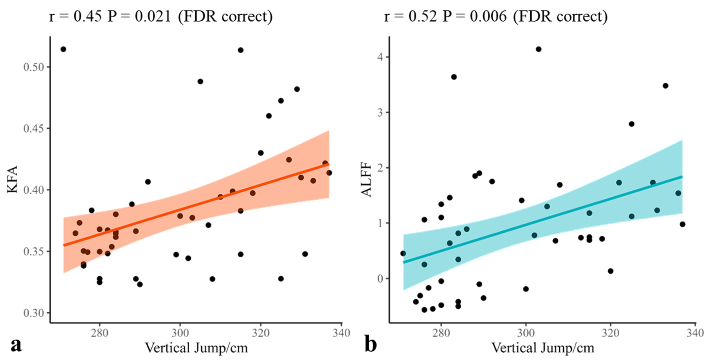
The correlations between specific brain measures and physical performance in basketball athletes were assessed using Spearman correlation analysis (Supplementary Table S3 for all results). Specifically, there was a positive correlation between the KFA value in the cerebellum and vertical jump score (r = 0.45, p = 0.021 FDR corrected, Figure 3). Additionally, the ALFF value in cerebellar Crus I showed positive correlations with vertical jump score (r = 0.52, p = 0.006, FDR-corrected, Figure 3). Furthermore, the ReHo value in the right cerebellar Crus II to lobule VII b area in basketball athletes was positively correlated with years of sport training (r = 0.30, p = 0.037 uncorrected) and years of basketball training (r = 0.31, p = 0.032 uncorrected). Lastly, no correlations were found between the RK within significantly altered brain regions and the basketball athletes’ training experience and physical abilities. Pearson correlation results were also presented in Table S4.

4. Discussion

This study examined cerebellar adaptations in basketball athletes using multimodal MRI. Compared with matched non-athletes, basketball athletes exhibited (i) greater GMV in the cerebellar Crus I, (ii) extensive microstructural alterations in both cerebellar cortex and peduncles, and (iii) enhanced functional activity in the Crus I/II regions. Significantly, some of these indices were positively correlated with vertical jump performance, a core basketball-specific motor ability. These findings suggest that basketball athletes exhibit multidimensional cerebellar enhancement and that such adaptations may partly be linked to their athletic performance.
In basketball athletes, gray matter enlargement was primarily observed in the cerebellar Crus I. An increase in gray matter volume usually indicates better functional performance. Functionally, Crus I has been consistently identified as a core hub of the cerebellum [35], mainly engaged in both higher-order cognitive processes [1,36] and motor functions [37]. The structural changes in this region may bring a faster and more effective cerebellum internal model for athletes and results in better motor and cognition performance. This inference aligns well with the integrated cognitive–motor demands of basketball, where athletes must perform precise movements while making rapid tactical decisions [20]. Such structural enhancement may therefore be functionally meaningful for athletes’ optimizing performance in highly dynamic game contexts.
Functional alterations were also observed in Crus I and Crus II. The increase in the ALFF signal in Crus I converges with the structural findings, reinforcing the idea that this region may represent a consistent site of cerebellar adaptation in basketball athletes. Crus II, functionally adjacent to Crus I, is similarly involved in higher-order cognitive processes that are closely linked to motor functions [1,38]. Enhanced regional coherence is associated with faster local information processes. The functional enhancement of this region may allow athlete form superior cerebellar function, thereby supporting the efficient integration of motor and cognitive performance [39,40]. The functional findings were highly consistent with the structural results. Cerebellar adaptations in athletes seemed to be themed on motor-cognitive integration, which directly mirrors the core requirements of basketball.
DKI analysis revealed extensive microstructural alterations in basketball athletes. At the white matter level, increased KFA and decreased RK generally indicate more ordered and efficient neural fiber structures [41,42,43]. These changes were predominantly located in the cerebellar peduncles, which constitute the only pathways through which the cerebellum communicates with other brain regions [44]. This finding suggests that the structural connectivity between the cerebellum and the broader brain network may be enhanced in basketball athletes. This enhancement provides an opportunity for athletes to perform a faster and more extensive information exchange between cerebellum and cerebral cortex. Such structural remodeling may underlie our previous observation of an enhanced cerebellar connection to relevant cerebral cortical areas during cognitive tasks [2,45].
At the cortical level, increased KFA and decreased RK imply the emergence of novel oriented microstructures [41,42,43], potentially reflecting processes such as the expansion of parallel fibers that enable faster and more efficient information processing. These cortical alterations were also concentrated around Crus I and lobule VI. Notably, lobule VI belongs to both motor and cognitive representations of the cerebellum [38], whose functions are highly relevant to the demands of basketball. The recurrent involvement of Crus I across structural, microstructural, and functional analyses further supports the notion that it is a core locus of adaptive changes in the cerebellum of basketball athletes.
A key finding of this study was that cerebellar KFA and ALFF values in basketball athletes were positively correlated with vertical jump performance—a sport-specific skill reflecting lower-limb power and intermuscular coordination. Although these associations were of moderate strength, this result is consistent with prior studies that suggest a potential link between physical abilities and improved neurophysiological function [21,46]. From a neural perspective, vertical jump performance may be modulated by cerebellar–cortical circuits regulation [6], whose functional activity has been associated with not only motor performance [3], but also with cognitive functions [39]. This raises the possibility that adaptive cerebellar changes could be related not only to motor performance (e.g., vertical jump) but also to cognitive abilities (e.g., in-game decision-making). Alternatively, these relationships may reflect broader improvements in neuromuscular efficiency or coordination rather than direct cerebellar modulation of power output. The correlations were not found in general physical fitness and training indexes, implying that cerebellar adaptations are likely more closely related to sport-specific demands than to general fitness or training duration, although this interpretation should be viewed with caution. Taken together, these findings tentatively suggest that cerebellar adaptations may contribute to the refinement of sport-specific skills. It will be important for future work to directly investigate the link between these cerebellar changes and specific task performance using longitudinal or task-based designs to better clarify their causal and functional significance.
Overall, basketball athletes demonstrated widespread cerebellar adaptations, encompassing structural, microstructural, and functional levels. These changes were centered on the Crus I, which not only modulates motor control but is also consistently recruited during cognitive tasks. Crus I may represent a consistent site of adaptation of cerebellar adaptations in basketball athletes, contributing to athletes’ advantages in both motor execution. While our behavioral assessments were limited to motor outcomes, it is plausible that cerebellar adaptations extend to cognitive domains such as working memory and decision-making, which are equally critical in basketball. Future studies incorporating cognitive tasks will be necessary to further test this possibility.
Several limitations warrant consideration. First, while DKI parameters suggest microstructural reorganization, the biological substrates of these changes (e.g., synaptic density vs. glial alterations) remain speculative and necessitate histopathological validation. Second, the cross-sectional design precludes causal inferences regarding training-induced plasticity; longitudinal or training-intervention designs that could track cerebellar plasticity over time are needed. Third, the relationship between cerebellar adaptation and specific sport performance remains incompletely understood. The functional implications of observed adaptations should be further tested using task-based fMRI paradigms and standardized cognitive-motor assessments to assess whether the same cerebellar regions support basketball-specific decision-making, not only physical performance. Lastly, a larger sample size is necessary to further validate the results of this study and compare them with other sports programs.

5. Conclusions

This multimodal study provides the first evidence that long-trained basketball athlete performed coordinated neural remodeling in the cerebellum, especially in Crus I. These findings not only delineate a map of cerebellar adaptation underlying exceptional athletic performance but, more profoundly, corroborate the cerebellum’s functional role as a hub for cognitive-motor integration. This work offers novel experimental insights and a theoretical framework for understanding experience-dependent plasticity in the human brain. These adaptations likely underpin the superior sensorimotor coordination, split-second decision-making, and kinematic precision demanded by elite basketball performance. Future work should explore whether cerebellar biomarkers could guide talent development or injury rehabilitation in sports medicine.

Supplementary Materials

The following supporting information can be downloaded at: https://www.mdpi.com/article/10.3390/brainsci15111221/s1, Table S1: Physical fitness test method; Table S2: The definitions of diffusion measures; Table S3: All results of correlation analysis; Table S4: All results of partial correlation analysis; Figure S1: Dribbling test diagram.

Author Contributions

Conceptualization and Methodology: X.D. and Y.Q. Formal analysis: Y.Q. and J.D. Investigation: Y.Q., Y.W. and X.L. Funding acquisition: X.D. Resources: W.F. and B.Z. Supervision: X.D. Writing—draft preparation: X.D. and Y.Q. Writing—review and editing: all authors. All authors have read and agreed to the published version of the manuscript.

Funding

This study was funded by the Social Sciences and Humanities project of the Ministry of Education, 23A10277004; National Natural Science Foundation of China, 81571658.

Institutional Review Board Statement

The study has complied with the Declaration of Helsinki (DoH)—Ethical Principles for Medical Research Involving Human Participants (1964) and its latest amendments adopted by the 75th General Assembly of the World Medical Association (WMA) in Finland on 19 October 2024. The protocol was approved by the Shanghai University of Sport Scientific Research Ethics Committee (Project No. 102772023RT052, 1 June 2023).

Informed Consent Statement

Informed consent was obtained from all subjects involved in the study.

Data Availability Statement

The datasets generated during and/or analyzed during the current study are available from the corresponding author upon reasonable request.

Acknowledgments

We extend our sincere gratitude to all individuals who participated in the research and to those who assisted in the data collection process.

Conflicts of Interest

The authors declare no conflicts of interest.

Abbreviations

The following abbreviations are used in this manuscript:
ADaxial diffusivity
AKaxial kurtosis
ALFFAmplitude of Low-Frequency Fluctuations
DCDegree Centrality
DKIDiffusion Kurtosis Imaging
DTIDiffusion Tensor Imaging
FAfractional anisotropy
fALFFfractional ALFF
GMVgray matter volume
KFAkurtosis fractional anisotropy
MDmean diffusivity
MKmean kurtosis
MRIMagnetic Resonance Imaging
RDradial diffusivity
ReHoRegional Homogeneity
RKradial kurtosis
TBSSTract-Based Spatial Statistics
TFCEThreshold-Free Cluster Enhancement

References

  1. King, M.; Hernandez-Castillo, C.R.; Poldrack, R.A.; Ivry, R.B.; Diedrichsen, J. Functional Boundaries in the Human Cerebellum Revealed by a Multi-Domain Task Battery. Nat. Neurosci. 2019, 22, 1371–1378. [Google Scholar] [CrossRef]
  2. Qi, Y.; Wang, Y.; Li, X.; Fang, W.; Du, X. Characteristics of Positive and Negative Networks in Working Memory Task of Basketball Athletes. Psychol. Sport Exerc. 2025, 80, 102880. [Google Scholar] [CrossRef]
  3. Warthen, K.G.; Walker, N.C.; Wicklund, B.D.; Gonzalez, M.M.; Ramirez, N.; Gee, S.C.; Al-Dasouqi, H.; Madore, M.R. Neuromodulation of the Cerebellum for Motor Applications: A Systematic Review. J. Integr. Neurosci. 2024, 23, 195. [Google Scholar] [CrossRef]
  4. Ito, M. Control of Mental Activities by Internal Models in the Cerebellum. Nat. Rev. Neurosci. 2008, 9, 304–313. [Google Scholar] [CrossRef]
  5. Koziol, L.F.; Budding, D.; Andreasen, N.; D’Arrigo, S.; Bulgheroni, S.; Imamizu, H.; Ito, M.; Manto, M.; Marvel, C.; Parker, K.; et al. Consensus Paper: The Cerebellum’s Role in Movement and Cognition. Cerebellum 2014, 13, 151–177. [Google Scholar] [CrossRef]
  6. Schmahmann, J.D.; Guell, X.; Stoodley, C.J.; Halko, M.A. The Theory and Neuroscience of Cerebellar Cognition. Annu. Rev. Neurosci. 2019, 42, 337–364. [Google Scholar] [CrossRef] [PubMed]
  7. Yao, Z.-F.; Fu, H.-L.; Liang, C.-W.; Li, Y.-J.; Wang, C.-H. Electrophysiological Differences in Inhibitory Control Processing between Collegiate Level Soccer Players and Non-Athletes in the Absence of Performance Differences. Brain Cogn. 2024, 178, 106179. [Google Scholar] [CrossRef] [PubMed]
  8. Carey, M.R. The Cerebellum. Curr. Biol. 2024, 34, R7–R11. [Google Scholar] [CrossRef] [PubMed]
  9. Wang, B.; LeBel, A.; D’Mello, A.M. Ignoring the Cerebellum Is Hindering Progress in Neuroscience. Trends Cogn. Sci. 2025, 29, 318–330. [Google Scholar] [CrossRef]
  10. Carrillo, J.; Cheng, S.-Y.; Ko, K.W.; Jones, T.A.; Nishiyama, H. The Long-Term Structural Plasticity of Cerebellar Parallel Fiber Axons and Its Modulation by Motor Learning. J. Neurosci. 2013, 33, 8301–8307. [Google Scholar] [CrossRef] [PubMed]
  11. Kang, N. Increased Cerebellar Gray Matter Volume in Athletes: A Voxel-Wise Coordinate-Based Meta-Analysis. Res. Q. Exerc. Sport 2022, 94, 597–608. [Google Scholar] [CrossRef] [PubMed]
  12. Lee, K.J.; Rhyu, I.J.; Pak, D.T.S. Synapses Need Coordination to Learn Motor Skills. Rev. Neurosci. 2014, 25, 223–230. [Google Scholar] [CrossRef] [PubMed]
  13. Zhang, K.; Jan, Y.-K.; Liu, Y.; Zhao, T.; Zhang, L.; Liu, R.; Liu, J.; Cao, C. Exercise Intensity and Brain Plasticity: What’s the Difference of Brain Structural and Functional Plasticity Characteristics Between Elite Aerobic and Anaerobic Athletes? Front. Hum. Neurosci. 2022, 16, 757522. [Google Scholar] [CrossRef]
  14. Cao, L.; Zhang, Y.; Huang, R.; Li, L.; Xia, F.; Zou, L.; Yu, Q.; Lin, J.; Herold, F.; Perrey, S.; et al. Structural and Functional Brain Signatures of Endurance Runners. Brain Struct. Funct. 2021, 226, 93–103. [Google Scholar] [CrossRef]
  15. Jang, D.C.; Shim, H.G.; Kim, S.J. Intrinsic Plasticity of Cerebellar Purkinje Cells Contributes to Motor Memory Consolidation. J. Neurosci. 2020, 40, 4145–4157. [Google Scholar] [CrossRef]
  16. Manto, M.; Huisman, T.A.G.M. The Cerebellum: Handbook of Clinical Neurology Series; Elsevier: San Diego, CA, USA, 2018; ISBN 978-0-444-63956-1. [Google Scholar]
  17. Li, W.; Kong, X.; Ma, J. Effects of Combat Sports on Cerebellar Function in Adolescents: A Resting-State fMRI Study. Br. J. Radiol. 2022, 95, 20210826. [Google Scholar] [CrossRef]
  18. Jalanko, P.; Säisänen, L.; Kallioniemi, E.; Könönen, M.; Lakka, T.A.; Määttä, S.; Haapala, E.A. Associations between Physical Fitness and Cerebellar Gray Matter Volume in Adolescents. Scand. J. Med. Sci. Sports 2024, 34, e14513. [Google Scholar] [CrossRef]
  19. Wenzel, U.; Taubert, M.; Ragert, P.; Krug, J.; Villringer, A. Functional and Structural Correlates of Motor Speed in the Cerebellar Anterior Lobe. PLoS ONE 2014, 9, e96871. [Google Scholar] [CrossRef]
  20. Terner, Z.; Franks, A. Modeling Player and Team Performance in Basketball. Annu. Rev. Stat. Its Appl. 2021, 8, 1–23. [Google Scholar] [CrossRef]
  21. Ishihara, T.; Miyazaki, A.; Tanaka, H.; Matsuda, T. Identification of the Brain Networks That Contribute to the Interaction between Physical Function and Working Memory: An fMRI Investigation with over 1,000 Healthy Adults. NeuroImage 2020, 221, 117152. [Google Scholar] [CrossRef] [PubMed]
  22. Reuben, D.B.; Magasi, S.; McCreath, H.E.; Bohannon, R.W.; Wang, Y.-C.; Bubela, D.J.; Rymer, W.Z.; Beaumont, J.; Rine, R.M.; Lai, J.-S.; et al. Motor Assessment Using the NIH Toolbox. Neurology 2013, 80, S65–S75. [Google Scholar] [CrossRef]
  23. Wen, N.; Dalbo, V.J.; Burgos, B.; Pyne, D.B.; Scanlan, A.T. Power Testing in Basketball: Current Practice and Future Recommendations. J. Strength Cond. Res. 2018, 32, 2677–2691. [Google Scholar] [CrossRef]
  24. Essentials of Strength Training and Conditioning, 4th ed.; Haff, G., Triplett, N.T., National Strength & Conditioning Association, Eds.; Human Kinetics: Champaign, IL, USA; Windsor, ON, Canada; Leeds, UK, 2015; ISBN 978-1-4925-0162-6. [Google Scholar]
  25. Diedrichsen, J.; Balsters, J.H.; Flavell, J.; Cussans, E.; Ramnani, N. A Probabilistic MR Atlas of the Human Cerebellum. NeuroImage 2009, 46, 39–46. [Google Scholar] [CrossRef]
  26. Yan, C.G.; Wang, X.D.; Zuo, X.N.; Zang, Y.F. DPABI: Data Processing & Analysis for (Resting-State) Brain Imaging. Neuroinformatics 2016, 14, 339–351. [Google Scholar] [CrossRef]
  27. Zou, Q.H.; Zhu, C.Z.; Yang, Y.; Zuo, X.N.; Long, X.Y.; Cao, Q.J.; Wang, Y.F.; Zang, Y.F. An Improved Approach to Detection of Amplitude of Low-Frequency Fluctuation (ALFF) for Resting-State fMRI: Fractional ALFF. J. Neurosci. Methods 2008, 172, 137–141. [Google Scholar] [CrossRef] [PubMed]
  28. Buckner, R.L.; Sepulcre, J.; Talukdar, T.; Krienen, F.M.; Liu, H.; Hedden, T.; Andrews-Hanna, J.R.; Sperling, R.A.; Johnson, K.A. Cortical Hubs Revealed by Intrinsic Functional Connectivity: Mapping, Assessment of Stability, and Relation to Alzheimer’s Disease. J. Neurosci. 2009, 29, 1860–1873. [Google Scholar] [CrossRef] [PubMed]
  29. Tournier, J.-D.; Smith, R.; Raffelt, D.; Tabbara, R.; Dhollander, T.; Pietsch, M.; Christiaens, D.; Jeurissen, B.; Yeh, C.-H.; Connelly, A. MRtrix3: A Fast, Flexible and Open Software Framework for Medical Image Processing and Visualisation. NeuroImage 2019, 202, 116137. [Google Scholar] [CrossRef]
  30. Cao, Z.; Lin, H.; Gao, F.; Shen, X.; Zhang, H.; Zhang, J.; Du, L.; Lai, C.; Ma, X.; Wu, D. Microstructural Alterations in Projection and Association Fibers in Neonatal Hypoxia–Ischemia. J. Magn. Reson. Imaging 2023, 57, 1131–1142. [Google Scholar] [CrossRef]
  31. Veraart, J.; Fieremans, E.; Novikov, D.S. Diffusion MRI Noise Mapping Using Random Matrix Theory. Magn. Reson. Med. 2016, 76, 1582–1593. [Google Scholar] [CrossRef] [PubMed]
  32. Kellner, E.; Dhital, B.; Kiselev, V.G.; Reisert, M. Gibbs-ringing Artifact Removal Based on Local Subvoxel-shifts. Magn. Reson. Med. 2016, 76, 1574–1581. [Google Scholar] [CrossRef]
  33. Tabesh, A.; Jensen, J.H.; Ardekani, B.A.; Helpern, J.A. Estimation of Tensors and Tensor-derived Measures in Diffusional Kurtosis Imaging. Magn. Reson. Med. 2011, 65, 823–836. [Google Scholar] [CrossRef]
  34. Van Baarsen, K.M.; Kleinnijenhuis, M.; Jbabdi, S.; Sotiropoulos, S.N.; Grotenhuis, J.A.; Van Cappellen Van Walsum, A.M. A Probabilistic Atlas of the Cerebellar White Matter. NeuroImage 2016, 124, 724–732. [Google Scholar] [CrossRef]
  35. Boonstra, J.T. The Cerebellar Connectome. Behav. Brain Res. 2025, 482, 115457. [Google Scholar] [CrossRef] [PubMed]
  36. Habas, C. Functional Connectivity of the Cognitive Cerebellum. Front. Syst. Neurosci. 2021, 15, 642225. [Google Scholar] [CrossRef]
  37. Henschke, J.U.; Pakan, J.M. Disynaptic Cerebrocerebellar Pathways Originating from Multiple Functionally Distinct Cortical Areas. eLife 2020, 9, e59148. [Google Scholar] [CrossRef]
  38. Guell, X.; Gabrieli, J.D.E.; Schmahmann, J.D. Triple Representation of Language, Working Memory, Social and Emotion Processing in the Cerebellum: Convergent Evidence from Task and Seed-Based Resting-State fMRI Analyses in a Single Large Cohort. NeuroImage 2018, 172, 437–449. [Google Scholar] [CrossRef]
  39. Clark, S.V.; Semmel, E.S.; Aleksonis, H.A.; Steinberg, S.N.; King, T.Z. Cerebellar-Subcortical-Cortical Systems as Modulators of Cognitive Functions. Neuropsychol. Rev. 2021, 31, 422–446. [Google Scholar] [CrossRef]
  40. Schmahmann, J.D. The Cerebellum and Cognition. Neurosci. Lett. 2019, 688, 62–75. [Google Scholar] [CrossRef] [PubMed]
  41. Hansen, B. An Introduction to Kurtosis Fractional Anisotropy. Am. J. Neuroradiol. 2019, 40, 1638–1641. [Google Scholar] [CrossRef] [PubMed]
  42. Glenn, G.R.; Helpern, J.A.; Tabesh, A.; Jensen, J.H. Quantitative Assessment of Diffusional Kurtosis Anisotropy: KURTOSIS ANISOTROPY. NMR Biomed. 2015, 28, 448–459. [Google Scholar] [CrossRef]
  43. Jensen, J.H.; Helpern, J.A.; Ramani, A.; Lu, H.; Kaczynski, K. Diffusional Kurtosis Imaging: The Quantification of Non-gaussian Water Diffusion by Means of Magnetic Resonance Imaging. Magn. Reson. Med. 2005, 53, 1432–1440. [Google Scholar] [CrossRef] [PubMed]
  44. De Benedictis, A.; Rossi-Espagnet, M.C.; De Palma, L.; Carai, A.; Marras, C.E. Networking of the Human Cerebellum: From Anatomo-Functional Development to Neurosurgical Implications. Front. Neurol. 2022, 13, 806298. [Google Scholar] [CrossRef] [PubMed]
  45. Fang, W.; Qi, Y.; Wang, Y.; Li, X.; Du, X.; Shao, B. Long-Term Basketball Training Shapes Cerebellar-Frontal Integration for Enhanced Cognitive Control. BMC Sports Sci. Med. Rehabil. 2025, 17, 253. [Google Scholar] [CrossRef]
  46. Meijer, A.; Königs, M.; Vermeulen, G.T.; Visscher, C.; Bosker, R.J.; Hartman, E.; Oosterlaan, J. The Effects of Physical Activity on Brain Structure and Neurophysiological Functioning in Children: A Systematic Review and Meta-Analysis. Dev. Cogn. Neurosci. 2020, 45, 100828. [Google Scholar] [CrossRef] [PubMed]
Figure 1. Significant differences in GMV, ALFF, and ReHo between basketball athletes and non-athletes (FDR-corrected p < 0.05). (a) Compared with non-athletes, the athletes showed significantly greater GMV in the bilateral cerebellar Crus I. (b) The athletes exhibited significantly higher ALFF values in the right cerebellar Crus I than non-athletes. (c) Compared with non-athletes, the athletes showed significant higher ReHo values in the right cerebellar crus II to lobule VII b. Red indicates higher values in athletes compared to the control group.
Brainsci 15 01221 g001
Figure 2. TBSS results indicated that KFA was higher and RK was lower in basketball athletes than non-athletes. Permutation test with 5000 iterations; p < 0.05 TFCE correction. The skeleton mask is shown in green. Areas of RK reduction are shown in blue. Areas of increased KFA are shown in red.
Brainsci 15 01221 g002
Figure 3. Individual KFA/ALFF values for the surviving clusters of basketball athletes were extracted for Spearman correlation with physical abilities. (a) The KFA value in the cerebellum was positively correlated with the vertical jump score. (b) The ALFF value in the cerebellar Crus I was positively correlated with the vertical jump scores. KFA: kurtosis fractional anisotropy, ALFF: Amplitude of Low-Frequency Fluctuations.
Brainsci 15 01221 g003
Table 1. Demographics and physical abilities of both groups.
Basketball AthletesNon-Athletesp
Gender (F, M)25, 3026, 290.847
Age (M ± SD)20.75 ± 1.8320.87 ± 1.980.727
Educated Years14.75 ± 1.8214.80 ± 1.670.870
Years of Sport Training9.25 ± 3.22--
Years of Basketball Training8.44 ± 2.69--
Training time (hours/week)7.21 ± 4.50--
Vertical Jump (cm)299.29 ± 23.36--
Dribbling (s)39.93 ± 2.19--
Shooting percentage (%)0.68 ± 0.14--
Agility (s)16.74 ± 2.4724.77 ± 5.92<0.001
Gait Speed (m/s)1.52 ± 0.441.51 ± 0.120.182
Explosive power (cm)33.79 ± 7.3933.04 ± 8.430.704
Table 2. MRI scanning parameters.
Imaging MethodsGeometry ParametersContrast Parameters
T1-weighted imagingFOV = 256 mm × 256 mm
Slice number = 192
Voxel size = 1 × 1 × 1 mm3
TR = 2530 ms
TE = 2.98 ms
TI = 1100 ms
flip angle = 7°
Diffusion-weighted imaging (DWI)FOV = 224 mm × 224 mm
Slice number = 74
Voxel size = 2 × 2 × 2 mm3
TR = 5000 ms
TE = 95 ms
b = 0, 1000, 2000 s/mm2
directions = 30
flip angle = 90°
BOLD weighted imagingFOV = 192 mm × 192 mm
Slice number = 58
interslice gap = 20%
Voxel size = 2 × 2 × 2 mm3
TR = 2000 ms,
TE = 30 ms,
flip angle = 90°
Field map for DWIFOV = 224 mm × 224 mm
Slice number = 74
Voxel size = 2 × 2 × 2 mm3
TR = 735 ms,
TE = 4.92 & 7.38 ms
flip angle = 90°
Field map for fMRIFOV = 192 mm × 192 mm
Slice number = 58
interslice gap = 20%
Voxel size = 2 × 2 × 2 mm3
TR = 571 ms,
TE = 4.92 & 7.38 ms
flip angle = 90°
FOV: field of view, TR: repetition time, TE: echo time, TI: inversion time.
Table 3. GMV differences between basketball athletes and non-athletes.
ClusterAreaSizeMNI (mm)Peak Tp
GMV 1Left Crus I79−23−73−354.210.023
GMV 2Right Crus I11328−73−333.990.012
GMV: gray matter volume.
Table 4. Resting-state functional brain activity differences between basketball athletes and non-athletes.
ClusterAreaSizeMNI (mm)Peak Tp
ALFF Right Crus I4836−80−313.870.002
ReHoRight Crus II and right VII b5822−68−413.650.038
ALFF: Amplitude of Low-Frequency Fluctuations, ReHo: Regional Homogeneity.
Table 5. DKI metrics difference between basketball athletes and non-athletes.
ClusterAreaSizeMNI (mm)Peak Tp
RK  475812−53−255.090.023
 White matter (CWMA)      
 Right Middle Cerebellar Peduncle592     
 Right Inferior Cerebellar Peduncle512     
 Left Inferior Cerebellar Peduncle424     
 Left Middle Cerebellar Peduncle320     
 Gray matter (SUIT)      
 Right Crus I746     
 Right VI452     
 Left VI419     
 Left Crus I301     
 Right Crus II174     
 Right V127     
 Right IX94     
 Vermis VI86     
 Vermis IX53     
KFA 137833−59−433.860.031
 White matter (CWMA)      
 Right Middle Cerebellar Peduncle389     
 Right Inferior Cerebellar Peduncle154     
 Gray matter (SUIT)      
 Right Crus I249     
 Right VI151     
 Left VI140     
 Left Crus I101     
 Right Crus II58     
DKI: Diffusion kurtosis Image, KFA: kurtosis fractional anisotropy, RK: radial kurtosis; CWMA: white matter atlas, SUIT: gay matter atlas.
Disclaimer/Publisher’s Note: The statements, opinions and data contained in all publications are solely those of the individual author(s) and contributor(s) and not of MDPI and/or the editor(s). MDPI and/or the editor(s) disclaim responsibility for any injury to people or property resulting from any ideas, methods, instructions or products referred to in the content.