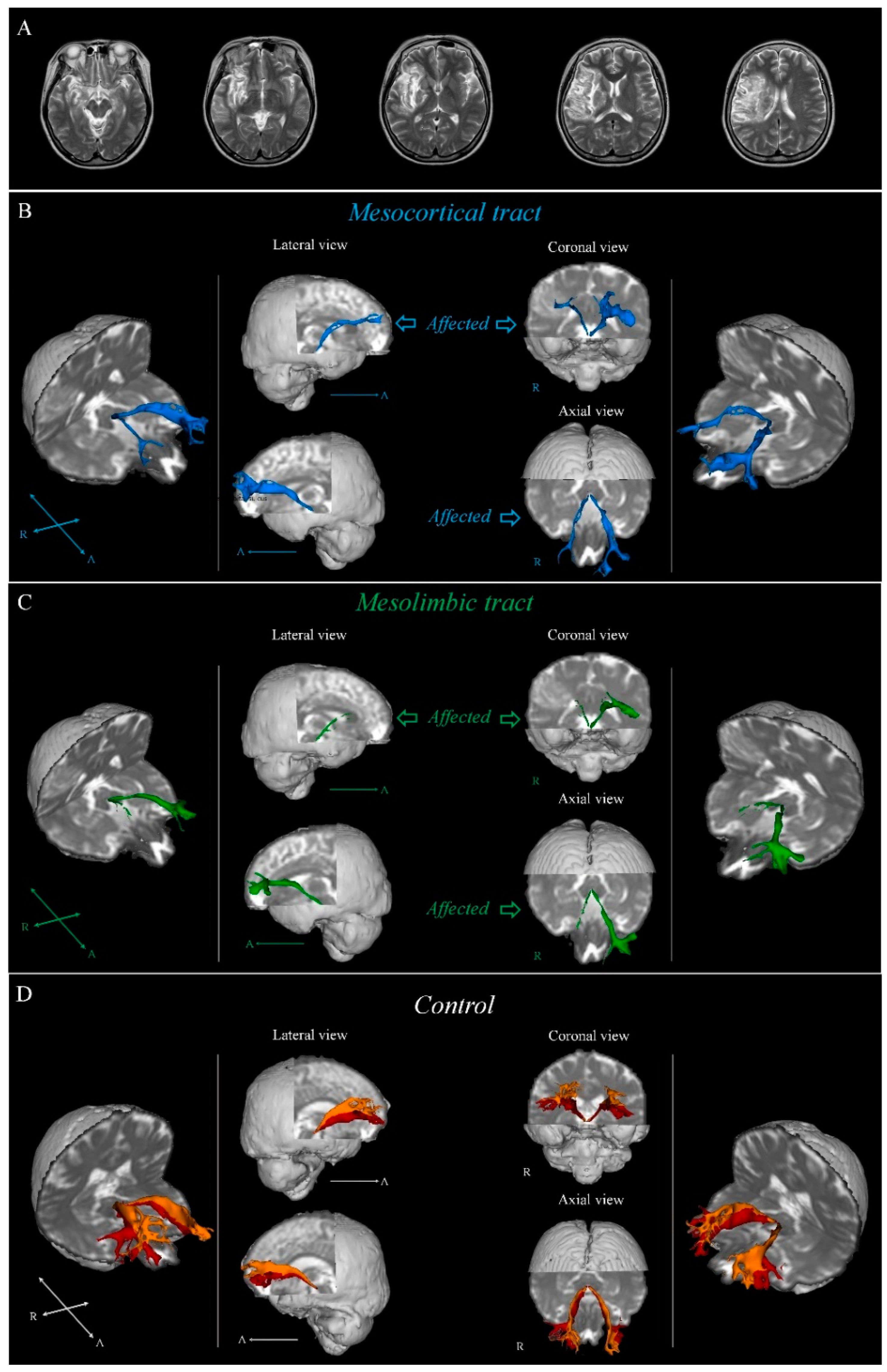
Neural Injury of the Dopaminergic Pathways in Patients with Middle Cerebral Artery Territory Infarct: A Diffusion Tensor Imaging Study
Abstract
:1. Introduction
2. Methods
2.1. Subjects
2.2. Data Acquisition
2.3. Fiber Tracking
2.4. Statistical Analysis
3. Results
4. Discussion
5. Conclusions
Author Contributions
Funding
Institutional Review Board Statement
Informed Consent Statement
Acknowledgments
Conflicts of Interest
Abbreviations
| VTA | Ventral tegmental area |
| NAc | Nucleus accumbens |
| MCT | Mesocortical tract |
| MLT | Mesolimbic tract |
| MCA | middle cerebral artery |
| FA | fractional anisotropy |
| MD | mean diffusivity |
| TV | Tract Volume |
| DTI | diffusion tensor imaging |
| DTT | diffusion tensor tractography |
| FMRIB | Functional Magnetic Resonance Imaging of the Brain |
References
- Lozano, R.; Naghavi, M.; Foreman, K.; Lim, S.; Shibuya, K.; Aboyans, V.; Abraham, J.; Adair, T.; Aggarwal, R.; Ahn, S. Global and regional mortality from 235 causes of death for 20 age groups in 1990 and 2010: A systematic analysis for the Global Burden of Disease Study 2010. Lancet 2012, 380, 2095–2128. [Google Scholar] [CrossRef] [PubMed]
- Murphy, S.L.; Kochanek, K.D.; Xu, J.; Arias, E. Mortality in the United States, 2020. 2021. Available online: https://stacks.cdc.gov/view/cdc/112079 (accessed on 21 April 2023).
- Murphy, T.H.; Corbett, D. Plasticity during stroke recovery: From synapse to behaviour. Nat. Rev. Neurosci. 2009, 10, 861–872. [Google Scholar] [CrossRef] [PubMed]
- Nogles, T.E.; Galuska, M.A. Middle Cerebral Artery Stroke. In StatPearls; StatPearls Publishing: Treasure Island, FL, USA, 2022. [Google Scholar]
- Dalley, A.F.; Agur, A. Moore’s Clinically Oriented Anatomy, 7th ed.; Lippincott Williams and Wilkins: Philadelphia, PA, USA, 2021. [Google Scholar]
- Corbetta, M.; Ramsey, L.; Callejas, A.; Baldassarre, A.; Hacker, C.D.; Siegel, J.S.; Astafiev, S.V.; Rengachary, J.; Zinn, K.; Lang, C.; et al. Common behavioral clusters and subcortical anatomy in stroke. Neuron 2015, 85, 927–941. [Google Scholar] [CrossRef] [PubMed] [Green Version]
- Ikemoto, S. Dopamine reward circuitry: Two projection systems from the ventral midbrain to the nucleus accumbens–olfactory tubercle complex. Brain Res. Rev. 2007, 56, 27–78. [Google Scholar] [CrossRef] [Green Version]
- Nestler, E.J.; Hyman, S.E.; Holtzman, D.M.; Malenka, R.C. Molecular Neuropharmacology: A Foundation for Clinical Neuroscience, 3rd ed.; McGraw-Hill Medical: New York, NY, USA, 2015. [Google Scholar]
- Meyer-Lindenberg, A.; Miletich, R.S.; Kohn, P.D.; Esposito, G.; Carson, R.E.; Quarantelli, M.; Weinberger, D.R.; Berman, K.F. Reduced prefrontal activity predicts exaggerated striatal dopaminergic function in schizophrenia. Nat. Neurosci. 2002, 5, 267–271. [Google Scholar] [CrossRef]
- Nestler, E.J.; Carlezon Jr, W.A. The mesolimbic dopamine reward circuit in depression. Biol. Psychiatry 2006, 59, 1151–1159. [Google Scholar] [CrossRef]
- Kronenberg, G.; Balkaya, M.; Prinz, V.; Gertz, K.; Ji, S.; Kirste, I.; Heuser, I.; Kampmann, B.; Hellmann-Regen, J.; Gass, P.; et al. Exofocal dopaminergic degeneration as antidepressant target in mouse model of poststroke depression. Biol. Psychiatry 2012, 72, 273–281. [Google Scholar] [CrossRef]
- Oestreich, L.K.; Wright, P.; O’Sullivan, M.J. Hyperconnectivity and altered interactions of a nucleus accumbens network in post-stroke depression. Brain Commun. 2022, 4, fcac281. [Google Scholar] [CrossRef]
- Supekar, K.; Kochalka, J.; Schaer, M.; Wakeman, H.; Qin, S.; Padmanabhan, A.; Menon, V. Deficits in mesolimbic reward pathway underlie social interaction impairments in children with autism. Brain 2018, 141, 2795–2805. [Google Scholar] [CrossRef] [Green Version]
- Behrens, T.E.; Berg, H.J.; Jbabdi, S.; Rushworth, M.F.; Woolrich, M.W. Probabilistic diffusion tractography with multiple fibre orientations: What can we gain? Neuroimage 2007, 34, 144–155. [Google Scholar] [CrossRef]
- Behrens, T.E.; Johansen-Berg, H.; Woolrich, M.; Smith, S.; Wheeler-Kingshott, C.; Boulby, P.; Barker, G.; Sillery, E.; Sheehan, K.; Ciccarelli, O.; et al. Non-invasive mapping of connections between human thalamus and cortex using diffusion imaging. Nat. Neurosci. 2003, 6, 750–757. [Google Scholar] [CrossRef]
- Kunimatsu, A.; Aoki, S.; Masutani, Y.; Abe, O.; Hayashi, N.; Mori, H.; Masumoto, T.; Ohtomo, K. The optimal trackability threshold of fractional anisotropy for diffusion tensor tractography of the corticospinal tract. Magn. Reson. Med. Sci. 2004, 3, 11–17. [Google Scholar] [CrossRef] [Green Version]
- Smith, S.M.; Jenkinson, M.; Woolrich, M.W.; Beckmann, C.F.; Behrens, T.E.; Johansen-Berg, H.; Bannister, P.R.; De Luca, M.; Drobnjak, I.; Flitney, D.E.; et al. Advances in functional and structural MR image analysis and implementation as FSL. Neuroimage 2004, 23, S208–S219. [Google Scholar] [CrossRef] [Green Version]
- Nakamura-Palacios, E.M.; Lopes, I.B.C.; Souza, R.A.; Klauss, J.; Batista, E.K.; Conti, C.L.; Moscon, J.A.; de Souza, R.S.M. Ventral medial prefrontal cortex (vmPFC) as a target of the dorsolateral prefrontal modulation by transcranial direct current stimulation (tDCS) in drug addiction. J. Neural Transm. 2016, 123, 1179–1194. [Google Scholar] [CrossRef]
- Assaf, Y.; Pasternak, O. Diffusion tensor imaging (DTI)-based white matter mapping in brain research: A review. J. Mol. Neurosci. 2008, 34, 51–61. [Google Scholar] [CrossRef]
- Neil, J.J. Diffusion imaging concepts for clinicians. J. Magn. Reson. Imaging 2008, 27, 1–7. [Google Scholar] [CrossRef]
- Pagani, E.; Agosta, F.; Rocca, M.A.; Caputo, D.; Filippi, M. Voxel-based analysis derived from fractional anisotropy images of white matter volume changes with aging. Neuroimage 2008, 41, 657–667. [Google Scholar] [CrossRef]
- Bennett, I.J.; Madden, D.J.; Vaidya, C.J.; Howard, D.V.; Howard, J.H., Jr. Age-related differences in multiple measures of white matter integrity: A diffusion tensor imaging study of healthy aging. Hum. Brain Mapp. 2010, 31, 378–390. [Google Scholar] [CrossRef] [Green Version]
- Inglese, M.; Ge, Y. Quantitative MRI: Hidden age-related changes in brain tissue. Top Magn. Reson. Imaging 2004, 15, 355–363. [Google Scholar] [CrossRef]
- Seo, J.P.; Koo, D.K. Degeneration of nigrostriatal pathway in patients with middle cerebral infarct: A diffusion tensor imaging study. Medicine 2023, 102, e33370. [Google Scholar] [CrossRef]
- Busto, R.; Dietrich, W.D.; Globus, M.Y.T.; Valdés, I.; Scheinberg, P.; Ginsberg, M.D. Small differences in intraischemic brain temperature critically determine the extent of ischemic neuronal injury. J. Cereb. Blood Flow Metab. 1987, 7, 729–738. [Google Scholar] [CrossRef] [PubMed] [Green Version]
- Gower, A.; Tiberi, M. The intersection of central dopamine system and stroke: Potential avenues aiming at enhancement of motor recovery. Front. Synaptic Neurosci. 2018, 10, 18. [Google Scholar] [CrossRef] [PubMed] [Green Version]
- Unger, E.L.; Wiesinger, J.A.; Hao, L.; Beard, J.L. Dopamine D2 receptor expression is altered by changes in cellular iron levels in PC12 cells and rat brain tissue. J. Nutr. 2008, 138, 2487–2494. [Google Scholar] [CrossRef] [PubMed] [Green Version]
- Bahk, J.Y.; Li, S.; Park, M.S.; Kim, M.O. Dopamine D1 and D2 receptor mRNA up-regulation in the caudate–putamen and nucleus accumbens of rat brains by smoking. Prog. Neuropsychopharmacol. Biol. Psychiatry 2002, 26, 1095–1104. [Google Scholar] [CrossRef]
- Kwak, S.Y.; Yeo, S.S.; Choi, B.Y.; Chang, C.H.; Jang, S.H. Corticospinal tract change in the unaffected hemisphere at the early stage of intracerebral hemorrhage: A diffusion tensor tractography study. Eur. Neurol. 2010, 63, 149–153. [Google Scholar] [CrossRef]
- Block, F.; Dihne, M.; Loos, M. Inflammation in areas of remote changes following focal brain lesion. Prog. Neurobiol. 2005, 75, 342–365. [Google Scholar] [CrossRef]
- Buss, A.; Pech, K.; Merkler, D.; Kakulas, B.A.; Martin, D.; Schoenen, J.; Noth, J.; Schwab, M.E.; Brook, G.A. Sequential loss of myelin proteins during Wallerian degeneration in the human spinal cord. Brain 2005, 128, 356–364. [Google Scholar] [CrossRef]
- Forno, L.S. Reaction of the substantia nigra to massive basal ganglia infarction. Acta Neuropathol. 1983, 62, 96–102. [Google Scholar] [CrossRef]
- Ogawa, T.; Yoshida, Y.; Okudera, T.; Noguchi, K.; Kado, H.; Uemura, K. Secondary thalamic degeneration after cerebral infarction in the middle cerebral artery distribution: Evaluation with MR imaging. Radiology 1997, 204, 255–262. [Google Scholar] [CrossRef]

| Control | Experimental | |
|---|---|---|
| Sex (male/female) | 25/15 | 20/16 |
| Mean age (year) | 55.65 (12.79) | 55.89 (8.80) |
| Lesion side (right/left) | - | 21/15 |
| Onset duration (days) | - | 21.4615 (1.7791) |
| Condition | Mean | F | p | Post-Hoc | ||
|---|---|---|---|---|---|---|
| MCT | FA | Control | 0.3881 (0.0490) | 5.791 | 0.004 * | C > U, A |
| Unaffected side | 0.3883 (0.0314) | U > C, A | ||||
| Affected side | 0.3573 (0.0569) | C, U > A | ||||
| TV | Control | 520.81 (357.37) | 17.019 | <0.001 * | C > U, A | |
| Unaffected side | 411.78 (374.44) | U > C, A | ||||
| Affected side | 139.50 (152.69) | C, U > A | ||||
| MLT | FA | Control | 0.3822 (0.0411) | 4.474 | 0.013 * | C > U, A |
| Unaffected side | 0.3867 (0.0469) | U > C, A | ||||
| Affected side | 0.3580 (0.0529) | C, U > A | ||||
| TV | Control | 556.74 (349.46) | 32.379 | <0.001 * | C > U, A | |
| Unaffected side | 289.64 (232.62) | C > U > A | ||||
| Affected side | 118.03 (123.85) | C, U > A | ||||
Disclaimer/Publisher’s Note: The statements, opinions and data contained in all publications are solely those of the individual author(s) and contributor(s) and not of MDPI and/or the editor(s). MDPI and/or the editor(s) disclaim responsibility for any injury to people or property resulting from any ideas, methods, instructions or products referred to in the content. |
© 2023 by the authors. Licensee MDPI, Basel, Switzerland. This article is an open access article distributed under the terms and conditions of the Creative Commons Attribution (CC BY) license (https://creativecommons.org/licenses/by/4.0/).
Share and Cite
Seo, J.P.; Ryu, H.J. Neural Injury of the Dopaminergic Pathways in Patients with Middle Cerebral Artery Territory Infarct: A Diffusion Tensor Imaging Study. Brain Sci. 2023, 13, 927. https://doi.org/10.3390/brainsci13060927
Seo JP, Ryu HJ. Neural Injury of the Dopaminergic Pathways in Patients with Middle Cerebral Artery Territory Infarct: A Diffusion Tensor Imaging Study. Brain Sciences. 2023; 13(6):927. https://doi.org/10.3390/brainsci13060927
Chicago/Turabian StyleSeo, Jeong Pyo, and Heun Jae Ryu. 2023. "Neural Injury of the Dopaminergic Pathways in Patients with Middle Cerebral Artery Territory Infarct: A Diffusion Tensor Imaging Study" Brain Sciences 13, no. 6: 927. https://doi.org/10.3390/brainsci13060927
APA StyleSeo, J. P., & Ryu, H. J. (2023). Neural Injury of the Dopaminergic Pathways in Patients with Middle Cerebral Artery Territory Infarct: A Diffusion Tensor Imaging Study. Brain Sciences, 13(6), 927. https://doi.org/10.3390/brainsci13060927


