Article

Tolerability and Effectiveness of Cathodal Transcranial Direct Current Stimulation in Children with Refractory Epilepsy: A Case Series

by
Soumya Ghosh
1,2,* and
Lakshmi Nagarajan
1,3
1
Children’s Neuroscience Service, Department of Neurology, Perth Children’s Hospital, Nedlands, WA 6009, Australia
2
Perron Institute for Neurological and Translational Science, University of Western Australia, Nedlands, WA 6009, Australia
3
School of Medicine, University of Western Australia, Nedlands, WA 6009, Australia
*
Author to whom correspondence should be addressed.
Brain Sci. 2023, 13(5), 760; https://doi.org/10.3390/brainsci13050760
Submission received: 23 April 2023 / Accepted: 29 April 2023 / Published: 4 May 2023
(This article belongs to the Section Developmental Neuroscience)

Abstract

:
There are limited treatment options for drug-resistant epilepsy (DRE) in children. We performed a pilot study to investigate the tolerability and effectiveness of cathodal transcranial direct current stimulation (tDCS) in DRE. Twelve children with DRE of varied etiology underwent three to four daily sessions of cathodal tDCS. The seizure frequency at 2 weeks before and after tDCS was obtained from seizure diaries; clinic reviews at 3 and 6 months assessed any longer-term benefits or adverse effects. The spike wave index (SWI) was analyzed in the EEGs done immediately before and after tDCS on the first and last day of tDCS. One child remained seizure free for a year after tDCS. One child had reduced frequency of ICU admissions for status epilepticus for 2 weeks, likely due to reduced severity of seizures. In four children, an improvement in alertness and mood was reported for 2–4 weeks after tDCS. There was no benefit following tDCS in the other children. There were no unexpected or serious adverse effects in any child. Benefit was seen in two children, and the reasons for the lack of benefit in the other children need further study. It is likely that tDCS stimulus parameters will need to be tailored for different epilepsy syndromes and etiologies.

1. Introduction

Up to 30% of adults and children with epilepsy have drug-resistant epilepsy (DRE) [1,2]. For those who are not candidates for surgical treatments, therapeutic options are often limited (e.g., ketogenic diet and vagal nerve stimulation) and may not achieve seizure freedom [3,4,5,6,7]. Non-invasive brain stimulation (NIBS) techniques such as tDCS are being evaluated for the treatment of epilepsy [8,9], and cathodal tDCS has been reported to reduce seizure frequency in adults and children with epilepsy [10,11,12,13,14,15,16,17,18,19,20,21,22,23,24,25,26,27]. No serious adverse effects have been reported after therapeutic NIBS for epilepsy [9,28].
Most studies have reported some improvement in seizures after tDCS in adults and children with focal epilepsy [11,12,14,16,17,21,25,26,27]. Positive results (a decrease in seizure frequency) were also reported in those with Lennox–Gastaut syndrome (LGS) [13], Rasmussen’s encephalitis [16,21], epilepsia partialis continua [29], epileptic spasms [10], and Ohtahara syndrome [19]. Only rare studies have reported the failure of tDCS to improve seizure frequency [30,31]. Follow-up has been relatively short, with most successful trials reporting improved seizure frequency over 1 month [11,12,13,14,17,25,32] and some at 2–6 months [20,21,27]. It is not clear if the improvement of seizure frequency is longer lasting and associated with improved function or quality of life.
Successful trials used a wide range of stimulus parameters. One to fourteen daily sessions were used [12,13,14,17,30,32], stimulus currents were from 1 to 2 mA, and the stimulus duration was 20–40 min for each session [12,13,17,25,32]. A distinctive tDCS protocol was used by a few studies with a session consisting of two bouts of 9–20 min separated by 20 min [11,14,27,33]. Two studies used two or three cycles of tDCS (5–6 days of tDCS in each cycle repeated every 1–2 months) over 2–6 months [26,27]. Most studies have positioned the cathode over the region with the most intense EEG epileptiform activity [12,14,16,17,30,32]. This was found to be more effective than other areas of stimulation [10]. Two studies used customized montages of up to eight electrodes (multichannel tDCS) designed to simultaneously apply maximal cathodal stimulation over the seizure focus using modelling based on stereo EEG or surface EEG [25,27]. Anodal positions have varied in different studies and included the contralateral supra-orbital region, contralateral shoulder, and contralateral hemisphere [11,12,14,17,21]. The two trials reporting negative results used high-density tDCS (HD-tDCS) [30,31], although another study reported a benefit with HD-tDCS [29]. Thus, the relationship between the stimulus parameters and the magnitude and duration of the benefit is not clear [9].
There are fewer studies in children. In a randomized trial, Auvichayapat et al. [12] found a marginal but significant decrease in seizure frequency after a single tDCS session of 1 mA cathodal stimulation 20 min long in children with DRE. In children with LGS, tDCS (2 mA for 20 min) over five consecutive days was found to reduce seizure frequency up to 4 weeks [13]. Shelyakin et al. [18] reported a marked decrease in seizure frequency up to 6 months after tDCS (one to 15 sessions, 0.3–0.7 mA for 20–40 min) in children with cerebral palsy and epilepsy. Yang et al. [10] found tDCS to be more effective in children (than adults) with epileptic spasms. Yook et al. [15] reported a reduction in seizure frequency for 2 months in a child with cortical dysplasia after 10 sessions of tDCS. Two studies using multichannel tDCS showed an improvement of seizure frequency for up to 6 months in children and adults with refractory focal epilepsy [25,27].
Due to the variability of tDCS efficacy and parameters of stimulation in previous studies and the lack of clarity in the optimal parameters, especially in children, we undertook a pilot study to assess the tolerability and improvement in seizure burden and quality of life in children with DRE to guide in planning a controlled trial. Changes in the interictal epileptiform discharges were also evaluated by recording video EEG (vEEG) immediately before and after tDCS.

2. Material and Methods

An open label case study was performed on 12 children, aged 3 to 17 years, with DRE. The study was conducted according to the guidelines of the Declaration of Helsinki, approved by the Child and Adolescent Health Service Human Research Ethics Committee (Approval No. RGS0000002430) and registered with the Australian New Zealand Clinical Trials Registry (ACTRN12618001821280). The children were recruited from the neurology clinics of Perth Children’s Hospital and Princess Margaret Hospital for Children. Written informed consent was obtained from their parents or guardians (and assent from the children when appropriate). The inclusion criteria were children older than 2 years with DRE. DRE was defined as a failure of adequate trials of two or more tolerated and appropriately chosen and used antiepileptic drug schedules (whether as monotherapies or in combination) to achieve sustained seizure freedom [34]. The exclusion criteria were children less than 2 years, those with skin inflammation/allergies affecting the scalp, children in whom EEG could not be undertaken, and children with significant neuro-behavioral disorders.

2.1. tDCS

All patients received active tDCS treatment. Previous studies have shown benefits with many different stimulation protocols. We chose a simple protocol (for logistical reasons and its applicability in different clinical settings and varying resources) that was similar to that used by several previous successful trials [12,13,14,18]. The treatment consisted of three or four consecutive days of cathodal tDCS delivered using a NeuroConn DC stimulator MC (neuroConn Gmbh, Ilmenau, Germany). Although a 4-day course of tDCS was initially planned, it was not always possible due to the availability of children and EEG/tDCS facilities during any given week. Square surface electrodes (25 cm2) coated with EEG electrode gel were used for tDCS. The cathodes were applied over one or two EEG sites (international 10–20 system) with the most frequent epileptiform activity seen in ictal and interictal EEG recordings (Figure 1) based on assessments made by a child neurologist experienced in reading children’s EEGs. Anodes were positioned over the ipsilateral or contralateral supra-orbital region of the forehead depending on comfort and accessibility during EEG recording. Stimulation intensity and duration were 0.8 mA for younger children (<10 years) or 1 mA for older children (0.32–0.4 A/m2) for 20 min. However, two of the children aged 12 and 13 years received 0.8 mA of tDCS, instead of 1 mA of tDCS as planned. All children had video EEGs for 20 min before starting tDCS, during tDCS, and immediately after tDCS, on the first and last day of tDCS. No changes were made to antiseizure medications (ASMs) throughout the study. A tDCS questionnaire (see attachment 1) was completed by the child (or parent) before and after each session of tDCS.

2.2. Outcome Measures

Parents were asked to complete a Quality of Life in Childhood Epilepsy Questionnaire [35] (QOLCE, see attachment 2) before starting tDCS [35]. Parents were requested to keep a seizure diary (see attachment 3) 2 weeks prior to, during, and 2 weeks after tDCS. A neurological review was undertaken at 3 months and 6 months after tDCS, which included a structured interview (see attachment 4) of the parents and children to document the overall impression of changes in seizure frequency and severity, quality of life, behavior, motor and verbal performance, and sleep after tDCS.

2.3. EEG

Video EEG (vEEG) was recorded digitally, using the Compumedics equipment and Profusion software (Compumedics Limited, Victoria, Australia). Electrodes were applied in accordance with the International 10–20 system. Assessments of previous vEEG recordings (standard one-hour daytime recordings and prolonged 1- to 4-day vEEG studies in the epilepsy monitoring unit) by an epileptologist (LN) experienced in reading EEGs were used to determine the most active focus of the epileptiform activity (interictal and ictal). This was used to determine the application of the cathodal electrode over one or two locations. Two locations were chosen for children who had more than one focus of frequent epileptiform discharges on EEG.
vEEGs were recorded on the first and last day of tDCS (20 min before, during, and 20 min after tDCS) (Figure 1). The EEGs were reviewed for qualitative assessment (background, state, and epileptiform activity) and quantitative analysis of spikes (spike wave index, SWI) by neurologists (LN and SG) experienced in reading children’s EEGs. The SWI was calculated by observing spikes in the derivation with the most frequent spike activity in 15 s epochs of EEG. Epochs with and without spikes were counted. The SWI was equal to the number of epochs with spikes divided by the total number of epochs reviewed (SWI = number of epochs with spikes/number of epochs with spikes + number of epochs without spikes) and expressed as a percentage. SWI% was calculated separately for the awake/drowsy and sleep states, in the EEG before and after tDCS. There was some artefact in the EEG recorded during tDCS making the SWI assessment unreliable.

2.4. Statistical Analysis

The statistical analysis was performed only on SWI% measures in EEG before and after tDCS on the first day using SPSS software (SPSSv20 for Windows, SPSS Inc, Chicago, IL, USA). The Shapiro–Wilk test was applied to check for normality of the data. The statistical analysis was performed on SWI% before and after tDCS on the first day using a non-parametric test (Wilcoxon signed-rank test). The significance was set to <0.05. Statistical testing was not performed to compare the SWI% on the first and last day of tDCS due to the difference in the number of tDCS days between children.

3. Results

The clinical details of the 12 children in the study (six boys and six girls) are described in Table 1. All children had DRE. Their ages varied from 3 to 17 years, and most had seizure onset within the first 2.5 years of life. Most children (9/12) were developmentally delayed, and two were severely disabled (cases 4 and 9). The majority had focal or multifocal epilepsy with varying etiology including cortical dysplasia, post-infection, post-surgical scarring, and genetic cause. In two children, the diagnosis was Rassmussen’s encephalitis, and no cause had been found in two others. Most had frequent daily seizures, and seizure types included focal seizures, focal seizures with secondary generalization, and generalized seizures. Two children (cases 4 and 7) required frequent hospital and ICU admissions for convulsive status epilepticus (CSE). All children had been trialed on multiple ASMs (6–14), and most had tried one or more other interventions. These included steroids, folinic acid, pyridoxine, biotin, intravenous immunoglobulin (IVIG), ketogenic diet, and vagal nerve stimulation (VNS). Two children had undergone epilepsy surgery (cases 7 and 12), and another had surgery for craniosynostosis (case 11). The overall quality of life (question 9.1 on the QOLCE) was reported as good by the parent(s) of five children, fair for three, and poor for four.

3.1. Stimulus Parameters

The stimulus parameters and clinical outcomes (seizure frequency and clinic reviews) are detailed in Table 2. All children were on one or more ASMs at the time of tDCS (range one to five, mean 2.75). Cathodes were positioned over one or two of the EEG coordinates that showed the most frequent epileptiform discharges. Anodes were located over the ipsilateral or contralateral (and midline) supraorbital regions.
One child (case 7) had a seizure before the first day of tDCS and a seizure before and after the last day of tDCS (based on the vEEG recording). This was not unexpected as he had been having innumerable daily seizures at that time, and the seizure semiology was the same. None of the other children had any significant adverse effects during or after tDCS. Four children had mild irritation or itchiness during the stimulation, three reported a mild transient headache after the stimulation, and one child said he felt slightly wobbly on his feet for a few minutes after the stimulation.

3.2. Outcomes

Outcomes are detailed in Table 2. One child (case 12) remained seizure free for a year after tDCS, but seizures returned after that. One child (case 4) had frequent admissions to the ICU due to convulsive status epilepticus (CSE) prior to tDCS. Convulsive status epilepticus is defined as a generalized tonic–clonic convulsion lasting more than 30 min or repeated tonic–clonic convulsions over a 30 min period without the recovery of consciousness between each convulsion. She had fewer days of admission in the ICU for CSE for 2 weeks after tDCS, probably due to the reduced severity of CSE. As a result of this unexpected benefit, she received repeated monthly tDCS for 2 months (on a compassionate basis) after the first treatment, resulting in continuing benefits (reduced frequency of CSE) for 6 months. Based on her medical records, she spent 21 days in the ICU 6 months prior to tDCS and 8 days for the next 6 months after tDCS. Parents of four children (cases 1, 3, 7, and 12) reported a transient improvement in alertness and mood for 2–4 weeks. There was no benefit in the other children. At the clinic review at 3 and 6 months, parents reported improved QOL in two children (cases 4 and 12). There was no obvious difference in the outcomes between those that received 3 or 4 days of tDCS. None of the children (or their parents) reported any worsening of seizure frequency or severity after tDCS, nor did they report any other adverse effects.

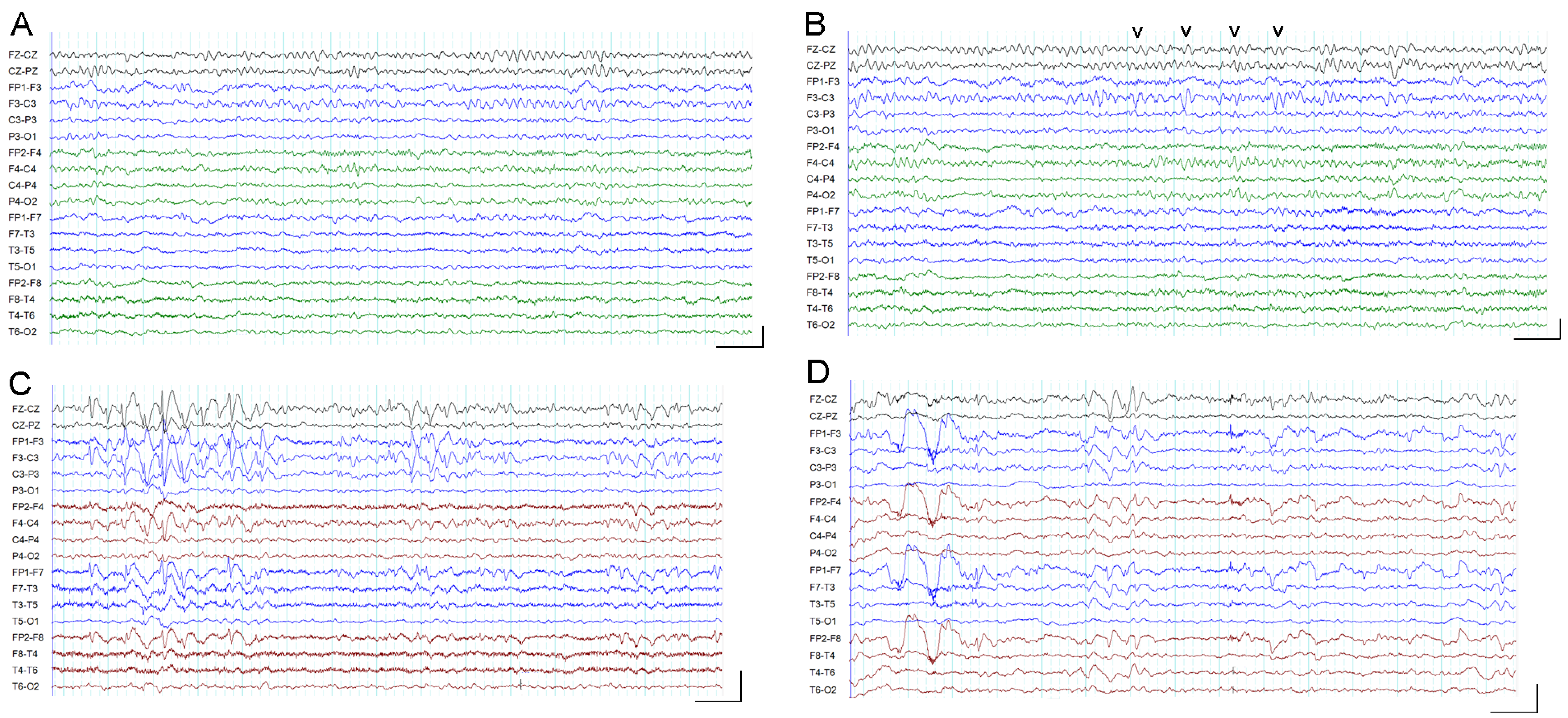
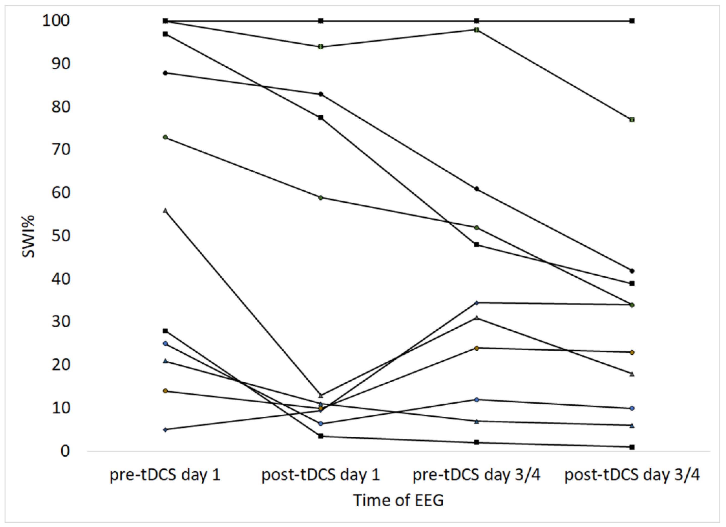
3.3. EEG and Spike Wave Index

The spike wave index (expressed as a percentage, SWI%) was calculated in the EEG before and after tDCS, and the results are shown in Table 3. Most of the recordings were in the wake state, and further analysis was performed for SWI% values in the wake recording (Figure 1A,B). Figure 2 shows line graphs of the values at different times, and there was a clear trend for reduction in SWI% in most children from before to after tDCS on the first and last day. In a few children (cases 2, 4, 5, 11, and 12), there was reduction in SWI% from the post-tDCS value on the first day to the pre-tDCS value on the third or fourth day. In the three children with SWI% values of 100% before and after tDCS, a reduction in the spike density was observed after tDCS (on visual analysis) but was not reflected in the SWI% values due to the high spike burden in the EEG (see Figure 1C,D).
Non-parametric statistical analysis of the SWI data before and after tDCS on the first day showed a significant reduction in the SWI% after tDCS (Wilcoxon signed-rank test, Z = −2.599, p = 0.009).
An increase in the background β activity was noted in the EEGs after tDCS in most children, and in one child, there was less asymmetry after tDCS (Table 3).

4. Discussion

Many studies have explored the use of NIBS for the treatment of seizures in patients with DRE [8,9,28]. Cathodal tDCS and low-frequency repetitive Transcranial magnetic stimulation (LF-rTMS) have been used to reduce cortical excitability underlying the stimulating electrodes [8,28,36]. The neural mechanisms by which TMS and tDCS affect cortical excitability are different; while TMS causes depolarization of neurons in the underlying cortex and can elicit action potentials, tDCS causes subthreshold depolarization or hyperpolarization of neurons [8,28]. The long-term effects of rTMS and tDCS are believed to be mediated by neuroplastic mechanisms, by modulating long-term potentiation (LTP) and long-term depression (LTD) like phenomena [8,28].
Most studies using cathodal tDCS have shown benefits in adults and children with focal epilepsy. However, tDCS treatment in our study showed benefits in only two children, even though our treatment parameters were similar to those in many previous studies.
There are many limitations of the study. This was a pilot study to help plan a controlled trial and therefore did not include a placebo/sham arm. Many previous investigators have performed pilot studies to evaluate the tolerability, electrophysiological markers, and effectiveness of tDCS to optimize the design of subsequent controlled trials [8,26,27]. The sample size was small but similar to previous open label trials and based on sample size calculations for pilot studies [8,25,26,27,37]. We included children with varying ages and durations of seizures to assess the safety and tolerability in a wide variety of epilepsies. Many of the children had innumerable seizures every day, making accurate counts of seizures difficult. The clinical profile of the children varied considerably, as did the severity and frequency of seizures. In some children, the frequency, severity, and types of seizures varied considerably from week to week and month to month, making it difficult to establish a clear baseline over short periods. We tried to overcome this by reviewing them in the clinic at 3 and 6 months to confirm changes, if any, were reliably present over a longer duration.
In our study, there was seizure freedom in one child for a year after tDCS (case 12), and one child (case 4) had fewer ICU admissions for CSE for 2 weeks after tDCS, probably due to the reduced severity of seizures. The only child to have a long-term benefit in our study also had the lowest frequency of seizures at baseline. There was no meaningful benefit for the other children, although parents reported transient improvements in alertness and mood in three of them. This result most closely matches that of Auvichayapat et al. [12], who found a marginal but significant decrease in seizure frequency after a single tDCS session. The increase in the number of tDCS sessions in our study did not appear to improve the outcome. We observed a significant reduction in the EEG epileptiform discharges after tDCS that was also reported by them. Other studies [15,18,19] have reported better outcomes with treatment protocols similar to ours. For example, Auvichayapat et al. [13] found a 90% decrease in seizure frequency for over a month in children with Lennox–Gastaut syndrome after five sessions of 2 mA tDCS, Shelyakin et al. [18] found a decrease in seizure frequency for up to 6 months following one to 15 sessions of 0.3–0.7 mA cathodal tDCS, and Fregni et al. [32] showed a 44% reduction in seizure frequency for 1 month following one session of 1 mA cathodal tDCS. There are several reasons why we may have been unable to show similar benefit. Many of the children in our study had multiple seizure types that appeared to be less responsive to tDCS [10]. tDCS may be less effective in children with epileptic encephalopathies [31]. The high seizure burden in some of the children (innumerable seizures per day) may have made a modest reduction unverifiable. Most children were on multiple ASMs, which may have interacted with tDCS. Other successful studies have used multichannel tDCS (based on modelling), more tDCS sessions, or repeated cycles of tDCS, which may be more effective in controlling seizures [8,10,25,26,27].
The pilot study also suggested ways in which we may be able to overcome some of these limitations in future trials. We plan to use a controlled study design to overcome the limitations including the variability in seizure frequency over days, weeks, and months. We plan to use a crossover design so that each patient acts as his or her control. We plan to use other scoring tools to look for a reduction in epilepsy burden apart from seizure frequency and guided by the seizure patterns (for example, the frequency of the use of rescue medications for prolonged seizures, the number of school days missed due to seizures, the number of injuries sustained after convulsive seizures, and the number of seizure-related admissions to hospital). We plan to recruit children with DRE who have a homogenous clinical profile in a single study (for example, children with normal development and focal epilepsy or children with a specific type of developmental and epileptic encephalopathy). Protocols need to be more carefully planned (based on the availability of resources and patient convenience) so that there is better adherence to stimulation parameters (for example, intensity, duration, and location) and outcome measures. Other features of EEG apart from epileptiform discharges (for example, background changes and changes in functional connectivity) may be useful in understanding tDCS effects and outcomes. It would be useful to trial newer tDCS protocols that may have a longer effect such as a single session consisting of two bouts of 9–20 min separated by 20 min [11,14,27,33]).
The effects of tDCS on epilepsy surrogate markers have been commonly obtained from the frequency of EEG epileptiform discharges. Our study showed an immediate reduction in EEG epileptiform discharges after tDCS. Some studies have reported reductions in EEG epileptiform discharges for up to 4 weeks after a single or multiple tDCS sessions [12,13,32]. Other studies have found no changes in EEG epileptiform discharges after tDCS [17,21,26]. A reduction in epileptiform activity has not been uniformly related to a reduction in seizure frequency, as was also seen in our study. It is possible that the improvements in EEG epileptiform discharges in our study were transient and therefore not beneficial in changing seizure outcomes. Changes in background EEG after tDCS were observed in this study and have been reported in previous studies [18]. We found an increase in background β activity in 10 of the 12 children. The amount of beta activity is thought to reflect increased vigilance and alertness [38]. The effects of background EEG changes following tDCS have not been explored. Other studies have evaluated functional connectivity in different frequency bands (alpha, beta, and theta bands) and related reductions in connectivity to seizure reduction after tDCS [26,27].
The few studies that have assessed QOL did not find any improvement after tDCS despite the reduction in seizure frequency [11,12]. In our study, four children were observed to be more alert and happier after the tDCS for a short period, but an improvement in QOL was reported in only two children (cases 4 and 12). None of the children in our study reported any serious adverse symptoms after tDCS, and it appears to be safe and well tolerated [9,39]. Although a seizure was observed after tDCS in one child in our study, this likely reflected the seizure burden at that time. Seizures have been reported by others during or after tDCS in children with frequent seizures [11,21].
There are some limitations of the therapeutic uses of tDCS. Although there are demonstrable changes in cortical excitability for up to an hour after a single session of tDCS, the effects of repeated stimulation over days and weeks are less well understood [40,41]. The effects of tDCS (i.e., excitatory or inhibitory) may be affected by the polarity and intensity of stimulation [42]. Although adverse effects are usually mild, seizures have been rarely reported to occur during tDCS, and there may be worsening of seizure frequency in a few children after tDCS [11,21,25]. A potential limitation of tDCS is its limited spatial accuracy. As the current passes through the brain from the cathode to the anode and modulates neural activity simultaneously underneath the cathode and the anode, it can be difficult to relate the effects of tDCS to a specific brain region. Thus, tDCS not only affects the brain regions directly under the electrodes but may also modulate functional connectivity between remote but functionally associated brain areas or influence the within-network connectivity [43].

5. Conclusions

This open label pilot study suggests that the role of tDCS in DRE in childhood, particularly in those with a high seizure burden, remains unclear. tDCS is well tolerated and appeared to make some children (30%) happier and more alert for a brief period. Impacts on seizure frequency or severity were seen in two (17%); one child became seizure free for a year, and another had fewer ICU admissions for 2 weeks. There was a significant reduction in SWI between the pre-tDCS and post-tDCS EEGs on the first and last day of tDCS. This reduction did not lead to the reduction in seizure frequency, as has been reported before [12]. Our study adds to the literature that suggests tDCS may have a promising role in DRE, but further study is needed to find the optimal stimulus paradigm for the different epilepsy etiologies and electroclinical syndromes.

Author Contributions

Both S.G. and L.N. were involved in conceptualization, methodology, software, validation, formal analysis, investigation, resources, data curation, visualization, project administration, and funding acquisition; S.G. wrote the original draft; and L.N. and S.G. were involved in review and editing. All authors have read and agreed to the published version of the manuscript.

Funding

This research received no external funding.

Institutional Review Board Statement

The study was conducted according to the guidelines of the Declaration of Helsinki, approved by the Child and Adolescent Health Service Human Research Ethics Committee (Approval No. RGS0000002430), and registered with the Australian New Zealand Clinical Trials Registry (ACTRN12618001821280).

Informed Consent Statement

The children were recruited from the neurology clinics of Perth Children’s Hospital and Princess Margaret Hospital for Children. Written informed consent was obtained from their parents or guardians (and assent from the children when appropriate).

Data Availability Statement

No other data are available due to patient privacy.

Acknowledgments

We thank Princess Margaret Hospital Foundation for the donation of the tDCS equipment used in the study. We thank the children and their parents for participating in the study. We thank the neuroscientists Linda Palumbo, Christina Mitrovic, and Emma Sivwright for helping with the study.

Conflicts of Interest

The authors declare no conflict of interest.

Abbreviations

ASMAntiseizure medications
CSEConvulsive status epilepticus
EEGElectroencephalogram
DREDrug-resistant epilepsy
NIBSNon-invasive brain stimulation
QOLQuality of life
SWISpike wave index
tDCSTranscranial direct current stimulation
vEEGVideo electroencephalogram

References

  1. Geerts, A.; Arts, W.F.; Stroink, H.; Peeters, E.; Brouwer, O.; Peters, B.; Laan, L.; Van Donselaar, C. Course and outcome of childhood epilepsy: A 15-year follow-up of the Dutch Study of Epilepsy in Childhood. Epilepsia 2010, 51, 1189–1197. [Google Scholar] [CrossRef]
  2. Sultana, B.; Panzini, M.-A.; Carpentier, A.V.; Comtois, J.; Rioux, B.; Gore, G.; Bauer, P.R.; Kwon, C.-S.; Jetté, N.; Josephson, C.B.; et al. Incidence and Prevalence of Drug-Resistant Epilepsy: A Systematic Review and Meta-analysis. Neurology 2021, 96, 805–817. [Google Scholar] [CrossRef] [PubMed]
  3. Ben-Menachem, E. AAN/AES Guidelines on Use of New AEDS. Epilepsy Curr. 2005, 5, 30–32. [Google Scholar] [CrossRef]
  4. French, J.A. Refractory Epilepsy: Clinical Overview. Epilepsia 2007, 48 (Suppl. S1), 3–7. [Google Scholar] [CrossRef] [PubMed]
  5. Lim, M.E.; Bowen, J.M.; Snead, O.C.; Elliott, I.; Donner, E.; Weiss, S.K.; Otsubo, H.; Ochi, A.; Drake, J.; Rutka, J.T.; et al. Access to surgery for paediatric patients with medically refractory epilepsy: A systems analysis. Epilepsy Res. 2013, 107, 286–296. [Google Scholar] [CrossRef] [PubMed]
  6. Morrison-Levy, N.; Go, C.; Ochi, A.; Otsubo, H.; Drake, J.; Rutka, J.; Weiss, S.K. Children with autism spectrum disorders and drug-resistant epilepsy can benefit from epilepsy surgery. Epilepsy Behav. 2018, 85, 200–204. [Google Scholar] [CrossRef]
  7. Rosati, A.; De Masi, S.; Guerrini, R. Antiepileptic Drug Treatment in Children with Epilepsy. CNS Drugs 2015, 29, 847–863. [Google Scholar] [CrossRef] [Green Version]
  8. Ghosh, S.; Nagarajan, L. Non-invasive brain stimulation in childhood epilepsy. J. Int. Child Neurol. Assoc. 2016, 16, 113. [Google Scholar] [CrossRef]
  9. Sudbrack-Oliveira, P.; Barbosa, M.Z.; Thome-Souza, S.; Razza, L.B.; Gallucci-Neto, J.; Valiengo, L.D.C.L.; Brunoni, A.R. Transcranial direct current stimulation (tDCS) in the management of epilepsy: A systematic review. Seizure 2021, 86, 85–95. [Google Scholar] [CrossRef] [PubMed]
  10. Yang, D.; Du, Q.; Huang, Z.; Li, L.; Zhang, Z.; Zhang, L.; Zhao, X.; Zhao, X.; Li, T.; Lin, Y.; et al. Transcranial Direct Current Stimulation for Patients With Pharmacoresistant Epileptic Spasms: A Pilot Study. Front. Neurol. 2019, 10, 50. [Google Scholar] [CrossRef]
  11. Yang, D.; Wang, Q.; Xu, C.; Fang, F.; Fan, J.; Li, L.; Du, Q.; Zhang, R.; Wang, Y.; Lin, Y.; et al. Transcranial direct current stimulation reduces seizure frequency in patients with refractory focal epilepsy: A randomized, double-blind, sham-controlled, and three-arm parallel multicenter study. Brain Stimul. 2020, 13, 109–116. [Google Scholar] [CrossRef] [PubMed] [Green Version]
  12. Auvichayapat, N.; Rotenberg, A.; Gersner, R.; Ngodklang, S.; Tiamkao, S.; Tassaneeyakul, W.; Auvichayapat, P. Transcranial Direct Current Stimulation for Treatment of Refractory Childhood Focal Epilepsy. Brain Stimul. 2013, 6, 696–700. [Google Scholar] [CrossRef] [PubMed]
  13. Auvichayapat, N.; Sinsupan, K.; Tunkamnerdthai, O.; Auvichayapat, P. Transcranial Direct Current Stimulation for Treatment of Childhood Pharmacoresistant Lennox–Gastaut Syndrome: A Pilot Study. Front. Neurol. 2016, 7, 66. [Google Scholar] [CrossRef] [PubMed] [Green Version]
  14. Zoghi, M.; O’Brien, T.J.; Kwan, P.; Cook, M.J.; Galea, M.; Jaberzadeh, S. Cathodal transcranial direct-current stimulation for treatment of drug-resistant temporal lobe epilepsy: A pilot randomized controlled trial. Epilepsia Open 2016, 1, 130–135. [Google Scholar] [CrossRef] [PubMed]
  15. Yook, S.-W.; Park, S.-H.; Seo, J.-H.; Kim, S.-J.; Ko, M.-H. Suppression of Seizure by Cathodal Transcranial Direct Current Stimulation in an Epileptic Patient—A Case Report -. Ann. Rehabil. Med. 2011, 35, 579–582. [Google Scholar] [CrossRef]
  16. Tekturk, P.; Erdogan, E.T.; Kurt, A.; Kocagoncu, E.; Küçük, Z.; Kinay, D.; Yapici, Z.; Aksu, S.; Baykan, B.; Karamursel, S. Transcranial direct current stimulation improves seizure control in patients with Rasmussen encephalitis. Epileptic Disord. 2016, 18, 58–66. [Google Scholar] [CrossRef] [PubMed]
  17. Assenza, G.; Campana, C.; Assenza, F.; Pellegrino, G.; Di Pino, G.; Fabrizio, E.; Fini, R.; Tombini, M.; Di Lazzaro, V. Cathodal transcranial direct current stimulation reduces seizure frequency in adults with drug-resistant temporal lobe epilepsy: A sham controlled study. Brain Stimul. 2017, 10, 333–335. [Google Scholar] [CrossRef]
  18. Shelyakin, A.M.; Preobrazhenskaya, I.G.; Kassil’, M.V.; Bogdanov, O.V. The effects of transcranial micropolarization on the severity of convulsive fits in children. Neurosci. Behav. Physiol. 2001, 31, 555–560. [Google Scholar] [CrossRef]
  19. Meiron, O.; Gale, R.; Namestnic, J.; Bennet-Back, O.; Gebodh, N.; Esmaeilpour, Z.; Mandzhiyev, V.; Bikson, M. Antiepileptic Effects of a Novel Non-invasive Neuromodulation Treatment in a Subject With Early-Onset Epileptic Encephalopathy: Case Report With 20 Sessions of HD-tDCS Intervention. Front. Neurosci. 2019, 13, 547. [Google Scholar] [CrossRef] [Green Version]
  20. San-Juan, D.; Calcáneo, J.D.D.D.C.; González-Aragón, M.F.; Maldonado, L.B.; Avellán, M.; Argumosa, E.V.G.; Fregni, F. Transcranial direct current stimulation in adolescent and adult Rasmussen’s encephalitis. Epilepsy Behav. 2011, 20, 126–131. [Google Scholar] [CrossRef]
  21. San-Juan, D.; López, D.A.E.; Gregorio, R.V.; Trenado, C.; Aragón, M.F.-G.; Morales-Quezada, L.; Ruiz, A.H.; Hernandez-González, F.; Alcaraz-Guzmán, A.; Anschel, D.J.; et al. Transcranial Direct Current Stimulation in Mesial Temporal Lobe Epilepsy and Hippocampal Sclerosis. Brain Stimul. 2016, 10, 28–35. [Google Scholar] [CrossRef] [PubMed]
  22. San-Juan, D.; Morales-Quezada, L.; Garduño, A.J.O.; Alonso-Vanegas, M.; González-Aragón, M.F.; López, D.A.E.; Gregorio, R.V.; Anschel, D.J.; Fregni, F. Transcranial Direct Current Stimulation in Epilepsy. Brain Stimul. 2015, 8, 455–464. [Google Scholar] [CrossRef] [PubMed]
  23. San-Juan, D.; Sarmiento, C.I.; González, K.M.; Barraza, J.M.O. Successful Treatment of a Drug-Resistant Epilepsy by Long-term Transcranial Direct Current Stimulation: A Case Report. Front. Neurol. 2018, 9, 65. [Google Scholar] [CrossRef] [PubMed] [Green Version]
  24. San-Juan, D. Cathodal Transcranial Direct Current Stimulation in Refractory Epilepsy: A Noninvasive Neuromodulation Therapy. J. Clin. Neurophysiol. 2021, 38, 503–508. [Google Scholar] [CrossRef]
  25. Kaye, H.L.; San-Juan, D.; Salvador, R.; Biagi, M.C.; Dubreuil-Vall, L.; Damar, U.; Pascual-Leone, A.; Ruffini, G.; Shafi, M.M.; Rotenberg, A. Personalized, Multisession, Multichannel Transcranial Direct Current Stimulation in Medication-Refractory Focal Epilepsy: An Open-Label Study. J. Clin. Neurophysiol. 2023, 40, 53–62. [Google Scholar] [CrossRef] [PubMed]
  26. Lin, L.-C.; Ouyang, C.-S.; Chiang, C.-T.; Yang, R.-C.; Wu, R.-C.; Wu, H.-C. Cumulative effect of transcranial direct current stimulation in patients with partial refractory epilepsy and its association with phase lag index-A preliminary study. Epilepsy Behav. 2018, 84, 142–147. [Google Scholar] [CrossRef]
  27. Daoud, M.; Salvador, R.; El Youssef, N.; Fierain, A.; Garnier, E.; Biagi, M.C.; Medina Villalon, S.; Wendling, F.; Benar, C.; Ruffini, G.; et al. Stereo-EEG based personalized multichannel transcranial direct current stimulation in drug-resistant epilepsy. Clin Neurophysiol. 2022, 137, 142–151. [Google Scholar] [CrossRef]
  28. Tsuboyama, M.; Kaye, H.L.; Rotenberg, A. Review of Transcranial Magnetic Stimulation in Epilepsy. Clin. Ther. 2020, 42, 1155–1168. [Google Scholar] [CrossRef]
  29. Grippe, T.C.; Brasil-Neto, J.P.; Boechat-Barros, R.; Cunha, N.S.; Oliveira, P.L. Interruption of Epilepsia Partialis Continua by Transcranial Direct Current Stimulation. Brain Stimul. 2015, 8, 1227–1228. [Google Scholar] [CrossRef]
  30. Karvigh, S.A.; Motamedi, M.; Arzani, M.; Roshan, J.H.N. HD-tDCS in refractory lateral frontal lobe epilepsy patients. Seizure 2017, 47, 74–80. [Google Scholar] [CrossRef] [Green Version]
  31. Meiron, O.; Gale, R.; Namestnic, J.; Bennet-Back, O.; David, J.; Gebodh, N.; Adair, D.; Esmaeilpour, Z.; Bikson, M. High-Definition transcranial direct current stimulation in early onset epileptic encephalopathy: A case study. Brain Inj. 2018, 32, 135–143. [Google Scholar] [CrossRef] [PubMed]
  32. Fregni, F.; Thome-Souza, S.; Nitsche, M.A.; Freedman, S.D.; Valente, K.D.; Pascual-Leone, A. A controlled clinical trial of cathodal DC polarization in patients with refractory epilepsy. Epilepsia 2006, 47, 335–342. [Google Scholar] [CrossRef]
  33. Monte-Silva, K.; Kuo, M.-F.; Liebetanz, D.; Paulus, W.; Nitsche, M.A. Shaping the Optimal Repetition Interval for Cathodal Transcranial Direct Current Stimulation (tDCS). J. Neurophysiol. 2010, 103, 1735–1740. [Google Scholar] [CrossRef] [Green Version]
  34. Kwan, P.; Arzimanoglou, A.; Berg, A.T.; Brodie, M.J.; Allen Hauser, W.; Mathern, G.; Moshé, S.L.; Perucca, E.; Wiebe, S.; French, J. Definition of drug resistant epilepsy: Consensus proposal by the ad hoc Task Force of the ILAE Commission on Therapeutic Strategies. Epilepsia 2010, 51, 1069–1077. [Google Scholar] [CrossRef] [PubMed]
  35. Connolly, A.; Sabaz, M.; Lawson, J.; Bye, A. Cairns quality of life in childhood epilepsy: Validating the qolce. J. Paediatr. Child Health 2005, 41, 157–158. [Google Scholar] [CrossRef] [PubMed]
  36. Woods, A.J.; Antal, A.; Bikson, M.; Boggio, P.S.; Brunoni, A.R.; Celnik, P.; Cohen, L.G.; Fregni, F.; Herrmann, C.S.; Kappenman, E.S.; et al. A technical guide to tDCS, and related non-invasive brain stimulation tools. Clin. Neurophysiol. 2016, 127, 1031–1048. [Google Scholar] [CrossRef] [Green Version]
  37. Connelly, L.M. Research considerations: Power analysis and effect size. Medsurg Nurs. Off. J. Acad. Med.-Surg. Nurses 2008, 17, 41–42. [Google Scholar]
  38. Steriade, M. Cellular Substrates of Brain Rhythms. In Electroencephalography: Basic Principles, Clinical Applications, and Related Fields, 3rd ed.; Niedermeyer, E., Lopes Da Silva, F., Eds.; Williams & Wilkins: Baltimore: Baltimore, MD, USA, 1993; pp. 27–62. [Google Scholar]
  39. Antal, A.; Alekseichuk, I.; Bikson, M.; Brockmöller, J.; Brunoni, A.; Chen, R.; Cohen, L.; Dowthwaite, G.; Ellrich, J.; Flöel, A.; et al. Low intensity transcranial electric stimulation: Safety, ethical, legal regulatory and application guidelines. Clin. Neurophysiol. 2017, 128, 1774–1809. [Google Scholar] [CrossRef] [Green Version]
  40. Nitsche, M.A.; Cohen, L.G.; Wassermann, E.M.; Priori, A.; Lang, N.; Antal, A.; Paulus, W.; Hummel, F.; Boggio, P.S.; Fregni, F.; et al. Transcranial direct current stimulation: State of the art 2008. Brain Stimul. 2008, 1, 206–223. [Google Scholar] [CrossRef]
  41. Monte-Silva, K.; Kuo, M.-F.; Hessenthaler, S.; Fresnoza, S.; Liebetanz, D.; Paulus, W.; Nitsche, M.A. Induction of Late LTP-Like Plasticity in the Human Motor Cortex by Repeated Non-Invasive Brain Stimulation. Brain Stimul. 2013, 6, 424–432. [Google Scholar] [CrossRef]
  42. Batsikadze, G.; Moliadze, V.; Paulus, W.; Kuo, M.-F.; Nitsche, M.A. Partially non-linear stimulation intensity-dependent effects of direct current stimulation on motor cortex excitability in humans. J. Physiol. 2013, 591, 1987–2000. [Google Scholar] [CrossRef] [PubMed]
  43. Polania, R.; Nitsche, M.A.; Paulus, W. Modulating functional connectivity patterns and topological functional organization of the human brain with transcranial direct current stimulation. Hum. Brain Mapp. 2011, 32, 1236–1249. [Google Scholar] [CrossRef] [PubMed]
Figure 1. EEG recordings. All EEG epochs are in a bipolar double banana montage. Calibration bars in each subfigure: horizontal, 1 s; vertical, 0.2 mV. (A,B) The 15 s epochs from wake EEG recording from case 12 before tDCS showing no epileptiform activity in (A) and epileptiform discharges in the F3-C3 derivations with phase reversal at C3 (see arrowheads) in (B). (C,D) The 15 s epochs from wake EEG recordings from case 8 before (C) and after (D) tDCS. Epileptiform discharges are seen in both epochs; however, there is a reduction in the amplitude and frequency of epileptiform discharges in (D). The symbols “v v v v” are the arrowheads referred to in description of Figure 1B. The horizontal and vertical bars at the bottom right corner of each subfigure are calibration bars mentioned in the caption.
Brainsci 13 00760 g001
Figure 2. Line graphs showing the spike wave index percent (SWI%) before and after the first and last (third or fourth) day of tDCS in individual cases. Abbreviations: SWI: spike wave index; tDCS: transcranial direct current stimulation.
Brainsci 13 00760 g002
Table 1. Clinical details of children participating in the trial.
Case No. and GenderAge at tDCS (Years)Age of Onset of SeizuresSeizure TypesInterictal Epileptiform DischargesEpilepsy ClassificationEtiology of EpilepsyDevelopmentNo. of ASMs TriedOther TreatmentsSeizure Frequency
1F173 mFS, FS with SGFocal RHFECortical dysplasiaDelayed13Steroids, folinic acid, pyridoxineVariable, often >20/day
2M172 y 5 mGTC, AtS, FSFocal RH > LHFERassmussen’s forme frusteDelayed9IVIG, ketogenic dietVariable, upto 80/day
3F1712 yGTC, MS, AS, FS, NCSEGeneralized, focal occ RH + LHGGE + IPOENot identifiedRegression11VNS, ketogenic dietVariable, two to three GTCS/day, innumerable MS/day
4F43 mGTC, AAS, MS, FS, CSEGeneralized, multifocalFE + GEDravet syndromeDelayed11Pyridoxine, prednisolone, ketogenic diet, IVIG, biotinInnumerable per day, frequent status, in and out of hospital
5F36 yFS, FS with SGFocal RHFERassmussen’s form frusteWNL6Regular IVIG every 4 weeks, alternating with pulsed dexamethazone for 3 days every 4 weeks 25–30/day
6F32 y 2 mFS, FS with SGFocal RH, ESES RHFECortical dysplasiaWNL7None>50/day
7M31 y 2 mFS, TS, FS with SGFocal LHFECortical dyspasiaDelayed16Ketogenic diet, IVIG, IV methyl prednisolone, epilepsy surgeryInnumerable seizures per day, frequent status, in and out of hospital
8F102 y 2 mFS, TS, AtS, AASMultifocal LH > RHFEHSV encephalitisDelayed14VNS, steroids, IVIG10 seizures per month, up to two to three per day
9M76 mFS, TS, AAS, GTCMultifocal RH + LHFE + GENot identifiedDelayed7None30 seizures per month
10M128 mFS, FS with SG, HSFocal LHFECortical dysplasiaDelayed11NoneFrom one per week to one to two per day
11M1612 y 6 mFS, AASMultifocal RH + LHFEFocal scarring following cranial surgeryWNL9NoneOne to two per day
12M1310 mFS, FS with SGFocal LHFECortical dysplasiaDelayed13Epilepsy surgery One per week
Abbreviations: Seizure types: AAS: atypical absence seizure; AS: absence seizure; AtS: atonic seizure; CSE: convulsive status epilepticus; FS: focal seizure; FS with SG: focal seizure with secondary generalization; GTCS: generalized tonic–clonic seizure; HS: hyperkinetic seizure; MS: myoclonic seizure; NCSE, non-convulsive status epilepticus; TS: tonic seizure. Other abbreviations: ESES: electrical status epilepticus of sleep; IV: intravenous; IVIG: intravenous immunoglobulin; LH: left hemisphere; RH: right hemisphere; VNS: vagal nerve stimulation; WNL: within normal limits.
Table 2. Clinical features at tDCS, stimulus parameters, and outcomes in all children.
Case No. and GenderAge at tDCS (Years)Antiseizure Medications at tDCSDuration of Epilepsy at tDCS (Years)Days of tDCSStimulation Amplitude (mA)Cathode LocationAnode LocationNo. of Seizures in 2 Weeks Pre-tDCSNo. of Seizures in 2 Weeks Post-tDCSOther Improvements After tDCS at 2 WeeksAdverse Effects of tDCS
1F17LCM, OXC, ZON1741F4LSO44More alert, happier, less moodyHeadache, irritation
2M17LCM, TPM741P4, T4RSO, midline SOInnumerable (up to 80/day)InnumerableNone Irritation
3F17CLON, LTG, LEV, TPM, VPA 731C1, C2LSO, RSO~40~40Happier and spoke more clearly Headache
4F4TPM, PB, RUF, LEV330.8C1, C2LSO, RSOInnumerableInnumerableReduced ICU admissions for status for several months, more alertNone
5F3LTG, PER, immunotherapy740.8C4RSO~20~20NoneItchiness
6F3SUL, LEV140.8C4, T4RSO, midline SO>50>50NoneIrritation
7M3LTG, RTG, PTY, NZP 230.8F3LSOInnumerableInnumerableNoneSeizure
8F10ZON, CLOB, LCM841F3, F4LSO, RSO76NoneNone
9M7VPA 730.8F5, F6LSO, RSO1314More alert, sleeps better, walking better None
10M12LEV, LTG1140.8F3LSO1413None Unsteady gait
11M16SUL, LTG431F3, F4LSO, RSO~20~20NoneNone
12M13PB, LCM, CBZ1340.8Fp1, F3LSO, midline SO10More alert, playing sportHeadache
Abbreviations: ASMs: antiseizure medications; CBZ: carbamazepine; CLB: clobazam; CLON: clonazepam; LCM: lacosamide; LTG: lamotrigine; LEV: levatiracetam; NZP: nitrazepam; OXC: oxcarbazepine; PER: perampanel; PB: phenobarbitone; PTY: phenytoin; RTG: retigabine; RUF: rufinamide; SUL: sulthiame; TPM: topiramate; VPA: valproic acid; ZON: zonisamide. Other abbreviations: LSO: left supraorbital; SO: supra-orbital; RSO: right supra-orbital. EEG coordinates based on the 10–20 system: C1, C2, C4, Fp1, F3, F4, F5 (between F3 and F7), F6 (between F4 and F8), P4, T4.
Table 3. Spike wave index percent (SWI%) values and other observations in EEG recordings taken before and after tDCS on the first and last day of tDCS.
Case No. First Day of tDCSLast Day of tDCS
and Before tDCSAfter tDCS Before tDCSAfter tDCS
GenderAwakeSleepAwakeSleep AwakeSleepAwakeSleep
SWI%SWI%SWI%SWI%Other ObservationsSWI%SWI%SWI%SWI%Other Observations
1F25 6.4 ↑β activity post-tDCS12 10 ↑β activity post-tDCS
2M88 83 ↑β activity post-tDCS61 42
3F100100 100 100 ↓spike density post-tDCS
4F97 77.5 ↑β activity post-tDCS48 39
5F28 3.5 2 1 ↑β activity post-tDCS
6F10010094 ↑β activity, ↓spike density post-tDCS98 77
7M512.59.510Seizure before tDCS34.5 34 Seizure before and after tDCS
8F100 100 ↑β activity, ↓spike density post-tDCS100 100 ↑β activity, ↓spike density post-tDCS
9M56 13 ↑β activity, ↓spike density post-tDCS316718 ↑β activity, ↓spike density post-tDCS
10M14 10 ↓spike density post-tDCS24 23 ↑β activity post-tDCS
11M21 11 ↑β activity post-tDCS7 6
12M738059 ↑β activity, less asymmetry post-tDCS52573438↑β activity, less asymmetry post-tDCS
Abbreviations: SWI: spike wave index; tDCS: transcranial direct current stimulation.
Disclaimer/Publisher’s Note: The statements, opinions and data contained in all publications are solely those of the individual author(s) and contributor(s) and not of MDPI and/or the editor(s). MDPI and/or the editor(s) disclaim responsibility for any injury to people or property resulting from any ideas, methods, instructions or products referred to in the content.

