
The Potential of Naturalistic Eye Movement Tasks in the Diagnosis of Alzheimer’s Disease: A Review


Abstract
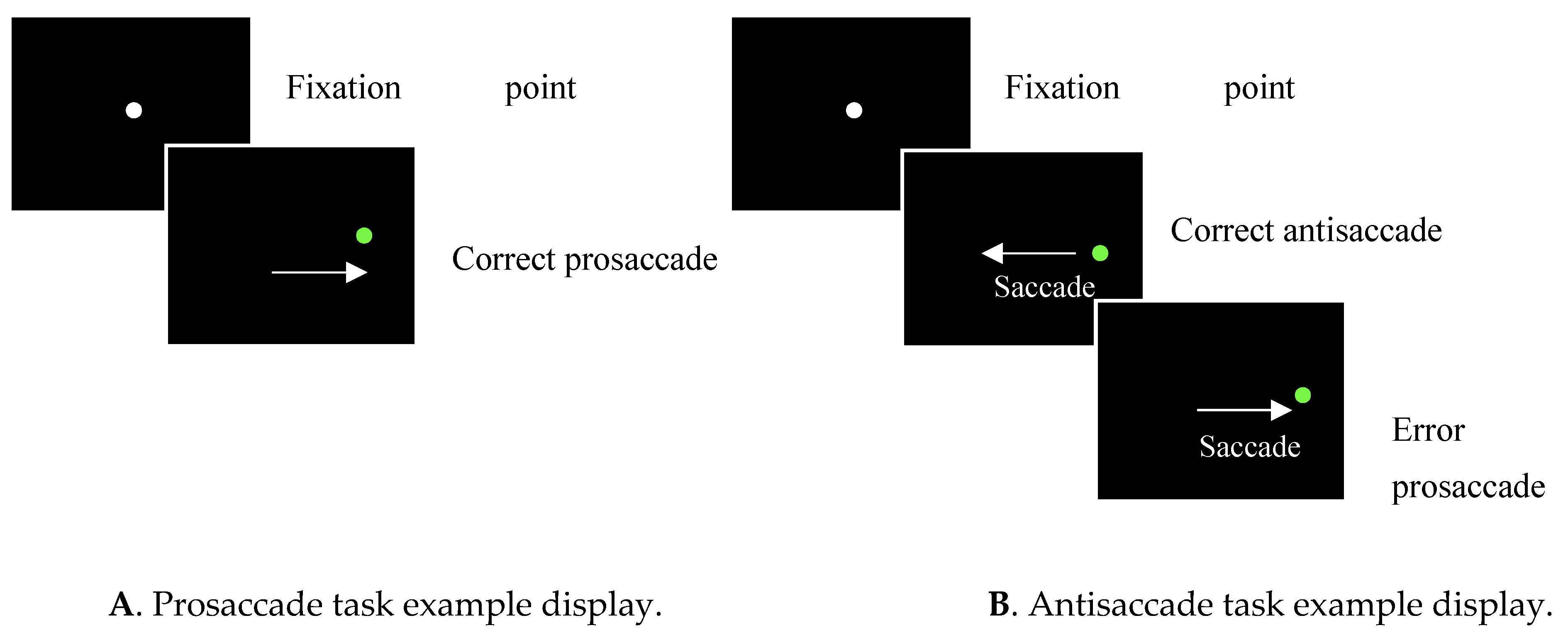
:1. Introduction
2. Materials and Methods
2.1. Data Sources
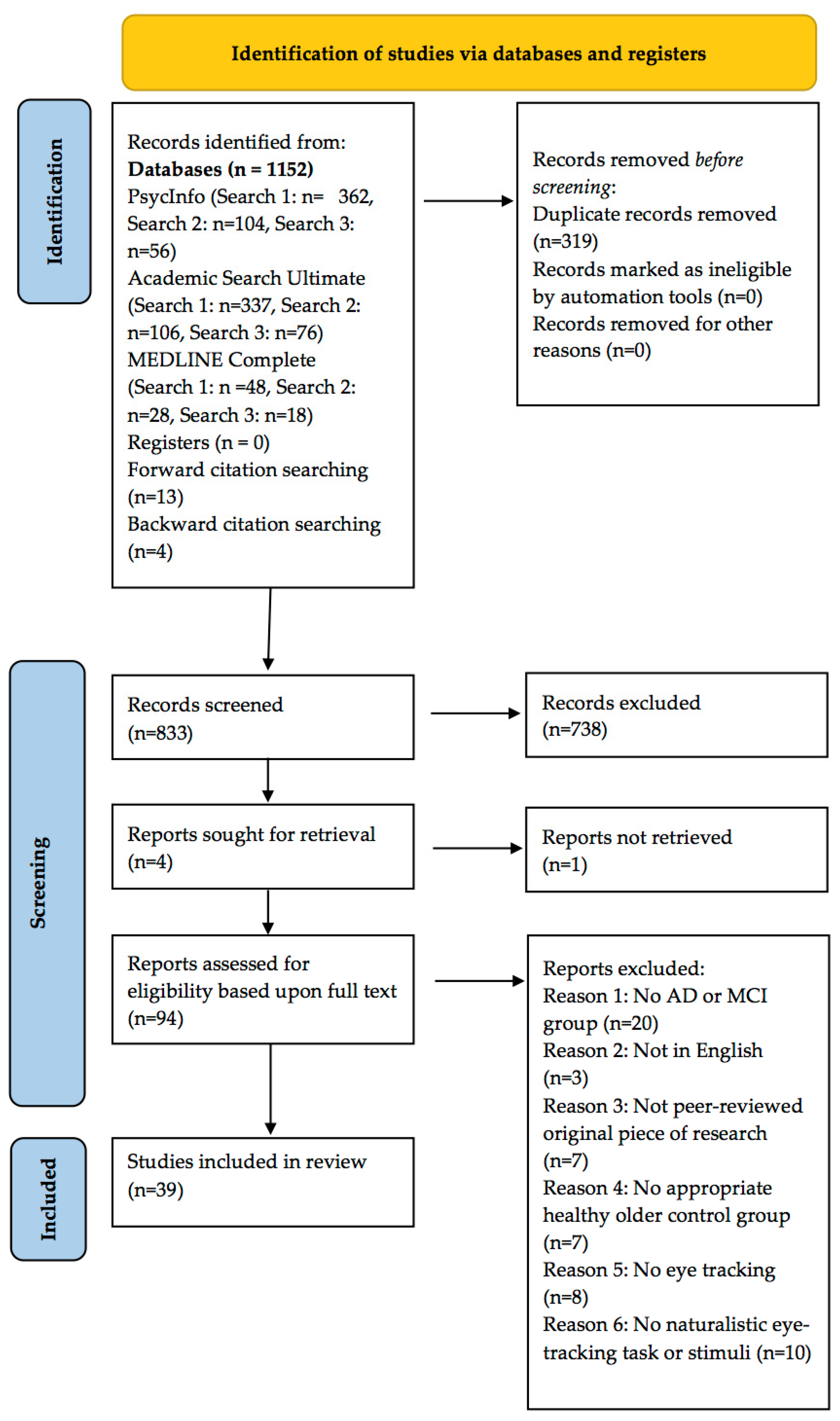
2.2. Screening
Inclusion Criteria
2.3. Data Extraction
2.4. Quality Assessment
3. Results
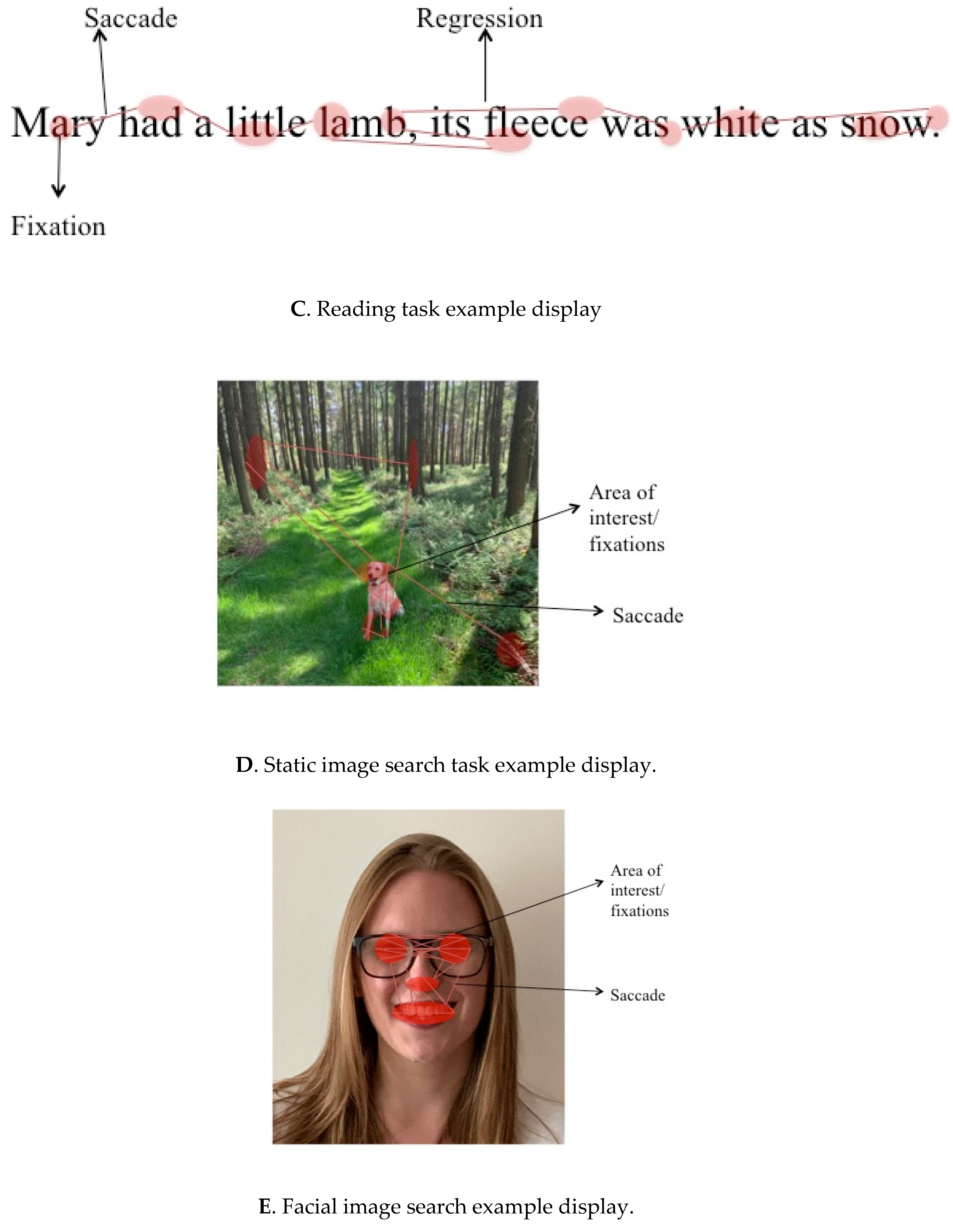
3.1. Reading Tasks
3.2. Studies Employing Goal-Directed Paradigms with Naturalistic Stimuli
3.3. Studies Employing Naturalistic Tasks
3.3.1. Eye Movement Behaviours during Static Image Search
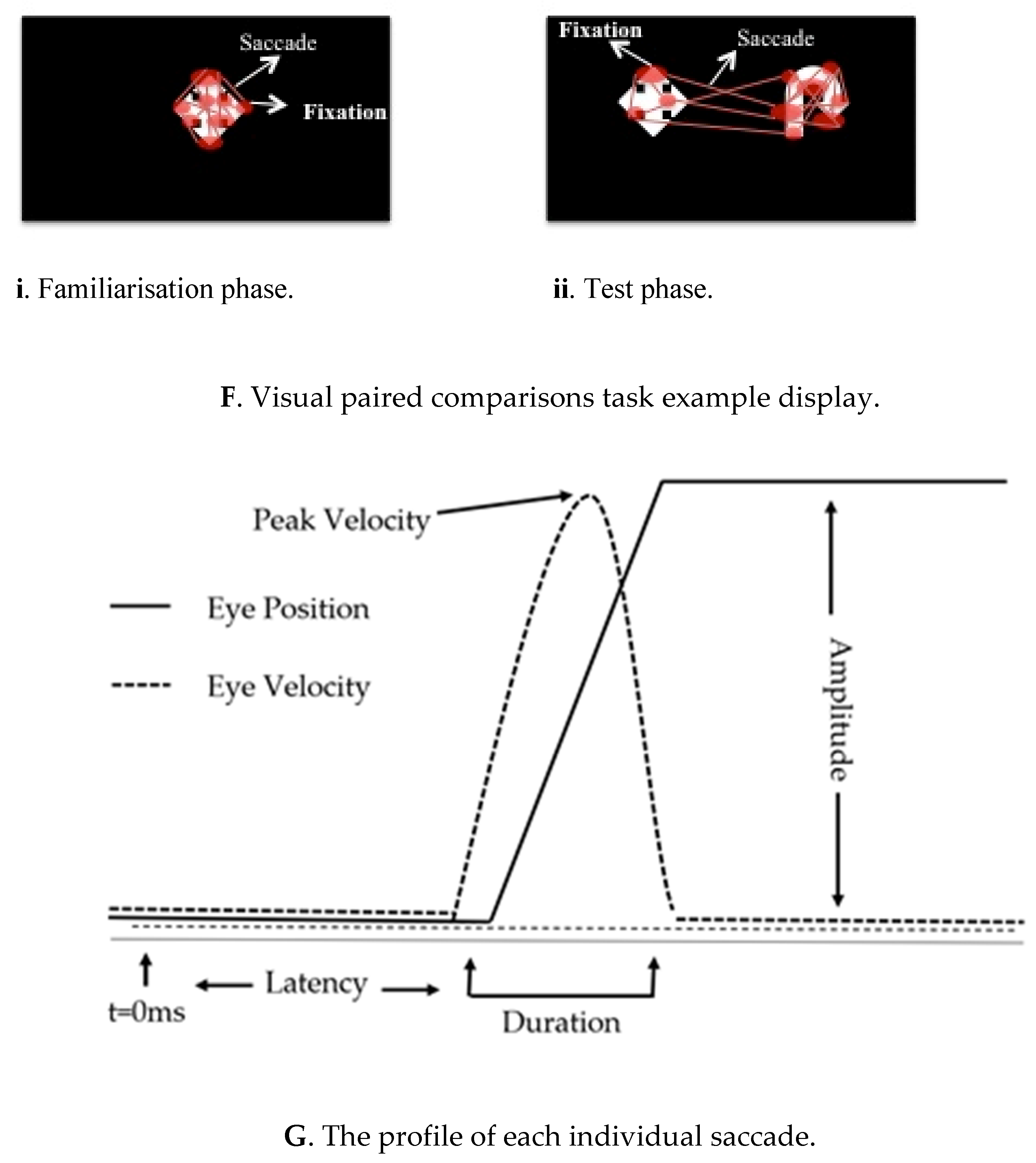
3.3.2. Eye Movement Behaviours during Visual Paired Comparison Tasks
3.3.3. Eye Movement Behaviours during Facial Processing
3.4. Eye Movement Behaviours during Every-Day Tasks and Real-Life Simulations
3.5. Analyses of the Specificity and Sensitivity of Eye Movements in Diagnostic Practices
4. Discussion
Supplementary Materials
Author Contributions
Funding
Institutional Review Board Statement
Informed Consent Statement
Data Availability Statement
Acknowledgments
Conflicts of Interest
Appendix A

| Database | Search ID | Search String |
|---|---|---|
| APA PsycInfo | S1 | (DE “Alzheimer’s Disease” OR DE “Cognitive Aging” OR DE “Cognitive Impairment” OR DE “Mild Cognitive Impairment” OR DE “Healthy Aging” OR DE “Older Adulthood” OR DE “Geriatrics”) OR TI (Alzheimer* OR “cognitive aging” OR “cognitive ageing” OR “cognitive impair*” OR “mild cognitive impairment” OR “AD” OR “MCI” OR ((“cognitive ability” OR cog*) N3 (impair*)) OR amnestic OR non-amnestic OR “non amnestic” OR “healthy aging” OR “healthy ageing” OR “older adult*” OR “elder*” OR “healthy cognitive aging” OR “healthy cognitive ageing”) OR AB (Alzheimer* OR “cognitive aging” OR “cognitive ageing” OR “cognitive impair*” OR “mild cognitive impairment” OR “AD” OR “MCI” OR ((“cognitive ability” OR cog*) N3 (impair*)) OR amnestic OR non-amnestic OR “non amnestic” OR “healthy aging” OR “healthy ageing” OR “older adult*” OR “elder*” OR “healthy cognitive aging” OR “healthy cognitive ageing”) |
| S2 | DE “Eye Movements” OR TI (“eye track*” OR “eye-track*” OR Oculomotor OR Ocularmotor OR “memory guided” OR “memory-guided” OR saccad* OR pro-saccad* OR prosaccade* OR “pro saccad*” OR anti-saccad* OR antisaccad* OR “anti saccad*” OR ((eye* OR retina* OR ocular* OR optic*) N3 (mov* OR track*))) OR AB (“eye track*” OR “eye-track*” OR Oculomotor OR Ocularmotor OR “memory guided” OR “memory-guided” OR saccad* OR pro-saccad* OR prosaccade* OR “pro saccad*” OR anti-saccad* OR antisaccad* OR “anti saccad*” OR ((eye* OR retina* OR ocular* OR optic*) N3 (mov* OR track*))) | |
| S3 | TI (natural* OR real* OR tea OR tea-making OR television OR TV OR watch* OR read* OR video* OR view*) OR AB (natural* OR real* tea OR tea-making OR television OR TV OR watch* OR read* OR video* OR view*) | |
| S4 | S1 AND S2 AND S3 | |
| S5 | (DE “Emotion Recognition” OR DE “Facial Affect Recognition” OR DE “Face Perception”) OR TI (“emotion* recognition” OR “emotion* processing OR “emotion* perception” OR “affect recognition” OR “affect processing” OR “affect perception” OR “face perception” OR “face processing” OR “expression processing” OR “expression recognition” OR “expression perception” OR (face N3 processing))) OR AB (“emotion* recognition” OR “emotion* processing OR “emotion* perception” OR “affect recognition” OR “affect processing” OR “affect perception” OR “face perception” OR “face processing” OR “expression processing” OR “expression recognition” OR “expression perception” OR (face N3 processing))) | |
| S6 | (DE “Locomotion” OR DE “Exercise” OR DE “Physical Activity”) OR TI (“locomotion” OR “exercise” OR “physical activity” OR “walk*” OR “run*” OR “jog*” OR “stairs” OR “travel*”) OR AB (“locomotion” OR “exercise” OR “physical activity” OR “walk*” OR “run*” OR “jog*” OR “stairs” OR “travel*”)) | |
| S7 | (S1 AND S2 AND S5) OR (S1 AND S2 AND S6) | |
| S8 | “VPC” OR “paired comparison*” OR “paired-comparison*” OR “free view*” OR “free-view*” OR “visual scan*” OR ((“natural” OR “scene”) AND (“view*” OR “vision”)) | |
| S9 | S1 AND S2 AND S8 | |
| Academic Search Ultimate | S1 | ((DE “MILD cognitive impairment” OR DE “AMNESTIC mild cognitive impairment”) OR (DE “COGNITIVE aging” OR DE “OLDER People” OR DE “CENTENERIANS” OR DE “OLD-old” OR DE “AGING” OR DE “OLD age” OR DE “AGE factors in cognition” OR DE “INFLUENCE of age on ability”)) OR TI (Alzheimer* OR “cognitive aging” OR “cognitive ageing” OR “cognitive impair*” OR “mild cognitive impairment” OR “AD” OR “MCI” OR ((“cognitive ability” OR cog*) N3 (impair*)) OR amnestic OR non-amnestic OR “non amnestic” OR “healthy aging” OR “healthy ageing” OR “older adult*” OR “elder*” OR “healthy cognitive aging” OR “healthy cognitive ageing”) OR AB (Alzheimer* OR “cognitive aging” OR “cognitive ageing” OR “cognitive impair*” OR “mild cognitive impairment” OR “AD” OR “MCI” OR ((“cognitive ability” OR cog*) N3 (impair*)) OR amnestic OR non-amnestic OR “non amnestic” OR “healthy aging” OR “healthy ageing” OR “older adult*” OR “elder*” OR “healthy cognitive aging” OR “healthy cognitive ageing”) |
| S2 | (DE “EYE movements” OR DE “EYE movement measurements” OR DE “EYE tracking” OR DE “SACCADIC eye movements”) OR TI (“eye track*” OR “eye-track*” OR Oculomotor OR Ocularmotor OR “memory guided” OR “memory-guided” OR saccad* OR pro-saccad* OR prosaccade* OR “pro saccad*” OR anti-saccad* OR antisaccad* OR “anti saccad*” OR ((eye* OR retina* OR ocular* OR optic*) N3 (mov* OR track*))) OR AB (“eye track*” OR “eye-track*” OR Oculomotor OR Ocularmotor OR “memory guided” OR “memory-guided” OR saccad* OR pro-saccad* OR prosaccade* OR “pro saccad*” OR anti-saccad* OR antisaccad* OR “anti saccad*” OR ((eye* OR retina* OR ocular* OR optic*) N3 (mov* OR track*))) | |
| S3 | TI (natural* OR real* OR tea OR tea-making OR television OR TV OR watch* OR read* OR video* OR view*) OR AB (natural* OR real* tea OR tea-making OR television OR TV OR watch* OR read* OR video* OR view*) | |
| S4 | S1 AND S2 AND S3 | |
| S5 | (DE “LOCOMOTION” OR DE “LOCOMOTOR control”)) OR TI ((“locomot*” OR “exercise” OR “physical activity” OR “walk*” OR “run*” OR “jog*” OR “stairs” OR “travel*”)) OR AB ((“locomot*” OR “exercise” OR “physical activity” OR “walk*” OR “run*” OR “jog*” OR “stairs” OR “travel*”) | |
| S6 | (DE “FACIAL expression”) AND (DE “FACE perception” OR DE “FACE perception testing”)) OR TI ((“emotion* recognition” OR “emotion* processing OR “emotion* perception” OR “affect recognition” OR “affect processing” OR “affect perception” OR “face perception” OR “face processing” OR “expression processing” OR “expression recognition” OR “expression perception” OR (face N3 processing)))) OR AB ((“emotion* recognition” OR “emotion* processing OR “emotion* perception” OR “affect recognition” OR “affect processing” OR “affect perception” OR “face perception” OR “face processing” OR “expression processing” OR “expression recognition” OR “expression perception” OR (face N3 processing))) | |
| S7 | (S1 AND S2 AND S5) OR (S1 AND S2 AND S6) | |
| S8 | “VPC” OR “paired comparison*” OR “paired-comparison*” OR “free view*” OR “free-view*” OR “visual scan*” OR ((“natural” OR “scene”) AND (“view*” OR “vision”)) | |
| S9 | S1 AND S2 AND S8 | |
| MEDLINE Complete | S1 | (MH “Cognitive Aging” OR MH “Cognitive Dysfunction” OR MH “Alzheimer Disease” OR MH “Frail Elderly OR MH “Healthy Aging” OR MH “Aging”) OR TI (Alzheimer* OR “cognitive aging” OR “cognitive ageing” OR “cognitive impair*” OR “mild cognitive impairment” OR “AD” OR “MCI” OR ((“cognitive ability” OR cog*) N3 (impair*)) OR amnestic OR non-amnestic OR “non amnestic” OR “healthy aging” OR “healthy ageing” OR “older adult*” OR “elder*” OR “healthy cognitive aging” OR “healthy cognitive ageing”) OR AB (Alzheimer* OR “cognitive aging” OR “cognitive ageing” OR “cognitive impair*” OR “mild cognitive impairment” OR “AD” OR “MCI” OR ((“cognitive ability” OR cog*) N3 (impair*)) OR amnestic OR non-amnestic OR “non amnestic” OR “healthy aging” OR “healthy ageing” OR “older adult*” OR “elder*” OR “healthy cognitive aging” OR “healthy cognitive ageing”) |
| S2 | (MH “Eye Movements” OR MH “Eye Movement Measurements” OR MH “Eye-Tracking Technology” OR MH “Saccades”) OR TI (“eye track*” OR “eye-track*” OR Oculomotor OR Ocularmotor OR “memory guided” OR “memory-guided” OR saccad* OR pro-saccad* OR prosaccade* OR “pro saccad*” OR anti-saccad* OR antisaccad* OR “anti saccad*” OR ((eye* OR retina* OR ocular* OR optic*) N3 (mov* OR track*)))OR AB (“eye track*” OR “eye-track*” OR Oculomotor OR Ocularmotor OR “memory guided” OR “memory-guided” OR saccad* OR pro-saccad* OR prosaccade* OR “pro saccad*” OR anti-saccad* OR antisaccad* OR “anti saccad*” OR ((eye* OR retina* OR ocular* OR optic*) N3 (mov* OR track*))) | |
| S3 | TI (natural* OR real* OR tea OR tea-making OR television OR TV OR watch* OR read* OR video* OR view*) OR AB (natural* OR real* tea OR tea-making OR television OR TV OR watch* OR read* OR video* OR view*) | |
| S4 | S1 AND S2 AND S3 | |
| S5 | ((MH “Locomotion”) OR (MH “Movement”) OR (MH “Motor Activity”) OR (MH “Exercise”) OR (MH “Walking”) OR (MH “Stair Climbing”) OR (MH “Running”) OR (MH “Jogging”)) OR TI ((“locomot*” OR “exercise” OR “physical activity” OR “walk*” OR “run*” OR “jog*” OR “stairs” OR “travel*”)) OR AB ((“locomot*” OR “exercise” OR “physical activity” OR “walk*” OR “run*” OR “jog*” OR “stairs” OR “travel*”)) | |
| S6 | ((MH “Facial Recognition” OR MH “Facial Expression”)) OR TI ((“emotion* recognition” OR “emotion* processing OR “emotion* perception” OR “affect recognition” OR “affect processing” OR “affect perception” OR “face perception” OR “face processing” OR “expression processing” OR “expression recognition” OR “expression perception” OR (face N3 processing)))) OR AB ((“emotion* recognition” OR “emotion* processing OR “emotion* perception” OR “affect recognition” OR “affect processing” OR “affect perception” OR “face perception” OR “face processing” OR “expression processing” OR “expression recognition” OR “expression perception” OR (face N3 processing)))) | |
| S7 | (S1 AND S3 AND S4) OR (S2 AND S3 AND S4) | |
| S8 | “VPC” OR “paired comparison*” OR “paired-comparison*” OR “free view*” OR “free-view*” OR “visual scan*” OR ((“natural” OR “scene”) AND (“view*” OR “vision”)) | |
| S9 | S1 AND S2 AND S8 |
Appendix B
| Study | Participant Group Studied, n (of Which Females) | Participant Age (SD) | Diagnostic Criteria | Cognitive Tests, Group: Score (SD) | Task Type | Paradigm and Dependent Measures | Eye Tracking Device | Main Results | Conclusion |
| Daffner et al. (1992) [70] | AD 12 (7) HOC 10 (7) | 73.1 (4.7) 71 (6.4) | NINCDS-ADRDA criteria; CT scan. | BDS, AD: 11 (7.1), HOC: 0.9 (0.88) | Naturalistic task | Free viewing of static images containing incongruous elements. Fixation duration overall and on ROIs. Frequency of fixations on ROIs. | Applied Science Laboratories Model 3000. |
| AD: discrepancy between image types may be attributable to the incongruous element of the lion image being overtly visible in comparison to the horse image. AD: diminished visual exploration of stimuli overall and give less attention to incongruous elements. AD: have impaired ability to recognise incongruous stimuli/diminished novelty-seeking drive. |
| LaBar et al. (2000) [45] | AD 9 (5) HOC 9 (7) HYC 24 (13) | 76 (4) 67 (5) 26 (4) | NINCDS-ADRDA; neurological/neuropsychological examinations. | MMSE, AD: 24 (4), HOC: 29 (2) WMS-LM, AD: 11 (6), HOC: 30 (5) | Naturalistic task | Free search of emotionally valenced static images. Latency of first saccade, duration of sustained attention. | Infrared oculography (ISCAN). |
| AD: can direct attention to negatively valenced content in a normal manner. |
| Lueck, Mendez, and Perryman (2000) [52] | AD 14 (10) HOC 14 (6) | 75.14 (4.44) 72.43 (6.66) | NINCDS-ADRDA; presence of predominant bilateral temporoparietal hypometabolism on SPECT or PET scans. | MMSE, AD: 18.79 (3.31), HOC c CDRS, AD: 1 (0.44), HOC: 0.0 CERAD (Verbal fluency), AD: 10.21 (4.85), HOC c CERAD (Mini-BNT), AD: 11.36 (2.82), HOC c | Reading task | Silent reading. Portion of text read, forward saccades, saccadic regressions, fixation duration, saccadic duration. | Ober2 (Permobil). |
| AD: altered eye movements during reading present in early stages. Correlation between decreased amount read with increasing dementia severity potentially reflects disturbed lexical-semantic access. |
| Ogrocki et al. (2000) [48] | AD 17 (10) HOC 15 (10) | 73.9 (7.8) 72.7 (4.1) | NINCDS-ADRDA; DSM-IV; neurological assessment; laboratory tests; neuropsychological assessment. | MMSE, AD: 21.8 (3.8), HOC: 29.2 (0.7) | Naturalistic task | Emotion identification. Total fixations and number of fixations within ROI. Fixation Duration. Emotion identification accuracy. | RK-426PC Pupil/corneal reflection tracking system (ISCAN). |
| AD: allocate attention differently than HOC during face viewing. AD: possibility of abnormal visual exploration strategies contributing to emotion identification deficits. |
| Mapstone et al. (2001) [46] | AD 13 (9) HOC 13 (9) HYC 11 (5) | 75.7 (5.7) 73.9 (4) 27.4 (3.9) | NINCDS-ADRDA; neurological/neuropsychological examinations. | MMSE, AD: 24.3 (3.1), HOC: 28.2 (1.5) WMS-LM Memory, AD: 2.1 (3.7), HOC: 25.6 (9.3) | Eye movement behaviours during every-day tasks and real-life simulations | Car driving simulation. Number of fixations, percentage of fixations inside the ROI, fixation duration. | Infrared Eye Tracking System (ISCAN, RK-426PC). |
| HOC/AD: unable to covertly attend to peripheral distractors when driving, instead directing gaze towards them, suggesting deficit in ability to switch between covert and overt attention. |
| Mosimann et al. (2004) [65] | AD 24 (13) HOC 24 (9) | 74.3 (6.3) 72.9 (6.9) | DSM-IV; and NINCDS-ADRDA; CT/MRI scans. | MMSE, AD: 20.1 (5.4), HOC: 29.1 (0.8) | Naturalistic task | Clock reading fixation duration, saccade length, exploration time. | Infrared eye tracking EyeLink (SRResearch). |
| AD: impaired ability to strategise focus on relevant aspects of clock suggesting selective attention impairment. |
| Crutcher et al. (2009) [79] | MCI 6 HOC 15 PD 4 | 70 (8.1) 67.5 (5.6) 63.8 (6.4) | MCI: standardised assessment by 3 clinicians; evidence of memory decline and possibly other cognitive domains with a severity insufficient to meet DSM-III-R criteria for dementia. | MMSE, MCI: 27.5 (2.8); HOC: 29.1 (1.3); PD: 29.0 (0.8) | Naturalistic task | VPC Task. Total number of fixations. Total looking time. % looking time on novel stimuli. | Applied Science Laboratories (ASL) Model 5000 remote pan/tilt camera system. | Familiarisation phase:
| MCI: comparable performance on 2 s delay but impaired performance on the 2 min delay suggests presence of a recognition memory deficit. |
| Forde et al. (2010) [25] | ADS 1 (0) AD 1 (1) HOC 2 (1) | 31 59 50, 30 c | Diagnosed by clinicians 3 years prior; MRI showing mild temporal atrophy. | d WAIS IQ, ADS: 58. WMS-VMI, ADS: 58 WMS-ACI, ADS: 63. MMSE, AD: 21 e | Behaviours during every-day tasks and real-life simulations | Tea Making. Number of fixations during ORAs. Durations of ORAs. Fixations on objects between ORAs. Orientating eye movements. Number of looks per object. | No eye tracker model provided. |
| AD: demonstrated comparable tea-making ability and eye movement patterns to HOC. |
| Lagun et al. (2011) [78] | MCI 10 AD 20 HOC 30 | 72.2 (6.9) 72.4 (10) 70.9 (7.1) | Formal diagnosis of MCI or AD established by neuropsychological battery and review by 3 clinicians. | MMSE, not reported. | Naturalistic task | VPC task. AUC analysis. | ASL eye tracker (120 Hz sampling rate). |
| VPC performance can distinguish between AD, MCI and HOC. Machine learning methods can aid in automatic detection of cognitive impairment |
| Fernández et al. (2013) [39] | HOC 20 (12) AD 20 (12) | 71 (6.1) 69 (7.2) | DSM-IV; MRI (n = 12) or CT (n = 8) scans; biochemical analysis; physical/neurological examination. | MMSE, AD: 23.2 (0.7), HOC: 27.8 (1.0) ACE-R, AD: 82.4 (2.1) | Reading task | Reading (sentences). Total, first-pass, second-pass, single fixations, and regressions. Skipped words. Saccade amplitude and duration. | EyeLink 1000 Desktop Mount (SRResearch). |
| AD: differences in eye movement patterns during reading suggestive of impaired retrieval and memory. AD: Increased second-pass fixations and regressions suggest impairment in word processing and an inability to direct attention according to the word just read. |
| Zola et al. (2013) [81] | AD 20 (10) aMCI 32 (14) HOC 60 (40) After 3 years, participants were re-assessed and divided based on whether their diagnosis had changed to either aMCI or AD. Converters 17 Non-converters 75 | 72.2 (10.2) 70.2 (8.0) 69.7 (7.2) | aMCI: Alzheimer’s Disease Centers UDS neuropsychological test battery. AD: criteria not provided. | MMSE, AD: 22.2 (5.0), aMCI 27.3 (1.8), HOC 29.2 (1.1). | Naturalistic task | Visual paired comparison. Comparisons between those whose diagnosis converted to aMCI/AD and those whose did not in the 3 years between testing. Percentage looking time to novel stimuli. Total looking time. Total number of fixations. | Applied Science Laboratories Model 6000 camera. |
Familiarisation phase:
| Scores on the VPC can predict change in diagnosis from aMCI to AD or from HOC to aMCI up to 3 years before a change in clinical diagnosis. |
| Brandão et al. (2014) [63] | AD 5 (3) HOC 10 (7) | 78.31 (6.65) 80.92 (5.51) | Diagnosed by two neurologists based on NINCDS-ADRDA criteria. | MMSE, AD: 20.91 (4.25), HOC: 28.37 (1.02) | Naturalistic task | Recalling life events using static visual cues (on-topic versus off-topic). Fixation duration. | Mobile head-mounted eye tracker (SMI HED 50 Hz). |
| AD: no difference in fixation duration for on-topic versus off-topic cues suggests deficits in inhibiting irrelevant stimuli. AD: greater tendency to fixate on experimenter’s face suggests discourse processing deficit and overreliance on communicative partner. |
| Boucart et al. (2014a) [67] | PCA 6 (3) AD 14 (8) HOC 15 (10) HYC 10 (7) | 65.4 (5) 71.5 (10) 66 (7) | IWG research criteria; hippocampal atrophy on MRI; neuropsychological assessment; CSF biomarker assays; PET/SPECT. | MMSE, PCA: 22.5 (3.61), AD: 23.3 (1.34) DRS, PCA: 114.5 (13.63), AD: 112.42 (24.55) | Studies employing goal-directed paradigms with naturalistic stimuli | Saccadic categorisation task. Response accuracy. Saccade latencies Response time. | Red-m pupil-tracking system (Senso-Motoric Instruments). |
| AD: demonstrate a speed-accuracy tradeoff to compensate for decreased cognitive control or to reduce errors. |
| Fernández et al. (2014a) [40] | AD 18 (11) HOC 40 (29) | 69 (7.2) 71 (6.1) | DSM-IV. | MMSE, AD: 23.2 (0.7), HOC: 27.8 (1.0) | Reading task | Reading (sentences). Skipping rates, first-pass, and second-pass fixations. Regressions and intra-word regressions. Fixation duration. Word predictability effects. Saccade amplitude. | EyeLink 2K Desktop Mount (SRResearch). |
| AD: results suggest word processing deficit and inability to shift attention according to the word just read. Unaffected by word predictability suggesting impaired retrieval mechanism. |
| Boucart et al. (2014b) [68] | AD 17 (8) HOC 23 (15) HYC 24 (17) | 70.2 (3.1) 72 (7.5) 28.2 (2) | Neuropsychological assessment, MRI, CSF biomarkers, SPECT or PET. | MMSE, AD: 23.4 (0.8), HOC: 29.46 (0.5) DRS, AD: 126.9 (6.2) | Studies employing goal-directed paradigms with naturalistic stimuli | Saccadic choice task. Latency, amplitude, and duration of first saccade. Accuracy. | iViewX (Senso-Motoric Instruments). |
| AD: more difficulty discriminating animals from distractors within scenes, suggests deficits in detecting relevant information. |
| Fernández et al. (2014b) [41] | AD 20 (12) HOC 40 (29) | 69 (7.3) 71 (6.1) | DSM-IV; physical/neurological examination; APOE e3/e4 genotype; thyroid test; MRI (n = 12), CT (n = 8); biochemical analysis. | MMSE, AD: 24.2 (0.8), HOC: 27.8 (1.0) ACE-R, AD: 84.4 (1.1) | Reading task | Reading (sentences). Word predictability. Fixation duration. | EyeLink 1000 Desktop Mount (SRResearch). |
| AD: unaffected by predictability suggesting impaired retrieval mechanism. Increased fixation duration suggests difficulty in processing meaning. |
| Chau et al. (2015) [77] | AD 41 (19) HOC 24 (12) | 79.2 (6.7) 76.2 (6.4) | DSM-IV; NINCDS-ADRDA. | MMSE, AD: 22.2 (4.0) HOC: 28.1 (2.0) | Naturalistic task | VPC task. Relative fixation time. Fixation time within images (ROI). Average fixation duration. | The VAST (EL-MAR Inc.). |
| AD: spent less time fixating on novel stimuli than HOC suggesting a decreased capacity for novelty preference and selective attention. |
| Fernández et al. (2015a) [42] | pAD 20 (12) HOC 40 (29) | 69 (7.3) 71 (6.1) | DSM-IV. | MMSE, AD: 24.2 (0.8), HOC: 27.8 (1.0) ACE-R, AD: 84.4 (1.1) | Reading task | Reading (proverbs) Fixation duration Word predictability. | EyeLink 1000 Desktop Mount (SRResearch). |
| AD: general reading preserved, but semantic content processing impaired. |
| Fernandez et al. (2015b) [75] | AD 35 (22) HOC 35 (24) | 68 (6.4) 70 (6.2) | DSM-IV; physical/neurological examination; APOE e3/e4 genotype; thyroid test; biochemical analysis; MRI (n = 27), CT (n = 8). | No cognitive tests described. | Reading task | Reading (sentences). Total number of fixations. First-pass fixations. Second-pass fixations. | EyeLink 1000 Desktop Mount (SRResearch). |
| AD: show an impaired ability to use sentence context for predicting upcoming words. Suggests impairments in the recognition and retrieval of words. |
| Lenoble et al. (2015) [76] | AD 20 (14) HOC 28 (18) HYC 26 (13) | 71.4 (5.8) 69.1 (7.1) 26.7 (2.3) | NINCDS-ADRDA/R criteria. | MMSE, AD: 23.8 (1.1), HOC: 29.1 (0.6) | Studies employing goal-directed paradigms with naturalistic stimuli | Saccadic choice task. Latency of first saccade. Accuracy. | Red-M; Senso-Motoric instruments. |
| AD: saccades to naturalistic images are only affected by the nature of the image |
| Shakespeare et al. (2015) [73] | PCA 7 (5) AD 8 (4) HOC 19 (14) | 58.9 (6.3), 69.7 (4.7), 63.1 (5.2) | PCA: clinical criteria for PCA [99,100]; diagnosis of AD; score in the normal range on the RMT for words; Biomarker neuropathology. AD: Dubois criteria; impaired range on the RMT for words; biomarker neuropathology. | MMSE: PCA 22.6 (2.57); AD 22.6 (4.50); HOC c | Studies employing goal-directed paradigms with naturalistic stimuli | Exploratory scanning of naturalistic visual scenes/visual search task. Fixation duration. Saccadic amplitude. Fixation position. Fixations in ROI. Scanpath consistency. | Eyelink II (SR Research). |
Exploratory scanning:
| AD: lack of modulation of scanpaths suggests poor perception and memory dysfunction. |
| Suzuki et al. (2015) [83] | AD 1 (1) PCA 1 (1) HOC 1 (1) | No participant ages provided. | No diagnostic criteria provided. | No diagnostic criteria provided. | Naturalistic task | Locomotion. Average fixation duration. Average resultant acceleration of left foot from start steeping to the completion of each task. | SMI ETG eye tracker. |
| AD: variability in the open room task due to secondary visuospatial impairments and deficits in memory and executive function. |
| Yong et al. (2015) [47] | PCA 15 (9) AD 6 (4) HOC 6 (4) | 61 (6.6) 62 (7.5) 61.3 (4.6) | NIAAAC. | MMSE, PCA: 19.0 (4.2), AD: 22.8 (5.3) c | Reading task | Reading (passages). Mean reading time. Number of saccades. Number of fixations. | EyeLink II (SRResearch). |
| AD: no differences in patterns of eye movements when reading compared to HOC. |
| Fernández et al. (2016) [43] | AD 35 (22) HOC 35 (24) | 68 (6.4) 70 (6.2) | DSM-IV; physical/neurological examination; APOE e3/e4 genotype; thyroid test; MRI (n = 12), CT (n = 8); biochemical analysis. | MMSE, AD: 25.3 (0.9), HOC: 28.8 (1.0) ACE-R, AD: 84.4 (1.1) | Reading task | Reading (sentences). Predictability effects. Mean fixation duration. Change in fixation duration following max jump. | EyeLink 1000 Desktop Mount (SRResearch). |
| AD: impairment in max jump suggests impaired prediction and retrieval of upcoming words. |
| Vallejo et al. (2016) [66] | AD 18 (10) HOC 20 (10) | 74.3 (7.6) 72.2 (3.4) | ICD (10th edition) criteria; CERAD neuropsychological battery; MRI; BADS; Functional Activities Questionnaire. | MoCA, AD: 19.4 (4.5), HOC: 28.5 (1.1) | Studies employing goal-directed paradigms with naturalistic stimuli | Go-NoGo visual search task of naturalistic scenes. Percentage of fixations in eccentricity areas, mean fixation time. Mean distance between gaze position and target position at target onset. | Integrated eye camera (Octopus 900). |
| AD: attending to central cues requires voluntary attentional control suggesting impaired selective attention. AD: longer time to detect targets suggests difficulty attending to relevant parts of space and covertly shifting attention to the periphery as well as an impaired ability to enact precise and quick eye movements. |
| Dragan et al. (2017) [38] | HYC 17 (12) HOC 10 (9) pMCI 8 (5) AD 9 (4) | 22.8 (3.1) 66.4 b 69 b 69.1 (7.8) | NIAAAC; Score of 12–23 on ADAS-cog11; Score of 0.5–1 on CDRS. | MoCA, HOC: 28.1, pMCI: 23.1, AD: No data | Naturalistic task | Visual search of natural scenes (Experiment 1: Flicker Change Detection Memory Task; Experiment 2: Target Detection Memory Task). Fixation location and duration. | Lab-iView X infrared eye-tracking system (Sen-soMotoric Instruments). | Experiment 1:
| AD: impaired scanning and memory-guided search of natural scenes. |
| Fraser et al. (2017) [44] | MCI 27 (14) HOC 30 (21) | 70.3 (5.8) 68.0 (7.5) | Neuropsych-ological examination; MRI, blood tests; lumbar punctures. | MMSE, MCI: 28.2 (1.3), HOC: 29.6 (0.6) | Reading task | Reading (short texts and comprehension). First-pass, later-pass, multi-fixations, and re-fixations. | EyeLink 1000 Desktop Mount. |
| MCI: greater tendency to skip words and return to them later compared with HOC. |
| Kawagoe et al. (2017) [50] | aMCI 18 (10) HOC 18 (13) | 77.61 (5.32) 74.05 (16.66) | NIAAAC; neuropsychological tests; psychological assessments; assessments of activities of daily living; MRI or CT; SPECT; blood count and metabolic panel. | MMSE, aMCI: 24.22 (3.90), HOC: 28.11 (1.64) WMS-LM I, aMCI: 2.50 (2.03), HOC: 9.22 (3.70) WMS-LM II, aMCI: 1.00 (1.88), HOC: 7.66 (4.02) | Naturalistic | Perception and short-term memory of faces and houses. Fixation duration. Number of fixations. | Tobii TX300 (Tobii Technology). |
| aMCI: face-specific impairments evidenced by proportion of correct responses, especially in memory conditions. Results indicated face-specific deficits seen in the aMCI group was exacerbated when the memory load of the task was increased. |
| Bourgin et al. (2018) [74] | AD 18 (9) HOC 33 (18) | 74 (9) 71 (7) | NIAAAC; MRI; neurological examination. | MMSE, AD: 24.57 (3.41), HOC: 29.28 (0.98) | Studies employing goal-directed paradigms with naturalistic stimuli | Prosaccade tasks using naturalistic stimuli. (Please note this paper also employed antisaccade task paradigms however the results are not incorporated here as this is not a naturalistic task). Saccadic error rate. Saccadic reaction Time. | Eyelink 1000 eye tracker (SR Research). |
| AD: results suggest impairment in early emotional attention (rather than an impairment of working memory) when the emotional stimulus is distracting/when there is no complex cognitive process involved and attention is relying on early orientation mechanisms. Lack of effect of emotional valence suggests over-processing of stimuli and an impairment in selectivity. |
| Lenoble et al. (2018) [71] | AD 12 (7) HOC 12 (6) HYC 12 (6) | 71.7 (5.9) 70.2 (6.8) 25.9 (3.1) | Neuropsychological assessment; MRI; CSF biomarkers or SPECT; PET scan. | MMSE AD: 23.1 (1.1), HOC: 29.3 (0.6) | Naturalistic task (free-viewing) and artificial task involving naturalistic stimuli (implicit/explicit saccadic choice task) | Free-viewing and implicit/explicit saccadic choice task. First saccade accuracy and latency. | Red-M Senso-Motoric Instruments: Teltow Germany. | Free viewing:
| AD: bias towards incongruent object/background scenes suggests an unconscious capture of attention by incongruent stimuli. Indicative of poor inhibitory control. |
| McCade et al. (2018) [49] | naMCI 18 (11) aMCI 14 (9) HOC 18 (11) | 63.78 (8.16) 67.93 (7.70) 64.61 (8.37) | Agreement of two neuropsychologists and one Old Age Psychiatrist; decrements below age-based norms in at least two cognitive domains; GDS. aMCI: clear evidence of memory storage (i.e., delayed recall) deficits on neuropsychological tests + impairments in at least one other cognitive domain. -naMCI deficits on multiple cognitive domains other than memory. | MMSE, naMCI: 28.61 (1.24), aMCI: 26.64 (1.91), HOC: 29.11 (0.88) | Naturalistic task | Free visual search of images of faces. Mean percentage of time fixating on facial regions. | Tobii X120. |
| NaMCI/aMCI: comparable eye movement behaviours despite worse cognitive test and emotion recognition performance. |
| Yong et al. (2018) [51] | AD 10 (6) PCA 8 (4) HOC 12 (6) | 66.2 (5.0) 64.1 (6.1) 63.7 (4.1) | NIAAAC; Molecular pathology amyloid imaging (n = 5). | MMSE, AD: 18.6 (4.9). | Naturalistic task | Visually guided navigation Fixation on target Time spent fixating on target. | SensoMotoric Eyetracking Glasses 1. |
| AD: weak effect of motion lights suggests motion perception may be preserved in AD but only at certain frequencies. Longer initial fixation during cued condition suggests that the environmental incongruence of the cues may require increased processing for those with memory impairments. |
| Fraser et al. (2019) [84] | MCI 26 (14) HOC 29 (21) | 70.6 (5.8) 67.8 (7.7) | Global Deterioration Scale (GDS); CDRS. | MMSE: MCI: 28.2 (1.4) HOC: 29.6 (0.6) | AUC of reading task | Reading (silently and aloud) AUC analysis. | EyeLink 1000 Desktop Mount with monocular eye-tracking sampling rate 1000 Hz. |
| Reading and speaking tasks can aid in the classification and detection of cognitive decline. Machine learning models incorporating multiple measures (cascaded approach) outperformed classifier trained based on a neuropsychological battery. |
| Haque et al. (2019) [80] | AD 22 MCI 27 HOC 77 | 76 (7.0) 69.5 (9.5) 64.5 (7.5) | Standardized neuropsychological testing; neurological examination; brain imaging; and bloodwork. | MoCA AD: 13.5 (5) MCI: 21.3 (4) HOC: 26.7 (2) | Naturalistic task | Visual comparison task. Number of fixations in ROI. Viewing time in ROI. | EyeTribe Infrared Scanner sampled at 30 Hz. |
| The task demonstrated performance differences between HOC and people with MCI. The multivariate model of memory performance on the task predicted MCI and AD with high sensitivity showing potential to be used as a diagnostic tool for AD and MCI. |
| Oyama et al. (2019)[72] | MCI 26 (17) Dementia 27 (16) HOC 27 (18) | 75.2 (8.2) 75.4 (9.5) 71.5 (11.1) | Physical and neurological examinations; neuropsychological assessment; MRI; blood tests; MCI: Petersen criteria [101]; AD: DSM-IV. | MMSE, MCI: 25.7 (3.0), HOC: 28.7 (1.6) FAB, MCI: 13.4 (2.4), HOC: 13.6 (1.8) ADAS-Cog, MCI: 9.4 (3.4), HOC: 4.4 (1.3) CDRS, MCI: 0.5 (0.2), HOC: 0 (0.0) | Naturalistic task | Cognitive assessment tasks. Average percentage fixation duration in ROI. | GazefinderNP-100, (JVC KENWOOD). |
| MCI: eye-tracking cognitive assessment was able to diagnose MCI with accuracy comparable to MMSE. |
| Barral et al. (2020) [85] | AD 68 (34) HOC 73 (51) | 71.6 (9.26) 64.9 (9.93) | Diagnoses made by expert clinicians with cognitive testing, clinical data, and neuroimaging and laboratory data. | MoCA: AD: 20.25 (5.44) HOC: 27.15 (2.73) | AUC | Cookie Theft picture description task. AUC analysis. | Tobii-Pro X3-120. |
| Eye tracking is a useful classification tool for identifying cognitive impairment in people with AD. |
| Davis and Sikoriskii (2020) [64] | AD,7 (4) a HOC 8 (4) | 76.57 (5.03) 75.00 (1.20) | NIAAAC; NINCDS-ADRDA; Score of 0.5–1 on CDRS. | MMSE, AD: 26.43 (2.30), HOC: 29.00 (1.20) MoCA, AD: 19.00 (3.51), HOC: 25.13 (2.41) | Eye movement behaviours during every-day tasks and real-life simulations | Wayfinding in a virtual retirement community. Percentage and duration of fixations. | Eye-tracking glasses (Applied Science Industries Mobile Eye-XG). |
| AD: difficulty identifying and attending to salient cues during visual wayfinding. |
| Nie et al. (2020) [82] | MCI 80 (62) HOC 170 (131) Note. This became HOC 57 and MCI 26 at the 1 year follow up. | 73.0 (4.4) 71.1 (4.1) | MCI: definite memory decline (MoCA >1.5 SD of age-appropriate norms); symptom severity not meeting DSM-IV criteria for dementia; possible impairment of other cognitive domains. | MoCA, MCI: 20.9 (3.2), HOC: 25.8 (2.5) | Naturalistic task/AUC | Visual paired comparison task. Fixation duration on the novel image at test and re-test (2 weeks later). AUC analysis. | Applied Science Laboratories Model 5000 camera. |
| Fixation duration on novel stimuli in a VPC task can accurately distinguish MCI from HOC. |
| Coco et al. (2021) [69] | MCI 27 (7) HOC 23 (14) | 72.48 (8.99) 68.08 (9.66) | International guidelines [28,102,103]; MMSE ≥18; family and medical history interviews; MRI and genetic data (when available). | MMSE, MCI: 24.58 (3.45), HOC: 28.74 (1.66) | Artificial task with naturalistic stimuli | 2-alternative forced-choice paradigm. Recognition accuracy. Semantic interference effects. Entropy during encoding and recognition. Scan pattern similarity during encoding and recognition. Fixation position and saliency map correspondence. | EyeTribe eye tracker. |
| MCI: show a significantly reduced semantic interference effect compared to HOC. May reflect inefficient access to semantic knowledge although this effect was skewed by low-performing MCI participants. MCI: needed to explore scenes more widely during recognition than HOC which is indicative of reduced focal attention. MCI: showed some oculomotor patterns similar to that of HOC. |
Appendix C
- (1)
- Is the hypothesis/aim/objective of the study clearly described?
- (2)
- Are the main outcomes to be measured clearly described in the introduction or methods section? If the main outcomes are first mentioned in the Results section, the question should be answered ‘no’.
- (3)
- Are the characteristics of the participants included in the study clearly described? Inclusion and/or exclusion criteria should be given. In case studies, a case-definition and the source for controls should be given.
- (4)
- Are the distributions of principal confounders in each group of subjects to be compared clearly described? A list of the principal confounders is provided.
- (5)
- Are the main findings of the study clearly described? Simple outcome data (including denominators and numerators) should be reported for all major findings so that the reader can check the major analyses and conclusions. (This question does not cover statistical tests which are considered below).
- (6)
- Have the characteristics of participants lost to exclusion been described? This should be answered ‘yes’ where there were no losses to exclusion or where losses to exclusion were so small that findings would be unaffected by their inclusion. This should not be answered ‘no’ where a study does not report the number of patients lost to exclusion.
- (7)
- Have actual probability values been reported (e.g., 0.035 rather than <0.05) for the main outcomes except where the probability value is less than 0.001?
- (8)
- Were the subjects who participated in the study representative of the entire population from which they were recruited? The study must identify the source population for participants and describe how the participants were selected. Participants would be representative if they comprised the entire source population, and unselected sample of consecutive participants, or a random sample. Random sampling is only feasible where a list of all members of the relevant population exists. Validation that the sample was representative would include demonstrating that the distribution of the main confounding factors was the same in the study sample and the source population.
- (9)
- Were the statistical tests used to assess the main outcomes appropriate? The statistical techniques used must be appropriate for the data. For example, non-parametric methods should be used for small sample sizes. Where little statistical analysis has been undertaken but where there is no evidence of bias, the question should be answered ‘yes’. If the distribution of data (normal or not) is not described, it must be assumed that the estimates used were appropriate and the question should be answered ‘yes’.
- (10)
- Were the main outcome measures used accurate (valid and reliable)? For studies where the outcome measures are clearly described, the question should be answered ‘yes’. For studies which refer to other work or that demonstrates the outcome measures are accurate, the question should be answered as ‘yes’.
- (11)
- Was there adequate adjustment for the confounding in the analyses from which the main findings were drawn? This question should be answered ‘no’ if: the distribution of known confounders in the different experimental groups was not described; or the distribution of known confounders differed between experimental groups but was not taken into account in the analyses. In non-randomised studies, if the effect of the main confounders was not investigated or confounding was demonstrated but no adjustment was made in the final analyses, the question should be answered ‘no’.
- (12)
- Were losses of participants to exclusion taken into account? If the numbers of participants lost to exclusion are not reported, the question should be answered as ‘unable to determine’. If the proportion lost to exclusion was too small to affect the main findings, the question should be answered ‘yes’.
- (13)
- Did the study give sufficient justification for the sample size used?
References
- Kumar, A.; Singh, A. A review on Alzheimer’s disease pathophysiology and its management: An update. Pharmacol. Rep. 2015, 67, 195–203. [Google Scholar] [CrossRef] [PubMed]
- Dias, E.C.; Segraves, M.A. Muscimol-induced inactivation of monkey frontal eye field: Effects on visually and memory-guided saccades. J. Neurophysiol. 1999, 81, 2191–2214. [Google Scholar] [CrossRef] [PubMed]
- Baddeley, A.D.; Baddeley, H.A.; Bucks, R.S.; Wilcock, G.K. Attentional control in Alzheimer’s disease. Brain 2001, 124, 1492–1508. [Google Scholar] [CrossRef] [PubMed] [Green Version]
- Perry, R.J.; Hodges, J.R. Attention and executive deficits in Alzheimer’s disease: A critical review. Brain 1999, 122, 383–404. [Google Scholar] [CrossRef] [Green Version]
- McKhann, G.; Drachman, D.; Folstein, M.; Katzman, R.; Price, D.; Stadlan, E.M. Clinical diagnosis of Alzheimer’s disease: Report of the NINCDS-ADRDA Work Group* under the auspices of Department of Health and Human Services Task Force on Alzheimer’s Disease. Neurology 1984, 34, 939. [Google Scholar] [CrossRef] [Green Version]
- Welsh, K.A.; Butters, N.; Hughes, J.P.; Mohs, R.C.; Heyman, A. Detection and staging of dementia in Alzheimer’s disease: Use of the neuropsychological measures developed for the Consortium to Establish a Registry for Alzheimer’s Disease. Arch. Neurol. 1992, 49, 448–452. [Google Scholar] [CrossRef]
- Hodges, J.R.; Patterson, K. Is semantic memory consistently impaired early in the course of Alzheimer’s disease? Neuroanatomical and diagnostic implications. Neuropsychologia 1995, 33, 441–459. [Google Scholar] [CrossRef]
- Nasreddine, Z.; Phillips, N.; Bédirian, V.; Charbonneau, S.; Whitehead, V.; Collin, I.; Cummings, J.; Chertkow, H. The Montreal Cognitive Assessment, MoCA: A brief screening tool for mild cognitive impairment. J. Am. Geriatr. Soc. 2005, 53, 695–699. [Google Scholar] [CrossRef]
- Hutton, S.B.; Ettinger, U. The antisaccade task as a research tool in psychopathology: A critical review. Psychophysiology 2006, 43, 302–313. [Google Scholar] [CrossRef] [Green Version]
- Garbutt, S.; Matlin, A.; Hellmuth, J.; Schenk, A.K.; Johnson, J.K.; Rosen, H.; Boxer, A.L. Oculomotor function in frontotemporal lobar degeneration, related disorders and Alzheimer’s disease. Brain 2008, 131, 1268–1281. [Google Scholar] [CrossRef] [Green Version]
- Crawford, T.J.; Higham, S.; Mayes, J.; Dale, M.; Shaunak, S.; Lekwuwa, G. The role of working memory and attentional disengagement on inhibitory control: Effects of aging and Alzheimer’s disease. Age 2013, 35, 1637–1650. [Google Scholar] [CrossRef] [Green Version]
- Wollenberg, L.; Deubel, H.; Szinte, M. Visual attention is not deployed at the endpoint of averaging saccades. PLoS Biol. 2018, 16, e2006548. [Google Scholar] [CrossRef] [PubMed] [Green Version]
- Anderson, T.J.; MacAskill, M.R. Eye movements in patients with neurodegenerative disorders. Nat. Rev. Neurol. 2013, 9, 74–85. [Google Scholar] [CrossRef] [PubMed]
- Abel, L.A.; Unverzagt, F.; Yee, R.D. Effects of stimulus predictability and interstimulus gap on saccades in Alzheimer’s disease. Dement. Geriatr. Cogn. Disord. 2002, 13, 235–243. [Google Scholar] [CrossRef] [PubMed]
- Levy, N.K.; Lavidor, M.; Vakil, E. Prosaccade and antisaccade paradigms in persons with Alzheimer’s disease: A meta-analytic review. Neuropsychol. Rev. 2018, 28, 16–31. [Google Scholar] [CrossRef] [PubMed]
- Everling, S.; Fischer, B. The antisaccade: A review of basic research and clinical studies. Neuropsychologia 1998, 36, 885–899. [Google Scholar] [CrossRef]
- Crawford, T.J.; Hill, S.; Higham, S. The inhibitory effect of a recent distracter. Vis. Res. 2005, 45, 3365–3378. [Google Scholar] [CrossRef]
- Crawford, T.J.; Taylor, S.; Mardanbegi, D.; Polden, M.; Wilcockson, T.W.; Killick, R.; Sawyer, P.; Gellersen, H.; Leroi, I. The effects of previous error and success in Alzheimer’s disease and mild cognitive impairment. Sci. Rep. 2019, 9, 1–10. [Google Scholar] [CrossRef]
- Boxer, A.L.; Garbutt, S.; Rankin, K.P.; Hellmuth, J.; Neuhaus, J.; Miller, B.L.; Lisberger, S.G. Medial versus lateral frontal lobe contributions to voluntary saccade control as revealed by the study of patients with frontal lobe degeneration. J. Neurosci. 2006, 26, 6354–6363. [Google Scholar] [CrossRef]
- Kaufman, L.D.; Pratt, J.; Levine, B.; Black, S.E. Executive deficits detected in mild Alzheimer’s disease using the antisaccade task. Brain Behav. 2012, 2, 15–21. [Google Scholar] [CrossRef]
- Zola, S.; Levey, A.; Lah, J.; Ouslander, J. P1-075 Behavioral tasks and eye-tracking technology for early diagnosis of Alzheimer’s disease in patients with mild cognitive impairment (MCI). Neurobiol. Aging 2004, 25, S116. [Google Scholar] [CrossRef]
- Polden, M.; Crawford, T.J. Active Visual Inhibition is Preserved in the Presence of a Distracter: A Cross-cultural, Ageing and Dementia Study. Cortex 2021, 142, 169–185. [Google Scholar] [CrossRef] [PubMed]
- Beltrán, J.; García-Vázquez, M.S.; Benois-Pineau, J.; Gutierrez-Robledo, L.M.; Dartigues, J.F. Computational techniques for eye movements analysis towards supporting early diagnosis of Alzheimer’s disease: A review. Comput. Math. Methods Med. 2018, 2018, 2676409. [Google Scholar] [CrossRef]
- Zeni, S.; Laudanna, I.; Baruffaldi, F.; Heimler, B.; Melcher, D.; Pavani, F. Increased overt attention to objects in early deaf adults: An eye-tracking study of complex naturalistic scenes. Cognition 2020, 194, 104061. [Google Scholar] [CrossRef] [PubMed]
- Forde, E.M.E.; Rusted, J.; Mennie, N.; Land, M.; Humphreys, G.W. The eyes have it: An exploration of eye movements in action disorganisation syndrome. Neuropsychologia 2010, 48, 1895–1900. [Google Scholar] [CrossRef]
- Stern, E. Individual differences in the learning potential of human beings. npj Sci. Learn. 2017, 2, 1–7. [Google Scholar] [CrossRef] [Green Version]
- Petersen, R.C.; Smith, G.E.; Waring, S.C.; Ivnik, R.J.; Tangalos, E.G.; Kokmen, E. Mild cognitive impairment: Clinical characterization and outcome. Arch. Neurol. 1999, 56, 303–308. [Google Scholar] [CrossRef]
- Gauthier, S.; Reisberg, B.; Zaudig, M.; Petersen, R.C.; Ritchie, K.; Broich, K.; Belleville, S.; Brodaty, H.; Bennett, D.; Chertkow, H.; et al. Mild cognitive impairment. Lancet 2006, 367, 1262–1270. [Google Scholar] [CrossRef]
- Petersen, R.C. Mild cognitive impairment as a diagnostic entity. J. Intern. Med. 2004, 256, 183–194. [Google Scholar] [CrossRef]
- Petersen, R.C. Clinical practice. Mild cognitive impairment. N. Engl. J. Med. 2011, 364, 2227–2234. [Google Scholar] [CrossRef] [Green Version]
- Busse, A.; Hensel, A.; Gühne, U.; Angermeyer, M.C.; Riedel-Heller, S.G. Mild cognitive impairment: Long-term course of four clinical subtypes. Neurology 2006, 67, 2176–2185. [Google Scholar] [CrossRef]
- Petersen, R.C.; Bennett, D. Mild cognitive impairment: Is it Alzheimer’s disease or not? J. Alzheimer’s Dis. 2005, 7, 241–245. [Google Scholar] [CrossRef]
- Fischer, P.; Jungwirth, S.; Zehetmayer, S.; Weissgram, S.; Hoenigschnabl, S.; Gelpi, E.; Krampla, W.; Tragl, K.H. Conversion from subtypes of mild cognitive impairment to Alzheimer dementia. Neurology 2007, 68, 288–291. [Google Scholar] [CrossRef] [PubMed]
- Ward, A.; Tardiff, S.; Dye, C.; Arrighi, H.M. Rate of conversion from prodromal Alzheimer’s disease to Alzheimer’s dementia: A systematic review of the literature. Dement. Geriatr. Cogn. Disord. Extra 2013, 3, 320–332. [Google Scholar] [CrossRef] [PubMed]
- Wilcockson, T.D.; Mardanbegi, D.; Xia, B.; Taylor, S.; Sawyer, P.; Gellersen, H.W.; Leroi, I.; Killick, R.; Crawford, T.J. Abnormalities of saccadic eye movements in dementia due to Alzheimer’s disease and mild cognitive impairment. Aging 2019, 11, 5389. [Google Scholar] [CrossRef]
- Seligman, S.C.; Giovannetti, T. The potential utility of eye movements in the detection and characterization of everyday functional difficulties in mild cognitive impairment. Neuropsychol. Rev. 2015, 25, 199–215. [Google Scholar] [CrossRef] [PubMed]
- Topor, M.; Pickering, J.S.; Barbosa Mendes, A.; Bishop, D.V.M.; Büttner, F.C.; Henderson, E.L.; Kalandadze, T.; Nitschke, F.; Staaks, J.; van den Akker, O.; et al. Non-Interventional, Reproducible, and Open (NIRO) Systematic Review Guidelines v1. Open Sci. Framew. 2020, 31, 222. [Google Scholar] [CrossRef]
- Dragan, M.C.; Leonard, T.K.; Lozano, A.M.; McAndrews, M.P.; Ng, K.; Ryan, J.D.; Tang-Wai, D.; Wynn, J.; Hoffman, K.L. Pupillary responses and memory-guided visual search reveal age-related and Alzheimer’s-related memory decline. Behav. Brain Res. 2017, 322, 351–361. [Google Scholar] [CrossRef]
- Fernández, G.; Mandolesi, P.; Rotstein, N.P.; Colombo, O.; Agamennoni, O.; Politi, L.E. Eye movement alterations during reading in patients with early Alzheimer disease. Investig. Ophthalmol. Vis. Sci. 2013, 54, 8345–8352. [Google Scholar] [CrossRef] [Green Version]
- Fernández, G.; Laubrock, J.; Mandolesi, P.; Colombo, O.; Agamennoni, O. Registering eye movements during reading in Alzheimer’s disease: Difficulties in predicting upcoming words. J. Clin. Exp. Neuropsychol. 2014, 36, 302–316. [Google Scholar] [CrossRef]
- Fernández, G.; Manes, F.; Rotstein, N.P.; Colombo, O.; Mandolesi, P.; Politi, L.E.; Agamennoni, O. Lack of contextual-word predictability during reading in patients with mild Alzheimer disease. Neuropsychologia 2014, 62, 143–151. [Google Scholar] [CrossRef]
- Fernández, G.; Castro, L.R.; Schumacher, M.; Agamennoni, O.E. Diagnosis of mild Alzheimer disease through the analysis of eye movements during reading. J. Integr. Neurosci. 2015, 14, 121–133. [Google Scholar] [CrossRef] [PubMed]
- Fernández, G.; Manes, F.; Politi, L.E.; Orozco, D.; Schumacher, M.; Castro, L.; Agamennoni, O.; Rotstein, N.P. Patients with mild Alzheimer’s disease fail when using their working memory: Evidence from the eye tracking technique. J. Alzheimer’s Dis. 2016, 50, 827–838. [Google Scholar] [CrossRef] [PubMed]
- Fraser, K.C.; Lundholm Fors, K.; Kokkinakis, D.; Nordlund, A. An analysis of eye-movements during reading for the detection of mild cognitive impairment. In Proceedings of the 2017 Conference on Empirical Methods in Natural Language Processing, Copenhagen, Denmark, 7–11 September 2017; pp. 1016–1026. [Google Scholar] [CrossRef] [Green Version]
- LaBar, K.S.; Mesulam, M.M.; Gitelman, D.R.; Weintraub, S. Emotional curiosity: Modulation of visuospatial attention by arousal is preserved in aging and early-stage Alzheimer’s disease. Neuropsychologia 2000, 38, 1734–1740. [Google Scholar] [CrossRef] [Green Version]
- Mapstone, M.; Rösler, A.; Hays, A.; Gitelman, D.R.; Weintraub, S. Dynamic allocation of attention in aging and Alzheimer disease: Uncoupling of the eye and mind. Arch. Neurol. 2001, 58, 1443–1447. [Google Scholar] [CrossRef] [PubMed] [Green Version]
- Yong, K.X.; Rajdev, K.; Shakespeare, T.J.; Leff, A.P.; Crutch, S.J. Facilitating text reading in posterior cortical atrophy. Neurology 2015, 85, 339–348. [Google Scholar] [CrossRef] [Green Version]
- Ogrocki, P.K.; Hills, A.C.; Strauss, M.E. Visual exploration of facial emotion by healthy older adults and patients with Alzheimer disease. Neuropsychiatry Neuropsychol. Behav. Neurol. 2000, 13, 271–278. [Google Scholar] [PubMed]
- McCade, D.L.; Guastella, A.J.; Chen, N.T.M.; Lewis, S.J.G.; Naismith, S.L. Visual processing of emotional faces is preserved in mild cognitive impairment. J. Alzheimer’s Dis. 2018, 66, 397–405. [Google Scholar] [CrossRef]
- Kawagoe, T.; Matsushita, M.; Hashimoto, M.; Ikeda, M.; Sekiyama, K. Face-specific memory deficits and changes in eye scanning patterns among patients with amnestic mild cognitive impairment. Sci. Rep. 2017, 7, 14344. [Google Scholar] [CrossRef]
- Yong, K.X.; McCarthy, I.D.; Poole, T.; Suzuki, T.; Yang, B.; Carton, A.M.; Holloway, C.; Papadosifos, N.; Boampong, D.; Langham, J.; et al. Navigational cue effects in Alzheimer’s disease and posterior cortical atrophy. Ann. Clin. Transl. Neurol. 2018, 5, 697–709. [Google Scholar] [CrossRef]
- Lueck, K.L.; Mendez, M.F.; Perryman, K.M. Eye movement abnormalities during reading in patients with Alzheimer disease. Neuropsychiatry Neuropsychol. Behav. Neurol. 2000, 13, 77–82. [Google Scholar]
- Daffner, K.R.; Mesulam, M.; Cohen, L.G.; Scinto, L.F. Mechanisms underlying diminished novelty-seeking behavior in patients with probable Alzheimer’s disease. Neuropsychiatry Neuropsychol. Behav. Neurol. 1999, 12, 58–66. [Google Scholar]
- Adams, J.; Hillier-Brown, F.C.; Moore, H.J.; Lake, A.A.; Araujo-Soares, V.; White, M.; Summerbell, C. Searching and synthesising ‘grey literature’ and ‘grey information’ in public health: Critical reflections on three case studies. Syst. Rev. 2016, 5, 1–11. [Google Scholar] [CrossRef] [Green Version]
- Paez, A. Gray literature: An important resource in systematic reviews. J. Evid. -Based Med. 2017, 10, 233–240. [Google Scholar] [CrossRef] [PubMed]
- Page, M.J.; McKenzie, J.E.; Bossuyt, P.M.; Boutron, I.; Hoffmann, T.C.; Mulrow, C.D.; Shamseer, L.; Tetzlaff, J.; Akl, E.; Brennan, S.; et al. The PRISMA 2020 statement: An updated guideline for reporting systematic reviews. BMJ 2021, 372, n71. [Google Scholar] [CrossRef]
- Ferretti, L.; McCurry, S.M.; Logsdon, R.; Gibbons, L.; Teri, L. Anxiety and Alzheimer’s disease. J. Geriatr. Psychiatry Neurol. 2001, 14, 52–58. [Google Scholar] [CrossRef] [PubMed]
- Santiago, J.A.; Potashkin, J.A. The impact of disease comorbidities in Alzheimer’s disease. Front. Aging Neurosci. 2021, 13, 38. [Google Scholar] [CrossRef] [PubMed]
- Emre, M. Dementia associated with Parkinson’s disease. Lancet Neurol. 2003, 2, 229–237. [Google Scholar] [CrossRef]
- Rogers, J.M.; Panegyres, P.K. Cognitive impairment in multiple sclerosis: Evidence-based analysis and recommendations. J. Clin. Neurosci. 2007, 14, 919–927. [Google Scholar] [CrossRef] [PubMed]
- Armstrong, T.; Olatunji, B.O. Eye tracking of attention in the affective disorders: A meta-analytic review and synthesis. Clin. Psychol. Rev. 2012, 32, 704–723. [Google Scholar] [CrossRef] [Green Version]
- Stock, L.; Krüger-Zechlin, C.; Deeb, Z.; Timmermann, L.; Waldthaler, J. Natural reading in Parkinson’s disease with and without mild cognitive impairment. Front. Aging Neurosci. 2020, 12, 120. [Google Scholar] [CrossRef] [PubMed]
- Brandão, L.; Monção, A.M.; Andersson, R.; Holmqvist, K. Discourse intervention strategies in Alzheimer’s disease: Eye-tracking and the effect of visual cues in conversation. Dement. Neuropsychol. 2014, 8, 278–284. [Google Scholar] [CrossRef] [PubMed] [Green Version]
- Davis, R.; Sikorskii, A. Eye Tracking Analysis of Visual Cues during Wayfinding in Early Stage Alzheimer’s Disease. Dement. Geriatr. Cogn. Disord. 2020, 49, 91–97. [Google Scholar] [CrossRef] [PubMed]
- Mosimann, U.P.; Felblinger, J.; Ballinari, P.; Hess, C.W.; Müri, R.M. Visual exploration behaviour during clock reading in Alzheimer’s disease. Brain 2004, 127, 431–438. [Google Scholar] [CrossRef] [Green Version]
- Vallejo, V.; Cazzoli, D.; Rampa, L.; Zito, G.A.; Feuerstein, F.; Gruber, N.; Muri, R.; Mosimann, U.; Nef, T. Effects of Alzheimer’s disease on visual target detection: A “Peripheral Bias”. Front. Aging Neurosci. 2016, 8, 200. [Google Scholar] [CrossRef]
- Boucart, M.; Calais, G.; Lenoble, Q.; Moroni, C.; Pasquier, F. Differential processing of natural scenes in posterior cortical atrophy and in Alzheimer’s disease, as measured with a saccade choice task. Front. Integr. Neurosci. 2014, 8, 60. [Google Scholar] [CrossRef]
- Boucart, M.; Bubbico, G.; Szaffarczyk, S.; Pasquier, F. Animal spotting in Alzheimer’s disease: An eye tracking study of object categorization. J. Alzheimer’s Dis. 2014, 39, 181–189. [Google Scholar] [CrossRef]
- Coco, M.I.; Merendino, G.; Zappalà, G.; Della Sala, S. Semantic interference mechanisms on long-term visual memory and their eye-movement signatures in mild cognitive impairment. Neuropsychology 2021, 35, 498–513. [Google Scholar] [CrossRef]
- Daffner, K.R.; Scinto, L.F.M.; Weintraub, S.; Guinessey, J.E.; Mesulam, M.M. Diminished curiosity in patients with probable Alzheimer’s disease as measured by exploratory eye movements. Neurology 1992, 42, 320. [Google Scholar] [CrossRef] [Green Version]
- Lenoble, Q.; Corveleyn, X.; Szaffarczyk, S.; Pasquier, F.; Boucart, M. Attentional capture by incongruent object/background scenes in patients with Alzheimer disease. Cortex 2018, 107, 4–12. [Google Scholar] [CrossRef]
- Oyama, A.; Takeda, S.; Ito, Y.; Nakajima, T.; Takami, Y.; Takeya, Y.; Yamamoto, K.; Sugimoto, K.; Shimizu, H.; Shimamura, M.; et al. Novel method for rapid assessment of cognitive impairment using high-performance eye-tracking technology. Sci. Rep. 2019, 9, 1–9. [Google Scholar] [CrossRef] [PubMed]
- Shakespeare, T.J.; Pertzov, Y.; Yong, K.X.; Nicholas, J.; Crutch, S.J. Reduced modulation of scanpaths in response to task demands in posterior cortical atrophy. Neuropsychologia 2015, 68, 190–200. [Google Scholar] [CrossRef] [Green Version]
- Bourgin, J.; Guyader, N.; Chauvin, A.; Juphard, A.; Sauvée, M.; Moreaud, O.; Silvert, L.; Hot, P. Early emotional attention is impacted in Alzheimer’s disease: An eye-tracking study. J. Alzheimer’s Dis. 2018, 63, 1445–1458. [Google Scholar] [CrossRef]
- Fernández, G.; Schumacher, M.; Castro, L.; Orozco, D.; Agamennoni, O. Patients with mild Alzheimer’s disease produced shorter outgoing saccades when reading sentences. Psychiatry Res. 2015, 229, 470–478. [Google Scholar] [CrossRef]
- Lenoble, Q.; Bubbico, G.; Szaffarczyk, S.; Pasquier, F.; Boucart, M. Scene categorization in Alzheimer’s disease: A saccadic choice task. Dement. Geriatr. Cogn. Disord. Extra 2015, 5, 1–12. [Google Scholar] [CrossRef] [PubMed]
- Chau, S.A.; Herrmann, N.; Eizenman, M.; Chung, J.; Lanctôt, K.L. Exploring visual selective attention towards novel stimuli in Alzheimer’s disease patients. Dement. Geriatr. Cogn. Disord. Extra 2015, 5, 492–502. [Google Scholar] [CrossRef] [PubMed]
- Lagun, D.; Manzanares, C.; Zola, S.M.; Buffalo, E.A.; Agichtein, E. Detecting cognitive impairment by eye movement analysis using automatic classification algorithms. J. Neurosci. Methods 2011, 201, 196–203. [Google Scholar] [CrossRef] [Green Version]
- Crutcher, M.D.; Calhoun-Haney, R.; Manzanares, C.M.; Lah, J.J.; Levey, A.I.; Zola, S.M. Eye tracking during a visual paired comparison task as a predictor of early dementia. Am. J. Alzheimer’s Dis. Other Dement. 2009, 24, 258–266. [Google Scholar] [CrossRef] [Green Version]
- Haque, R.U.; Manzanares, C.M.; Brown, L.N.; Pongos, A.L.; Lah, J.J.; Clifford, G.D.; Levey, A.I. VisMET: A passive, efficient, and sensitive assessment of visuospatial memory in healthy aging, mild cognitive impairment, and Alzheimer’s disease. Learn. Mem. 2019, 26, 93–100. [Google Scholar] [CrossRef] [Green Version]
- Zola, S.M.; Manzanares, C.M.; Clopton, P.; Lah, J.J.; Levey, A.I. A behavioral task predicts conversion to mild cognitive impairment and Alzheimer’s disease. Am. J. Alzheimer’s Dis. Other Dement. 2013, 28, 179–184. [Google Scholar] [CrossRef]
- Nie, J.; Qiu, Q.; Phillips, M.; Sun, L.; Yan, F.; Lin, X.; Xiao, S.; Li, X. Early diagnosis of mild cognitive impairment based on eye movement parameters in an aging Chinese population. Front. Aging Neurosci. 2020, 12, 221. [Google Scholar] [CrossRef] [PubMed]
- Suzuki, T.; Yong, K.; Yang, B.; Carton, A.; McCarthy, I.; Papadosifos, N.; Boampong, D.; Holloway, C.; Tyler, N.; Crutch, S. Locomotion and eye behaviour under controlled environment in individuals with Alzheimer’s disease. In Proceedings of the 2015 37th Annual International Conference of the IEEE Engineering in Medicine and Biology Society (EMBC), Milan, Italy, 25–29 August 2015; pp. 6594–6597. [Google Scholar] [CrossRef]
- Fraser, K.C.; Lundholm Fors, K.; Eckerström, M.; Öhman, F.; Kokkinakis, D. Predicting MCI status from multimodal language data using cascaded classifiers. Front. Aging Neurosci. 2019, 11, 205. [Google Scholar] [CrossRef] [Green Version]
- Barral, O.; Jang, H.; Newton-Mason, S.; Shajan, S.; Soroski, T.; Carenini, G.; Conati, C.; Field, T. Non-Invasive Classification of Alzheimer’s Disease Using Eye Tracking and Language. In Proceedings of the 5th Machine Learning for Healthcare Conference, Durham, NC, USA, 7 August 2020; pp. 813–841. [Google Scholar]
- Downs, S.H.; Black, N. The feasibility of creating a checklist for the assessment of the methodological quality both of randomised and non-randomised studies of health care interventions. J. Epidemiol. Community Health 1998, 52, 377–384. [Google Scholar] [CrossRef] [PubMed] [Green Version]
- Rayner, K.; Pollatsek, A.; Ashby, J.; Clifton, C., Jr. Psychology of Reading; Psychology Press: Sussex, UK, 2012. [Google Scholar]
- Fletcher, W.A.; Sharpe, J.A. Saccadic eye movement dysfunction in Alzheimer’s disease. Ann. Neurol. Off. J. Am. Neurol. Assoc. Child Neurol. Soc. 1986, 20, 464–471. [Google Scholar] [CrossRef] [PubMed]
- Fagan, J.F., III. Memory in the infant. J. Exp. Child Psychol. 1970, 9, 217–226. [Google Scholar] [CrossRef]
- Manns, J.R.; Stark, C.E.; Squire, L.R. The visual paired-comparison task as a measure of declarative memory. Proc. Natl. Acad. Sci. USA 2000, 97, 12375–12379. [Google Scholar] [CrossRef] [PubMed] [Green Version]
- Myszczynska, M.A.; Ojamies, P.N.; Lacoste, A.M.; Neil, D.; Saffari, A.; Mead, R.; Hautbergue, G.M.; Holbrook, J.D.; Ferraiuolo, L. Applications of machine learning to diagnosis and treatment of neurodegenerative diseases. Nat. Rev. Neurol. 2020, 16, 440–456. [Google Scholar] [CrossRef] [PubMed]
- Rycroft, S.S.; Giovannetti, T.; Shipley, T.F.; Hulswit, J.; Divers, R.; Reilly, J. Windows to functional decline: Naturalistic eye movements in older and younger adults. Psychol. Aging 2018, 33, 1215. [Google Scholar] [CrossRef] [PubMed]
- Purser, J.L.; Fillenbaum, G.G.; Pieper, C.F.; Wallace, R.B. Mild cognitive impairment and 10-year trajectories of disability in the Iowa established populations for epidemiologic studies of the elderly cohort. J. Am. Geriatr. Soc. 2005, 53, 1966–1972. [Google Scholar] [CrossRef]
- Higgins, J.P.; Altman, D.G.; Gøtzsche, P.C.; Jüni, P.; Moher, D.; Oxman, A.D.; Savović, J.; Schulz, K.; Weeks, L.; Sterne, J.A. The Cochrane Collaboration’s tool for assessing risk of bias in randomised trials. BMJ 2011, 343, d5928. [Google Scholar] [CrossRef] [Green Version]
- Smart, R.G. Subject selection bias in psychological research. Can. Psychol. Psychol. Can. 1966, 7, 115. [Google Scholar] [CrossRef]
- Ferguson, C.J.; Heene, M. A vast graveyard of undead theories: Publication bias and psychological science’s aversion to the null. Perspect. Psychol. Sci. 2012, 7, 555–561. [Google Scholar] [CrossRef] [PubMed] [Green Version]
- Bublak, P.; Redel, P.; Sorg, C.; Kurz, A.; Förstl, H.; Müller, H.J.; Schneider, W.; Finke, K. Staged decline of visual processing capacity in mild cognitive impairment and Alzheimer’s disease. Neurobiol. Aging 2011, 32, 1219–1230. [Google Scholar] [CrossRef] [PubMed]
- Metz, C.E. Basic principles of ROC analysis. Semin. Nucl. Med. 1978, 8, 283–298. [Google Scholar] [CrossRef]
- Mendez, M.; Ghajarania, M.; Perryman, K.M. Posterior cortical atrophy: Clinical characteristics and differences compared to Alzheimer’s disease. Dement. Geriatr. Cogn. Disord. 2002, 14, 33–40. [Google Scholar] [CrossRef]
- Tang-Wai, D.F.; Graff-Radford, N.R.; Boeve, B.F.; Dickson, D.W.; Parisi, J.E.; Crook, R.; Petersen, R.C. Clinical, genetic, and neuropathologic characteristics of posterior cortical atrophy. Neurology 2004, 63, 1168–1174. [Google Scholar] [CrossRef]
- Winblad, B.; Palmer, K.; Kivipelto, M.; Jelic, V.; Fratiglioni, L.; Wahlund, L.O.; Petersen, R.C. Mild cognitive impairment–beyond controversies, towards a consensus: Report of the International Working Group on Mild Cognitive Impairment. J. Intern. Med. 2004, 256, 240–246. [Google Scholar] [CrossRef]
- Arnáiz, E.; Almkvist, O.; Ivnik, R.J.; Tangalos, E.G.; Wahlund, L.O.; Winblad, B.; Petersen, R.C. Mild cognitive impairment: A cross-national comparison. J. Neurol. Neurosurg. Psychiatry 2004, 75, 1275–1280. [Google Scholar] [CrossRef] [Green Version]
- Petersen, R.C. Mild cognitive impairment. Contin. Lifelong Learn. Neurol. 2016, 22, 404–418. [Google Scholar] [CrossRef]
- American Psychiatric Association. Diagnostic and Statistical Manual of Mental Disorders, 4th ed.; American Psychiatric Association: Washington, DC, USA, 1994. [Google Scholar]
- American Psychiatric Association. Diagnostic and Statistical Manual of Mental Disorders, 3rd ed.; American Psychiatric Association: Washington, DC, USA, 1980. [Google Scholar]
- Blessed, G.; Tomlinson, B.E.; Roth, M. The association between quantitative measures of dementia and of senile change in the cerebral grey matter of elderly subjects. Br. J. Psychiatry 1968, 114, 797–811. [Google Scholar] [CrossRef]
- Folstein, M.; McHugh, P. Mini mental state a practical method for grading the cognitive state of patients for the clinician. J. Psychiatr. Res. 1975, 12, 189–198. [Google Scholar] [CrossRef]
- Wechsler, D. WMS-III Administration and Scoring Manual; The Psychological Corporation: San Antonio, TX, USA, 1997. [Google Scholar]
- Hughes, C.P.; Berg, L.; Danziger, W.; Coben, L.A.; Martin, R.L. A new clinical scale for the staging of dementia. Br. J. Psychiatry 1982, 140, 566–572. [Google Scholar] [CrossRef] [PubMed]
- Welsh, K.A.; Butters, N.; Mohs, R.C.; Beekly, D.; Edland, S.; Fillenbaum, G.; Heyman, A. The Consortium to Establish a Registry for Alzheimer’s Disease (CERAD). Part V. A normative study of the neuropsychological battery. Neurology 1994, 44, 609. [Google Scholar] [CrossRef] [PubMed]
- Mack, W.J.; Freed, D.M.; Williams, B.W.; Henderson, V.W. Boston Naming Test: Shortened versions for use in Alzheimer’s disease. J. Gerontol. 1992, 47, 154–158. [Google Scholar] [CrossRef]
- Wechsler, D. WAIS-R: Wechsler Adult Intelligence Scale-Revised; The Psychological Corporation: New York, NY, USA, 1981. [Google Scholar]
- Mioshi, E.; Dawson, K.; Mitchell, J.; Arnold, R.; Hodges, J.R. The Addenbrooke’s Cognitive Examination Revised (ACE-R): A brief cognitive test battery for dementia screening. Int. J. Geriatr. Psychiatry A J. Psychiatry Late Life Allied Sci. 2006, 21, 1078–1085. [Google Scholar] [CrossRef]
- Morris, J.C.; Weintraub, S.; Chui, H.C.; Cummings, J.; DeCarli, C.; Ferris, S.; Foster, N.; Galasko, D.; Graff-Radford, N.; Peskind, E.; et al. The Uniform Data Set (UDS): Clinical and cognitive variables and descriptive data from Alzheimer Disease Centers. Alzheimer Dis. Assoc. Disord. 2006, 20, 210–216. [Google Scholar] [CrossRef]
- Dubois, B.; Feldman, H.; Jacova, C.; Cummings, J.; DeKosky, S.; Barberger-Gateau, P.; Delacourte, A.; Frisoni, G.; Fox, N.; Galasko, D.; et al. Revising the definition of Alzheimer’s Disease: A new lexicon. Lancet Neurol. 2010, 9, 1118–1127. [Google Scholar] [CrossRef]
- Mattis, S. Dementia Rating Scale; NFER-Nelson: Berkshire, UK, 1973. [Google Scholar]
- McKhann, G.M.; Knopman, D.S.; Chertkow, H.; Hyman, B.T.; Jack, C.R., Jr.; Kawas, C.H.; Klunk, W.; Koroshetz, W.; Manly, J.; Mayeux, R.; et al. The diagnosis of dementia due to Alzheimer’s disease: Recommendations from the National Institute on Aging-Alzheimer’s Association workgroups on diagnostic guidelines for Alzheimer’s disease. Alzheimer’s Dement. 2011, 7, 263–269. [Google Scholar] [CrossRef] [Green Version]
- Warrington, E.K. Recognition Memory Test: Manual; NFER-Nelson: Berkshire, UK, 1984. [Google Scholar]
- Clifford, J.; Albert, M.; Knopman, D.; McKhann, G.; Sperling, R.; Carrillo, M.; Thies, B.; Phelps, C. Introduction to the recommendations from the National Institute on Aging-Alzheimer’s Association workgroups on diagnostic guidelines for Alzheimer’s Disease. Alzheimer’s Dement. 2011, 7, 257–262. [Google Scholar]
- World Health Organization. The International Statistical Classification of Diseases and Health Related Problems ICD-10: Tenth Revision; World Health Organization: Geneva, Switzerland, 2004. [Google Scholar]
- Bucks, R.S.; Ashworth, D.L.; Wilcock, G.K.; Siegfried, K. Assessment of activities of daily living in dementia: Development of the Bristol Activities of Daily Living Scale. Age Ageing 1996, 25, 113–120. [Google Scholar] [CrossRef] [Green Version]
- Reisberg, B.; Ferris, S.H.; De Leon, M.; Crook, T. Global deterioration scale (GDS). Psychopharmacol. Bull. 1987, 24, 661–663. [Google Scholar]
- Dubois, B.; Slachevsky, A.; Litvan, I.; Pillon, B.F.A.B. The FAB: A frontal assessment battery at bedside. Neurology 2000, 55, 1621–1626. [Google Scholar] [CrossRef] [PubMed] [Green Version]
- Rosen, W.G.; Mohs, R.C.; Davis, K.L. A new rating scale for Alzheimer’s disease. Am. J. Psychiatry 1984, 141, 1356–1364. [Google Scholar] [PubMed]


| Reference | Quality Assessment Rating (out of 26) |
|---|---|
| Fernández et al. [39] Fernández et al. [40] Fernández et al. [41] Fernández et al. [42] Fernández et al. [43] Fraser et al. [44] Yong et al. [47] Lueck et al. [52] Mapstone et al. [46] | 16 17 16 16 15 17 17 17 19 |
| Davis and Sikorskii [64] | 16 |
| Dragan et al. [38] | 19 |
| LaBar et al. [45] Brandão et al. [63] | 12 17 |
| Mosimann et al. [65] Vallejo et al. [66] Boucart et al. [67] Boucart et al. [68] | 19 18 22 19 |
| Coco et al. [69] | 20 |
| Daffner et al. [70] Lenoble et al. [71] Oyama et al. [72] Shakespeare et al. [73] Bourgin et al. [74] Kawagoe et al. [50] McCade et al. [49] Ogrocki et al. [48] Forde et al. [25] Yong et al. [51] | 20 18 16 18 20 23 19 17 13 19 |
| Lenoble et al. [76] | 19 |
| Fernández et al. [75] | 15 |
| Crutcher et al. [79] | 20 |
| Nie et al. [82] | 18 |
| Zola et al. [81] | 18 |
| Suzuki et al. [83] | 7 |
| Chau et al. [77] | 18 |
| Haque et al. [80] | 11 |
| Lagun et al. [78] | 17 |
| Fraser et al. [84] | 18 |
| Barral et al. [85] | 19 |
Publisher’s Note: MDPI stays neutral with regard to jurisdictional claims in published maps and institutional affiliations. |
© 2021 by the authors. Licensee MDPI, Basel, Switzerland. This article is an open access article distributed under the terms and conditions of the Creative Commons Attribution (CC BY) license (https://creativecommons.org/licenses/by/4.0/).
Share and Cite
Readman, M.R.; Polden, M.; Gibbs, M.C.; Wareing, L.; Crawford, T.J. The Potential of Naturalistic Eye Movement Tasks in the Diagnosis of Alzheimer’s Disease: A Review. Brain Sci. 2021, 11, 1503. https://doi.org/10.3390/brainsci11111503
Readman MR, Polden M, Gibbs MC, Wareing L, Crawford TJ. The Potential of Naturalistic Eye Movement Tasks in the Diagnosis of Alzheimer’s Disease: A Review. Brain Sciences. 2021; 11(11):1503. https://doi.org/10.3390/brainsci11111503
Chicago/Turabian StyleReadman, Megan Rose, Megan Polden, Melissa Chloe Gibbs, Lettie Wareing, and Trevor J. Crawford. 2021. "The Potential of Naturalistic Eye Movement Tasks in the Diagnosis of Alzheimer’s Disease: A Review" Brain Sciences 11, no. 11: 1503. https://doi.org/10.3390/brainsci11111503
APA StyleReadman, M. R., Polden, M., Gibbs, M. C., Wareing, L., & Crawford, T. J. (2021). The Potential of Naturalistic Eye Movement Tasks in the Diagnosis of Alzheimer’s Disease: A Review. Brain Sciences, 11(11), 1503. https://doi.org/10.3390/brainsci11111503