Using Fractal Dimension Analysis with the Desikan–Killiany Atlas to Assess the Effects of Normal Aging on Subregional Cortex Alterations in Adulthood

 ,
,
Abstract
1. Introduction
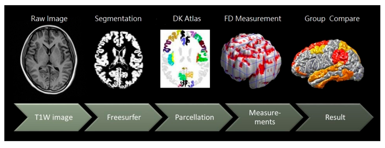
2. Materials and Methods
2.1. Participants
2.2. Image Acquisition and DK Cortical Atlas Parcellation
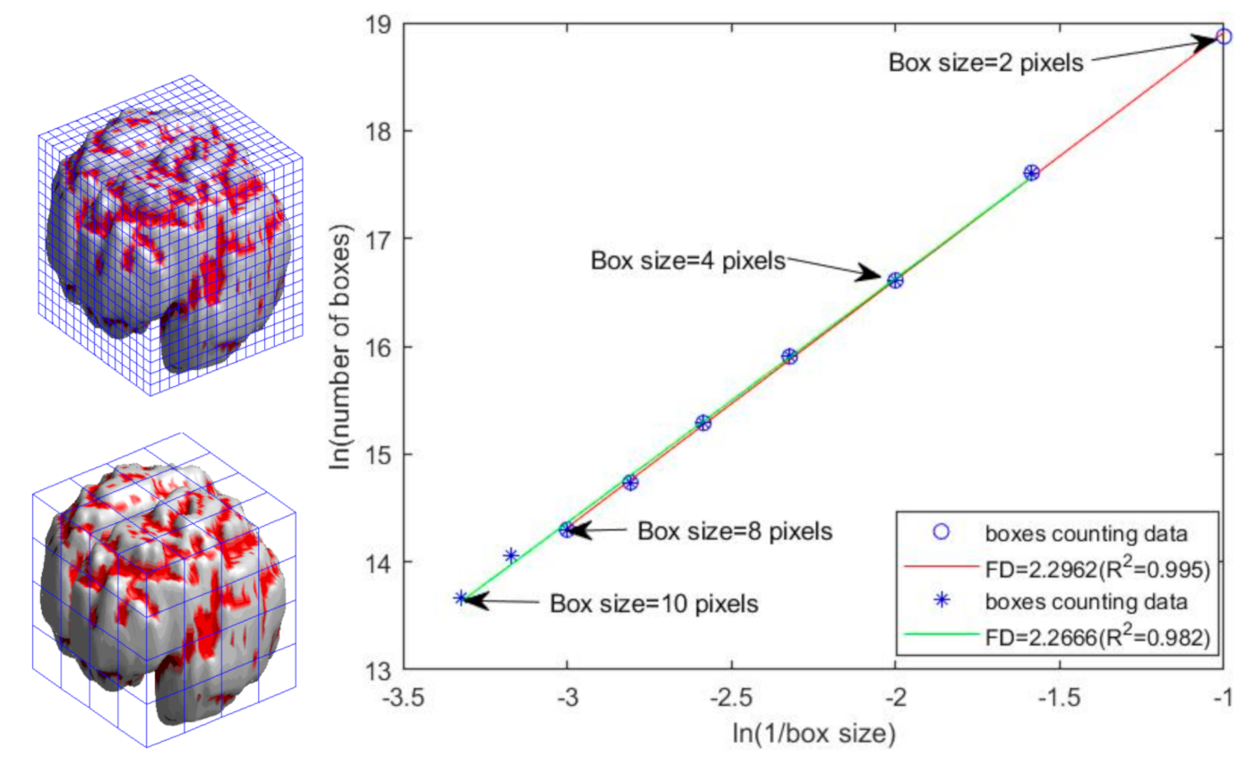
2.3. Three-Dimensional FD Measurement of Parcellated Regions
2.4. Statistical Analysis
3. Results
3.1. Women and Men Exhibited More Cortical Lateralization in Young and Middle Adulthood, Respectively
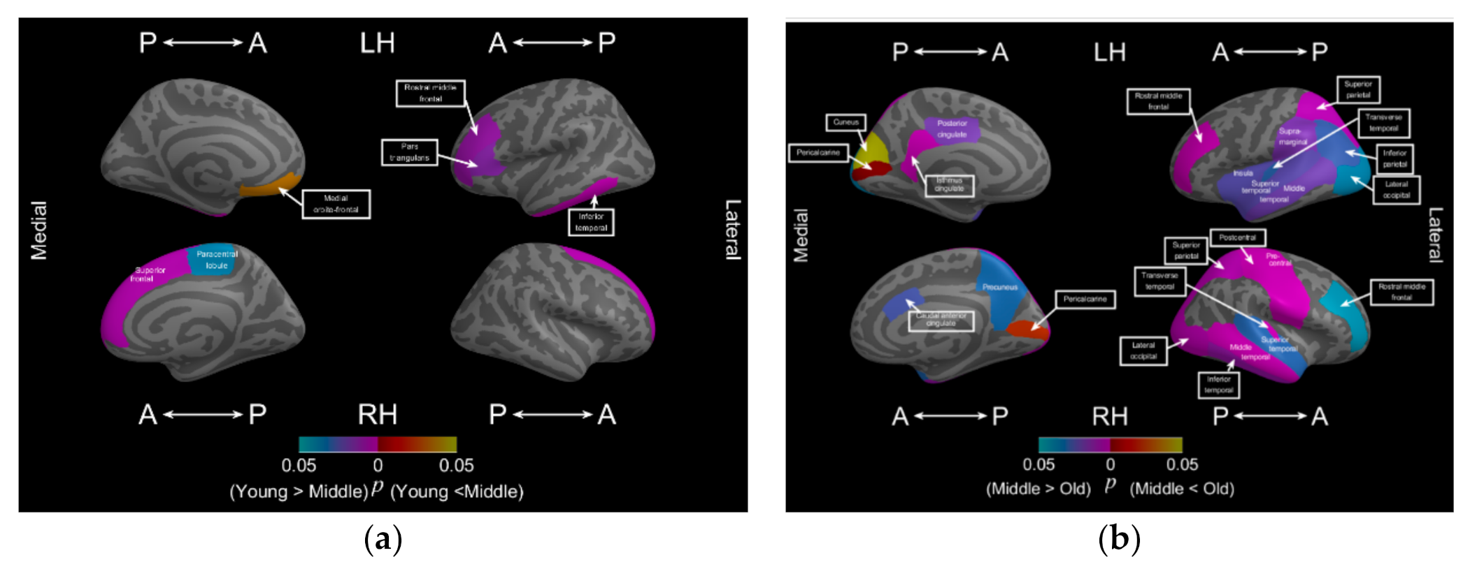
3.2. Aging Affects the Bilateral Frontal and Left Temporal Lobes Early
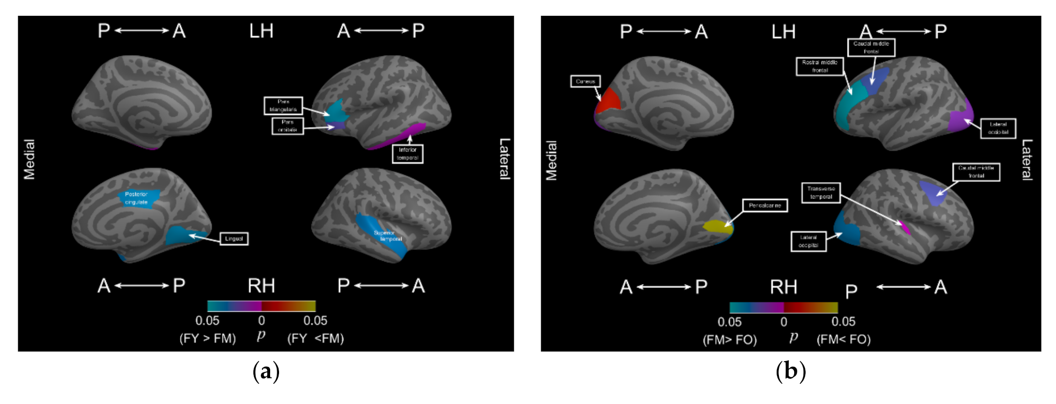
3.3. Men Exhibited a Decrease in Cortical Complexity Earlier and in More Subregions than Did Women across Two Age Periods of Adulthood
4. Discussion
4.1. Measuring FD Using the Cerebral Atlas Is a Superior Measure for Assessing Subregional Morphological Alterations in Normal Aging and Neurodegenerative Disease
4.2. Men Exhibited More Aging Effects on Cerebral Morphological Changes than Women
4.3. Measuring FD Using the DK Atlas to Develop a Prediagnosis System for Assessing Aging and Cortical Neurodegenerative Disease
5. Conclusions
Author Contributions
Funding
Institutional Review Board Statement
Informed Consent Statement
Data Availability Statement
Acknowledgments
Conflicts of Interest
References
- Fjell, A.M.; Walhovd, K.B.; Fennema-Notestine, C.; McEvoy, L.K.; Hagler, D.J.; Holland, D.; Brewer, J.B.; Dale, A.M. One-Year Brain Atrophy Evident in Healthy Aging. J. Neurosci. 2009, 29, 15223–15231. [Google Scholar] [CrossRef]
- Johansen-Berg, H. The future of Functionally-Related Structural Change Assessment. NeuroImage 2012, 62, 1293–1298. [Google Scholar] [CrossRef]
- Maguire, E.A.; Gadian, D.G.; Johnsrude, I.S.; Good, C.D.; Ashburner, J.; Frackowiak, R.S.J.; Frith, C.D. Navigation-Related Structural Change in the Hippocampi of Taxi Drivers. Proc. Natl. Acad. Sci. USA 2000, 97, 4398–4403. [Google Scholar] [CrossRef]
- Schmidt-Wilcke, T.; Rosengarth, K.; Luerding, R.; Bogdahn, U.; Greenlee, M. Distinct Patterns of Functional and Structural Neuroplasticity Associated with Learning Morse Code. NeuroImage 2010, 51, 1234–1241. [Google Scholar] [CrossRef]
- Hölzel, B.K.; Carmody, J.; Vangel, M.; Congleton, C.; Yerramsetti, S.M.; Gard, T.; Lazar, S.W. Mindfulness Practice Leads to Increases in Regional Brain Gray Matter Density. Psychiatry Res. Neuroimaging 2011, 191, 36–43. [Google Scholar] [CrossRef]
- Matsuda, H. Voxel-based Morphometry of Brain MRI in Normal Aging and Alzheimer’s Disease. Aging Dis. 2012, 4, 29–37. [Google Scholar]
- Coffey, C.E.; Lucke, J.F.; Saxton, J.A.; Ratcliff, G.; Unitas, L.J.; Bilig, B.; Bryan, R.N. Sex Differences in Brain Aging: A Quantitative Magnetic Resonance Imaging Study. Arch. Neurol. 1998, 55, 169–179. [Google Scholar]
- Guttmann, C.; Jolesz, F.A.; Kikinis, R.; Killiany, R.J.; Moss, M.B.; Sandor, T.; Albert, M.S. White Matter Changes with Normal Aging. Neurology 1998, 50, 972–978. [Google Scholar] [CrossRef]
- Cowell, P.E.; Turetsky, B.T.; Gur, R.C.; Grossman, R.I.; Shtasel, D.L.; Gur, R.E. Sex Differences in Aging of the Human Frontal and Temporal Lobe. J. Neurosci. 1994, 14, 4748–4755. [Google Scholar]
- Raz, N.; Gunning-Dixon, F.M.; Head, D.; Dupuis, J.H.; Acker, J.D. Neuroanatomical Correlates of Cognitive Aging: Evidence from Structural Magnetic Resonance Imaging. Neuropsychology 1998, 12, 95–114. [Google Scholar]
- Cho, S.; Jones, D.; Reddick, W.E.; Ogg, R.J.; Steen, R. Establishing norms for age-related changes in proton T1 of human brain tissue in vivo. Magn. Reson. Imaging 1997, 15, 1133–1143. [Google Scholar] [CrossRef]
- Jacobs, B.; Driscoll, L.; Schall, M. Life-span dendritic and spine changes in areas 10 and 18 of human cortex: A quantitative Golgi study. J. Comp. Neurol. 1997, 386, 661–680. [Google Scholar]
- Storsve, A.B.; Fjell, A.M.; Tamnes, C.K.; Westlye, L.T.; Overbye, K.; Aasland, H.W.; Walhovd, K.B. Differential Longitudinal Changes in Cortical Thickness, Surface Area and Volume Across the Adult Life Span: Regions of Accelerating and Decelerating Change. J. Neurosci. 2014, 34, 8488–8498. [Google Scholar]
- Long, X.; Liao, W.; Jiang, C.; Liang, D.; Qiu, B.; Zhang, L. Healthy Aging: An Automatic Analysis of Global and Regional Morphological Alterations of Human Brain. Acad. Radiol. 2012, 19, 785–793. [Google Scholar]
- Van Someren, E.J.W.; Oosterman, J.; Van Harten, B.; Vogels, R.; Gouw, A.; Weinstein, H.; Poggesi, A.; Scheltens, P.; Scherder, E. Medial Temporal Lobe Atrophy Relates More Strongly to Sleep-Wake Rhythm Fragmentation than to Age or Any Other Known Risk. Neurobiol. Learn. Mem. 2019, 160, 132–138. [Google Scholar] [CrossRef]
- Jack, C.R.; Petersen, R.C.; Xu, Y.; O’Brien, P.C.; Smith, G.E.; Ivnik, R.J.; Kokmen, E. Rate of Medial Temporal Lobe Atrophy in Typical Aging and Alzheimer’s Disease. Neurology 1998, 51, 993–999. [Google Scholar]
- Convit, A.; De Asis, J.; De Leon, M.; Tarshish, C.; De Santi, S.; Rusinek, H. Atrophy of the Medial Occipitotemporal, Inferior, and Middle Temporal Gyri in Non-Demented Elderly Predict Decline to Alzheimer’s Disease. Neurobiol. Aging 2000, 21, 19–26. [Google Scholar] [CrossRef]
- Xu, J.; Kobayashi, S.; Yamaguchi, S.; Iijima, K.; Okada, K.; Yamashita, K. Gender Effects on Age-Related Changes in Brain Structure. Am. J. Neuroradiol. 2000, 21, 112–118. [Google Scholar]
- Gur, R.E.; Gur, R.C. Gender Differences in Aging: Cognition, Emotions, and Neuroimaging Studies. Dialogues Clin. Neurosci. 2002, 4, 197. [Google Scholar]
- Arélin, K.; Mueller, K.; Barth, C.; Rekkas, P.V.; Kratzsch, J.; Burmann, I.; Villringer, A.; Sacher, J. Progesterone Mediates Brain Functional Connectivity Changes During the Menstrual Cycle—A Pilot Resting State MRI Study. Front. Neurosci. 2015, 9, 44. [Google Scholar] [CrossRef]
- Le, J.; Thomas, N.; Gurvich, C. Cognition, The Menstrual Cycle, and Premenstrual Disorders: A Review. Brain Sci. 2020, 10, 198. [Google Scholar] [CrossRef]
- Nguyen, T.-V.; McCracken, J.T.; Ducharme, S.; Cropp, B.F.; Botteron, K.N.; Evans, A.C.; Karama, S. Interactive Effects of Dehydroepiandrosterone and Testosterone on Cortical Thickness during Early Brain Development. J. Neurosci. 2013, 33, 10840–10848. [Google Scholar] [CrossRef]
- Henderson, V.W. Cognitive Changes after Menopause: Influence of Estrogen. Clin. Obstet. Gynecol. 2008, 51, 618–626. [Google Scholar] [CrossRef]
- Baek, H.-S.; Kim, G.-W.; Sundaram, T.; Park, K.; Jeong, G.-W. Brain Morphological Changes with Functional Deficit Associated With Sexual Arousal in Postmenopausal Women. Sex. Med. 2019, 7, 480–488. [Google Scholar] [CrossRef]
- Taylor, C.M.; Pritschet, L.; Olsen, R.K.; Layher, E.; Santander, T.; Grafton, S.T.; Jacobs, E.G. Progesterone Shapes Medial Temporal Lobe Volume Across the Human Menstrual Cycle. NeuroImage 2020, 220, 117125. [Google Scholar] [CrossRef]
- Ge, Y.; Grossman, R.I.; Babb, J.S.; Rabin, M.L.; Mannon, L.J.; Kolson, D.L. Age-Related Total Gray Matter and White Matter Changes in Normal Adult Brain. Part I: Volumetric MR Imaging Analysis. Am. J. Neuroradiol. 2002, 23, 1327–1333. [Google Scholar]
- Biasutti, M. A Comparative Analysis of Forms and Wikis as Tools for Online Collaborative Learning. Comput. Educ. 2017, 107, 158–171. [Google Scholar] [CrossRef]
- Lemaitre, H.; Goldman, A.L.; Sambataro, F.; Verchinski, B.A.; Meyer-Lindenberg, A.; Weinberger, D.R.; Mattay, V.S. Normal Age-Related Brain Morphometric Changes: Nonuniformity Across Cortical Thickness, Surface Area and Gray Matter Volume? Neurobiol. Aging 2012, 33, 617-e1. [Google Scholar] [CrossRef]
- Wu, Y.-T.; Shyu, K.-K.; Jao, C.-W.; Wang, T.-Y.; Soong, B.-W.; Wu, H.-M.; Wang, P.-S. Fractal Dimension Analysis for Quantifying Cerebellar Morphological Change of Multiple System Atrophy of the Cerebellar Type (MSA-C). NeuroImage 2010, 49, 539–551. [Google Scholar] [CrossRef]
- Mandelbrot, B. How Long Is the Coast of Britain? Statistical Self-Similarity and Fractional Dimension. Science 2006, 156, 636–638. [Google Scholar] [CrossRef]
- Ha, T.H.; Yoon, U.; Lee, K.J.; Shin, Y.-W.; Lee, J.-M.; Kim, I.Y.; Ha, K.S.; Kim, S.I.; Kwon, J.S. Fractal Dimension of Cerebral Cortical Surface in Schizophrenia and Obsessive–Compulsive Disorder. Neurosci. Lett. 2005, 384, 172–176. [Google Scholar] [CrossRef]
- Esteban, F.J.; Sepulcre, J.; De Mendizábal, N.V.; Goñi, J.; Navas, J.; De Miras, J.R.; Bejarano, B.; Masdeu, J.C.; Villoslada, P. Fractal Dimension and White Matter Changes in Multiple Sclerosis. NeuroImage 2007, 36, 543–549. [Google Scholar] [CrossRef]
- Esteban, F.J.; Sepulcre, J.; De Miras, J.R.; Navas, J.; De Mendizábal, N.V.; Goñi, J.; Quesada, J.M.; Bejarano, B.; Villoslada, P. Fractal Dimension Analysis of Grey Matter in Multiple Sclerosis. J. Neurol. Sci. 2009, 282, 67–71. [Google Scholar] [CrossRef]
- Desikan, R.S.; Ségonne, F.; Fischl, B.; Quinn, B.T.; Dickerson, B.C.; Blacker, D.; Buckner, R.L.; Dale, A.M.; Maguire, R.P.; Hyman, B.T.; et al. An Automated Labeling System for Subdividing the Human Cerebral Cortex on MRI Scans Into gyral Based Regions of Interest. NeuroImage 2006, 31, 968–980. [Google Scholar] [CrossRef]
- Shaper, A.G.; Wannamethee, S.G.; Walker, M. Body Weight: Implications for the Prevention of Coronary Heart Disease, Stroke, and Diabetes Mellitus in a Cohort Study of Middle Aged Men. BMJ 1997, 314, 1311. [Google Scholar] [CrossRef]
- Kim, J. Effect of Subjective Health Perception and Mental Health Status on the Quality of Life in the Late Middle Age. Int. J. Adv. Cult. Technol. 2020, 8, 18–27. [Google Scholar] [CrossRef]
- Geng, Z.; Liu, H.; Wang, L.; Zhu, Q.; Song, Z.; Chang, R.; Lv, H. A Voxel-Based Morphometric Study of Age- and Sex-Related Changes in White Matter Volume in the Normal Aging Brain. Neuropsychiatr. Dis. Treat. 2016, 12, 453–465. [Google Scholar] [CrossRef]
- Kiselev, V.G.; Hahn, K.R.; Auer, D.P. Is the Brain Cortex a Fractal? NeuroImage 2003, 20, 1765–1774. [Google Scholar] [CrossRef]
- Shan, Z.Y.; Liu, J.Z.; Glassa, J.O.; Gajjarc, A.; Lid, C.S.; Reddicka, W.E. Quantitative Morphologic Evaluation of White Matter in Survivors of Childhood Medulloblastoma. Magn. Reson. Imaging 2006, 24, 1015–1022. [Google Scholar]
- Madan, C.R.; Kensinger, E.A. Cortical complexity as a measure of age-related brain atrophy. NeuroImage 2016, 134, 617–629. [Google Scholar] [CrossRef]
- Marzi, C.; Giannelli, M.; Tessa, C.; Mascalchi, M.; Diciotti, S. Toward a More Reliable Characterization of Fractal Properties of the Cerebral Cortex of Healthy Subjects during the Lifespan. Sci. Rep. 2020, 10, 1–12. [Google Scholar] [CrossRef]
- Zhang, L.; Liu, J.Z.; Dean, D.; Sahgal, V.; Yue, G.H. A Three-Dimensional Fractal Analysis Method for Quantifying White Matter Structure in Human Brain. J. Neurosci. Methods 2006, 150, 242–253. [Google Scholar] [CrossRef]
- Benjamini, Y.; Hochberg, Y. Controlling the False Discovery Rate—A Practical and Powerful Approach to Multiple Testing. J. R. Stat. Soc. Ser. B Methodol. 1995, 57, 289–300. [Google Scholar]
- Collantoni, E.; Madan, C.R.; Meneguzzo, P.; Chiappini, I.; Tenconi, E.; Manara, R.; Favaro, A. Cortical Complexity in Anorexia Nervosa: A Fractal Dimension Analysis. J. Clin. Med. 2020, 9, 833. [Google Scholar] [CrossRef]
- Seitz, J.; Bühren, K.; Von Polier, G.G.; Heussen, N.; Herpertz-Dahlmann, B.; Konrad, K. Morphological Changes in the Brain of Acutely Ill and Weight-Recovered Patients with Anorexia Nervosa a Meta-Analysis and Qualitative Review. J. Child Adolesc. Psychiatry Psychother. 2014, 42, 7–18. [Google Scholar]
- Knopman, D.S.; Petersen, R.C. Mild Cognitive Impairment and Mild Dementia: A Clinical Perspective. Mayo Clin. Proc. 2014, 89, 1452–1459. [Google Scholar] [CrossRef]
- Reinvang, I.; Grambaite, R.; Espeseth, T. Executive Dysfunction in MCI: Subtype or Early Symptom. Int. J. Alzheimer’s Dis. 2012, 2012, 1–8. [Google Scholar] [CrossRef]
- Petersen, R.C.; Roberts, R.O.; Knopman, D.S.; Geda, Y.E.; Cha, R.H.; Pankratz, V.S.; Boeve, B.F.; Tangalos, E.G.; Ivnik, R.J.; Rocca, W.A. Prevalence of Mild Cognitive Impairment is Higher in Men: The Mayo Clinic Study of Aging. Neurology 2010, 75, 889–897. [Google Scholar] [CrossRef]
- Xu, B.; Xiong, F.; Tian, R.; Zhan, S.; Gao, Y.; Qiu, W.; Wang, R.; Ge, W.; Ma, C. Temporal Lobe in Human Aging: A Quantitative Protein Profiling Study of Samples from Chinese Human Brain Bank. Exp. Gerontol. 2016, 73, 31–41. [Google Scholar] [CrossRef]
- Rosen, A.C.; Gabrieli, J.D.; Stoub, T.; Prull, M.W.; O’Hara, R.; Yesavage, J.; Detoledo-Morrell, L. Relating Medial Temporal Lobe Volume to Frontal fMRI Activation for Memory Encoding in Older Adults. Cortex 2005, 41, 595–602. [Google Scholar] [CrossRef]
- Boyle, R.; Jollans, L.; Rueda-Delgado, L.M.; Rizzo, R.; Yener, G.G.; McMorrow, J.P.; Knight, S.; Carey, D.; Robertson, I.H.; Emek-Savaş, D.D.; et al. Brain-Predicted Age Difference Score is Related to Specific Cognitive Functions: A Multi-Site Replication Analysis. Brain Imaging Behav. 2020, 1–19. [Google Scholar] [CrossRef]
- Aycheh, H.M.; Seong, J.-K.; Shin, J.-H.; Na, D.L.; Kang, B.; Seo, S.W.; Sohn, K.-A. Biological Brain Age Prediction Using Cortical Thickness Data: A Large Scale Cohort Study. Front. Aging Neurosci. 2018, 10, 252. [Google Scholar] [CrossRef]
- Cole, J.H.; Ritchie, S.J.; Bastin, M.; Hernández, M.C.V.; Maniega, S.M.; Royle, N.; Corley, J.; Pattie, A.; Harris, S.; Zhang, Q.; et al. Brain Age Predicts Mortality. Mol. Psychiatry 2018, 23, 1385–1392. [Google Scholar] [CrossRef]


| Group | Young (Number/Mean Age ± std) | Middle | Old |
|---|---|---|---|
| Female | 30/39.8 ± 3.6 | 41/53.94 ± 4.44 | 41/67.23 ± 5.34 |
| Male | 36/38.93 ± 7.22 | 57/53.61 ± 4.25 | 53/67.8 ± 4.52 |
| Total | 66/39.45 ± 6.08 | 98/53.74 ± 4.32 | 94/67.35 ± 4.87 |
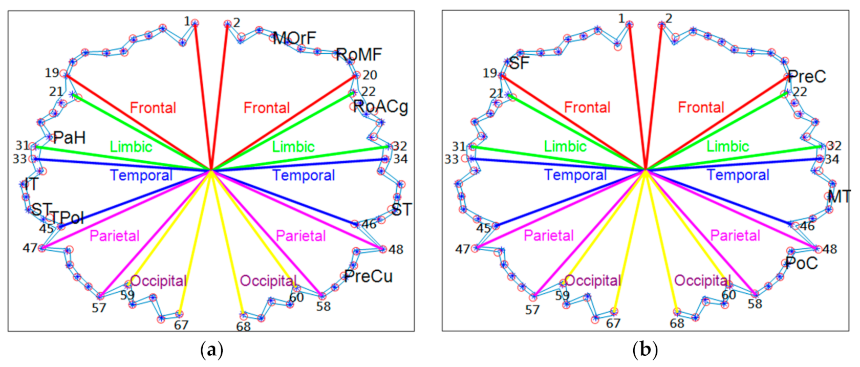
| Frontal | ROI | Abbreviation | Temporal | ROI | Abbreviation |
|---|---|---|---|---|---|
| 1, 2 | Caudal middle frontal | CMF | 33, 34 | Bankssts | B |
| 3, 4 | Frontal pole | FPol | 35, 36 | Entorhinal | En |
| 5, 6 | Lateral orbitofrontal | LOrF | 37, 38 | Inferior temporal | IT |
| 7, 8 | Medial orbitofrontal | MOrF | 39, 40 | Middle temporal | MT |
| 9, 10 | Pars opercularis | Op | 41, 42 | Superior temporal | ST |
| 11, 12 | Pars orbitalis | Or | 43, 44 | Temporal pole | TPol |
| 13, 14 | Pars triangularis | Tr | 45, 46 | Transverse temporal | TrT |
| 15, 16 | Rostral middle frontal | RoMF | Parietal | ||
| 17, 18 | Superior frontal | SF | 47, 48 | Inferior parietal | IP |
| 19, 20 | Precentral gyrus | PreC | 49, 50 | Paracentral | PaC |
| Limbic | 51, 52 | Postcentral | PoC | ||
| 21, 22 | Caudal anterior cingulate | CACg | 53, 54 | Precuneus | PreCu |
| 23, 24 | Rostral anterior cingulate | RoACg | 55, 56 | Superior parietal | SP |
| 25, 26 | Isthmus cingulate | IstCg | 57, 58 | Supra marginal | SM |
| 27, 28 | Insula | Ins | Occipital | ||
| 29, 30 | Parahippocampal | PaH | 59, 60 | Pericalcarine | PerCa |
| 31, 32 | Posterior cingulate | PoCg | 61, 62 | Fusiform | Fu |
| 63, 64 | Cuneus | Cu | |||
| 65, 66 | Lateral occipital | LO | |||
| 67, 68 | Lingual | Lg |
| Lobes | Index | FY (L) | FY(R) | FM(L) | FM(R) | FO(L) | FO (R) |
|---|---|---|---|---|---|---|---|
| Frontal Lobe | CMF | 2.0806 * | 2.1507 | 2.0718 ** | 2.1259 | 2.0650 *** | 2.1163 |
| FPol | 2.2815 | 2.2993 | 2.2933 | 2.2886 | 2.2806 | 2.2731 | |
| LOrF | 2.2873 | 2.2770 | 2.2848 | 2.2636 ** | 2.2768 | 2.2573 *** | |
| MOrF | 2.2125 * | 2.2448 | 2.2373 | 2.2379 | 2.2240 | 2.2380 | |
| Op | 2.1693 * | 2.2057 | 2.1624 ** | 2.1919 | 2.1565 *** | 2.2012 | |
| Or | 2.2693 | 2.2169 * | 2.2542 | 2.2180 ** | 2.2451 | 2.2114 *** | |
| Tr | 2.1662 | 2.1879 | 2.1351 ** | 2.1707 | 2.1368 *** | 2.1803 | |
| RoMF | 2.2512 | 2.2736 | 2.2306 ** | 2.2688 | 2.2261 *** | 2.2645 | |
| SF | 2.3137 | 2.3069 | 2.3141 | 2.3083 | 2.3114 | 2.3028 | |
| PreC | 2.1178 | 2.0897 | 2.1295 | 2.1084 | 2.1195 | 2.0936 | |
| Limbic Lobe | CACg | 2.4088 | 2.3993 | 3.4011 | 2.3946 | 2.3920 | 2.3916 |
| RoACg | 2.4058 | 2.4057 | 2.4055 | 2.4008 | 2.4012 | 2.3980 | |
| IstCg | 2.0447 | 2.0605 | 2.0154 ** | 2.0493 | 2.0342 | 2.0553 | |
| Ins | 2.2366 | 2.2221 | 2.2335 | 2.2275 | 2.2250 | 2.2319 | |
| PaH | 2.1695 | 2.1575 | 2.1519 | 2.1594 | 2.1545 | 2.1463 | |
| PoCg | 2.0932 | 2.0939 | 2.0817 | 2.0745 | 2.0891 | 2.0883 | |
| Temporal Lobe | B | 2.2901 | 2.2803 | 2.2914 | 2.2796 ** | 2.2823 | 2.2724 |
| En | 2.3350 | 2.3147 * | 2.3171 | 2.3104 | 2.3242 | 2.3023 *** | |
| IT | 2.3038 | 2.3158 | 2.2965 ** | 2.3096 | 2.2873 *** | 2.3018 | |
| MT | 2.0800 | 2.0214 * | 2.0606 | 2.0210 ** | 2.0503 | 2.0173 *** | |
| ST | 2.3170 * | 2.3389 | 2.3140 ** | 2.3255 | 2.3074 *** | 2.3242 | |
| TPol | 2.1664 | 2.1766 | 2.1869 | 2.1846 | 2.1940 | 2.1776 | |
| TrT | 2.0390 | 1.9603 * | 2.0298 | 1.9688 ** | 2.0157 | 1.9320 *** | |
| Parietal Lobe | IP | 2.3903 * | 2.4234 | 2.3947 ** | 2.4201 | 2.3891 *** | 2.4181 |
| PaC | 2.1032 | 2.0912 | 2.1002 | 2.0989 | 2.0834 | 2.0867 | |
| PoC | 2.2889 | 2.2679 * | 2.2791 | 2.2703 | 2.2844 | 2.2639 *** | |
| PreCu | 2.1777 * | 2.2242 | 2.1771 | 2.1932 | 2.1542 *** | 2.1844 | |
| SP | 2.3139 * | 2.3409 | 2.3158 | 2.3271 | 2.3065 *** | 2.3306 | |
| SM | 2.3508 | 2.3583 | 2.3498 | 2.3484 | 2.3436 | 2.3402 | |
| Occipital Lobe | PerCa | 2.3726 | 2.3717 | 2.3784 | 2.3589 ** | 2.3718 | 2.3541 *** |
| Fu | 2.0687 * | 2.1364 | 2.0853 ** | 2.1352 | 2.1085 *** | 2.1379 | |
| Cu | 2.3672 | 2.3688 | 2.3633 | 2.3682 | 2.3494 | 2.3557 | |
| LO | 2.2647 | 2.2663 | 2.2447 | 2.2459 | 2.2479 | 2.2506 | |
| Lg | 2.0355 * | 2.0975 | 2.0519 | 2.0648 | 2.0815 | 2.0935 | |
| Number of significant smaller sub-regions | 9 | 5 | 9 | 6 | 10 | 7 | |
| Lobes | Index | MY (L) | MY(R) | MM(L) | MM(R) | MO(L) | MO (R) |
|---|---|---|---|---|---|---|---|
| Frontal Lobe | CMF | 2.1162 | 2.1330 | 2.0865 ** | 2.1400 | 2.0611 *** | 2.1182 |
| FPol | 2.3051 | 2.3125 | 2.2979 | 2.2922 | 2.3028 | 2.2943 | |
| LOrF | 2.3086 | 2.2961 | 2.2991 | 2.2766 ** | 2.3063 | 2.2801 *** | |
| MOrF | 2.2320 * | 2.2634 | 2.2404 ** | 2.2578 | 2.2419 | 2.2481 | |
| Op | 2.1858 | 2.2054 | 2.1668 ** | 2.1889 | 2.1616 | 2.1760 | |
| Or | 2.2600 | 2.2214 * | 2.2591 | 2.2181 ** | 2.2588 | 2.2223 *** | |
| Tr | 2.1825 | 2.1892 | 2.1669 ** | 2.1936 | 2.1716 *** | 2.2020 | |
| RoMF | 2.2646 * | 2.2981 | 2.2424 ** | 2.2744 | 2.2351 *** | 2.2687 | |
| SF | 2.3363 | 2.3204 | 2.3208 | 2.3189 | 2.3158 | 2.3100 | |
| PreC | 2.1340 | 2.1090 | 2.1346 | 2.1203 | 2.1413 | 2.1136 *** | |
| Limbic Lobe | CACg | 2.4284 | 2.4255 | 2.4098 | 2.4115 | 2.4027 | 2.4061 |
| RoACg | 2.4190 | 2.4262 | 2.4154 | 2.4047 ** | 2.4108 | 2.4042 | |
| IstCg | 2.0524 | 2.0939 | 2.0351 ** | 2.0714 | 2.0343 *** | 2.0777 | |
| Ins | 2.2578 | 2.2412 | 2.2417 | 2.2303 ** | 2.2357 | 2.2237 | |
| PaH | 2.1893 | 2.1970 | 2.1758 | 2.1635 | 2.1754 | 2.1656 | |
| PoCg | 2.1118 | 2.0722 | 2.1116 | 2.0920 ** | 2.0892 | 2.0936 | |
| Temporal Lobe | B | 2.3176 | 2.3096 | 2.2938 | 2.2920 | 2.2967 | 2.2891 |
| En | 2.3509 | 2.3434 | 2.3311 | 2.3269 | 2.3304 | 2.3179 | |
| IT | 2.3146 | 2.3317 | 2.3130 ** | 2.3273 | 2.3076 | 2.3156 | |
| MT | 2.0693 | 2.0542 | 2.0662 | 2.0356 ** | 2.0538 | 2.0306 *** | |
| ST | 2.3387 | 2.3501 | 2.3278 ** | 2.3418 | 2.3211 | 2.3322 | |
| TPol | 2.1996 | 2.1899 | 2.1985 | 2.1925 | 2.1904 | 2.1808 | |
| TrT | 2.0899 | 1.9919 * | 2.0543 | 1.9845 ** | 2.0414 | 1.9616 *** | |
| Parietal Lobe | IP | 2.4268 * | 2.4455 | 2.4035 ** | 2.4342 | 2.3962 *** | 2.4298 |
| PaC | 2.1167 | 2.1198 | 2.1207 | 2.1036 ** | 2.1047 | 2.0924 | |
| PoC | 2.2906 | 2.2840 | 2.2842 | 2.2769 | 2.2816 | 2.2633 *** | |
| PreCu | 2.2137 | 2.2038 | 2.1912 ** | 2.2106 | 2.1789 | 2.1963 | |
| SP | 2.3510 | 2.3432 | 2.3283 ** | 2.3380 | 2.3255 | 2.3225 | |
| SM | 2.3697 | 2.3744 | 2.3526 ** | 2.3602 | 2.3421 | 2.3517 | |
| Occipital Lobe | PerCa | 2.3925 | 2.3813 | 2.3923 | 2.3739 ** | 2.3852 | 2.3714 *** |
| Fu | 2.1176 | 2.1530 | 2.1013 ** | 2.1455 | 2.1112 *** | 2.1469 | |
| Cu | 2.3717 | 2.3807 | 2.3666 ** | 2.3752 | 2.3648 | 2.3693 | |
| LO | 2.2782 | 2.2669 | 2.2705 | 2.2611 | 2.2766 | 2.2761 | |
| Lg | 2.0575 | 2.0531 | 2.0652 ** | 2.0961 | 2.0950 | 2.1152 | |
| Number of significant smaller sub-regions | 3 | 2 | 15 | 9 | 6 | 7 | |
| From Young to Middle Age Period | |
| Left Hemisphere | Right: Hemisphere |
| Frontal: Rostral middle frontal, Pars triangularis Temporal: Inferior temporal | Frontal: Superior frontal Parietal: paracentral |
| From Middle to Old Age Period | |
| Left Hemisphere | Right Hemisphere |
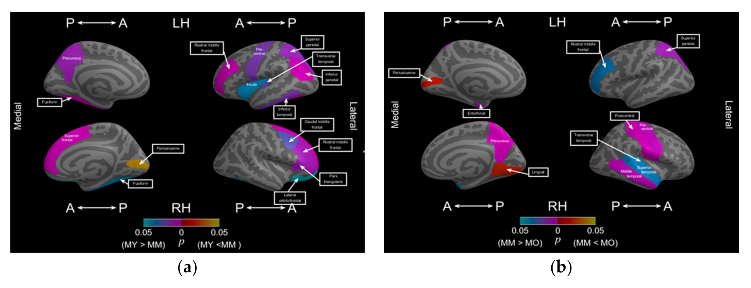
| Frontal: Rostral middle frontal, Limbic: Isthmus cingulate, Insula Posterior cingulate Temporal: Middle temporal, Superior temporal, Transverse temporal, Parietal: Inferior parietal, Supra marginal,Superior parietal Occipital: Lateral Occipital | Frontal: Rostral middle frontal, Precentral Limbic: Caudal anterior cingulate Temporal: Inferior temporal, Middle temporal Superior temporal, Transverse temporal, Parietal: Postcentral, Precuneus, Superior parietal Occipital: Lateral Occipital |
| Female | Young to Middle Age Period | |
| Left Hemisphere | Right Hemisphere | |
| Frontal: Pars triangularis, Pars orbitalis Temporal: Infer temporal | Limbic: Posterior cingulate Temporal: Superior temporal Occipital: Lingual | |
| Middle to Old Age Period | ||
| Left: | Right | |
| Frontal: rostral middle frontal, caudal middle frontal Occipital: Lateral occipital | Frontal: caudal middle frontal Temporal: Transverse temporal Occipital: Lateral Occipital | |
| Male | Young to Middle Age Period | |
| Left Hemisphere | Right Hemisphere | |
| Frontal: Rostral middle frontal, Precentral Limbic: Insula Temporal: Inferior Temporal Parietal: Inferior parietal, Precuneus, Superior parietal, Transverse temporal Occipital: Fusiform | Frontal: Caudal middle frontal, Lateral orbitofrontal, Rostral middle frontal, Pars triangulars, Superior Frontal Occipital: Fusiform | |
| Middle to Old Age Period | ||
| Left Hemisphere | Right Hemisphere | |
| Frontal: Rostral middle frontal Temporal: Entorhinal cortex Parietal: Superior parietal | Frontal: precentral Temporal: middle temporal, Superior temporal, Transverse temporal Parietal: Postcentral, precuneus | |
Publisher’s Note: MDPI stays neutral with regard to jurisdictional claims in published maps and institutional affiliations. |
© 2021 by the authors. Licensee MDPI, Basel, Switzerland. This article is an open access article distributed under the terms and conditions of the Creative Commons Attribution (CC BY) license (http://creativecommons.org/licenses/by/4.0/).
Share and Cite
Jao, C.-W.; Lau, C.I.; Lien, L.-M.; Tsai, Y.-F.; Chu, K.-E.; Hsiao, C.-Y.; Yeh, J.-H.; Wu, Y.-T. Using Fractal Dimension Analysis with the Desikan–Killiany Atlas to Assess the Effects of Normal Aging on Subregional Cortex Alterations in Adulthood. Brain Sci. 2021, 11, 107. https://doi.org/10.3390/brainsci11010107
Jao C-W, Lau CI, Lien L-M, Tsai Y-F, Chu K-E, Hsiao C-Y, Yeh J-H, Wu Y-T. Using Fractal Dimension Analysis with the Desikan–Killiany Atlas to Assess the Effects of Normal Aging on Subregional Cortex Alterations in Adulthood. Brain Sciences. 2021; 11(1):107. https://doi.org/10.3390/brainsci11010107
Chicago/Turabian StyleJao, Chi-Wen, Chi Ieong Lau, Li-Ming Lien, Yuh-Feng Tsai, Kuang-En Chu, Chen-Yu Hsiao, Jiann-Horng Yeh, and Yu-Te Wu. 2021. "Using Fractal Dimension Analysis with the Desikan–Killiany Atlas to Assess the Effects of Normal Aging on Subregional Cortex Alterations in Adulthood" Brain Sciences 11, no. 1: 107. https://doi.org/10.3390/brainsci11010107
APA StyleJao, C.-W., Lau, C. I., Lien, L.-M., Tsai, Y.-F., Chu, K.-E., Hsiao, C.-Y., Yeh, J.-H., & Wu, Y.-T. (2021). Using Fractal Dimension Analysis with the Desikan–Killiany Atlas to Assess the Effects of Normal Aging on Subregional Cortex Alterations in Adulthood. Brain Sciences, 11(1), 107. https://doi.org/10.3390/brainsci11010107