Case Report

Alopecia Totalis Treated with 1064 nm Picosecond Nd:YAG Laser: A Case Report

1
Department of Dermatology, Far Eastern Memorial Hospital, New Taipei 22060, Taiwan
2
Ferrari Skin and Hair Clinic, Taipei 11071, Taiwan
3
Graduate Institute of Applied Science and Engineering, College of Science and Engineering, Fu Jen Catholic University, New Taipei 24205, Taiwan
4
Department of Dermatology, Chang Gung Memorial Hospital, Linkou, Taoyuan 33305, Taiwan
5
College of Medicine, Chang Gung University, Taoyuan 33302, Taiwan
*
Authors to whom correspondence should be addressed.
Appl. Sci. 2019, 9(7), 1298; https://doi.org/10.3390/app9071298
Submission received: 25 February 2019 / Revised: 23 March 2019 / Accepted: 25 March 2019 / Published: 28 March 2019
(This article belongs to the Special Issue Ultrafast Laser Pulses)

Abstract

:
Alopecia areata (AA) is an autoimmune disorder causing nonscarring hair loss. Alopecia totalis (AT), the severe form of AA, is usually refractory to many first-line treatments including topical and intralesional corticosteroids and topical immunotherapy. Systemic corticosteroids, ultraviolet light phototherapy, and immunosuppressants may be effective but can cause side effects and high recurrence rates after discontinuing treatment. Janus kinase inhibitors and numerous types of lasers, such as 308 nm excimer laser or light, low-level laser therapy, and fractional lasers have been explored for alternative treatment of AA over the past few years with beneficial effects. Herein, we presented a case of AT treated with the novel 1064 nm picosecond Nd:YAG laser (ps-Nd:YAG) (Cutera, Brisbane, CA, USA) under a split-scalp study. A 25-year-old woman presenting with AT for two years was treated with topical and intralesional steroid and the 1064 nm Nd-YAG picosecond laser on the right half of the scalp. The laser treatment interval was once a month. The left half of the scalp was treated with topical and intralesional steroids alone. After treatment for six times, the right half of the scalp had greater improvement than the left half of the scalp. 1064 nm picosecond Nd:YAG laser therapy may be an effective alternative adjuvant treatment for AT.

1. Introduction

Alopecia areata (AA) is a polygenic autoimmune disease causing nonscarring hair loss mediated through chronic hair follicle inflammation. The lifetime risk of this condition is about 2.1% of the world population [1,2]. Although patchy AA (patchy hair loss involving <40% of the scalp) is the most common presentation, it can progress to complete loss of scalp hairs (alopecia totalis (AT)), or the entire body hairs (alopecia universalis (AU)) [1,3]. Although spontaneous remission may occur in patchy AA within one year, patients with more extensive hair loss, including AT and AU, do not commonly experience remission and are difficult to treat [1,4,5].
Traditionally, the first-line treatments for mild patchy AA include topical and intralesional steroids [6,7,8], but both increase the risk of skin atrophy over the treated sites [9]. Systemic steroids are used in moderate to severe AA [10]. Arguments against the use of systemic steroids include relapses, adverse side effects, and ineffectiveness in the case of AT or AU [8,11,12,13]. Topical immunotherapy with use of sensitizing agent, such as diphenylcyclopropenone (DPCP) or squaric acid dibutylester (SADBE), is the treatment option for severe AA (>50% scalp involvement) or chronic relapsing AA [8,14]. Dermatitis, blistering, urticaria, and depigmentation are potential adverse effects [1]. Oral Janus kinase inhibitors (tofacitinib, ruxolitinib, baricitinib) are being popularly investigated as potential treatment for AA in recent years. The response rate revealed in one meta-analysis was impressive (72.4%), with mean time to initial and complete hair growth about 2.2 and 6.7 months [8,15,16]. Various response rates of phototherapy for treatment of AA, including narrow-band ultraviolet (UV) B [17,18], topical psorlaen-UVA [19,20], and UVA-1 [21], have been reported. However, the long-term potential oncogenic effect on treated areas is a limiting factor of phototherapy [22].
Due to the side effects and high relapse rates of traditional medical treatment and phototherapy, numerous types of lasers have been explored for alternative treatment of AA [4], including the 308 nm excimer laser or light, low-level laser therapy (LLLT) or photomodulation therapy and fractional lasers. Each of these lasers/lights has different mechanisms and variable effects on hair growth.
Recently, novel picosecond duration lasers (ps-lasers) have been developed for the treatment of unwanted tattoos [23,24,25,26,27]. The picosecond pulse duration is 10 times shorter than the traditional q-switched nanosecond devices. This shorter pulse delivery creates more photomechanical shock than the nanosecond duration, with greater possibility of shattering ink or pigment, and thus requires less treatments to remove tattoos than the longer-nanosecond devices [28]. Since 2013, ps-lasers have become commercially available [23]. The most popular ones for clinical use are picosecond 755 nm alexandrite laser (ps-Alex laser) and picosecond 1064/532 nm Nd:YAG laser (ps-Nd:YAG laser).
To date, no studies have evaluated the effects of the novel 1064 nm picosecond Nd:YAG laser (ps-Nd:YAG) for the treatment of AA. The objective of this study was to evaluate the efficacy of 1064 nm ps-Nd:YAG laser on hair growth of one patient with AT.

2. Materials and Methods
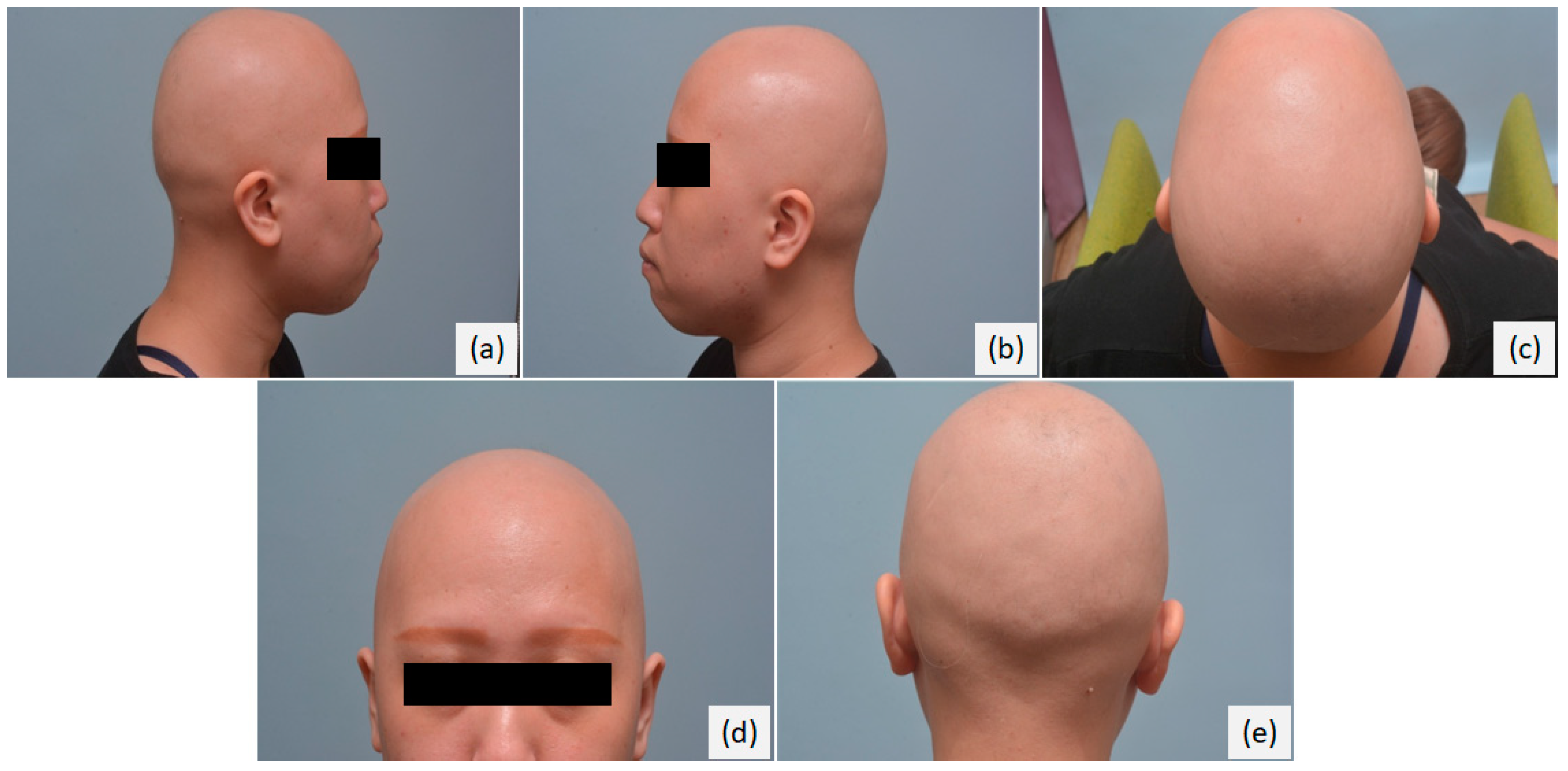
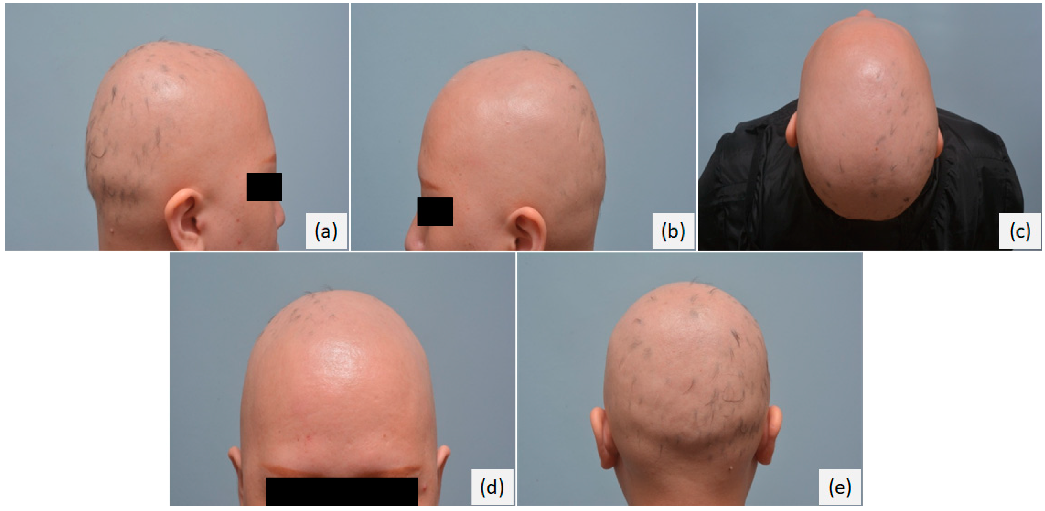
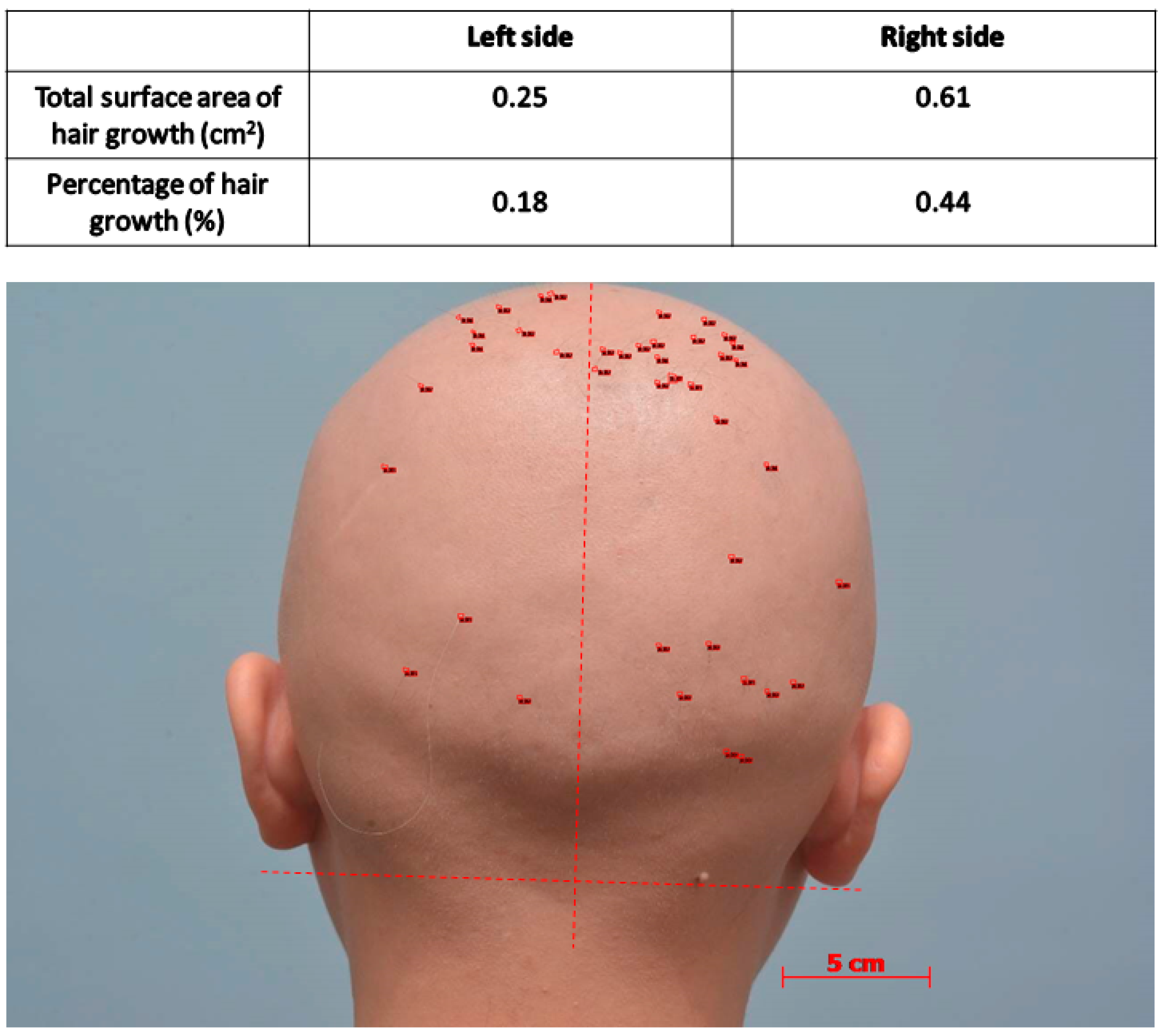
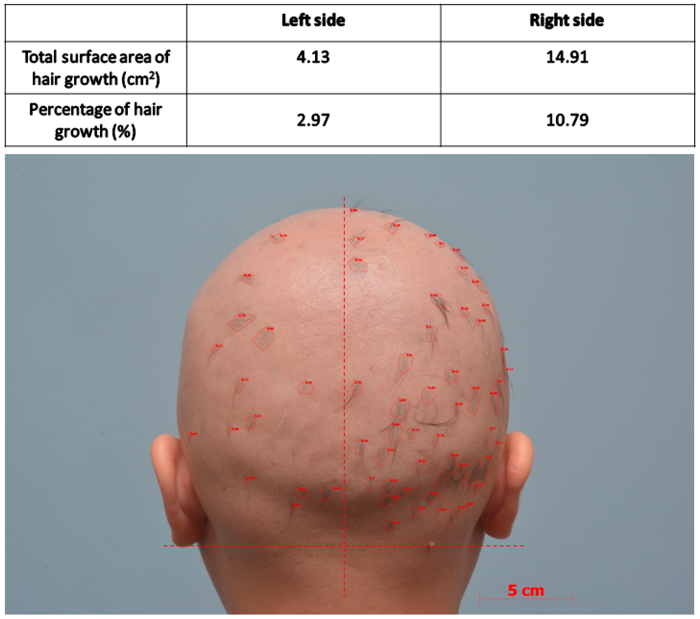
A 25-year-old woman presented with a two-year history of alopecia totalis (Figure 1). She had received treatments including topical steroid and intralesional steroid injections for many times but only scanty hair growth was noted. After obtaining an informed consent, we conducted a split-scalp therapeutic strategy. The right half of her scalp was treated with topical and intralesional steroids and the 1064 nm ps-Nd-Yag laser (Cutera, Brisbane, CA, USA) with Microlens array. The left side was treated with topical and intralesional steroids alone. The ps-laser treatment interval was once a month. A pulse energy of 1.0 J/cm2 was used, and one to three passes per session were performed. The treatment was well tolerated with only mild erythema after treatment.

3. Results

Multiple areas of focal hair growth on the right half of the scalp were observed after laser treatment for six months. In contrast, hair growth was barely noted on the left half of the scalp except few small tufts of hair growth on the left parietal scalp (Figure 2). We used the software (Axio Vision Rel 4.8) to estimate the total surface area and the percentage of hair growth on each side of the scalp before and after laser treatment (Figure 3 and Figure 4). The hair regrowth rate on the right side of the scalp was 10.35% (=10.79–0.44%). The hair regrowth rate on the left side of the scalp was 2.79% (=2.97–0.18%). The ratio of hair regrowth rate between laser- versus nonlaser-treated side was about 3.71.

4. Discussion

AA occurs as a result of T-cell mediated autoimmune reaction against hair follicles. The main histopathologic feature of AA is a lymphocytic infiltrate surrounding the follicular bulbs with a peribulbar or intrabulbar “swarm of bees” pattern [4]. Affected hair follicles terminate their anagen phase prematurely and regress via the induction of massive apoptosis of the lower portion of the follicle (the catagen phase), which results in a resting hair follicle (the telogen phase) [29]. Genome-wide association studies (GWASs) have greatly facilitated our understanding of the immune pathways involved in AA. Some genes found in AA patients were related to autophagy/apoptosis, regulatory T-cells (T regs) and Janus kinase signaling [15].
Anecdotal paradoxical hypertrichosis noted during laser epilation for hair removal has generated the interest in the use of laser to stimulate hair growth [30,31,32]. There is no study of the 1064 nm ps-Nd:YAG laser in inducing hair regrowth in AA. In our case report, 1064 nm ps-Nd:YAG laser provided beneficial effect for treatment of AT. The mechanism is unknown. It is proposed to be the induction of T-cell apoptosis and enhancement of hair growth. Similar to 308 nm excimer laser and LLLT reported in many studies, laser might promote hair growth by inducing T-cell apoptosis or decreasing inflammation [29]. Another possible mechanism may resemble the action of fractional laser, with a decrease in perifollicular lymphocytic infiltration through ‘scattering of perifollicular lymphocytes’ [29]. The 1064 nm ps-Nd:YAG laser treatment may result in a laser-induced optic breakdown, which are microinjuries, with pockets of intraepidermal necrosis, created without damage to the dermis below or ablative wound to the tissue surface. This process induces cavitation bubbles in the skin, which expand and may generate shock waves to disrupt the tissue and create a healing process that includes lymphocyte infiltrations [28]. These phenomena may scatter perifollicular lymphocyte infiltration and halt disease progression by arresting the hair follicles in the telogen stage of the hair cycle and increasing the anagen stage. Besides, minor trauma and the wound healing process itself can drive hair growth by de novo hair follicle neogenesis in skin [33,34].
In our study, this patient demonstrated greater hair regrowth on the laser treatment plus topical and intralesional steroid use side of the scalp compared with the side with topical and intralesional steroid use alone. It means the picosecond laser may induce a different mechanism to hair regrowth other than the anti-inflammatory effect of steroid. Besides, picosecond laser treatment may have synergistic effect with steroid use. Whether the cytokines or signaling pathways involved in modifying the immunology of the scalp is related to Janus kinase signaling is less understood and needs more investigation. Besides, there is no optimal therapeutic parameter yet, because this is the first study to investigate the picosecond laser treatment for AA. According to many previous reports which were treated by other laser modalities to AA, we modified the treatment protocols with medium energy, once a month in frequency for more than six months. Further studies need to be undertaken to determine the optimal therapeutic parameters.

5. Conclusions

AT, the severe form of alopecia areata, is difficult to treat and may have a large impact on patients’ quality of life, causing both cosmetic and psychosocial distress. Recently, various treatment modalities, including Janus kinase inhibitors and light therapies, have shown beneficial effects on AA. Here, we reported one case with AT who received a novel treatment with 1064 nm ps-Nd:YAG laser under a split-scalp strategy with good response. In conclusion, 1064 nm ps-Nd:YAG laser may be an effective alternative adjuvant treatment for alopecia areata, but more cohort studies should be conducted to support this hypothesis in the future.

Author Contributions

Conceptualization and methodology, T.-H.T.; writing—original draft preparation, S.-J.J.; writing—review and editing, T.-H.T., S.-H.W., and C.-C.C.

Funding

This research received no external funding.

Conflicts of Interest

The authors declared no conflict of interest.

References

  1. Pratt, C.H.; King, L.E., Jr.; Messenger, A.G.; Christiano, A.M.; Sundberg, J.P. Alopecia areata. Nat. Rev. Dis. Primers 2017, 3, 17011. [Google Scholar] [CrossRef] [PubMed]
  2. Mirzoyev, S.A.; Schrum, A.G.; Davis, M.D.P.; Torgerson, R.R. Lifetime incidence risk of alopecia areata estimated at 2.1% by Rochester Epidemiology Project, 1990–2009. J. Investig. Dermatol. 2014, 134, 1141–1142. [Google Scholar] [CrossRef] [PubMed]
  3. Hordinsky, M.K. Overview of alopecia areata. J. Investig. Dermatol. Symp. Proc. 2013, 16, S13–S15. [Google Scholar] [CrossRef] [PubMed]
  4. Mlacker, S.; Aldahan, A.S.; Simmons, B.J.; Shah, V.; McNamara, C.A.; Samarkandy, S.; Nouri, K. A review on laser and light-based therapies for alopecia areata. J. Cosmet. Laser Ther. 2017, 19, 93–99. [Google Scholar] [CrossRef]
  5. MacDonald Hull, S.P.; Wood, M.L.; Hutchinson, P.E.; Sladden, M.; Messenger, A.G.; British Association of Dermatologists. Guidelines for the management of alopecia areata. Br. J. Dermatol. 2003, 149, 692–699. [Google Scholar] [CrossRef]
  6. Charuwichitratana, S.; Wattanakrai, P.; Tanrattanakorn, S. Randomized double-blind placebo-controlled trial in the treatment of alopecia areata with 0.25% desoximetasone cream. Arch. Dermatol. 2000, 136, 1276–1277. [Google Scholar] [CrossRef]
  7. Kubeyinje, E.P. Intralesional triamcinolone acetonide in alopecia areata amongst 62 Saudi Arabs. East Afr. Med. J. 1994, 71, 674–675. [Google Scholar]
  8. Gupta, A.K.; Carviel, J.; Abramovits, W. Treating Alopecia Areata: Current Practices Versus New Directions. Am. J. Clin. Dermatol. 2017, 18, 67–75. [Google Scholar] [CrossRef]
  9. Darwin, E.; Arora, H.; Hirt, P.A.; Wikramanayake, T.C.; Jimenez, J.J. A review of monochromatic light devices for the treatment of alopecia areata. Lasers Med. Sci. 2018, 33, 435–444. [Google Scholar] [CrossRef]
  10. Jahn-Bassler, K.; Bauer, W.M.; Karlhofer, F.; Vossen, M.G.; Stingl, G. Sequential high- and low-dose systemic corticosteroid therapy for severe childhood alopecia areata. J. Dtsch. Dermatol. Ges. 2017, 15, 42–47. [Google Scholar] [CrossRef] [Green Version]
  11. Seiter, S.; Ugurel, S.; Tilgen, W.; Reinhold, U. High-dose pulse corticosteroid therapy in the treatment of severe alopecia areata. Dermatology 2001, 202, 230–234. [Google Scholar] [CrossRef] [PubMed]
  12. Friedli, A.; Labarthe, M.P.; Engelhardt, E.; Feldmann, R.; Salomon, D.; Saurat, J.H. Pulse methylprednisolone therapy for severe alopecia areata: An open prospective study of 45 patients. J. Am. Acad. Dermatol. 1998, 39, 597–602. [Google Scholar] [CrossRef]
  13. Senila, S.C.; Danescu, S.A.; Ungureanu, L.; Candrea, E.; Cosgarea, R.M. Intravenous methylprednisolone pulse therapy in severe alopecia areata. Indian J. Dermatol. Venereol. Leprol. 2015, 81, 95. [Google Scholar]
  14. Wiseman, M.C.; Shapiro, J.; MacDonald, N.; Lui, H. Predictive model for immunotherapy of alopecia areata with diphencyprone. Arch. Dermatol. 2001, 137, 1063–1068. [Google Scholar]
  15. Strazzulla, L.C.; Wang, E.H.C.; Avila, L.; Lo Sicco, K.; Brinster, N.; Christiano, A.M.; Shapiro, J. Alopecia areata: An appraisal of new treatment approaches and overview of current therapies. J. Am. Acad. Dermatol. 2018, 78, 15–24. [Google Scholar] [CrossRef]
  16. Phan, K.; Sebaratnam, D.F. JAK inhibitors for alopecia areata: A systematic review and meta-analysis. J. Eur. Acad. Dermatol. Venereol. 2019. [Google Scholar] [CrossRef]
  17. Kaur, S.; Mahajan, B.B.; Mahajan, R. Comparative Evaluation of Intralesional Triamcinolone Acetonide Injection, Narrow Band Ultraviolet B, and their Combination in Alopecia Areata. Int. J. Trichol. 2015, 7, 148–155. [Google Scholar] [CrossRef] [PubMed] [Green Version]
  18. Bayramgurler, D.; Demirsoy, E.O.; Akturk, A.S.; Kiran, R. Narrowband ultraviolet B phototherapy for alopecia areata. Photodermatol. Photoimmunol. Photomed. 2011, 27, 325–327. [Google Scholar] [CrossRef] [PubMed] [Green Version]
  19. Mohamed, Z.; Bhouri, A.; Jallouli, A.; Fazaa, B.; Kamoun, M.R.; Mokhtar, I. Alopecia areata treatment with a phototoxic dose of UVA and topical 8-methoxypsoralen. J. Eur Acad. Dermatol. Venereol. 2005, 19, 552–555. [Google Scholar] [CrossRef]
  20. Kamel, M.M.; Salem, S.A.; Attia, H.H. Successful treatment of resistant alopecia areata with a phototoxic dose of ultraviolet A after topical 8-methoxypsoralen application. Photodermatol. Photoimmunol. Photomed. 2011, 27, 45–50. [Google Scholar] [CrossRef]
  21. Herz-Ruelas, M.E.; Welsh, O.; Gomez-Flores, M.; Welsh, E.; Miranda-Maldonado, I.; Ocampo-Candiani, J. Ultraviolet A-1 phototherapy as an alternative for resistant alopecia areata. Int. J. Dermatol. 2015, 54, e445–e447. [Google Scholar] [CrossRef]
  22. Hofbauer, G. [Phototherapy and carcinogenesis]. Hautarzt 2013, 64, 349–353. [Google Scholar] [CrossRef]
  23. Ohshiro, T.; Ohshiro, T.; Sasaki, K.; Kishi, K. Picosecond pulse duration laser treatment for dermal melanocytosis in Asians: A retrospective review. Laser Ther. 2016, 25, 99–104. [Google Scholar] [CrossRef]
  24. Choi, M.S.; Seo, H.S.; Kim, J.G.; Choe, S.J.; Park, B.C.; Kim, M.H.; Hong, S.P. Effects of picosecond laser on the multi-colored tattoo removal using Hartley guinea pig: A preliminary study. PLoS ONE 2018, 13, e0203370. [Google Scholar] [CrossRef] [PubMed]
  25. Reiter, O.; Atzmony, L.; Akerman, L.; Levi, A.; Kershenovich, R.; Lapidoth, M.; Mimouni, D. Picosecond lasers for tattoo removal: A systematic review. Lasers Med. Sci. 2016, 31, 1397–1405. [Google Scholar] [CrossRef] [PubMed]
  26. Kauvar, A.N.B.; Keaney, T.C.; Alster, T. Laser Treatment of Professional Tattoos With a 1064/532-nm Dual-Wavelength Picosecond Laser. Dermatol. Surg. 2017, 43, 1434–1440. [Google Scholar] [CrossRef] [PubMed]
  27. Bernstein, E.F.; Schomacker, K.T.; Basilavecchio, L.D.; Plugis, J.M.; Bhawalkar, J.D. A novel dual-wavelength, Nd:YAG, picosecond-domain laser safely and effectively removes multicolor tattoos. Lasers Surg. Med. 2015, 47, 542–548. [Google Scholar] [CrossRef] [PubMed]
  28. Dierickx, C. Using normal and high pulse coverage with picosecond laser treatment of wrinkles and acne scarring: Long term clinical observations. Lasers Surg. Med. 2018, 50, 51–55. [Google Scholar] [CrossRef]
  29. Yoo, K.H.; Kim, M.N.; Kim, B.J.; Kim, C.W. Treatment of alopecia areata with fractional photothermolysis laser. Int. J. Dermatol. 2010, 49, 845–847. [Google Scholar] [CrossRef] [PubMed]
  30. Avci, P.; Gupta, G.K.; Clark, J.; Wikonkal, N.; Hamblin, M.R. Low-level laser (light) therapy (LLLT) for treatment of hair loss. Lasers Surg. Med. 2014, 46, 144–151. [Google Scholar] [CrossRef]
  31. Wikramanayake, T.C.; Rodriguez, R.; Choudhary, S.; Mauro, L.M.; Nouri, K.; Schachner, L.A.; Jimenez, J.J. Effects of the Lexington LaserComb on hair regrowth in the C3H/HeJ mouse model of alopecia areata. Lasers Med. Sci. 2012, 27, 431–436. [Google Scholar] [CrossRef] [PubMed]
  32. Bouzari, N.; Firooz, A.R. Lasers may induce terminal hair growth. Dermatol. Surg. 2006, 32, 460. [Google Scholar] [CrossRef] [PubMed]
  33. Ito, M.; Yang, Z.; Andl, T.; Cui, C.; Kim, N.; Millar, S.E.; Cotsarelis, G. Wnt-dependent de novo hair follicle regeneration in adult mouse skin after wounding. Nature 2007, 447, 316–320. [Google Scholar] [CrossRef] [PubMed]
  34. Chuong, C.M. Regenerative biology: New hair from healing wounds. Nature 2007, 447, 265–266. [Google Scholar] [CrossRef] [PubMed]
Figure 1. The pictures of patient before 1064 nm ps-Nd-Yag laser treatment; (a) right lateral view, (b) left lateral view, (c) vertex view, (d) anterior view, (e) posterior view.
Applsci 09 01298 g001
Figure 2. The pictures of patient after treatment with 1064 nm ps-Nd-Yag laser for six times; (a) right lateral view, (b) left lateral view, (c) vertex view, (d) anterior view, (e) posterior view.
Applsci 09 01298 g002
Figure 3. Total surface area and the percentage of hair growth on each side of the scalp before laser treatment. (Percentage of hair growth % = total surface area of hair growth on one side of the scalp/total surface area of the same side of scalp.)
Applsci 09 01298 g003
Figure 4. Total surface area and the percentage of hair growth on each side of the scalp after laser treatment. (Percentage of hair growth % = total surface area of hair growth on one side of the scalp/total surface area of the same side of scalp.)
Applsci 09 01298 g004

