Article

Solvent-Tuned Synthesis of Mesoporous Nickel Cobaltite Nanostructures and Their Catalytic Properties

Organic Optoelectronics Engineering Research Center of Fujian’s Universities, Fujian Jiangxia University, Fuzhou 350108, China
*
Author to whom correspondence should be addressed.
Appl. Sci. 2019, 9(6), 1100; https://doi.org/10.3390/app9061100
Submission received: 18 February 2019 / Revised: 12 March 2019 / Accepted: 13 March 2019 / Published: 15 March 2019
(This article belongs to the Section Materials Science and Engineering)

Abstract

:
In this paper, we prepared mesoporous nickel cobaltite (NiCo2O4) nanostructures with multi-morphologies by simple solvothermal and subsequent heat treatment. By adjusting the solvent type, mesoporous NiCo2O4 nanoparticles, nanorods, nanowires, and microspheres were easily prepared. The as-prepared products were systematically characterized by X-ray diffraction (XRD), transmission electron microscopy (TEM), scanning electron microscopy (SEM), X-ray photoelectron spectroscopy (XPS), and Brunauer–Emmett–Teller (BET) method. Furthermore, the catalytic activities towards the thermal decomposition of ammonium perchlorate (AP) of as-prepared NiCo2O4 nanostructures were investigated.

1. Introduction

It is well known that the properties of materials are closely related to their structures and morphologies [1]. Recently, the controlled synthesis of mesoporous structures has become one of the hotspots in the research of new functional materials [2]. The reason is that the mesoporous structure can provide electron or ion transport channels, more reactive sites, and transport channels of reactants, which makes this structure applied in many important fields [3]. As an important functional material, spinel nickel cobaltite (NiCo2O4) has been intensively applied in electrochemical and catalytic fields due to its good electronic conductivity, multiple convertible valence state, and excellent electrochemical and catalytic activity [4,5,6]. Hence, the fabrication of mesoporous nanostructure of NiCo2O4 has attracted much attention.
To date, various mesoporous NiCo2O4 nanostructures have been widely investigated and demonstrated significantly improved properties. For example, Huang et al. prepared mesoporous NiCo2O4 nanoparticles by the chemical deposition method and demonstrated their improved electrochemical activity towards urea electro-oxidation [7]. Karunakaran et al. synthesized mesoporous NiCo2O4 nanoparticles via an ascorbic acid-assisted co-precipitation method, which exhibited enhanced lithium storage properties [8]. Samantara et al. reported mesoporous NiCo2O4 nanorods grown on reduced graphene oxide sheets as dual-functional electrode material for supercapacitor and electrochemical oxygen evolution reactions [9]. Saraf et al. reported a facile two-step method to synthesize mesoporous NiCo2O4 nanorods with improved properties for glucose sensors and supercapacitors [10]. Wang et al. obtained mesoporous NiCo2O4 nanowires by hydrothermal impregnation method using SBA-15 as template. The mesoporous NiCo2O4 nanowires exhibited excellent pseudo capacitance and good cycling stability when used as supercapacitor electrode material [11]. Khalid et al. reported a microwave-assisted method to synthesize mesoporous NiCo2O4 microspheres and studied their promising application in asymmetric and symmetric supercapacitors [12]. Despite the considerable progress that has been made, the majority of methods can only prepare specific mesoporous NiCo2O4 nanomaterials, which makes the preparation of mesoporous NiCo2O4 nanomaterials with multi-morphologies complicated and costly. Therefore, it is crucial to find a simple approach to mesoporous NiCo2O4 nanomaterials with multi-morphologies.
In this work, based on simple solvothermal and subsequent heat treatment, we successfully synthesized mesoporous NiCo2O4 nanoparticles, nanorods, nanowires, and microspheres by adjusting the types of reaction solvents. When used as additives for ammonium perchlorate (AP), these mesoporous NiCo2O4 nanostructures exhibited apparent catalytic activities by accelerating the thermal decomposition of AP.

2. Materials and Methods

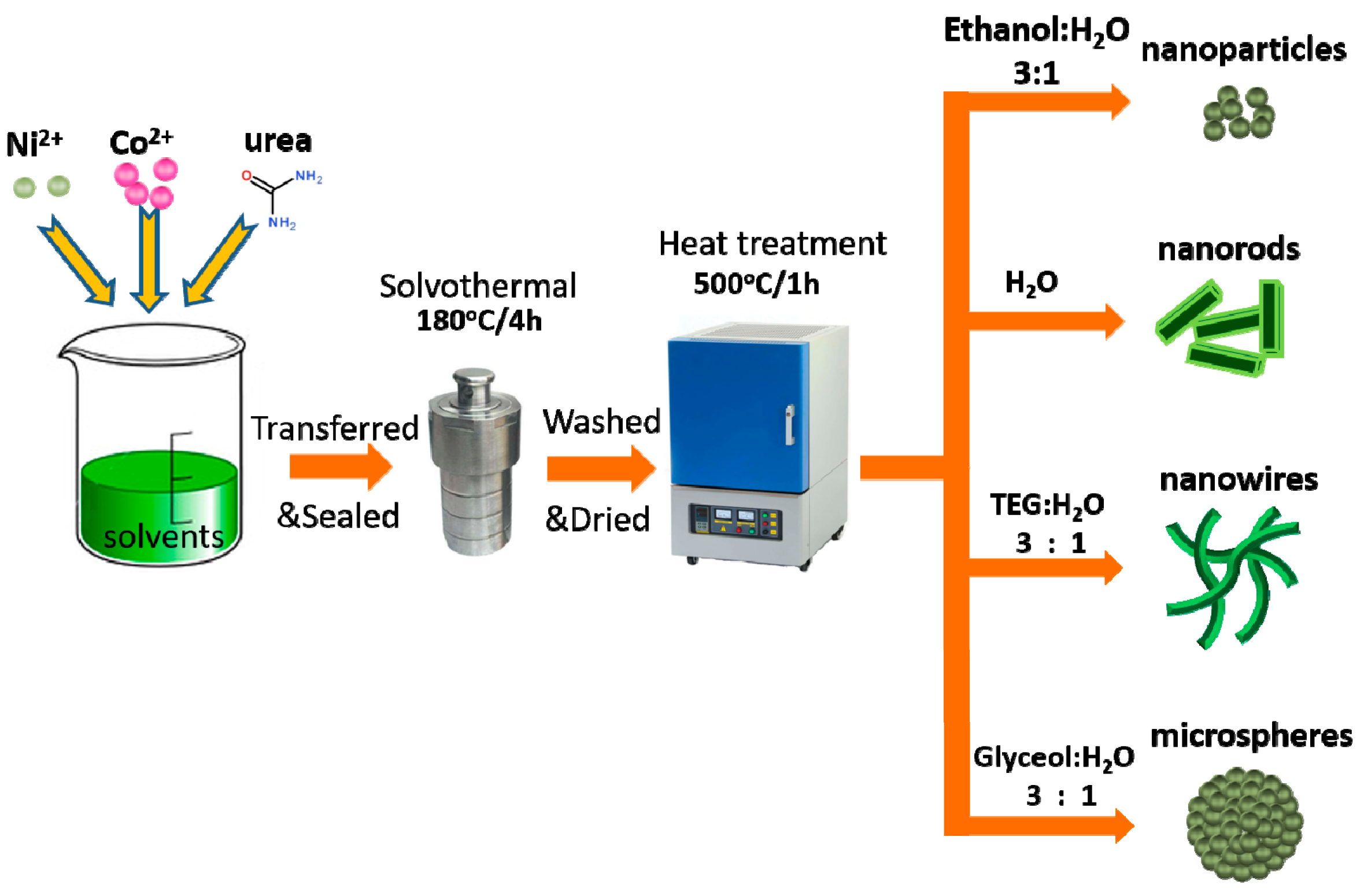
2.1. Sample Preparation

In the experiment, NiCo2O4 nanostructures were synthesized by simple solvothermal and subsequent heat treatment. Nickel acetate tetrahydrate (C4H6NiO4·4H2O) and cobalt acetate tetrahydrate (C4H6CoO4·4H2O) were used as Ni source and Co source, respectively. Urea (CO(NH2)2) was used as the mineralizer. Deionized water (H2O), ethanol, glycerol, and tetraetylene glycol (TEG) were used as reaction solvents. The typical synthesis process can be described as follows: 1 mmol C4H6NiO4·4H2O and 2 mmol C4H6CoO4·4H2O were dissolved in 40 mL prefabricated reaction solvents to form mixed solution of metal ions, then 7 mmol CO(NH2)2 was added to the mixed solution under vigorous stirring to form suspension. The suspension was sealed and transferred to a 60 mL Teflon-lined stainless-steel autoclave. The autoclave was then heated at 180 °C for 4 h. The resultant products were collected, washed by ethanol and water, and then dried in 80 °C for 8 h in air. Finally, the dried products were put into a furnace and heat-treated in air at 500 °C for 1 h, and then mesoporous NiCo2O4 nanostructures were obtained. In the experiment, the prefabricated reaction solvents are the mixture of ethanol and deionized water (volume ration = 3:1), deionized water, the mixture of glycerol and deionized water (volume ration = 3:1), or the mixture of TEG and deionized water (volume ration = 3:1). By adjusting the type of prefabricated reaction solvents, NiCo2O4 nanoparticles, nanorods, nanowires, and microspheres could be prepared. The synthesis process and experimental details are shown in Figure 1.

2.2. Sample Characterization

The phase structure and microstructure of the samples were measured by X-ray diffraction (XRD, MiniFlex II, Rigaku, Tokyo, Japan) and transmission electron microscopy (TEM, JEM-2010, JEOL, Tokyo, Japan), respectively. The morphology and elemental distribution of the samples were measured by scanning electron microscopy (SEM, JSM-6700-F, JEOL, Tokyo, Japan), and energy dispersive spectrometer (EDS, Oxford INCA Energy 250, Oxford Instruments, Abingdon, UK), respectively. Chemical composition and valence state of the samples were analyzed by X-ray photoelectron spectroscopy (XPS, ESCA-LAB250XI, Waltham, MA, USA) with a monochromatic Al Ka X-ray source. The pore size distribution and specific surface area were obtained by the surface area and porosity analyzer (ASAP2460, Micromeritics, Norcross, GA, USA), using Barrett–Joyner–Halenda (BJH) model and Brunauer–Emmett–Teller (BET) method, respectively.

2.3. Catalytic Activity Test

The catalytic activity of as-prepared NiCo2O4 nanostructures were evaluated by differential scanning calorimeter (DSC, Netzsch Model STA449F3). The DSC-TGA experiments were described as follows: AP (98 wt%) and as-prepared NiCo2O4 (2 wt%) nanostructures were mixed in agate mortar by grinding for 10 min to obtain the target samples, and then a total mass of 5 mg for each target sample was put into an open alumina crucible, which was heated at heating rates of 5–25 °C min−1 in N2 atmosphere over a temperature range of 30–500 °C.

3. Results and Discussion

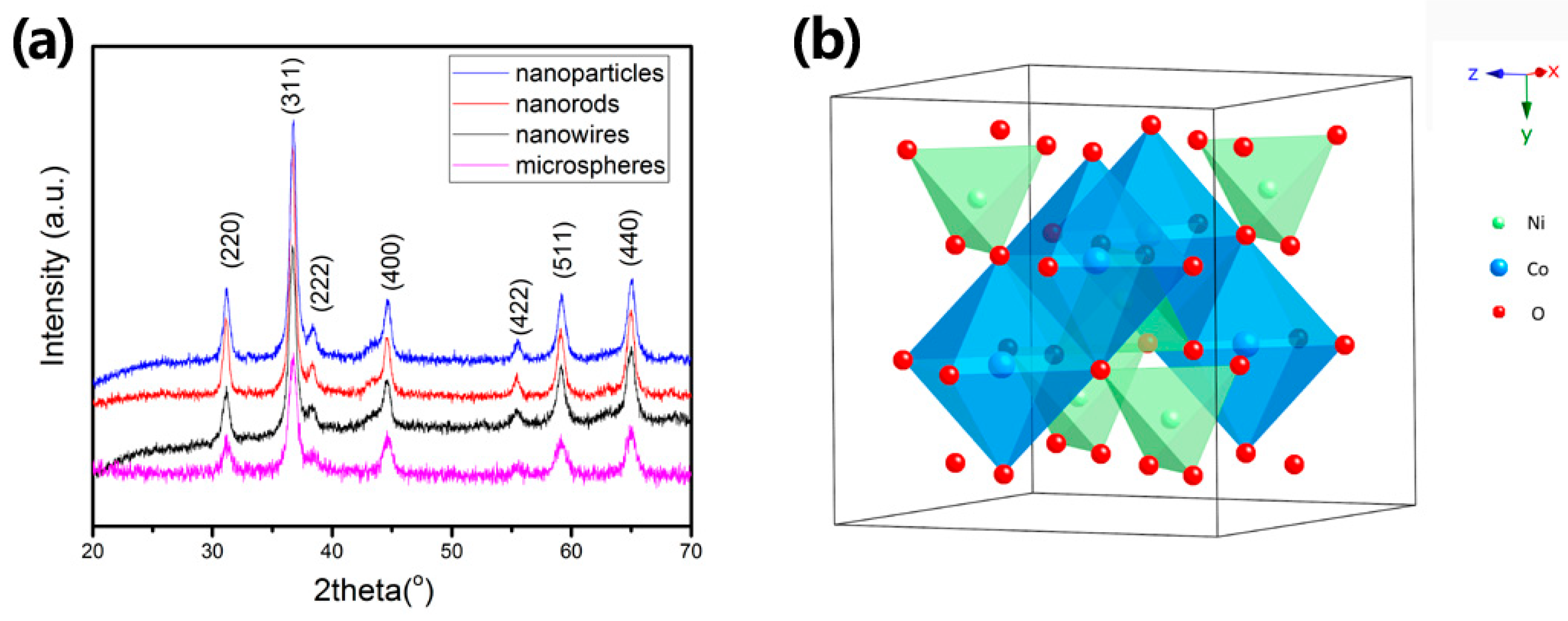
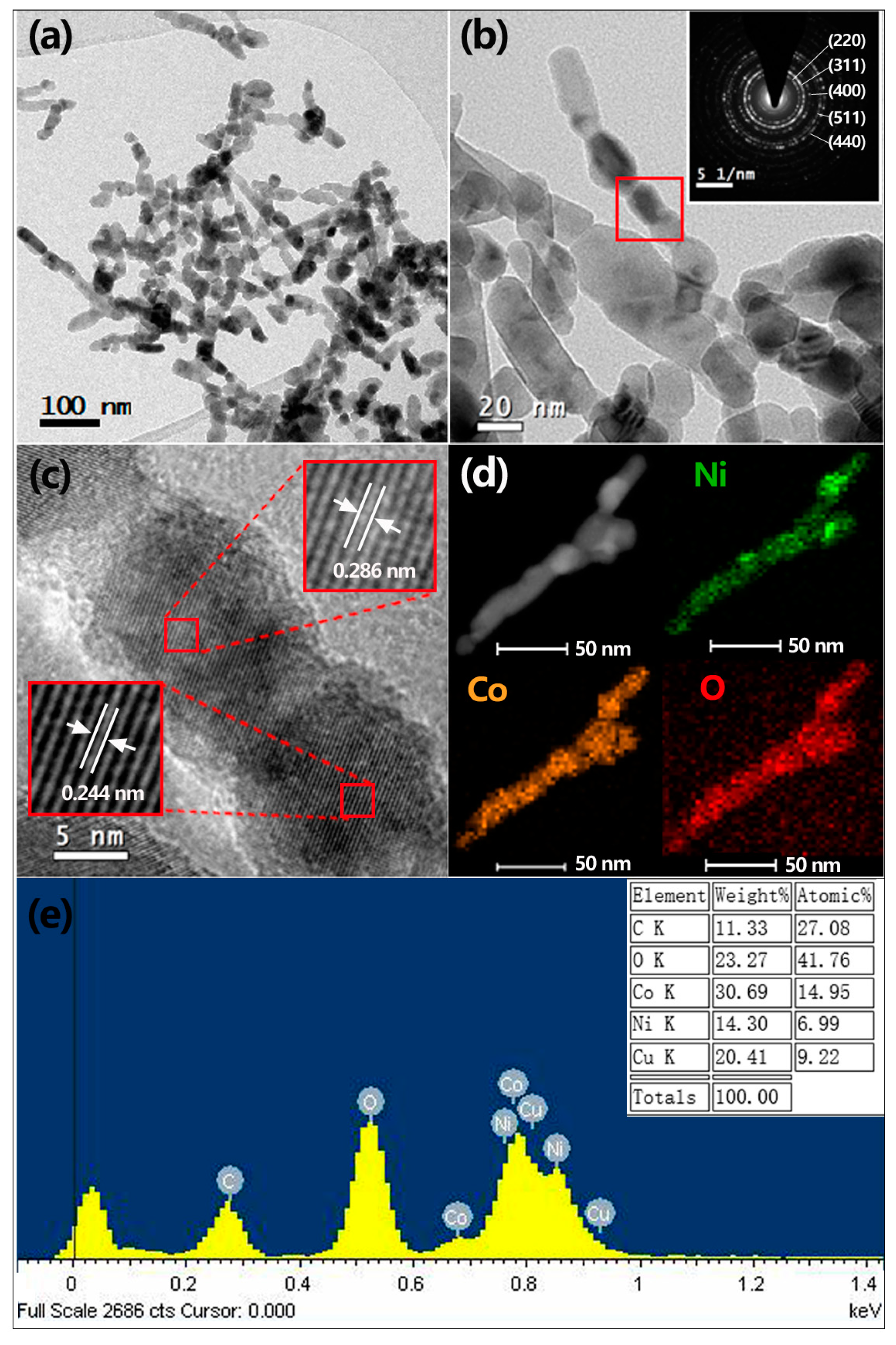
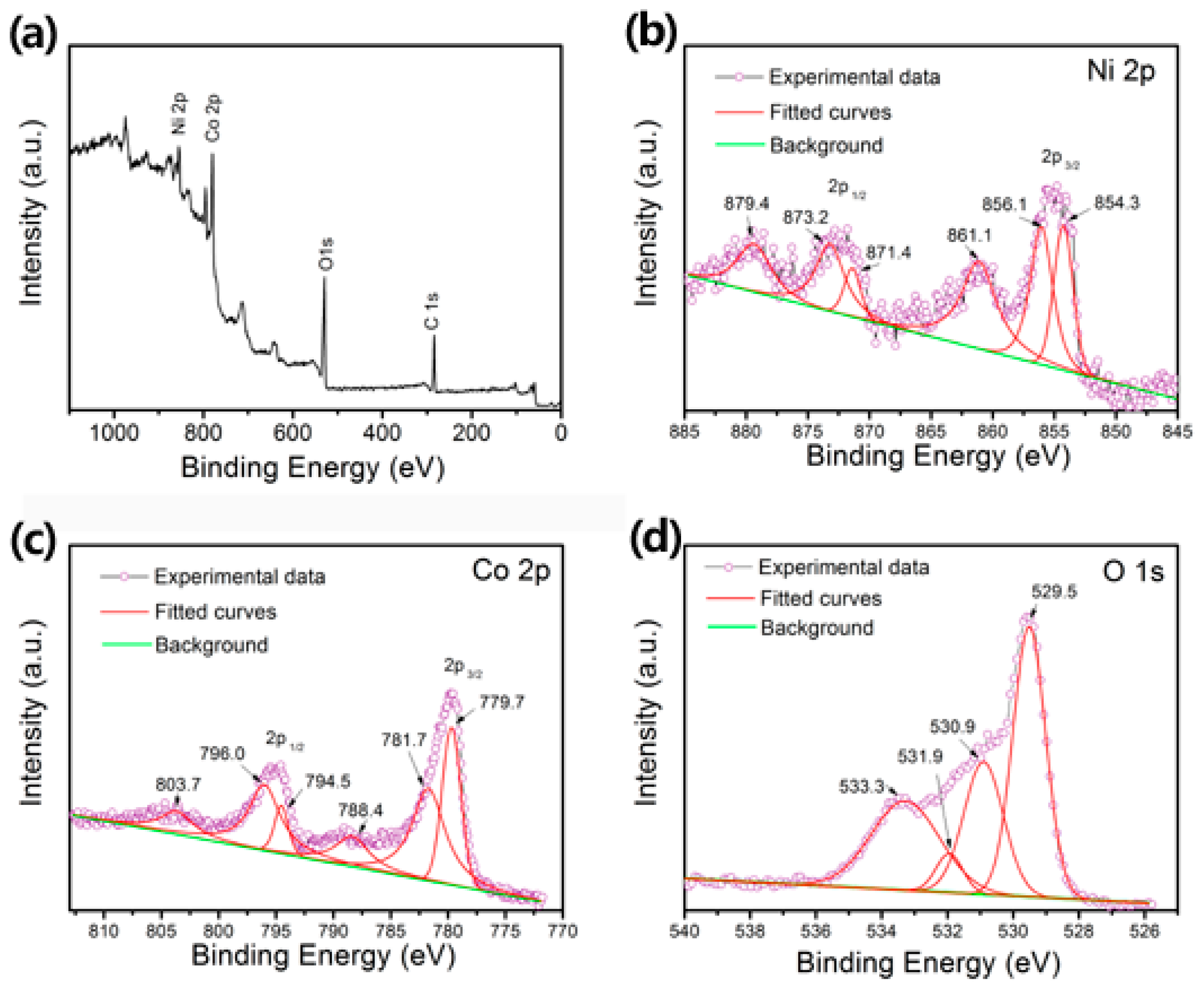
3.1. Morphology, Microstructure, and Composition Characterizations

The morphologies of as-prepared NiCo2O4 nanostructures were characterized by SEM, as shown in Figure 2. When using ethanol and deionized water (volume ratio = 3:1) as reaction solvent, the resultant product was composed of aggregates of nanoparticles with about 30–40 nm in diameter (Figure 2a). When using deionized water, the resultant product was composed of nanorods with about 30–40 nm in width and 90–120 nm in length (Figure 2b). When using TEG and deionized water (volume ratio = 3:1), the resultant product consisted of many nanowires with about 20–30 nm in width and 200–300 nm in length (Figure 2c). When using glycerol and deionized water (volume ratio = 3:1), some microspheres with about 0.3 µm in diameters are observed, which are composed of small nanoparticles (Figure 2d). Therefore, in our synthesis method, NiCo2O4 nanostructures with different morphologies can easily obtained by adjusting the type of reaction solvents.
The crystalline structure and phase composition of as-prepared NiCo2O4 nanostructures were analyzed by XRD (Cu Kα, λ = 0.15406 nm). Figure 3a shows that all samples exhibited similar diffraction peaks, which matched well with the standard diffraction data (PDF. No. 73-1702) for spinel structured NiCo2O4 phase [13]. All diffraction peaks correspond to the (220), (311), (333), (400), (422), (511), and (440) crystal planes of spinel structure NiCo2O4. The lattice parameters (a) of as-prepared NiCo2O4 nanoparticles, nanorods, nanowires, and microspheres were calculated by the XRD data, which are 0.8113, 0.8116, 0.8112, and 0.8111 nm, respectively. All calculated lattice parameters are closed to the lattice parameter a = 0.8114 nm in the standard data (PDF. No. 73-1702) for spinel structured NiCo2O4 phase. No other diffraction peaks of impurities phase were observed. It indicated that pure phase of spinel structured NiCo2O4 are obtained by our synthesis method. The typical spinel structure of NiCo2O4 is illustrated in Figure 3b.
To further investigate the microstructural information of as-prepared NiCo2O4 nanowires sample, TEM, high-resolution TEM (HRTEM), and selected area electron (SAED) patterns were measured and the results are shown in Figure 4. Figure 4a shows that as-prepared NiCo2O4 nanowires sample was composed of nanoparticles of 20–30 nm in diameter, which were connected to one another to form nanowire-like structure. The SAED pattern indicated good polycrystallinity of the sample (inset in Figure 4b). Figure 4c is the HRTEM image of enlarged part marked by red square in Figure 4b. It reveals distinct lattice lines with spacings of 0.286 and 0.244 nm according to the HRTEM measurement and subsequent calculation by the software of Digital Micrograph (Gatan Inc., Pleasanton, CA) applied in the HRTEM, which are corresponding to the (220) and (311) planes of spinel structure NiCo2O4, respectively [14]. The corresponding elemental mapping (Figure 4d) shows evenly distributed elements of Ni, Co, and O in the single nanowire, which supports the XRD results that the as-prepared NiCo2O4 nanowires are multi-component metal oxide, instead of metal oxide composites. The corresponding EDS spectrum shows that the atomic ratio of Ni:Co for the single NiCo2O4 nanowire is approximately 1:2 (Figure 4e). To investigate the chemical composition and element valence of as-prepared NiCo2O4 nanowires sample, the XPS analysis was performed and the results are shown in Figure 5. The survey spectrum showed the presence of the core-levels peaks of Ni 2p, Co 2p, and O 1s (Figure 5a). The Ni 2p spectrum (Figure 5b) exhibits several strong peaks, which are fitted to the 2p3/2 and 2p1/2 peaks and the satellite peaks. The peaks at 854.3 and 871.4 eV are associated with Ni2+ component and the peaks located at 856.1 and 873.2 eV are related with Ni3+ component in NiCo2O4 [15]. The peaks at 861.1 and 879.4 eV are the satellite peaks. The Co 2p spectrum (Figure 5c) consists of two spin-orbit doublets and two satellite peaks. The doublet peaks at 779.7 and 794.5 eV are characteristic of Co3+ component. The doublet peaks at 781.7 and 796.0 eV are characteristic of Co2+ component. The satellite peak at 788.4 and 803.7 eV are associated with Co3+ component and Co2+/ Co3+, respectively [16]. The O 1s spectrum (Figure 5d) can be fitted to four peaks at 529.5, 530.9, 531.9, and 533.3 eV, which correspond to metal-oxygen bonds, oxygen in hydroxyl groups, oxygen defects, and surface adsorbed water, respectively [17]. These results confirmed the formation of spinel NiCo2O4 phase, consistent with the XRD and TEM results.
Since the gases produced during AP thermal decomposition mainly react on the surface of additives, and the gases are diffused through open pores rather than closed pores, the properties of catalytic additives depend on the surface area and open porosities. The surface areas and open porosities of the samples were studied by the surface area and porosity analyzer, and the results are illustrated in Figure 6. The surface areas were calculated by BET method, which are 34.3, 39.3, 43.7, and 30.1 m2/g for as-prepared NiCo2O4 nanoparticles, nanorods, nanowires, and microspheres, respectively. The absorption-desorption isothermals curves of as-prepared NiCo2O4 nanostructures are shown in Figure 6a. All samples show a typical IV type isotherm with a H2 hysteresis loop according to IUPAC [18], indicating the mesoporous characteristic of the samples. Figure 6b shows the pore size distribution curves of the samples determined from the desorption branches. All samples exhibited mesoscale pores range from 2–20 nm, with a peak centered around 2.5 nm. Therefore, all as-prepared NiCo2O4 samples possess mesoporous structure.

3.2. Catalytic Activities

AP is a common oxidant for composite solid propellants and its thermal decomposition can be divided into two stages: low-temperature decomposition (LTD) stage and high-temperature decomposition (HTD) stage [19]. The LTD stage usually occurs around 290–330 °C and the HTD stage usually occurs around 350–450 °C. The LTD stage involves the reaction process taking place at the local sites on the surface of AP crystal. During the reaction process, AP is dissociated by proton transfer to form NH3 and HClO4, which will adsorb on the surface of AP and react with each other. Because NH3 adsorbed at low temperature cannot be completely oxidized by the decomposition products of HClO4, NH3 continuously covers the AP crystal surface with the decomposition process. If NH3 covers all the activation centers (reaction centers) on the surface of AP, the decomposition process will stop. With the increase of temperatures, the HTD stage begins. The adsorbed NH3 would release from the surface of AP and thus potential reaction centers are reactivated. At the same time, partial liquefaction of AP produces more NH3 and HClO4, which makes the reaction take place in the whole condensate phase, and the reaction becomes more intense. The catalytic activities of as-prepared mesoporous NiCo2O4 nanostructures towards AP thermal decomposition were analyzed by DSC, and the results are shown in Figure 7. As shown in Figure 7a, pure AP sample shows two endothermal peaks at about 242.2 °C and 411.6 °C. The first endothermal peak at 242.2 °C corresponds to the phase transformation of AP and the second endothermal peak corresponds to the HTD stage of AP. It is noted that no apparent peak corresponding to the LTD stage is observed, which may be ascribed to the particle size effect of AP crystal. Previous studies have reported that the LTD stage is closely related to the particle size of AP [20,21]. For AP crystals with large particle size, there is a large internal stress in the crystal itself. During LTD stage, the accumulation of adsorbed reaction products, such as NH3 and HClO4, increases the internal stress and promotes the breakdown of large crystals to form smaller crystals. The newly formed crystals can provide larger specific surface area to react, which makes LTD intensely. However, the internal stress of fine AP particles is small, and the accumulation of adsorbed reaction products is not enough to make AP particles break down to form smaller particles. Therefore, the initial decomposition of NH3 quickly covers the surface of small particles, so that no obvious first stage decomposition of fine AP particles can be seen. When 2 wt% as-prepared NiCo2O4 nanostructures were used as additives, all curves exhibited similar crystal transformation temperature at around 242.2 °C. It indicates that NiCo2O4 additives did not affect the crystal transformation of AP, consistent with previous reports [22]. However, the HTD peaks shifted towards lower temperature and became exothermic type instead of endothermic, as one observed in pure AP. It has been well documented that the HTD process of pure AP can be either exothermic or endothermic, depending on the use of lids [23,24]. When the sample container with lid was used, the HTD process was the exothermic type; while the sample container without lid was used, the HTD process would be the endothermic type. In our experiment, open alumina crucibles without lids were used, so the HTD process is endothermic for pure AP. After adding NiCo2O4 nanostructures as catalytic additives, the HTD process of AP were catalyzed by NiCo2O4 additives, which made the reaction intensify to release reaction heat and thus the HTD process became exothermic.
Figure 7b shows the HTD temperatures of all samples. In comparison with the HTD temperature of pure AP, those of the mixtures were greatly decreased. This indicates that all mesoporous NiCo2O4 nanostructures showed obvious catalytic activity towards the thermal decomposition of AP. In particular, the as-prepared NiCo2O4 nanowires exhibited the lowest HTD temperature of 312.3 °C, which decreased the HTD temperature of AP by 99.3 °C. The catalytic activity toward AP thermal decomposition of the as-prepared NiCo2O4 nanowires is much better than other reported metal oxides, such as CuO nanocomposites [25], Co3O4 nanowires [26], NiO nanostructures [27], and NiCo2O4 superstructures [28]. Therefore, the as-prepared NiCo2O4 nanowires can be an effective additive to catalyze the HTD process of AP thermal decomposition.
The activation energy of AP mixing with as-prepared NiCo2O4 nanowires was further studied by the thermal behavior of the mixture at different heating rates. For each given heating rate, a 5 mg mixture of AP and as-prepared NiCo2O4 nanowires was used separately. The results are shown in Figure 8. As can be seen, the crystal transformation temperature of AP is independent on heating rate, which remained as 242.2 °C (Figure 8a). Comparatively, the HTD temperature of the AP shifted toward higher temperature with the increasing heating rates from 10 to 30 K/min. According to the Kissinger correlation [29], the activation energy (Ea) can be calculated by fitting the slope of the plot of ln(β/Tp2) as a function of 1/Tp, where β is the heating rate and Tp is the HTD temperature. Figure 8b shows the plot of ln(β/Tp2) as a function of 1/Tp based on the data in Figure 8a. The Ea was calculated to be 317 kJ mol−1, which is larger than reported activation energy (100–130 kJ mol−1) of pure AP [30]. The increased activation energy for AP decomposition has been observed in several metal oxide additives such as CoO [24], CuO [31], and MgAl2O4 [32], which is ascribed to the kinetic compensation effect [31]. Though it appears that adding as-prepared NiCo2O4 nanowires increases rather than decreases the activation energy (energy barrier) of the AP decomposition reaction, we may conclude that the as-prepared NiCo2O4 nanowires is an effective additive which decreased the HTD temperature of AP by 99.3 °C. Though the reaction mechanism of AP thermal decomposition is complicated, it is anticipated that the excellent catalytic activity of as-prepared NiCo2O4 nanowires is due to their large surface and unique mesoporous nanostructures, which facilitates the gas diffusion and provides more active sites for AP thermal decomposition [30]. In addition, the multiple convertible valence state of Ni and Co atom could promote ammonia oxidation and the dissociation of ClO4 species [28].

4. Conclusions

In this study, mesoporous NiCo2O4 nanostructures were prepared by simple solvothermal and subsequent heat treatment. Mesoporous NiCo2O4 nanoparticles, nanorods, nanowires, and microspheres could be easily obtained by using ethanol/deionized water, deionized water, TEG/deionized water, and glycerol/deionized water as the reaction solvent, respectively. When used as catalytic additive for the thermal decomposition of AP, the as-prepared NiCo2O4 nanostructures apparently decreased the HTD temperature of AP thermal decomposition. Among all the products, the NiCo2O4 nanowires demonstrated the superior catalytic activity, which decreased the HTD temperature of AP by 99.3°C and exhibited the activation energy of 317 kJ/mol. Therefore, the method reported here would provide a straightforward and scalable way to synthesize mesoporous NiCo2O4 nanostructures with multi-morphologies, and the NiCo2O4 nanowires could be a promising catalytic additive for the thermal decomposition of AP.

Author Contributions

X.G. conceived, designed, performed the experiments, and wrote the paper; P.L., Y.Y., X.L., and D.C. analyzed the data.

Funding

This work was funded by Natural Science Foundation of Fujian Province (2017J01676), Key Research Foundation for Young Scholars of Fujian Education Department of China (JZ160486, JZ160484), Program for New Century Excellent Talents in Fujian Province University (Minjiaoke [2018] No. 47).

Conflicts of Interest

The authors declare no conflict of interest.

References

  1. Tan, C.L.; Cao, X.H.; Wu, X.J.; He, Q.Y.; Yang, J.; Zhang, X.; Chen, J.Z.; Zhao, W.; Han, S.K.; Nam, G.H.; et al. Recent advances in ultrathin two-dimensional nanomaterials. Chem. Rev. 2017, 117, 6225–6331. [Google Scholar] [CrossRef] [PubMed]
  2. Dong, C.Q.; Kou, T.Y.; Gao, H.; Peng, Z.Q.; Zhang, Z.H. Eutectic-derived mesoporous Ni-Fe-O nanowire network catalyzing oxygen evolution and overall water splitting. Adv. Energy Mater. 2018, 8, 1701347. [Google Scholar] [CrossRef]
  3. Liu, D.L.; Zhang, C.; Yu, Y.F.; Shi, Y.M.; Yu, Y.; Niu, Z.Q.; Zhang, B. Hydrogen evolution activity enhancement by tuning the oxygen vacancies in self-supported mesoporous spinel oxide nanowire arrays. Nano Res. 2018, 11, 603–613. [Google Scholar] [CrossRef]
  4. Bhagwan, J.; Nagaraju, G.; Ramulu, B.; Sekhar, S.C.; Yu, J.S. Rapid synthesis of hexagonal NiCo2O4 nanostructures for high-performance asymmetric supercapacitors. Electrochimi. Acta 2019, 299, 509–517. [Google Scholar] [CrossRef]
  5. Du, X.Q.; Shao, Q.Z.; Zhang, X.S. Metal tungtate dominated NiCo2O4@NiWO4 nanorods arrays as an efficient electrocatalyst for water splitting. Inter. J. Hydro. Energy 2019, 44, 2883–2888. [Google Scholar] [CrossRef]
  6. Jain, A.; Paul, B.J.; Kim, S.; Jain, V.K.; Kim, J.; Rai, A.K. Two-dimensional porous nanodisks of NiCo2O4 as anode material for high-performance rechargeable lithium-ion battery. J. All. Compd. 2019, 772, 72–79. [Google Scholar] [CrossRef]
  7. Ding, R.; Qi, L.; Jia, M.J.; Wang, H.Y. Facile synthesis of mesoporous spinel NiCo2O4 nanostructures as highly efficient electrocatalysts for urea electro-oxidation. Nanoscale 2014, 6, 1369–1376. [Google Scholar] [CrossRef] [PubMed]
  8. Karunakaran, G.; Maduraiveeran, G.; Kolesnikov, E.; Balasingam, S.K.; Viktorovich, L.D.; Ilinyh, I.; Gorshenkov, M.V.; Sasidharan, M.; Kuznetsov, D.; Kundu, M. Ascorbic acid-assisted eco-friendly synthesis of NiCo2O4 nanoparticles as an anode material for high-performance lithium-ion batteries. JOM 2018, 70, 1416–1422. [Google Scholar] [CrossRef]
  9. Samantara, A.K.; Kamila, S.; Ghosh, A.; Jena, B.K. Highly ordered 1D NiCo2O4 nanorods on graphene: An efficient dual functional hybrid material for electrochemical energy conversion and storage applications. Electrochimi. Acta 2018, 263, 147–157. [Google Scholar] [CrossRef]
  10. Saraf, M.; Natarajan, K.; Mobin, S.M. Multifunctional porous NiCo2O4 nanorods: Sensitive enzymeless glucose detection and supercapacitor properties with impedance spectroscopic investigations. New J. Chem. 2017, 41, 9299–9313. [Google Scholar] [CrossRef]
  11. Wang, Q.Y.; Jin, K.L.; Lv, Z.X.; Yang, D.; Xu, J.C.; Hong, B.; Wang, X.Q. Synthesis and pseudocapacitive performance of ordered mesoporous NiCo2O4 nanowires. Inter. J. Electrochem. Sci. 2018, 13, 11675–11683. [Google Scholar] [CrossRef]
  12. Khalid, S.; Cao, C.B.; Wang, L.; Zhu, Y.Q. Microwave assisted synthesis of porous NiCo2O4 microspheres: Application as high performance asymmetric and symmetric supercapacitors with large areal capacitance. Sci. Rep. 2016, 6, 22699. [Google Scholar] [CrossRef] [PubMed]
  13. Zhang, C.F.; Yu, J.S. Morphology-tuned synthesis of NiCo2O4-coated 3D graphene architectures used as binder-free electrodes for lithium-ion batteries. Chem. Eur. J. 2016, 22, 4422–4430. [Google Scholar] [CrossRef] [PubMed]
  14. Zhang, Y.Y.; Wang, J.X.; Ye, J.H.; Wan, P.P.; Wei, H.M.; Zhao, S.Q.; Li, T.F.; Hussain, S. NiCo2O4 arrays nanostructures on nickel foam: Morphology control and application for pseudocapacitors. Ceram. Inter. 2016, 42, 14976–14983. [Google Scholar] [CrossRef]
  15. Hao, C.; Zhou, S.; Wang, J.J.; Wang, X.H.; Gao, H.W.; Ge, C.W. Preparation of hierarchical spinel NiCo2O4 nanowires for high-performance supercapacitors. Ind. Eng. Chem. Res. 2018, 57, 2517–2525. [Google Scholar] [CrossRef]
  16. Marco, J.F.; Gancedo, R.; Gracia, M. Characterization of the nickel cobaltite, NiCo2O4, prepared by several methods: An XRD, XANE S, EXAFS, and XPS study. J. Solid State Chem. 2000, 153, 74–81. [Google Scholar] [CrossRef]
  17. Nguyen, V.H.; Shim, J.J. Three-dimensional nickel foam/graphene/NiCo2O4 as high-performance electrodes for supercapacitors. J. Power Sources 2015, 273, 110–117. [Google Scholar] [CrossRef]
  18. Sing, K.S.W. Reporting physisorption data for gas/solid systems with special reference to the determination of surface area and porosity. Pure Appl. Chem. 1985, 57, 603–619. [Google Scholar] [CrossRef]
  19. Guan, X.F.; Li, G.S.; Zhou, L.H.; Li, L.P.; Qiu, X.Q. Template-free approach to core-shell-structured Co3O4 microspheres. Chem. Lett. 2009, 38, 280–281. [Google Scholar] [CrossRef]
  20. Abdel-Wareth, W.; Xu, X. Particle size effect on ammonium perchlorate decomposition kinetics. J. Aerosp. Power 2012, 27, 1179–1184. [Google Scholar]
  21. Liu, Z.R.; Yin, C.M.; Kong, Y.H.; Zhao, F.Q.; Luo, Y.; Xiang, H. The thermal decomposition of ammonium perchlorate. Chin. J. Energ. Mater. 2000, 8, 75–79. [Google Scholar]
  22. Reid, D.L.; Russo, A.E.; Carro, R.V.; Stephens, M.A.; LePage, A.R.; Spalding, T.C.; Petersen, E.L.; Seal, S. Nanoscale additives tailor energetic materials. Nano Lett. 2007, 7, 2157–2161. [Google Scholar] [CrossRef]
  23. Vyazovkin, S.; Wight, C.A. Kinetics of thermal decomposition of cubic ammonium perchlorate. Chem. Mater. 1999, 11, 3386–3393. [Google Scholar] [CrossRef]
  24. Lang, A.J.; Vyazovkin, S. Effect of pressure and sample type on decomposition of ammonium perchlorate. Combust. Flame 2006, 145, 779–790. [Google Scholar] [CrossRef]
  25. Zhao, H.F.; Lv, J.; Sang, J.S.; Zhu, L.; Zheng, P.; Andrew, G.L.; Tan, L.H. A facile method to construct MXene/CuO nanocomposite with enhanced catalytic activity of CuO on thermal decomposition of Ammonium Perchlorate. Materials 2018, 11, 2457. [Google Scholar] [CrossRef] [PubMed]
  26. Zhou, H.; Lv, B.L.; Wu, D.; Xu, Y. Synthesis of polycrystalline Co3O4 nanowires with excellent ammonium perchlorate catalytic decomposition property. Mater. Res. Bull. 2014, 60, 492–497. [Google Scholar] [CrossRef]
  27. Zhao, Y.J.; Zhang, X.W.; Xu, X.M.; Zhao, Y.Z.; Zhou, H.P.; Li, J.B.; Jin, H.B. Synthesis of NiO nanostructures and their catalytic activity in the thermal decomposition of ammonium perchlorate. Cryst. Eng. Commun. 2016, 18, 4836–4843. [Google Scholar] [CrossRef]
  28. Wang, J.G.; Jin, L.N.; Qian, X.Y.; Dong, M.D. Preparation and catalytic activity comparison of porous NiO, Co3O4 and NiCo2O4 superstructures on the thermal decomposition of ammonium perchlorate. J. Nanosci. Nanotech. 2016, 16, 8635–8639. [Google Scholar] [CrossRef]
  29. Tang, G.; Tian, S.Q.; Zhou, Z.X.; Wen, Y.W.; Pang, A.M.; Zhang, Y.G.; Zeng, D.W.; Li, H.T.; Xie, C.S. ZnO micro/nanocrystals with tunable exposed (0001) facets for enhanced catalytic activity on the thermal decomposition of ammonium perchlorate. J. Phys. Chem. 2014, 118, 11833–11841. [Google Scholar] [CrossRef]
  30. Jia, Z.G.; Ren, D.P.; Wang, Q.Z.; Zhu, R.S. A new precursor strategy to prepare ZnCo2O4 nanorods and their excellent catalytic activity for thermal decomposition of ammonium perchlorate. Appl. Surf. Sci. 2013, 270, 312–318. [Google Scholar] [CrossRef]
  31. Ninan, K.N. A thermogravimetric study on the catalytic decomposition of ammonium perchlorate from activation energy normalized through kinetic compensation. Indian J. Chem. 1998, 37A, 295–302. [Google Scholar]
  32. Guan, X.F.; Li, L.P.; Zheng, J.; Li, G.S. MgAl2O4 nanoparticles: A new low-density additive for accelerating thermal decomposition of ammonium perchlorate. RSC Adv. 2011, 1, 1808–1814. [Google Scholar] [CrossRef]
Figure 1. Scheme of the synthesis process of nickel cobaltite (NiCo2O4) nanostructures.
Applsci 09 01100 g001
Figure 2. Scanning electron microscopy (SEM) images (secondary electron imaging mode) of as-prepared NiCo2O4 nanostructures obtained by different reaction solvents: (a) Ethanol and deionized H2O (3:1); (b) deionized H2O; (c) tetraetylene glycol (TEG) and deionized H2O (3:1); and (d) glycerol and deionized H2O (3:1).
Applsci 09 01100 g002
Figure 3. (a) X-ray diffraction (XRD) (Cu Kα, λ = 0.15406 nm) patterns of as-prepared NiCo2O4 nanostructures; (b) structural illustration of spinel NiCo2O4.
Applsci 09 01100 g003
Figure 4. (a) Low-magnification transmission electron microscopy (TEM) image; (b) high-magnification TEM image, (c) high-resolution TEM (HRTEM) image of enlarged part marked by red square in (b), (d) elemental mapping, and (e) energy dispersive spectrometer (EDS) spectrum of single NiCo2O4 nanowire.
Applsci 09 01100 g004
Figure 5. X-ray photoelectron spectroscopy (XPS) spectrum of as-prepared NiCo2O4 nanowires: (a) survey spectrum; and binding energy spectrum of (b) Ni 2p; (c) Co 2p; (d) O 1s.
Applsci 09 01100 g005
Figure 6. (a) N2 adsorption-desorption isotherm curves and (b) pore size distribution curves of the as-prepared NiCo2O4 nanostructures.
Applsci 09 01100 g006
Figure 7. (a) Differential scanning calorimeter (DSC) curves of ammonium perchlorate (AP) mixed with as-prepared NiCo2O4 nanostructures and pure AP at a heating rate of 10 K/min; (b) histogram of the corresponding high-temperature decomposition (HTD) temperatures.
Applsci 09 01100 g007
Figure 8. (a) DSC curves of AP mixed with as-prepared NiCo2O4 nanowires and pure AP at different heating rates; (b) the plot of ln(β/Tp2) as a function of 1/Tp.
Applsci 09 01100 g008

