Article

Predictive Capacity of the MADYMO Multibody Human Body Model Applied to Head Kinematics during Rugby Union Tackles

by
Gregory J. Tierney
1,2,* and
Ciaran Simms
2
1
School of Biomedical Sciences, Faculty of Biological Sciences, University of Leeds, Leeds LS2 9NL, UK
2
Trinity Centre for Bioengineering, Trinity College Dublin, D02 PN40 Dublin, Ireland
*
Author to whom correspondence should be addressed.
Appl. Sci. 2019, 9(4), 726; https://doi.org/10.3390/app9040726
Submission received: 25 January 2019 / Revised: 15 February 2019 / Accepted: 16 February 2019 / Published: 19 February 2019
(This article belongs to the Special Issue Accident Analysis and Prevention: Experimental & Numerical Approaches)

Abstract

:
Multibody models have not yet been evaluated for reconstructing head kinematics during sports impacts. Accordingly, the goal of this study was to utilise whole-body motion data from twenty upper and mid/lower trunk rugby shoulder tackles recorded in a marker-based 3D motion analysis laboratory to assess the MADYMO human body passive ellipsoid model for head kinematic reconstruction. Head linear and angular velocity during the tackle for the multibody model predictions and 3D motion laboratory measures were recorded for the ball carrier. Examined were the linear and angular velocity, as well as the absolute and percentage differences. For upper trunk tackles, the median percentage error (with quartiles) for the MADYMO predictions were 10% (6% to 45%) and 23% (16% to 39%) for change in head linear and angular velocity, respectively. For mid/lower trunk tackles, the median percentage error (with quartiles) for the MADYMO predictions were 46% (33% to 63%) and 60% (53% to 123%) for change in head linear and angular velocity, respectively. In conclusion, the model is currently unsuitable for reconstruction of head kinematics during individual rugby union tackle cases.

1. Introduction

The measurement of head kinematics during sports collisions is essential for understanding the mechanism of concussion injuries [1] and long-term brain health. However, measuring head kinematics is challenging with on-field measurement devices [2,3]. An alternative approach is to use multibody modelling. Multibody models have been used to study head kinematics during concussive direct-head impacts in unhelmeted sports including rugby union and Australian rules football [4,5,6] as well as inertial head loading during legal rugby union tackles [7]. McIntosh et al. [6] used a passive MADYMO (TASS International, Helmond, Netherlands, 2015, Version 7.6) facet human body model approach to calculate six degree-of-freedom head kinematics from concussive and non-concussive direct head impacts in unhelmeted sports. They reported that angular acceleration of the head in the coronal plane had the greatest association with a concussion, with tentative threshold values of 1747 rad/s2 and 2296 rad/s2 reported for a 50% and 75% chance of concussion, respectively. Tierney and Simms [7] recently used the MADYMO passive ellipsoid multibody human body model and demonstrated that tackles to the upper body can cause greater inertial head and neck loading than tackles to the lower body, particularly for the ball carrier. They also postulated that tackles to the upper body may be a catalyst for some of the chronic head and neck symptoms exhibited by a rugby player. Although the study reported numerical values for head kinematics and neck dynamics, Tierney and Simms [7] cautioned that their focus was on the trends reported rather than precise numerical values, as the models have not yet been validated for sporting collisions.
These studies used MADYMO passive human body models, which have numerous input parameters (geometry, mechanical/structural properties, contact characteristics and initial conditions) and kinematic outputs, are generally validated based on staged impact tests using cadavers [8,9,10]. Assumptions regarding these input parameters strongly influence the results of impact simulations [11]. Although a sensitivity analysis was conducted for each of the abovementioned multibody model sports head kinematic studies, their predictive capacity in sporting impacts has not been formally assessed [7]. In particular, there is no direct validation of these passive MADYMO human models for reconstruction of head kinematics, largely because direct validation data for sporting impacts is not available [1,7,12]. This currently limits the potential to develop our understanding of head kinematics and concussion during sporting collisions using model-based approaches, which can then examine “what if” scenarios to help guide prevention strategies.
Tackling is a highly technical, fast-paced and dynamic part of rugby union [13,14] with certain playing positions, in extreme cases, potentially making over 30 tackles per game [15]. Unfortunately, it is also the most common cause of injury and concussion [16,17,18]. The biomechanics of tackling is not yet fully understood in relation to head kinematics [7]. However, reconstructing a tackle in a controlled environment such as a marker-based 3D motion laboratory enables ball carrier and tackler whole body kinematics to be recorded during an impact. Although a real-life direct head impact might not be reconstructed using such an environment (player protection requirements), this approach provides a means to assess multibody models for player-to-player sports impact reconstruction for the first time and to assess head kinematics due to inertial loading. Accordingly, the goal of this exploratory study was to record rugby tackles using human volunteers in a marker-based 3D Vicon motion analysis laboratory to assess the predictive capacity of the MADYMO human body passive ellipsoid model for head kinematic reconstruction.

2. Materials and Methods

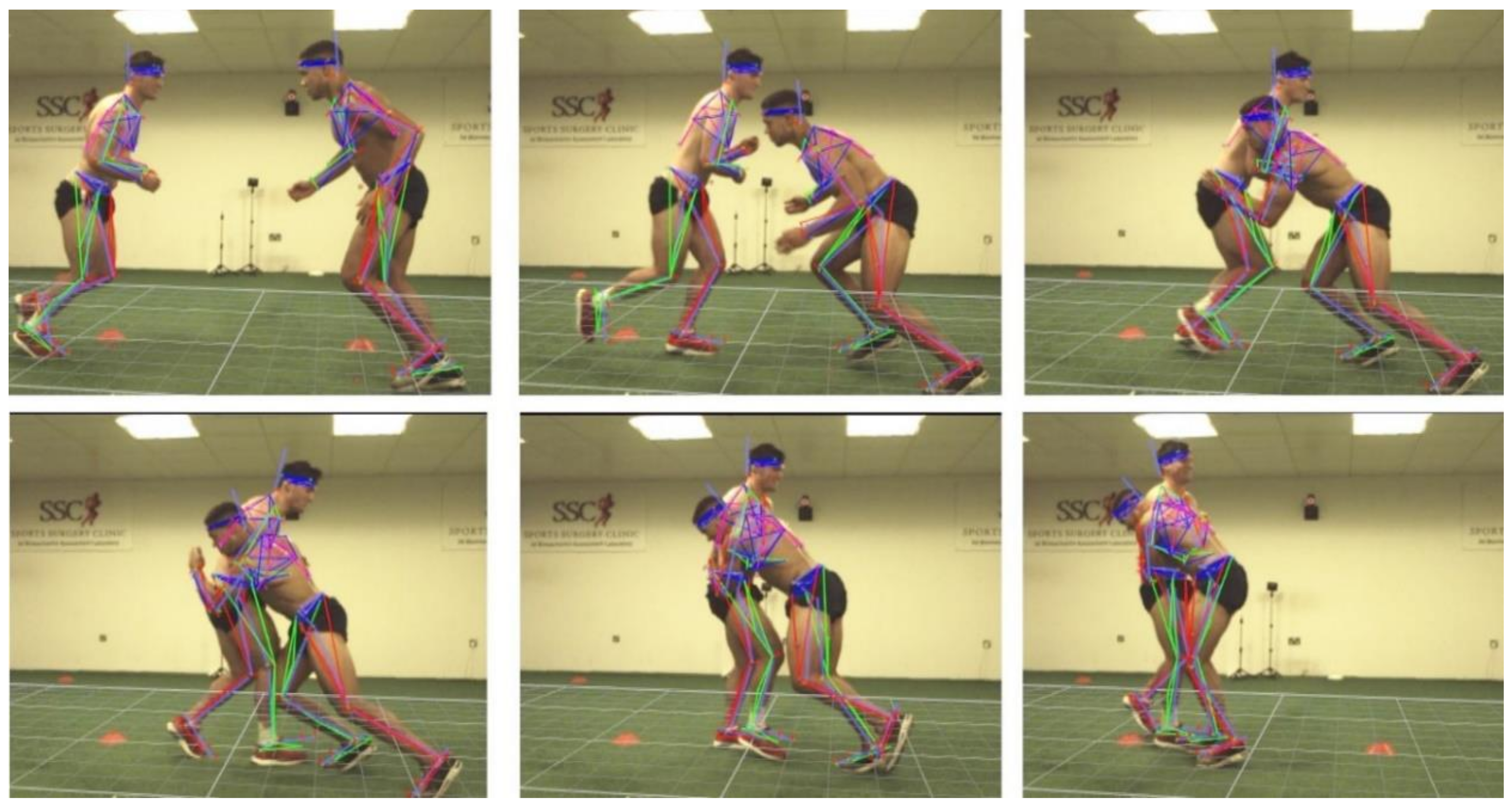
2.1. Staged Rugby Tackle Laboratory Trials

The staged rugby tackle laboratory trials are described in detail by Tierney et al. [19]. Four (two pairs) professional rugby union players performed twenty shoulder tackles (10 tackles per pair, where each player executed 5 tackles as the ball carrier and 5 tackles as the tackler) in a marker-based 3D motion laboratory. Ethical approval was given by the Trinity College Dublin Faculty of Health Sciences Ethics Committee. The players were initially positioned 2.5 m apart and initiated the tackles from a standing start. The tackler executed tackles to both the upper trunk and mid/lower trunk of the ball carrier [17,20,21].
A side, front and oblique camera view of every tackle was recorded with video cameras (Bonita 720C, Vicon, UK, 2015) recording at 66.6 Hz. The cameras were synchronised with a 10 camera infrared motion analysis system (Bonita-B10, Vicon, UK, 2015) recording at 200 Hz. Within a calibrated volume, the marker-based 3D motion system used these 10 infrared cameras to record the motion of spherical retro-reflective markers attached to the subject. The marker-based 3D motion system combined this information from all the cameras and thus recorded the 3D motion of each individual marker. Subjects wore reflective markers secured to the shoe or to the skin using tape at bony landmarks on the lower limbs, pelvis, trunk, arms and head. The markers were attached according to the plug-in-gait model protocol, while additional markers (C5, left and right ribcage and sacrum) were placed to allow an accurate reconstruction of the markers needed to apply the plug-in-gait model. The model used 43 reflective markers (10 mm radius) attached to each subject. This marker configuration enabled a three-dimensional description of the head, trunk, upper arm, forearm, pelvis, upper leg, shank and foot (see Figure 1). The plug-in-gait model calculated successive rotation angles for each of the abovementioned body regions, allowing the angular and linear kinematics to be computed for each body region [1]. A zero lag four-way low-pass filter with a 15 Hz cut-off frequency was applied to the plug-in-gait model data. Errors of less than 2% for both position and orientation tracking have been reported for the Vicon system [22].

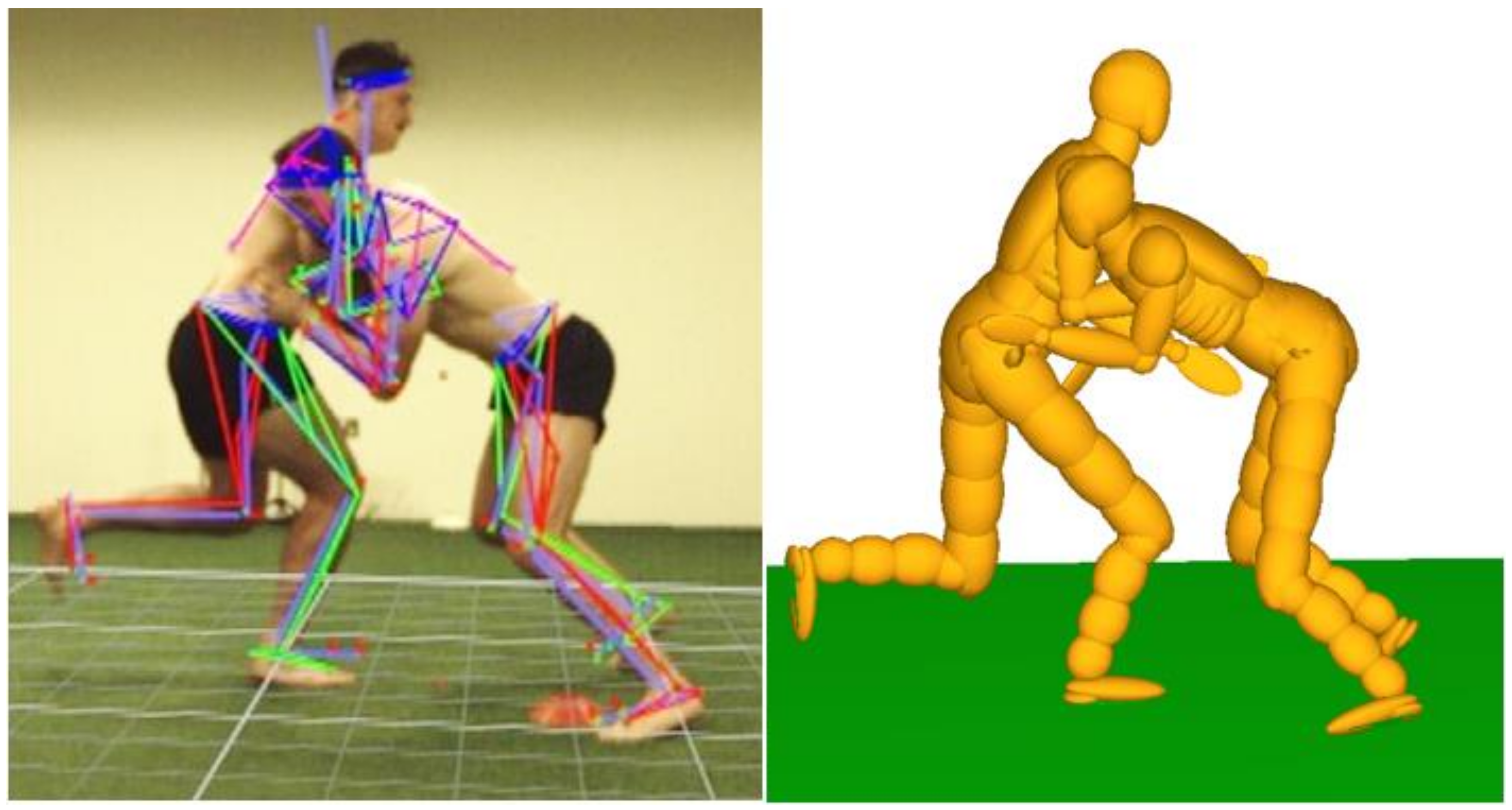
2.2. Multibody Model Reconstruction

The 50th percentile MADYMO human body ellipsoid model was used as a basis for simulating the staged rugby tackle laboratory trials (Figure 2). This model has 52 rigid bodies connected by kinematic joints, with ellipsoids for surface representation and contact evaluation. Using the MADYSCALE function, the model mass, moments of inertia and height were scaled based on the players’ height and mass. Player-to-player and player-to-ground contact evaluations using the built-in MADYMO contact stiffness functions were applied and an integration time step of 10 µs was used. Modelling muscle activation with a passive multibody model is a challenge [4,5,6]. Therefore, every simulation was run using the default unlocked joint condition which results in the joints of the model being free to articulate within the physiological range of motion with minimal resistance. This muscle activation condition has been regarded as a low awareness state [7]. This is a limitation of the model setup, given that both the tackler and ball carrier in these cases had a high level of awareness and therefore would have braced for the impacts accordingly.
The model was initially developed for vehicle pedestrian impact modelling and has been validated for various blunt impact locations (pelvis, abdomen, thorax and shoulder) [9,23,24,25,26,27,28]. There is currently no direct validation of this model for head angular velocity or acceleration, but it provides reasonable predictions for head translations, rotations, head impact time and head impact velocity in pedestrian collisions [10].
A customised Matlab script allowed the three-dimensional description of the head, trunk, upper arm, forearm, pelvis, upper leg, shank and foot gained from the staged rugby tackle laboratory trials to be utilised for positioning and orienting the model at the time of impact. This customised Matlab script also allowed angular velocity for each body region as well as the pelvis linear velocity to be calculated and used as initial conditions for the simulations. The simulations were run for 100 ms.

2.3. Statistical Analysis

Only the change in resultant head linear and angular velocity for the ball carrier were compared, as the ball carrier has been a focus of recent multibody model rugby tackle studies [7,19,29]. The change in head linear and angular velocity for the multibody model predictions (MADYMO) and 3D motion laboratory measures (Vicon) were recorded. The absolute and percentage differences were calculated for each trial.

3. Results

Of the twenty tackle cases recorded in the staged laboratory trials, nineteen could be reconstructed in the multibody modelling simulations (in one case, too many body markers fell off a participant during a tackle to generate valid kinematic measures). This resulted in eight mid/lower trunk tackles and 11 upper trunk tackles being executed. Table 1 reports the change in ball carrier head linear and angular velocity results for each tackle trial. For upper trunk tackles, the median percentage error (absolute value with quartiles) for the MADYMO predictions were 10% (6% to 45%) and 23% (16% to 39%) for change in head linear and angular velocity, respectively. For mid/lower trunk tackles, the median percentage error (absolute value with quartiles) for the MADYMO predictions were 46% (33% to 63%) and 60% (53% to 123%) for change in head linear and angular velocity, respectively. Table 1 also shows very high percentage errors, up to 336% and 366% for the change in head linear and angular velocity, respectively. The MADYMO simulations tend to respectively overpredict and underpredict the change in ball carrier head angular and linear velocity, for mid/lower trunk tackles (Figure 3). For upper trunk tackles (Figure 3), the results showed no clear trend with respect to underprediction or overprediction of the model.

4. Discussion

4.1. General

This study set out to use staged rugby tackle Vicon data to assess the predictive capacity of the MADYMO human body model for head kinematic reconstruction during inertial head loading impacts. The results demonstrated that the predictive capacity was limited in individual cases, with errors ranging up to 336% and 366% for the change in resultant head linear and angular velocity, respectively. The reasons for this may include: (1) the model contact and joint stiffness parameters, (2) the model shape and mass distribution and (3) the model player-to-player friction parameters do not realistically reproduce player-to-player impacts in rugby union.
The intensity of the staged tackles was significantly lower than during competitive play as the impact speeds of the ball carrier and tackler were around 2–3 times lower than average impact speeds in elite game environments (4.8 m/s and 5.6 m/s, respectively) [30]. This gave relatively low changes in ball carrier head linear and angular velocity in the motion laboratory trials, resulting in high percentage errors for the model predictions, even though the absolute errors could be considered small [1]. Thus, it is possible that the model may be better suited to assessing inertial loading in higher velocity impacts as it has been validated for vehicle-cadaver impacts (usually 40 km/h impacts [1,31]). The unlocked joint condition is also more representative of an unaware standing pedestrian than a trained rugby player bracing for contact. This may explain why the model has previously shown lower errors for head kinematic predictions during vehicle-cadaver collisions (maximum of 36% for head linear velocity predictions at the point of head-windscreen impact [10]). The models generally (with some exceptions) over-predicted peak ball carrier change in resultant head angular velocity but under-predicted peak ball carrier change in resultant head linear velocity. This suggests that the joint restraints torques in the model trunk and neck are too low and future emphasis should be on implementing active musculature in the models. The exceptions suggest muscle bracing is quite variable.
Future work could use this 3D motion laboratory data to optimise the numerous input parameters of the model and re-evaluate its accuracy for sports impact reconstruction. This should include optimising the contact friction properties, contact stiffness, joint stiffness and model shape. In recent years, the development of active human body models has become a promising prospect for multibody modelling [32]. These allow for active muscle behaviour to be exhibited by the model during an impact, however, they require muscle activation parameters as initial conditions, which are not yet fully known [33]. Examples of active human body models are the MADYMO active human body model [32], as well as the OpenSim rugby model [33]. It is recommended to assess these more sophisticated models for sports impact reconstruction using the 3D motion laboratory data gained from this study.
The McIntosh et al. [6] facet model approach only assessed direct head impacts. For their study, they refined and validated the contact properties of the head using published data from cadaver impacts tests/finite element simulations. They also suggested in a previous parametric study that neck stiffness had little influence on head kinematics for a direct head impact [4]. The study conducted post-processing by adjusting the initial position, body segment velocities and joint restraint torques of the models to match the kinematic behaviour of the players in the video event. Although post-processing had been conducted, it is recommended that the facet model should still be assessed using the 3D motion laboratory data obtained from this study.
The player-to-player configuration/orientation during the tackles influences the effective masses of both players. Therefore, the future focus should be placed on accurate whole-body player orientation during multibody model simulations from real-match events. Given that previous multibody modelling studies have used visually based approaches for player orientation [6], approaches such as model-based image-matching, which can extract body segment orientation data from multiple camera view videos, may be beneficial [1,12,34,35].

4.2. Limitations

The models were run using the default unlocked joint condition, which results in the joints of the model being free to articulate within the physiological range of motion with minimal resistance [7]. This muscle activation condition can be regarded as a low awareness state [7]. Although ball carriers can be visually unaware of impending tackles [36,37], both the tackler and ball carrier in these cases had a high level of awareness and therefore would have braced for the impacts accordingly. The plug-in-gait model treats the trunk of the player as one rigid body, whereas the multibody model treats it as two. The plug-in-gait model also treats the neck/head as one rigid body, whereas the multibody model treats it as two. Both of these can be considered insufficient given the articulation in the spine. The multibody model also has only two degrees of freedom for the shoulder joint, which potentially led to errors in the positioning of the players’ arms in the simulations. The plug-in gait model configuration is also not identical to the MADYMO model, which leads to some differences in initial linear velocities when matching initial angular velocities in the joints. The average difference at time zero between the linear velocity of the head for the multibody model and the 3D motion laboratory results was 0.2 m/s and 0.5 m/s for the ball carrier and tackler, respectively.
Impact validation data from high-velocity sports are currently unavailable. Clearly, a direct head impact could not be executed in the motion analysis laboratory and therefore, only indirect head impacts (inertial kinematics) could be assessed in this study. Approaches such as model-based image-matching [1,12,35] could enable whole body kinematic data to be extracted from real game direct head impacts. With the development of this approach, as well as instrumented mouthguards [2], direct head impact validation data may be available in the future.

5. Conclusions

This paper quantified the predictive capacity of the MADYMO human body model applied to staged rugby tackles, recorded in a 3D Vicon motion analysis laboratory. For upper trunk tackles, the median percentage error (absolute value with quartiles) for the MADYMO predictions were 10% (6% to 45%) and 23% (16% to 39%) for the change in head linear and angular velocity, respectively. For mid/lower trunk tackles, the median percentage error (absolute value with quartiles) for the MADYMO predictions were 46% (33% to 63%) and 60% (53% to 123%) for the change in head linear and angular velocity, respectively. These results demonstrated that the MADYMO human body ellipsoid model is currently unsuitable for detailed reconstruction of head kinematics in individual rugby union tackle cases.

Author Contributions

Conceptualization, G.T. and C.S.; methodology, G.T. and C.S.; software, G.T.; formal analysis, G.T.; data curation, G.T.; writing—original draft preparation, G.T.; writing—review and editing, G.T. and C.S.; supervision, C.S.; project administration, G.T. and C.S.; funding acquisition, G.T. and C.S.

Funding

This research was funded by the Irish Research Council (Grant NO: 202835).

Acknowledgments

The authors would like to thank Chris Richter and Katja Osterwald from the Santry Sports Surgery Clinic for their help in conducting the staged rugby tackles in the motion analysis laboratory.

Conflicts of Interest

The authors declare no conflict of interest.

References

  1. Tierney, G.J.; Joodaki, H.; Krosshaug, T.; Forman, J.L.; Crandall, J.R.; Simms, C.K. Assessment of model-based image-matching for future reconstruction of unhelmeted sport head impact kinematics. Sports Biomech. 2017, 17, 33–47. [Google Scholar] [CrossRef] [PubMed]
  2. Wu, L.C.; Nangia, V.; Bui, K.; Hammoor, B.; Kurt, M.; Hernandez, F.; Kuo, C.; Camarillo, D.B. In vivo evaluation of wearable head impact sensors. Ann. Biomed. Eng. 2016, 44, 1234–1245. [Google Scholar] [CrossRef] [PubMed]
  3. King, D.; Hume, P.A.; Brughelli, M.; Gissane, C. Instrumented mouthguard acceleration analyses for head impacts in amateur rugby union players over a season of matches. Am. J. Sports Med. 2015, 43, 614–624. [Google Scholar] [CrossRef] [PubMed]
  4. Fréchède, B.; McIntosh, A. Use of Madymo’s Human Facet Model to Evaluate the Risk of Head Injury in Impact. In Proceedings of the Conference on the Enhanced Safety of Vehicles, Lyon, France, 18–21 June 2007; pp. 18–21. [Google Scholar]
  5. Frechede, B.; McIntosh, A.S. Numerical reconstruction of real-life concussive football impacts. Med. Sci. Sports Exerc. 2009, 41, 390–398. [Google Scholar] [CrossRef] [PubMed]
  6. McIntosh, A.S.; Patton, D.A.; Fréchède, B.; Pierré, P.-A.; Ferry, E.; Barthels, T. The biomechanics of concussion in unhelmeted football players in australia: A case–control study. BMJ Open 2014, 4, e005078. [Google Scholar] [CrossRef] [PubMed]
  7. Tierney, G.J.; Simms, C.K. The effects of tackle height on inertial loading of the head and neck in rugby union: A multibody model analysis. Brain Inj. 2017, 31, 1925–1931. [Google Scholar] [CrossRef] [PubMed]
  8. Hardy, W.N.; Foster, C.D.; Mason, M.J.; Yang, K.H.; King, A.I.; Tashman, S. Investigation of head injury mechanisms using neutral density technology and high-speed biplanar X-ray. Stapp Car Crash J. 2001, 45, 337–368. [Google Scholar]
  9. TNO. Madymo Human Body Models Manual Release 7.6; TASS International: Helmond, The Netherlands, 2015; pp. 103–105. [Google Scholar]
  10. Elliott, J.; Lyons, M.; Kerrigan, J.; Wood, D.; Simms, C. Predictive capabilities of the MADYMO multibody pedestrian model: Three-dimensional head translation and rotation, head impact time and head impact velocity. Proc. Inst. Mech. Eng. Part K J. Multi-Body Dyn. 2012, 226, 266–277. [Google Scholar] [CrossRef]
  11. Parent, D.P.; Kerrigan, J.R.; Crandall, J.R. Comprehensive computational rollover sensitivity study, part 1: Influence of vehicle pre-crash parameters on crash kinematics and roof crush. Int. J. Crashworthiness 2011, 16, 633–644. [Google Scholar] [CrossRef]
  12. Tierney, G.J.; Joodaki, H.; Krosshaug, T.; Forman, J.L.; Crandall, J.R.; Simms, C.K. The kinematics of head impacts in contact sport: An initial assessment of the potential of model based image matching. In Proceedings of the ISBS-Conference Proceedings Archive, Tsukuba, Japan, 18–22 July 2016; pp. 108–111. [Google Scholar]
  13. Tierney, G.J.; Denvir, K.; Farrell, G.; Simms, C.K. The effect of technique on tackle gainline success outcomes in elite level rugby union. Int. J. Sports Sci. Coach 2018, 13, 16–25. [Google Scholar] [CrossRef]
  14. Tierney, G.J.; Denvir, K.; Farrell, G.; Simms, C.K. Does player time-in-game affect tackle technique in elite level rugby union? J. Sci. Med. Sport 2018, 21, 221–225. [Google Scholar] [CrossRef] [PubMed]
  15. Deutsch, M.; Kearney, G.; Rehrer, N. Time–motion analysis of professional rugby union players during match-play. J. Sports Sci. 2007, 25, 461–472. [Google Scholar] [CrossRef]
  16. Quarrie, K.L.; Hopkins, W.G. Tackle injuries in professional rugby union. Am. J. Sports Med. 2008, 36, 1705–1716. [Google Scholar] [CrossRef] [PubMed]
  17. Tierney, G.J.; Lawler, J.; Denvir, K.; McQuilkin, K.; Simms, C. Risks associated with significant head impact events in elite rugby union. Brain Inj. 2016, 30, 1350–1361. [Google Scholar] [CrossRef] [PubMed]
  18. Fuller, C.W.; Taylor, A.; Raftery, M. Epidemiology of concussion in men’s elite rugby-7s (sevens world series) and rugby-15s (rugby world cup, junior world championship and rugby trophy, pacific nations cup and english premiership). Br. J. Sports Med. 2015, 49, 478–483. [Google Scholar] [CrossRef]
  19. Tierney, G.J.; Richter, C.; Denvir, K.; Simms, C.K. Could lowering the tackle height in rugby union reduce ball carrier inertial head kinematics? J. Biomech. 2018, 72, 29–36. [Google Scholar] [CrossRef] [PubMed]
  20. Tierney, G.J.; Simms, C.K. The effect of intended primary contact location on tackler head impact risk. In Proceedings of the IRCOBI Conference Proceedings, Antwerp, Belgium, 13–15 September 2017; pp. 703–704. [Google Scholar]
  21. Tierney, G.J.; Simms, C.K. Can tackle height influence head injury assessment risk in elite rugby union? J. Sci. Med. Sport 2018, 21, 1210–1214. [Google Scholar] [CrossRef] [PubMed]
  22. Richards, J.G. The measurement of human motion: A comparison of commercially available systems. Hum. Mov. Sci. 1999, 18, 589–602. [Google Scholar] [CrossRef]
  23. Kajzer, J.; Cavallero, C.; Bonnoit, J.; Morjane, A.; Ghanouchi, S. Response of the knee joint in lateral impact: Effect of bending moment. In Proceedings of the IRCOBI Conference Proceedings, Eindhoven, The Netherlands, 8–10 September 1993; pp. 105–116. [Google Scholar]
  24. Yang, J.; Kajzer, J.; Cavallero, C.; Bonnoit, J. Computer Simulation of Shearing and Bending Response of the Knee Joint to a Lateral Impact; 0148-7191; SAE Technical Paper; SAE International: Warrendale, PA, USA, 1995. [Google Scholar]
  25. Bouquet, R.; Ramet, M.; Bermond, F.; Cesari, D. Thoracic and pelvis human response to impact. In Proceedings of the Conference on the Enhanced Safety of Vehicles, Munich, Germany, 23–26 May 1994; pp. 100–109. [Google Scholar]
  26. Viano, D.C. Biomechanical Responses and Injuries in Blunt Lateral Impact; 0148-7191; SAE Technical Paper; SAE International: Warrendale, PA, USA, 1989. [Google Scholar]
  27. Talantikite, Y.; Bouquet, R.; Ramet, M.; Guillemot, H.; Robin, S.; Voiglio, E. Human thorax behaviour for side impact: Influence of impact mass and velocities. In Proceedings of the Conference on the Enhanced Safety of Vehicles, Windsor, ON, Canada, 31 May–4 June 1998. [Google Scholar]
  28. Meyer, E.; Bonnoit, J. Le choc latéral sur l’épaule: Mise en place d’un protocole expérimental en sollicitation dynamique. Mémoire de DEA, Université Paris Val de Marne, Faculté de Médecine Pitié-Salpetrière, Ecole National Supérieure des Arts et Métiers de Paris, 1994. [Google Scholar]
  29. Tierney, G.J.; Lawler, J.; Simms, C.K. Upper and lower body tackles in rugby union: The effect on head kinematics. In Proceedings of the IRCOBI Conference Proceedings, Malaga, Spain, 14–16 September 2016; pp. 381–382. [Google Scholar]
  30. Hendricks, S.; Karpul, D.; Nicolls, F.; Lambert, M. Velocity and acceleration before contact in the tackle during rugby union matches. J. Sports Sci. 2012, 30, 1215–1224. [Google Scholar] [CrossRef] [PubMed]
  31. Forman, J.L.; Joodaki, H.; Forghani, A.; Riley, P.; Bollapragada, V.; Lessley, D.; Overby, B.; Heltzel, S.; Yarboro, S.; Weiss, D.B.; et al. Whole-body response for pedestrian impact with a generic sedan buck. Stapp Car Crash J. 2015, 59, 401–444. [Google Scholar]
  32. Meijer, R.; Van Hassel, E.; Broos, J.; Elrofai, H.; Van Rooij, L.; Van Hooijdonk, P. Development of a multi-body human model that predicts active and passive human behaviour. In Proceedings of the IRCOBI Conference Proceedings, Dublin, Ireland, 12–14 September 2012; pp. 622–636. [Google Scholar]
  33. Cazzola, D.; Trewartha, G.; Preatoni, E. Analysis of cervical spine loading in rugby scrummaging: A computer simulation approach. In Proceedings of the ISBS-Conference Proceedings Archive, Poitiers, France, 29 June–3 July 2015; pp. 1406–1408. [Google Scholar]
  34. Tierney, G.J.; Krosshaug, T.; Wilson, F.; Simms, C.K. An assessment of a novel approach for determining the player kinematics in elite rugby union players. In Proceedings of the IRCOBI Conference Proceedings, Lyon, France, 9–11 September 2015; pp. 180–181. [Google Scholar]
  35. Krosshaug, T.; Bahr, R. A model-based image-matching technique for three-dimensional reconstruction of human motion from uncalibrated video sequences. J. Biomech. 2005, 38, 919–929. [Google Scholar] [CrossRef] [PubMed]
  36. Tierney, G.J.; Denvir, K.; Farrell, G.; Simms, C.K. The effect of tackler technique on head injury assessment risk in elite rugby union. Med. Sci. Sports Exerc. 2017, 50, 603–608. [Google Scholar] [CrossRef] [PubMed]
  37. Tierney, G.J.; Denvir, K.; Farrell, G.; Simms, C.K. Does ball carrier technique influence tackler head injury assessment risk in elite rugby union? J. Sports Sci. 2019, 37, 262–267. [Google Scholar] [CrossRef] [PubMed]
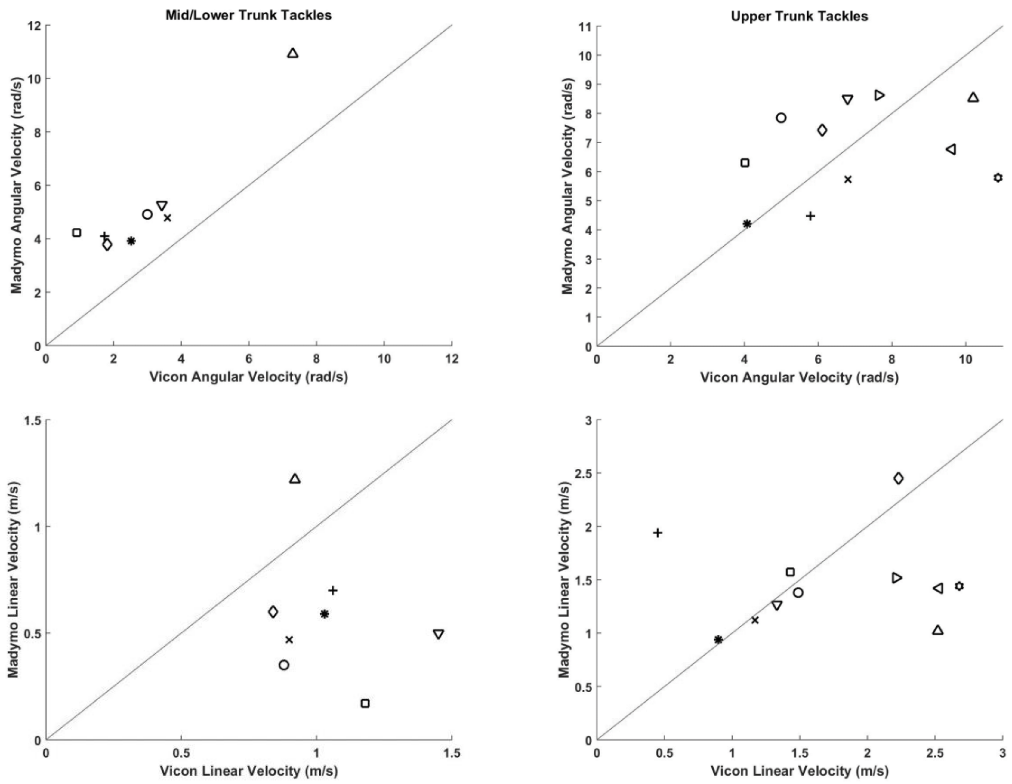
Figure 1. An image sequence of a tackle with the plug-in-gait model overlay.
Applsci 09 00726 g001
Figure 2. A staged tackle in the Vicon motion analysis laboratory (left) reconstructed in a multibody model simulation (right).
Applsci 09 00726 g002
Figure 3. Comparison between the predicted multibody model (MADYMO) and 3D motion laboratory (Vicon) peak ball carrier change in resultant head angular and linear velocity for upper and mid/lower trunk tackles. Each symbol corresponds to a tackle and is matched for both graphs.
Applsci 09 00726 g003
Table 1. The ball carrier change in head linear and angular velocity multibody model predictions (MADYMO) with 3D motion laboratory measures (Vicon) with absolute and percentage differences.
Ball Carrier Change in Resultant Head Linear Velocity (m/s)Change in Resultant Head Angular Velocity (rad/s)
Tackle IDClosing Speed (m/s)ViconMADYMODifference
(MADYMO-Vicon)
% Error (% of Vicon)ViconMADYMODifference
(MADYMO-Vicon)
% Error (% of Vicon)
Mid/Lower 24.041.030.59−0.44−432.523.921.4056
Mid/Lower 33.060.880.35−0.53−603.004.921.9264
Mid/Lower 43.701.060.70−0.35−331.734.092.36136
Mid/Lower 52.890.900.47−0.43−483.594.791.2033
Mid/Lower 64.201.450.50−0.95−653.425.281.8654
Mid/Lower 73.780.840.60−0.24−281.813.781.97109
Mid/Lower 83.850.921.220.30337.3010.913.6149
Mid/Lower 94.221.180.17−1.01−860.914.223.31366
Median (Absolute) 3.820.980.550.44462.764.511.9560
25th Percentile
(Absolute)
3.540.900.440.34331.794.051.7553
75th Percentile
(Absolute)
4.031.120.650.74633.515.102.84123
Upper 13.870.900.940.0454.074.210.144
Upper 23.951.491.38−0.11−75.007.852.8557
Upper 34.260.451.941.503365.794.48−1.31−23
Upper 44.051.171.12−0.05−46.815.73−1.09−16
Upper 54.381.331.27−0.06−46.808.511.7125
Upper 64.532.232.450.22106.117.431.3222
Upper 74.882.521.02−1.50−5910.208.52−1.67−16
Upper 84.541.431.570.14104.026.302.2857
Upper 95.182.211.52−0.69−317.648.630.9913
Upper 104.572.531.42−1.11−449.626.76−2.86−30
Upper 114.002.681.44−1.23−4610.875.79−5.08−47
Median (Absolute)4.381.491.420.22106.806.761.6723
25th Percentile
(Absolute)
4.031.251.200.0965.405.761.2016
75th Percentile
(Absolute)
4.562.381.551.17458.638.182.5739

