Evaluating the Overall Accuracy of Additional Learning and Automatic Classification System for CT Images


Abstract
Featured Application
Abstract
1. Introduction
2. Materials and Methods
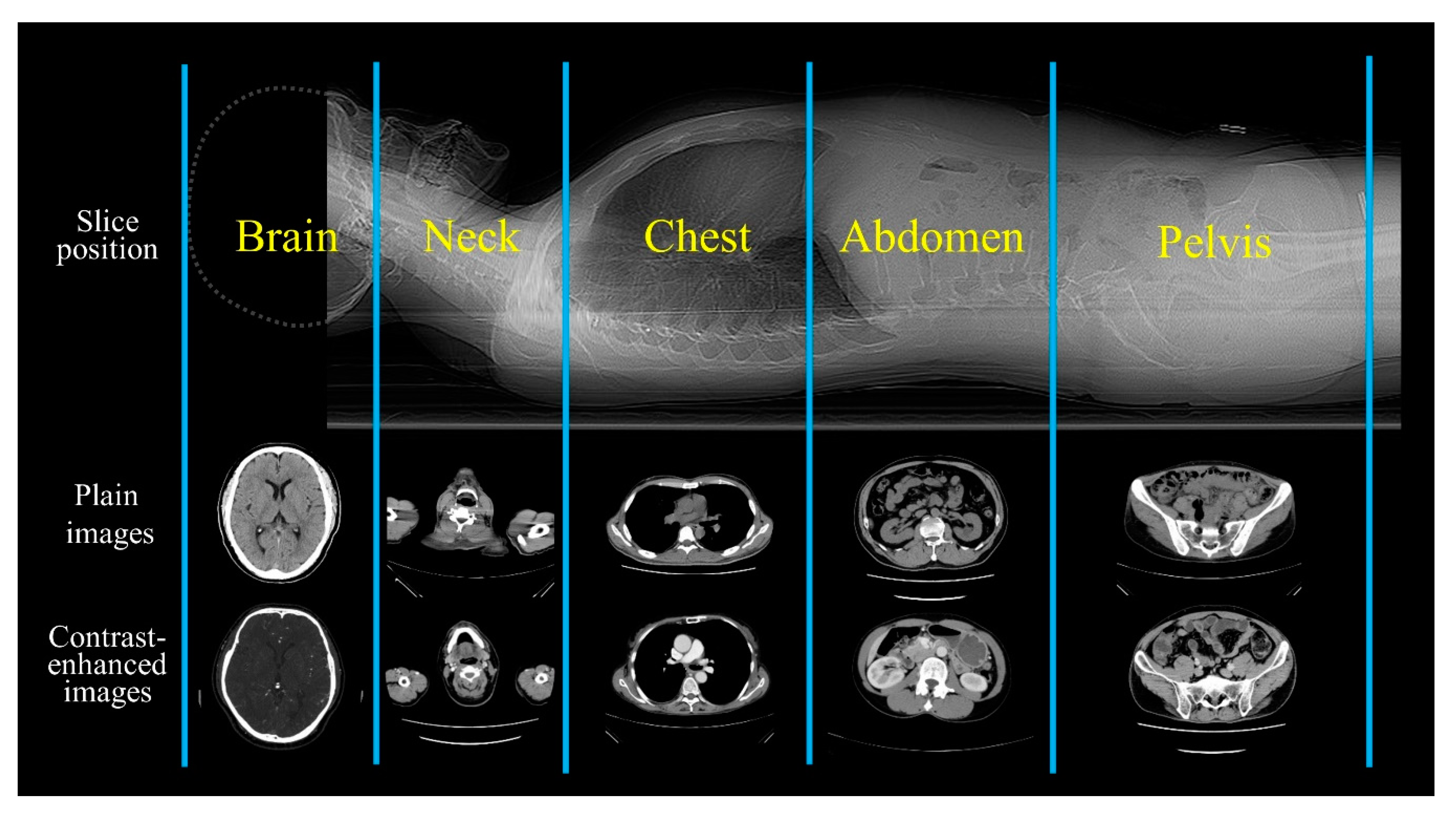
2.1. Subjects and CT Images
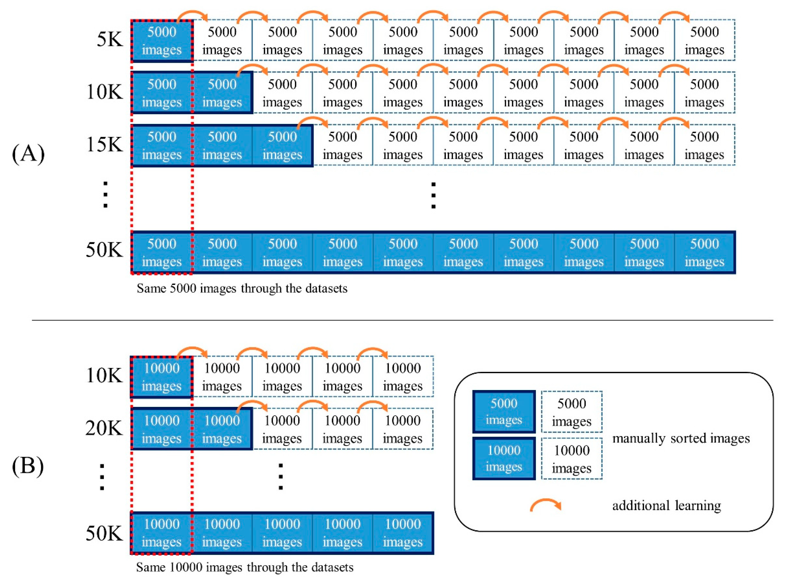
2.2. Datasets
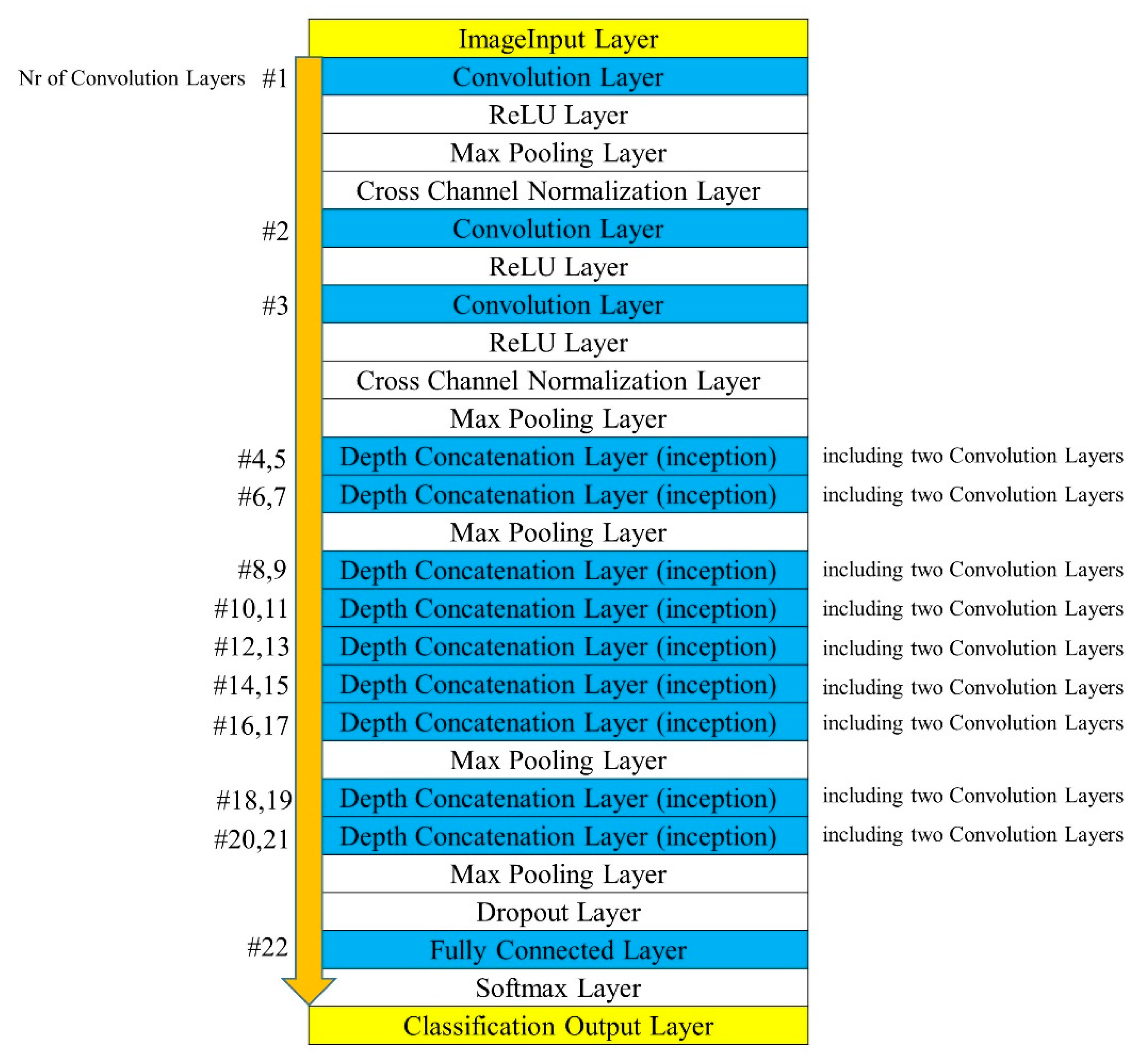
2.3. Preprocessing of Images for Creating the Models
2.4. Manual Training of the Images for Creating the Models
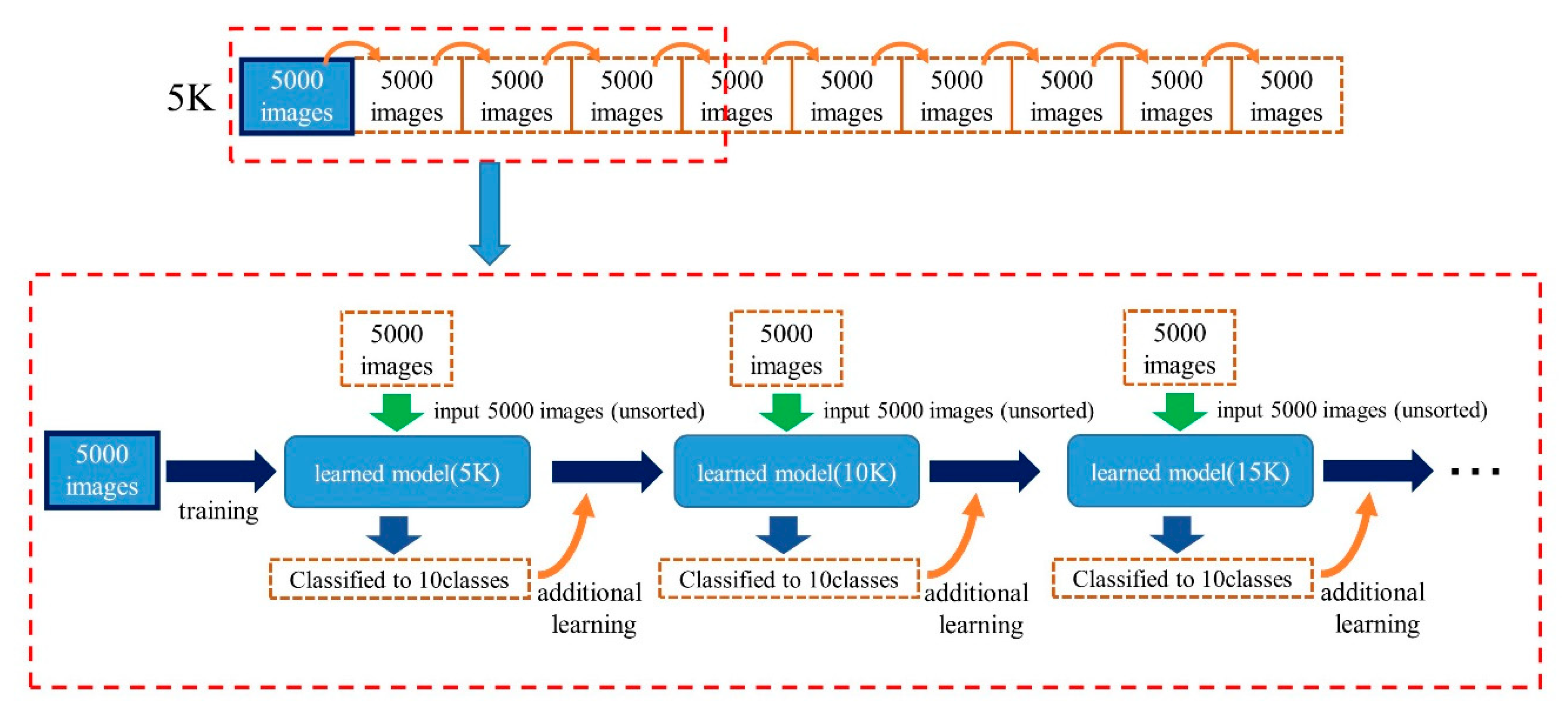
2.5. Automatic Training for Creating Models
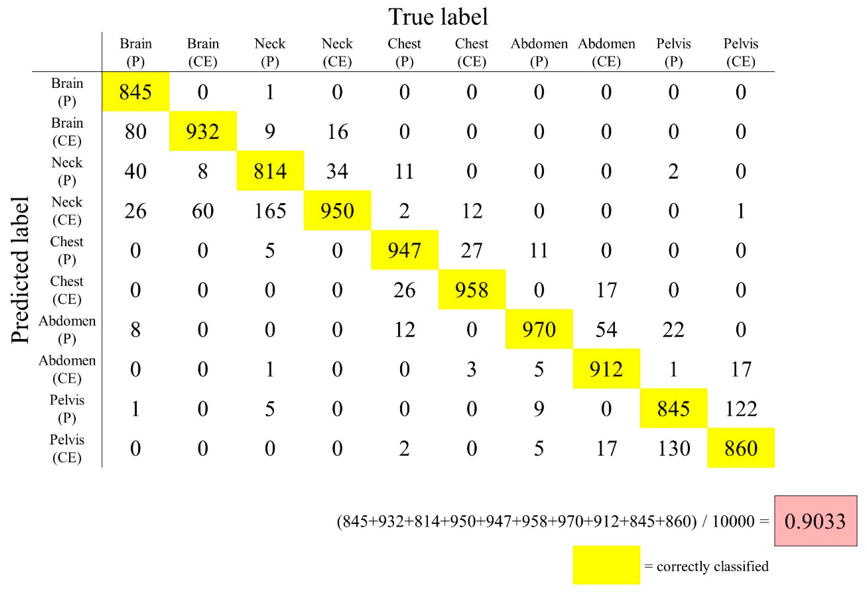
2.6. Evaluation of the Created Models
3. Results and Discussions
3.1. Reference Accuracy
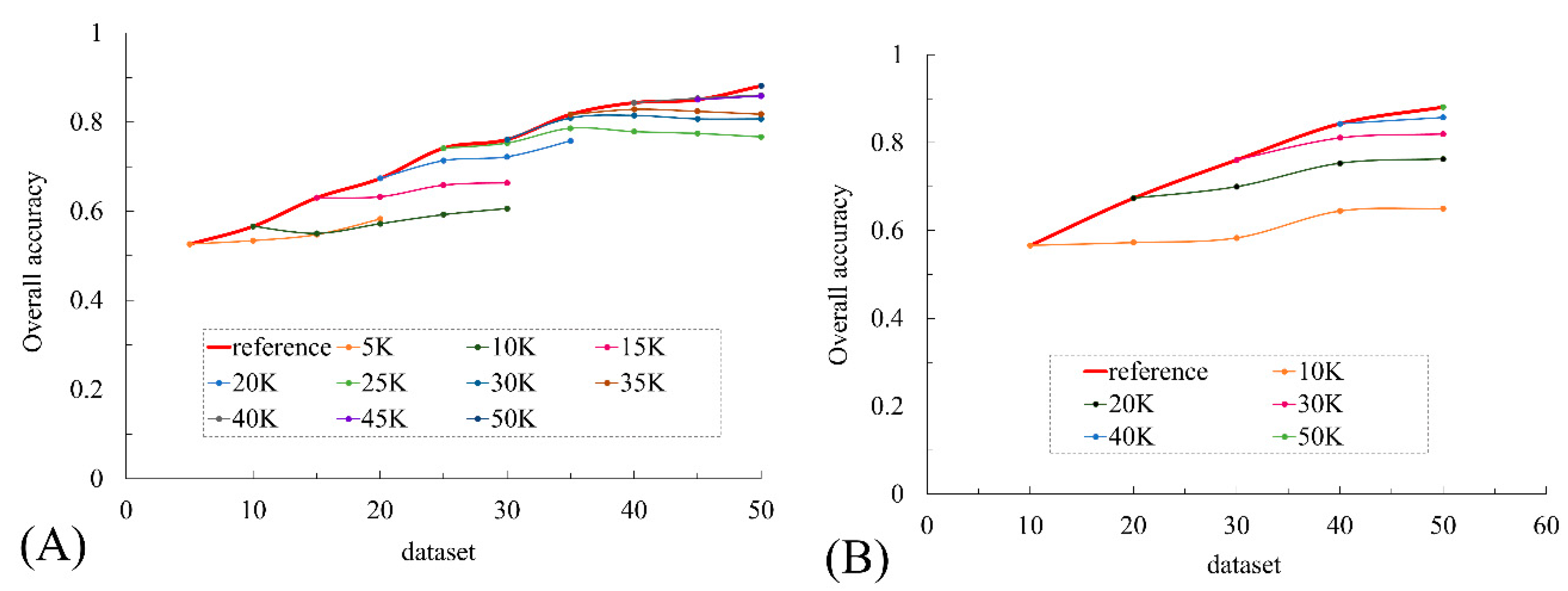
3.2. Manual Training
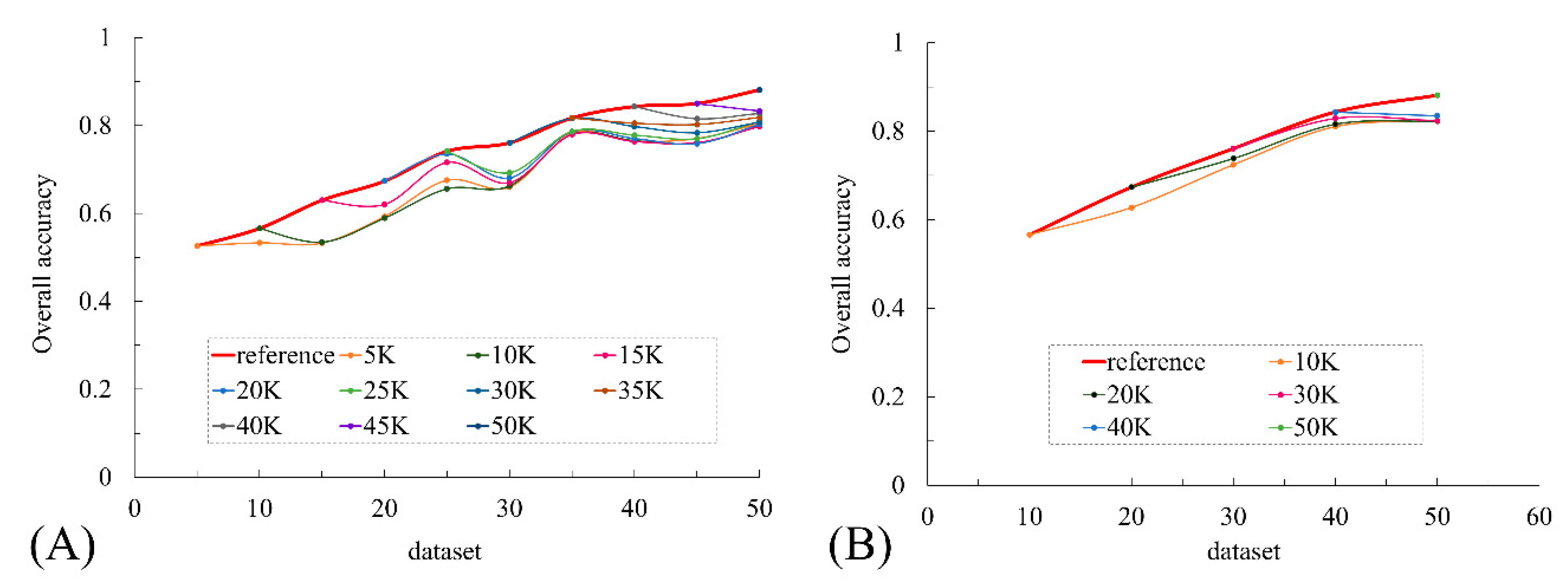
3.3. Automatic Training
4. Conclusions
Author Contributions
Funding
Acknowledgments
Conflicts of Interest
References
- LeCun, Y.; Bottou, L.; Bengio, Y.; Haffner, P. Gradient-Based Learning Applied to Document Recognition. Proc. IEEE 1998, 86, 2278–2324. [Google Scholar] [CrossRef]
- Krizhevsky, A.; Sutskever, I.; Hinton, G.E. Imagenet classification with deep convolutional neural networks. In Proceedings of the Advances in Neural Information Processing Systems, Lake Tahoe, NV, USA, 3–6 December 2012; pp. 1097–1105. [Google Scholar]
- Szegedy, C.; Liu, W.; Jia, Y.; Sermanet, P.; Reed, S.; Anguelov, D.; Erhan, D.; Vanhoucke, V.; Rabinovich, A. Going deeper with convolutions. In Proceedings of the IEEE Conference on Computer Vision and Pattern Recognition, Boston, MA, USA, 7–12 June 2015. [Google Scholar]
- Lakhani, P.; Sundaram, B. Deep Learning at Chest Radiography: Automated Classification of Pulmonary Tuberculosis by Using Convolutional Neural Networks. Radiology 2017, 284, 574–582. [Google Scholar] [CrossRef] [PubMed]
- Qayyum, A.; Anwar, S.M.; Awais, M.; Majid, M. Medical image retrieval using deep convolutional neural network. Neurocomputing 2017, 266, 8–20. [Google Scholar] [CrossRef]
- Gao, X.W.; Hui, R.; Tian, Z. Classification of CT brain images based on deep learning networks. Comput. Methods Programs Biomed. 2017, 138, 49–56. [Google Scholar] [CrossRef] [PubMed]
- Masood, A.; Sheng, B.; Li, P.; Hou, X.; Wei, X.; Qin, J.; Feng, D. Computer-Assisted Decision Support System in Pulmonary Cancer detection and stage classification on CT images. J. Biomed. Inform. 2018, 79, 117–128. [Google Scholar] [CrossRef] [PubMed]
- Zhao, X.; Liu, L.; Qi, S.; Teng, Y.; Li, J.; Qian, W. Agile convolutional neural network for pulmonary nodule classification using CT images. Int. J. Comput. Assist. Radiol. Surg. 2018, 13, 585–595. [Google Scholar] [CrossRef] [PubMed]
- Wachinger, C.; Reuter, M.; Klein, T. DeepNAT: Deep convolutional neural network for segmenting neuroanatomy. Neuroimage 2018, 170, 434–445. [Google Scholar] [CrossRef] [PubMed]
- Akkus, Z.; Galimzianova, A.; Hoogi, A.; Rubin, D.L.; Erickson, B.J. Deep Learning for Brain MRI Segmentation: State of the Art and Future Directions. J. Digit. Imaging 2017, 30, 449–459. [Google Scholar] [CrossRef] [PubMed]
- Ren, X.; Xiang, L.; Nie, D.; Shao, Y.; Zhang, H.; Shen, D.; Wang, Q. Interleaved 3D-CNNs for joint segmentation of small-volume structures in head and neck CT images. Med. Phys. 2018, 45, 2063–2075. [Google Scholar] [CrossRef] [PubMed]
- Avendi, M.R.; Kheradvar, A.; Jafarkhani, H. A combined deep-learning and deformable-model approach to fully automatic segmentation of the left ventricle in cardiac MRI. Med. Image Anal. 2016, 30, 108–119. [Google Scholar] [CrossRef] [PubMed]
- Sugimori, H. Classification of Computed Tomography Images in Different Slice Positions Using Deep Learning. J. Healthc. Eng. 2018, 2018, 9. [Google Scholar] [CrossRef] [PubMed]
- Kim, K.H.; Choi, S.H.; Park, S.-H. Improving Arterial Spin Labeling by Using Deep Learning. Radiology 2017, 287, 658–666. [Google Scholar] [CrossRef] [PubMed]
- Liu, F.; Jang, H.; Kijowski, R.; Bradshaw, T.; McMillan, A.B. Deep Learning MR Imaging–based Attenuation Correction for PET/MR Imaging. Radiology 2017, 286, 676–684. [Google Scholar] [CrossRef] [PubMed]
- Yasaka, K.; Akai, H.; Kunimatsu, A.; Abe, O.; Kiryu, S. Liver Fibrosis: Deep Convolutional Neural Network for Staging by Using Gadoxetic Acid–enhanced Hepatobiliary Phase MR Images. Radiology 2017, 287, 146–155. [Google Scholar] [CrossRef] [PubMed]
- Chen, M.C.; Ball, R.L.; Yang, L.; Moradzadeh, N.; Chapman, B.E.; Larson, D.B.; Langlotz, C.P.; Amrhein, T.J.; Lungren, M.P. Deep Learning to Classify Radiology Free-Text Reports. Radiology 2017, 286, 845–852. [Google Scholar] [CrossRef] [PubMed]
- Zheng, Q.; Yang, M.; Yang, J.; Zhang, Q.; Zhang, X. Improvement of Generalization Ability of Deep CNN via Implicit Regularization in Two-Stage Training Process. IEEE Access 2018, 6, 15844–15869. [Google Scholar] [CrossRef]
- Szegedy, C.; Vanhoucke, V.; Ioffe, S.; Shlens, J.; Wojna, Z. Rethinking the Inception Architecture for Computer Vision. In Proceedings of the IEEE Conference on Computer Vision and Pattern Recognition, Boston, MA, USA, 7–12 June 2015. [Google Scholar]
- Lin, M.; Chen, Q.; Yan, S. Network in Network. arXiv, 2013; arXiv:1312.4400. [Google Scholar]
- Kamaruddin, N.; Rajion, Z.A.; Yusof, A.; Aziz, M.E. Relationship between Hounsfield unit in CT scan and gray scale in CBCT. AIP Conf. Proc. 2016, 1791, 020005. [Google Scholar] [CrossRef]


| Class Name | Dataset | Validation Dataset | |||||||||||
|---|---|---|---|---|---|---|---|---|---|---|---|---|---|
| 5 K | 10 K | 15 K | 20 K | 25 K | 30 K | 35 K | 40 K | 45 K | 50 K | A | B | C | |
| Brain (P) | 500 | 1000 | 1500 | 2000 | 2500 | 3000 | 3500 | 4000 | 4500 | 5000 | 1000 | 1000 | 1000 |
| Brain (CE) | 500 | 1000 | 1500 | 2000 | 2500 | 3000 | 3500 | 4000 | 4500 | 5000 | 1000 | 1000 | 1000 |
| Neck (P) | 500 | 1000 | 1500 | 2000 | 2500 | 3000 | 3500 | 4000 | 4500 | 5000 | 1000 | 1000 | 1000 |
| Neck (CE) | 500 | 1000 | 1500 | 2000 | 2500 | 3000 | 3500 | 4000 | 4500 | 5000 | 1000 | 1000 | 1000 |
| Chest (P) | 500 | 1000 | 1500 | 2000 | 2500 | 3000 | 3500 | 4000 | 4500 | 5000 | 1000 | 1000 | 1000 |
| Chest (CE) | 500 | 1000 | 1500 | 2000 | 2500 | 3000 | 3500 | 4000 | 4500 | 5000 | 1000 | 1000 | 1000 |
| Abdomen (P) | 500 | 1000 | 1500 | 2000 | 2500 | 3000 | 3500 | 4000 | 4500 | 5000 | 1000 | 1000 | 1000 |
| Abdomen (CE) | 500 | 1000 | 1500 | 2000 | 2500 | 3000 | 3500 | 4000 | 4500 | 5000 | 1000 | 1000 | 1000 |
| Pelvis (P) | 500 | 1000 | 1500 | 2000 | 2500 | 3000 | 3500 | 4000 | 4500 | 5000 | 1000 | 1000 | 1000 |
| Pelvis (CE) | 500 | 1000 | 1500 | 2000 | 2500 | 3000 | 3500 | 4000 | 4500 | 5000 | 1000 | 1000 | 1000 |
| Total number of images | 5000 | 10,000 | 15,000 | 20,000 | 25,000 | 30,000 | 35,000 | 40,000 | 45,000 | 50,000 | 10,000 | 10,000 | 10,000 |
| Dataset Type | Group | Dataset | |||||||||
|---|---|---|---|---|---|---|---|---|---|---|---|
| 5 K | 10 K | 15 K | 20 K | 25 K | 30 K | 35 K | 40 K | 45 K | 50 K | ||
| Validation dataset | A | 0.6028 | 0.6532 | 0.7293 | 0.7914 | 0.8334 | 0.8369 | 0.8615 | 0.8947 | 0.8986 | 0.9033 |
| B | 0.4833 | 0.5352 | 0.5713 | 0.6166 | 0.6789 | 0.7056 | 0.7693 | 0.7877 | 0.7884 | 0.8422 | |
| C | 0.4927 | 0.5101 | 0.5899 | 0.613 | 0.7121 | 0.7397 | 0.8208 | 0.8472 | 0.8633 | 0.8974 | |
| mean | 0.5263 | 0.5662 | 0.6302 | 0.6737 | 0.7415 | 0.7607 | 0.8172 | 0.8432 | 0.8501 | 0.881 | |
© 2019 by the author. Licensee MDPI, Basel, Switzerland. This article is an open access article distributed under the terms and conditions of the Creative Commons Attribution (CC BY) license (http://creativecommons.org/licenses/by/4.0/).
Share and Cite
Sugimori, H. Evaluating the Overall Accuracy of Additional Learning and Automatic Classification System for CT Images. Appl. Sci. 2019, 9, 682. https://doi.org/10.3390/app9040682
Sugimori H. Evaluating the Overall Accuracy of Additional Learning and Automatic Classification System for CT Images. Applied Sciences. 2019; 9(4):682. https://doi.org/10.3390/app9040682
Chicago/Turabian StyleSugimori, Hiroyuki. 2019. "Evaluating the Overall Accuracy of Additional Learning and Automatic Classification System for CT Images" Applied Sciences 9, no. 4: 682. https://doi.org/10.3390/app9040682
APA StyleSugimori, H. (2019). Evaluating the Overall Accuracy of Additional Learning and Automatic Classification System for CT Images. Applied Sciences, 9(4), 682. https://doi.org/10.3390/app9040682