Article

Construction of Silver Quantum Dot Immobilized Zn-MOF-8 Composite for Electrochemical Sensing of 2,4-Dinitrotoluene

1
Department of Chemistry, Guru Jambheshwar University of Science and Technology, Hisar 125001, Haryana, India
2
Department of Bio and Nano Technology, Guru Jambheshwar University of Science and Technology, Hisar 125001, Haryana, India
3
Department of Regional Centre of Advanced Technologies and Materials, Department of Physical Chemistry, Faculty of Science, Palacky University, Šlechtitelů 27, 783 71 Olomouc, Czech Republic
*
Authors to whom correspondence should be addressed.
Appl. Sci. 2019, 9(22), 4952; https://doi.org/10.3390/app9224952
Submission received: 29 September 2019 / Revised: 3 November 2019 / Accepted: 14 November 2019 / Published: 18 November 2019

Abstract

:
In the present study, we report a highly effective electrochemical sensor for detecting 2,4-dinitrotoluene (2,4-DNT). The amperometric determination of 2,4-DNT was carried out using a gold electrode modified with zinc–metal organic framework-8 and silver quantum dot (Zn-MOF-8@AgQDs) composite. The synthesized nanomaterials were characterized by using transmission electron microscopy (TEM), Fourier transform infrared spectroscopy (FTIR) and X-ray powder diffraction (XRD). The synthesized nanocomposite proved to be efficient in electro-catalysis thereby reducing the 2,4-DNT. The unique combination present in Zn-MOF-8@AgQDs composite offered an excellent conductivity and large surface area enabling the fabrication of a highly sensitive (−0.238 µA µM−1 cm−2), selective, rapid and stable 2,4-DNT sensor. The dynamic linear range and limit of detection (LOD) was about 0.0002 µM to 0.9 µM and 0.041 µM, respectively. A 2,4-DNT reduction was also observed during the linear sweep voltammetry (LSV) experiments with reduction peaks at −0.49 V and −0.68 V. This is an unprecedented report with metal organic framework (MOF) composite for sensing 2,4-DNT. In addition, the presence of other species such as thiourea, urea, ammonia, glucose, and ascorbic acid displayed no interference in the modified electrode suggesting its practicability in various environmental applications.

Graphical Abstract

1. Introduction

Homeland security has become the principal challenge for most nations, conceivably due to the sudden outburst of heat and potential energy release from nitrogen-containing aromatic compounds. This has led to a rapid increase in research into the capability of detecting materials such as explosive compounds, biological weapons, flammable materials and chemical warfare agents, which can cause widespread communal harm even at trace levels [1,2]. Nitroaromatic compounds are the most common prevalent explosives, representing a prominent class [3]. The nitroaromatic explosive 2,4-dinitrotoluene (DNT) is widely used in antitank and antipersonnel landmines, and is responsible for extensive contamination of land and water. In addition, 2,4-dinitotoluene (DNT) is also a degradation product of trinitrotoluene (TNT) and is a significant environmental concern [4]; the vapor pressure of 2,4-DNT being 40–1000 times higher than that of TNT [5]. The US Environmental Protection Agency cites 2,4-DNT as a carcinogen [6] and considering its destructive potential, significant efforts have been devoted for the development of reliable, innovative, simple and efficient sensing devices.
In recent years, various researchers have attempted to detect explosives using strategies including fluorescence, liquid chromatography, mass spectrometry, ion mobility spectrometry, infrared absorption spectroscopy and electron capture detection [7,8,9,10,11,12,13,14,15,16]. However, these techniques are both expensive and time-consuming. The electrochemical technique has been extensively utilized to detect explosives due to its simplicity, high sensitivity and quick response time [17,18]. The electroactive nature of the nitro groups present in 2,4-DNT is responsible for the electrochemical sensing which can be performed by a stepwise 4-electron reduction for each NO2 group into NHOH followed by a further 2-electron reduction of the NHOH group into NH2 [19,20]. Recently, nanomaterials have also attracted remarkable interest, owing to their unique features [21,22,23,24] and are represented by conductive polymers, metal oxides, natural semiconductors and carbon nanomaterials for sensing toxic materials [25,26] with electrocatalytic frameworks for reduction of 2,4-DNT. In an earlier study, reduced graphene fabricated on a glassy carbon electrode (GCE) surface was used to detect 2,4-DNT via a stripping voltammetry technique in which the detection limit was 42 nmol L−1 and a carbon-screen–printed electrode for trace 2,4-DNT detection in field applications [1,27], including electrochemical sensing in sea water [28]. In another study, a GCE-rGO/SrTiO3 modified glassy carbon electrode (MGCE) showed rapid electronic communication for the electrochemical sensing of nitro-substituted 2,4-DNT [29] and a carbowax/PPy-based system [30]. A carbon nanotube (CNT)-based flexible sensor was developed for the trace detection of 2,4-DNT, displaying good response at 0.22 ppm concentration; such composite conducting materials provide a fast electron exchange between the electrode and the electrolytic solution.
Yet, the production of these materials is complex, with a low yield thus necessitating a need to develop novel materials for the rapid detection of 2,4-DNT. Metal organic frameworks (MOFs) are of broad research interest due to their porous, flexible nature which allows the entrapment of different types of guest molecules, thereby increasing their electrical conductivity and stability; they hold immense potential in diverse industrial applications, such as drug delivery, gas storage, separation, heterogeneous catalysis and chemical sensing [31,32,33,34,35,36,37].
In the present study, silver quantum dots immobilized on Zn-MOF-8 (Zinc–metal organic framework-8) were sonochemically synthesized and characterized using TEM, FTIR and XRD. The encapsulated Zn–MOF-8@AgQDs (silver quantum dots) were applied over the gold electrode to create a modified Zn–MOF-8@Ag/Au electrode for the electrochemical reduction of 2,4-DNT. The performance of this electrode was examined using impedance spectroscopy, cyclic voltammetry and linear sweep voltammetry wherein the electrode exhibited noteworthy features in terms of high surface area, better electron transfer, good sensitivity, stability and detection limit. This is an unprecedented finding for the electrochemical sensing of 2,4-DNT.

2. Materials and Methods

2.1. Chemicals and Reagents

Zinc acetate dihydrate [Zn(O₂CCH₃)₂ 2H₂O (99%)], naphthalene-2,6-dicarboxylic acid C12H8O4 (99%), dimethylformamide (CH3)2NC(O)H hydrazine and Nafion were purchased from Sigma-Aldrich (St. Louis, Missouri, USA). Dried methanol (max. 0.2% water), triethylamine, ethanol, 2,4-dinitrotoluene (C7H6N2O4), silver nitrate (AgNO3), NaBH4, KH2PO4, Na2HPO4, KCl, ammonia, urea, thiourea, KI, ascorbic acid, glucose and NaCl were purchased from Himedia Laboratories, West Chester, Pennsylvania, USA and used without purification.

2.2. Apparatus

The morphology and size of as-grown Zn-MOF-8 and its composite were analyzed by TEM Morgagni 268D (Fei Electron Optics, AIIMS, Delhi, India). The X-ray diffraction was performed by Rigaku Miniflex-II diffractometer, Tokyo,, Japan (Cu-Kɑ radiation, λ = 1.54 A). The FT-IR spectrum was analyzed by Shimazdu IR Affinity-I FT-IR spectrophotometer,(Nakagyo-Ku, Kyoto, Japan ). Ultrasonication was carried out in an ultrasonic water bath, Synergy Techno Support & Solution LLP, Mumbai, India(frequency of 40 KHz). Thermal analysis was performed using the TGA/DSC 3+ Stare system (Mettler Toledo, Leicester, UK) in the temperature range between 25–1000 °C with a heat rate flow of 10 °C min−1 in a static nitrogen atmosphere. Energy dispersive X-ray (EDX) acquired on Bruker–Quan Tax 200 in the textile technology department at Indian Institute of Technology, New Delhi, India ). The electrochemical experiments were performed on an electrochemical workstation PGSTAT302N (metrohm), µ-Autolab, Kanaalweg Utrecht, Netherlands, at room temperature with a three-electrode set up system: modified AuE as working electrode, Ag/AgCl as reference electrode and Pt as auxiliary electrode.

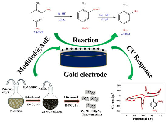
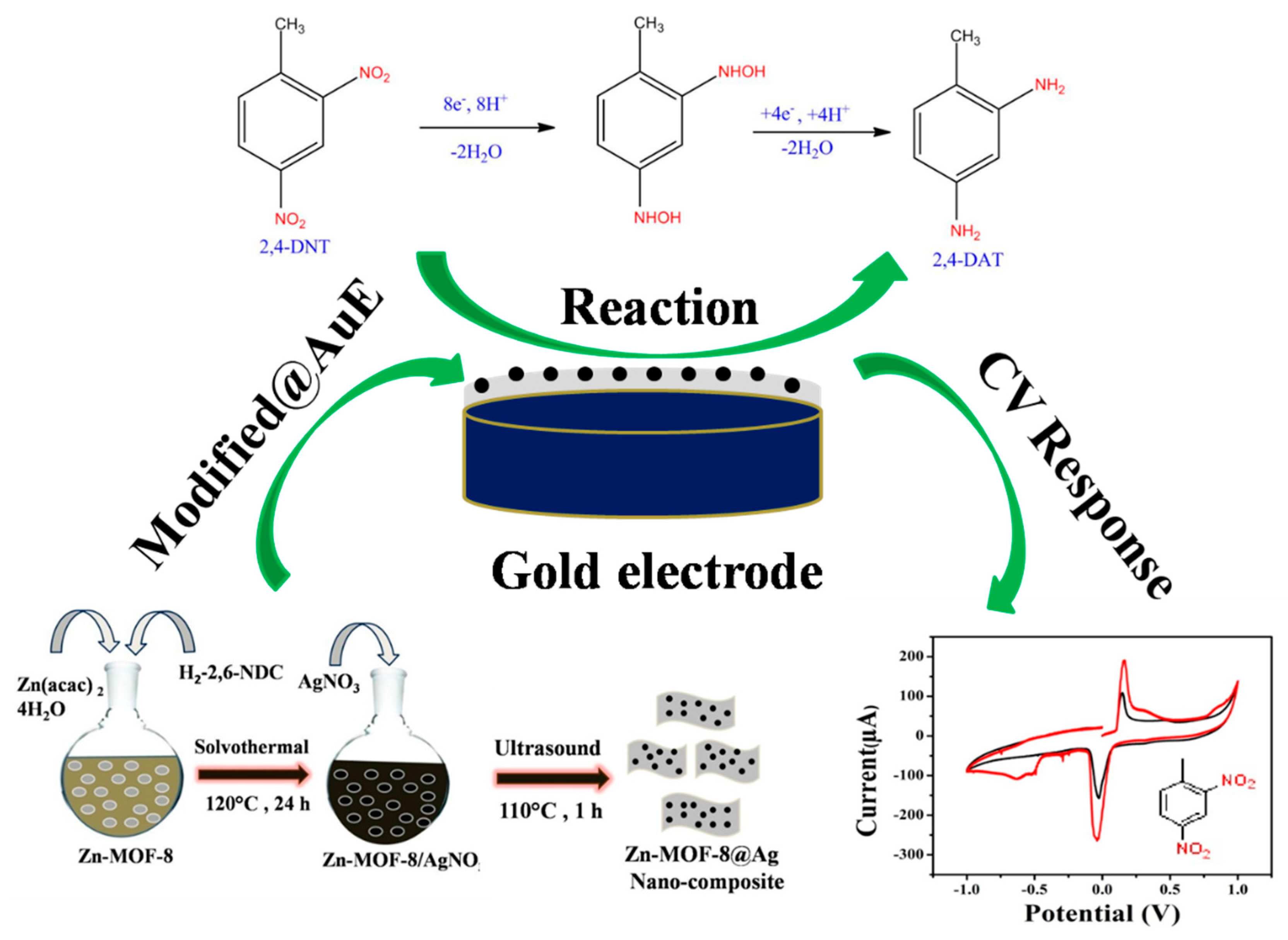
2.3. Synthesis of Nanostructure Zn-MOF-8 and Their Composites with Silver Quantum Dot Particles

The Zn–MOF-8 was synthesized using solvothermal method. For this, 3.188 mg (0.007 mmol) Zn(C4O4H6).2H2O was dissolved in 40 mL of dimethylformamide and mixed with 0.90 g 2,6-naphthalene dicarboxylic acid, dissolved in 3 mL triethylamine. The solution was poured into a round-bottom flask and incubated in an oil bath at 120 °C for 24 h, after which the product was stored at room temperature. The precipitate was isolated by filtration and washed at least three times with ethanol followed by double distilled water (DDW) and was dried under vacuum for 6 h. The Zn–MOF-8@Ag composite was prepared by dispersing 700 mg of Zn-MOF-8 in 22 mL of dry methanol under ambient conditions, to which 3 mL of AgNO3 (22.05 mg, 0.13 mmol) solution was added drop wise under vigorous stirring [38]. This was followed by addition of 5 mL sodium borohydride (NaBH4 37.1 mg) solution in distilled water under vigorous stirring for complete reduction of Ag+. The resultant solution was sonicated for 2 h and the ensuing product was washed with methanol followed by double distilled water (DDW). The obtained product was dried at 55 °C in an oven for 5 h and stored for further use.

2.4. Fabrication of Au/Zn-MOF-8@AgQDs Composite Modified Electrode

Before modification, the bare Au electrode was cleaned using alumina slurry (0.05 µm) and double distilled water followed by sonication to remove the adsorbed material on the electrode surface. The Zn–MOF-8@AgQDs dispersion was obtained by dispersing the composite material in double distilled water under sonication. About 3 µL of dispersed Zn–MOF-8@AgQDs dispersion was drop casted on the surface of gold electrode and allowed to dry in air for 1 h. After drying, the Zn-MOF-8@AgQDs modified gold electrode, Nafion (2 µL) solution was added over the electrode and further dried for 1 h. The fabricated gold electrode was used for the detection of 2,4-DNT.

3. Results and Discussion

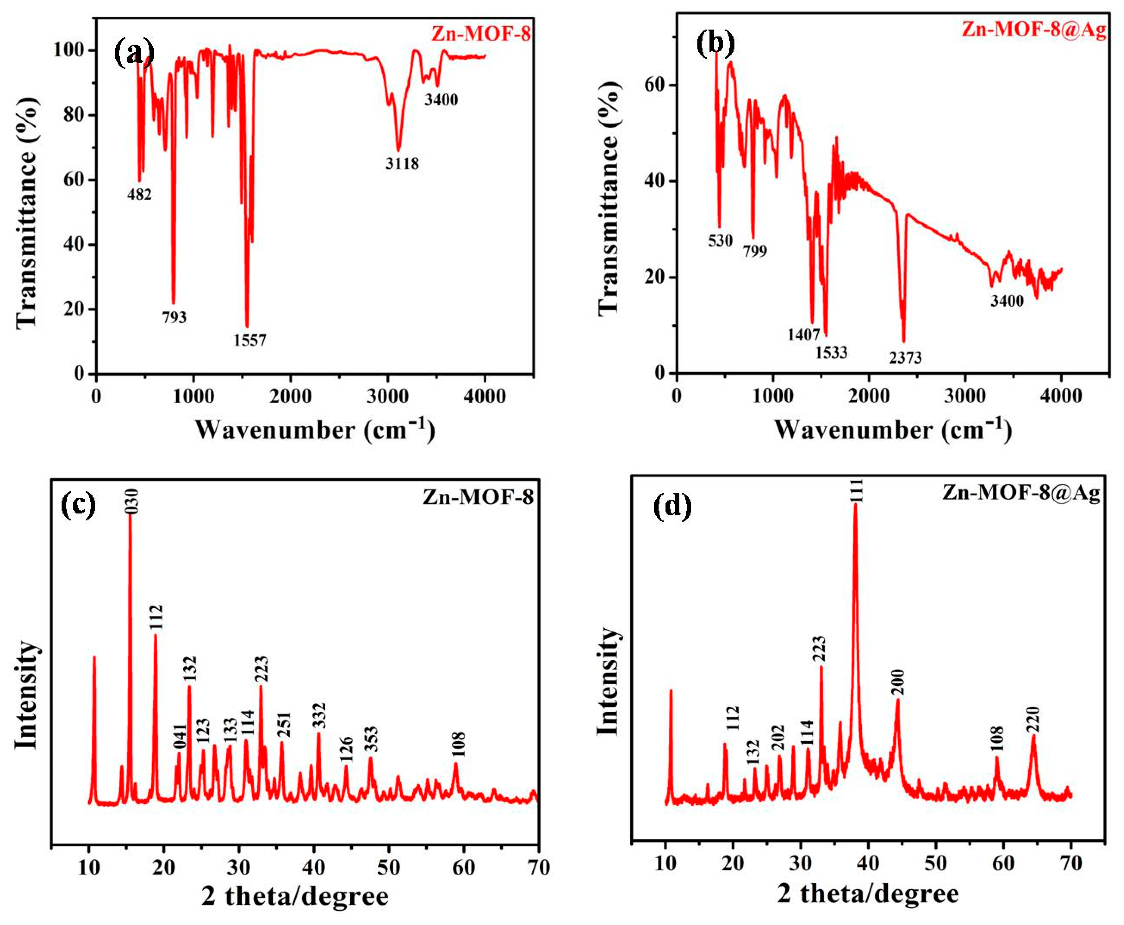
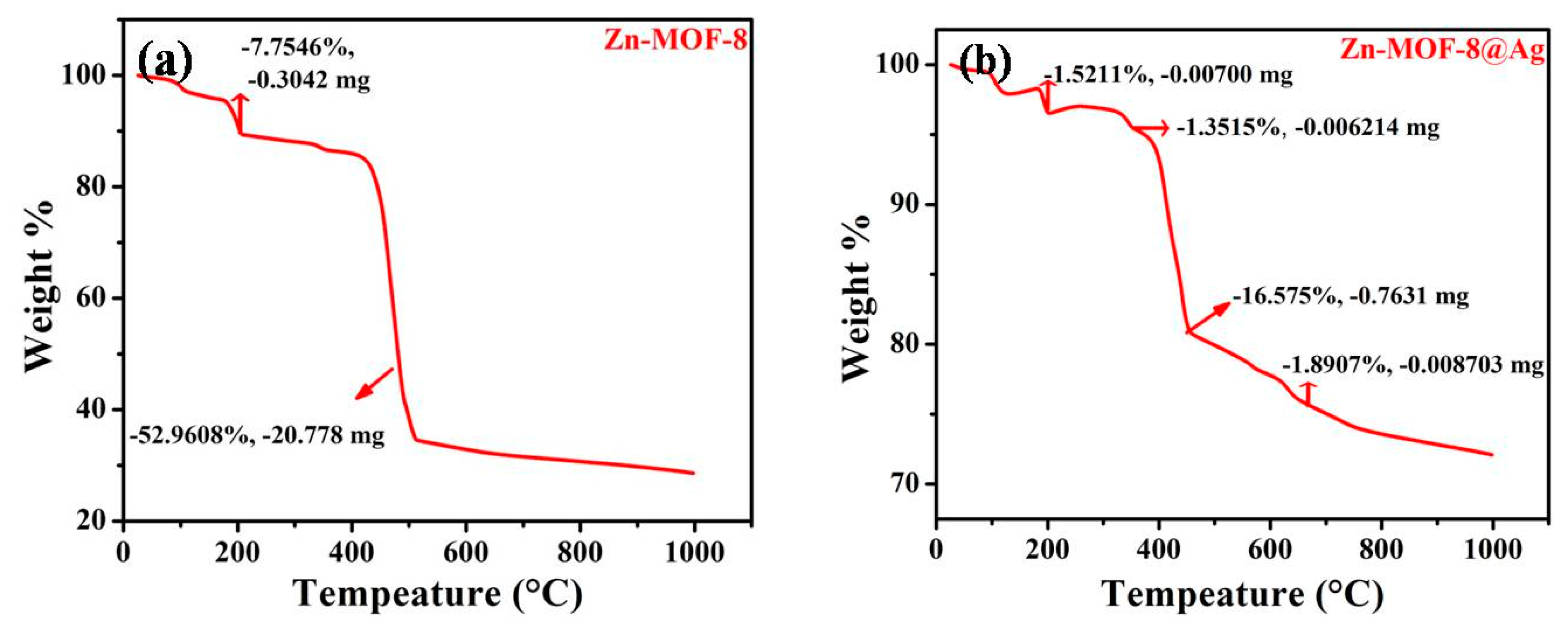
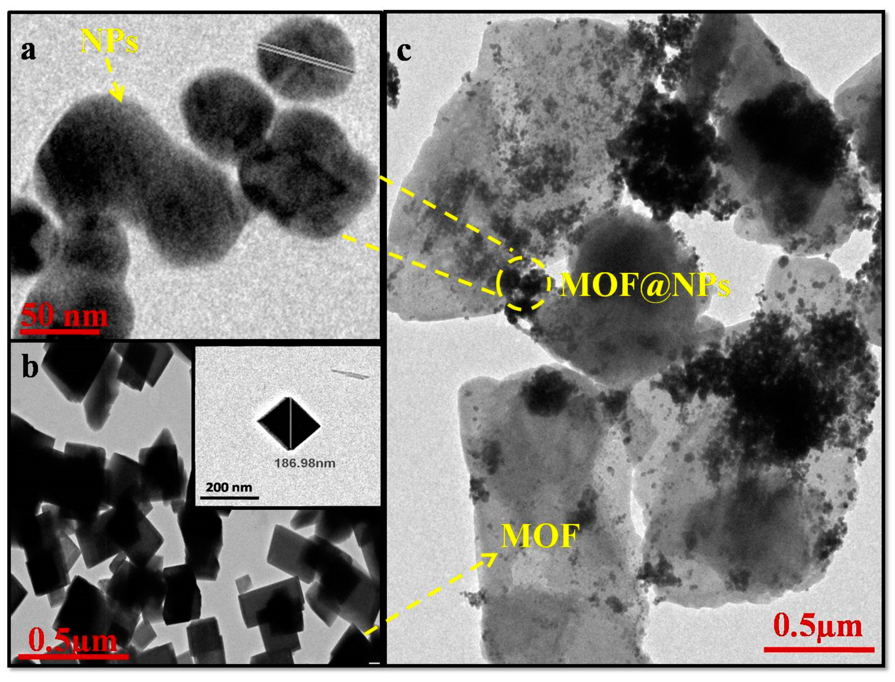
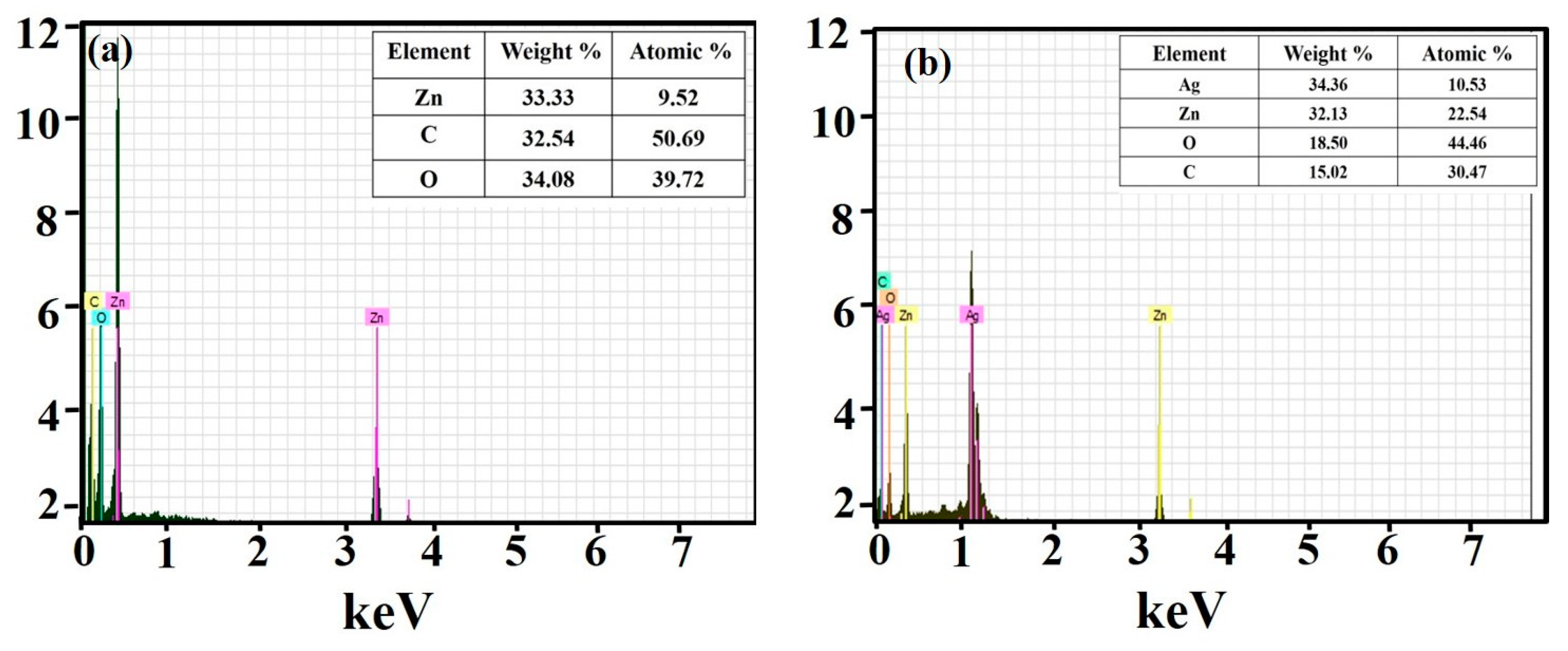
3.1. Characterization of Zn-MOF-8@AgQDs Composite

The chemical structure of the samples was investigated by FTIR spectroscopy. Figure 1a,b displays FTIR spectra of the prepared zinc metal complex and their composite in the range of 400–4000 cm−1. In Figure 1, the zinc–metal complex showed the absorption frequency at 3400 cm−1 which confirms the presence of hydroxyl group in the complex. Peaks appearing at 3118 cm−1 and 1612 cm−1 were assigned due to C–H stretching present in aromatic ring of the linker and C=O stretching lower than those of napthalenedicarboxylic acid, respectively which confirmed the coordination of Zn metal. The absorption band at 1557 cm−1 and 1492 cm−1 confirmed the aromatic C=C bending vibrations whereas the absorption at 927 cm−1, 792 cm−1 and 648 cm−1 was due to presence of small amount of dimethylformamide as residual solvent. The absorption peaks at 482 cm−1 correspond to Zn–O stretching which confirmed the structure of the metal coordination bond in the complex. There was no absorption peak at 1700 cm−1 which implies that carboxyl group of 2,6-naphthalenedicarboxylic acid was completely removed during the reaction. Figure 1b depicts the FTIR spectrum of ZnMOF–8@AgQDs composite indicating partial changes in the peak value due to encapsulation of Ag QDs into the pores of MOFs. The extra peak appearing at about 530 cm−1 corresponds to stretching of the Ag–O bond and the peak at 2373 cm−1 was due to air in the atmosphere. The crystallinity, structure and phase composition of Zn–MOF-8 and its composite were analyzed by XRD, which displayed patterns for Zn-MOF-8, similar to that of the single crystal structure of Zn–MOF-8 as prepared by solvothermal method. The number of well-defined diffraction peaks at various positions in the complex was confirmed by powder-XRD and the crystalline nature of the metal complexes was observed (Figure 1c,d). In Figure 1c, the sharp and intense peak in P-XRD confirmed the monoclinic crystal system with space group P21/c. For Zn–MOF-8@Ag composite (Figure 1d), the P-XRD pattern was comparable to that of Zn–MOF-8 crystal except for strong peaks observed at 38° and 44° and 60° which corresponds to the (111), (200) and (108) planes of silver QDs particles [39,40,41].
To investigate the thermal stability of the nanostructure metal complex and its composite, thermal gravimetric analysis (TGA) was performed in the temperature range from 25–1000 °C under nitrogen atmosphere (Figure 2). The TGA curve of Zn–MOF-8 revealed that weight loss initiated with the removal of water and dimethylformamide molecules around 100–200 °C whereas the second prominent mass loss was seen around 500–530 °C which is due to structural decomposition of organic linkers (Figure 2a). However, in the case of the composite, the weight loss started around 100–200 °C and the second weight loss occurred round 450 °C due to large amount of metallic species in the composite (Figure 2b). The third weight loss occurred around 650 °C which corresponded to the degradation of metal framework. The results clearly showed that the weight loss of Zn–MOF-8@AgQDs was detected at relatively higher temperature, which indicated high thermal stability of the MOF after incorporation of Ag nanoparticles.
The morphology and size of the metal complex and its composite were examined by TEM. TEM images of the Zn–MOF-8 nanoparticles and its QDs composite are shown in Figure 3. The Zn–MOF-8 showed cube-shaped morphologies with size in the range of ͂ 186 nm (Figure 3b). Moreover, Zn-MOF–8@Ag composite confirmed that Ag particles were adhering to the Zn–MOF-8 surface. Figure 3c reveals the strong binding of Ag QDs particles on Zn–MOF-8 surface resulting in a smooth pathway for the collection and transportation of electrons. The elemental composition of Zn–MOF-8 and Zn–MOF-8@Ag was studied by using energy dispersive X-ray analysis (EDXA). The EDX analysis depicted in Figure 4a,b shows the presence of Zn, O, C in Zn-MOF and Zn, Ag, C and O in Zn–MOF@AgQDs, respectively indicating the formation of nanocomposite. This EDX mapping confirmed that the AgQDs are well encapsulated by Zn–MOF-8 and atomic % of Ag within Zn–MOF-8 was determined, see atomic % inset in Figure 4a,b.

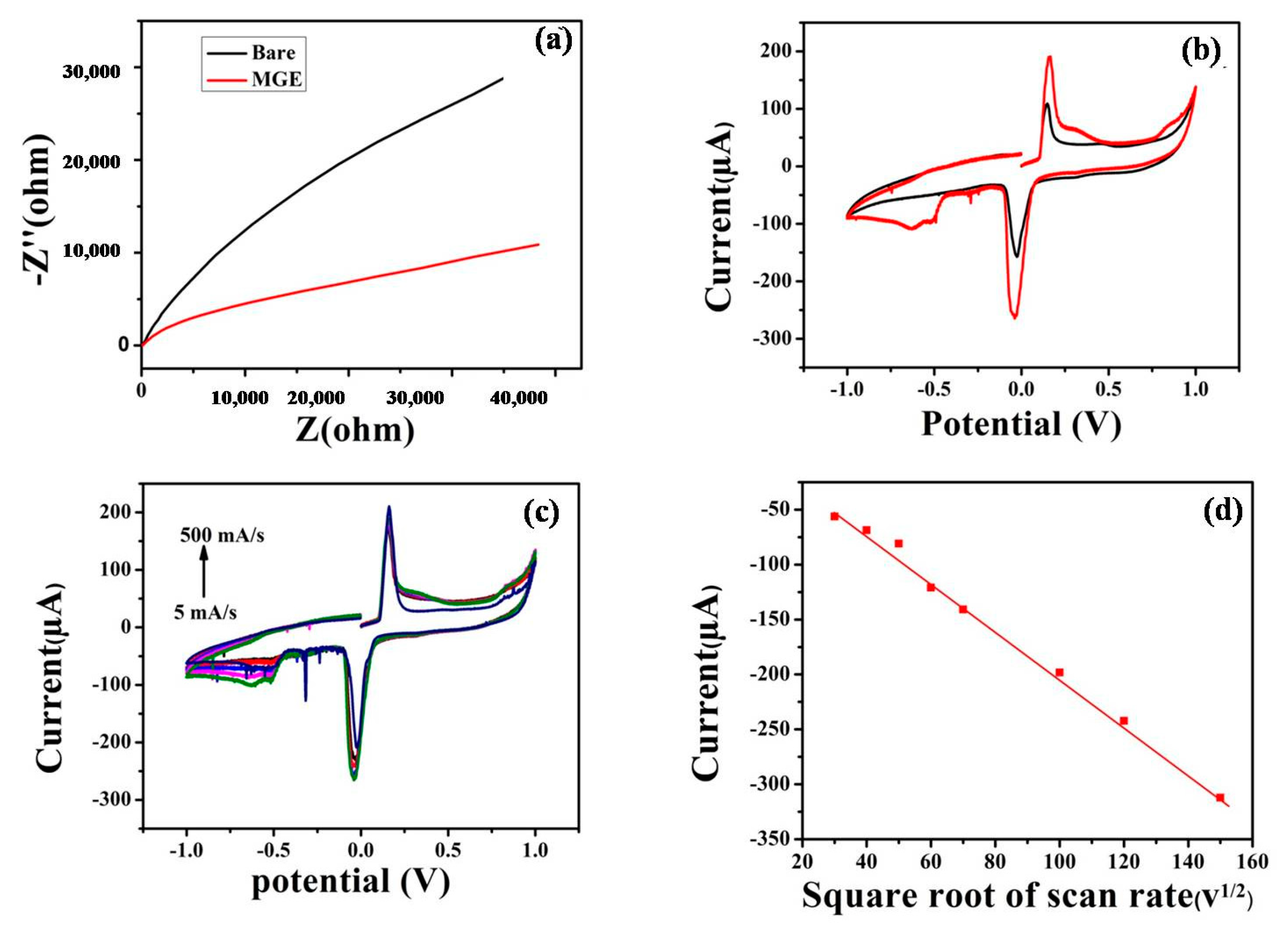
3.2. Electrochemical Impedance Spectroscopy (EIS) Study

Impedance spectroscopy, a rapidly developing electrochemical technique analyzes the charge transfer behavior of the modified electrode. The Nyquist plot of different electrodes: (a) bare, (b) modified, were plotted in the frequency range from 0.01 Hz to 100 Hz in phosphate buffer saline (PBS) of pH 7.0 (Figure 5a). The bare electrode exhibited higher charge transfer resistance than the modified Zn–MOF-8/Ag/Au electrode, which suggested better electron exchange of the modified electrode as compared to the bare electrode, affirming that the higher charge resistance limits the electron exchange system.

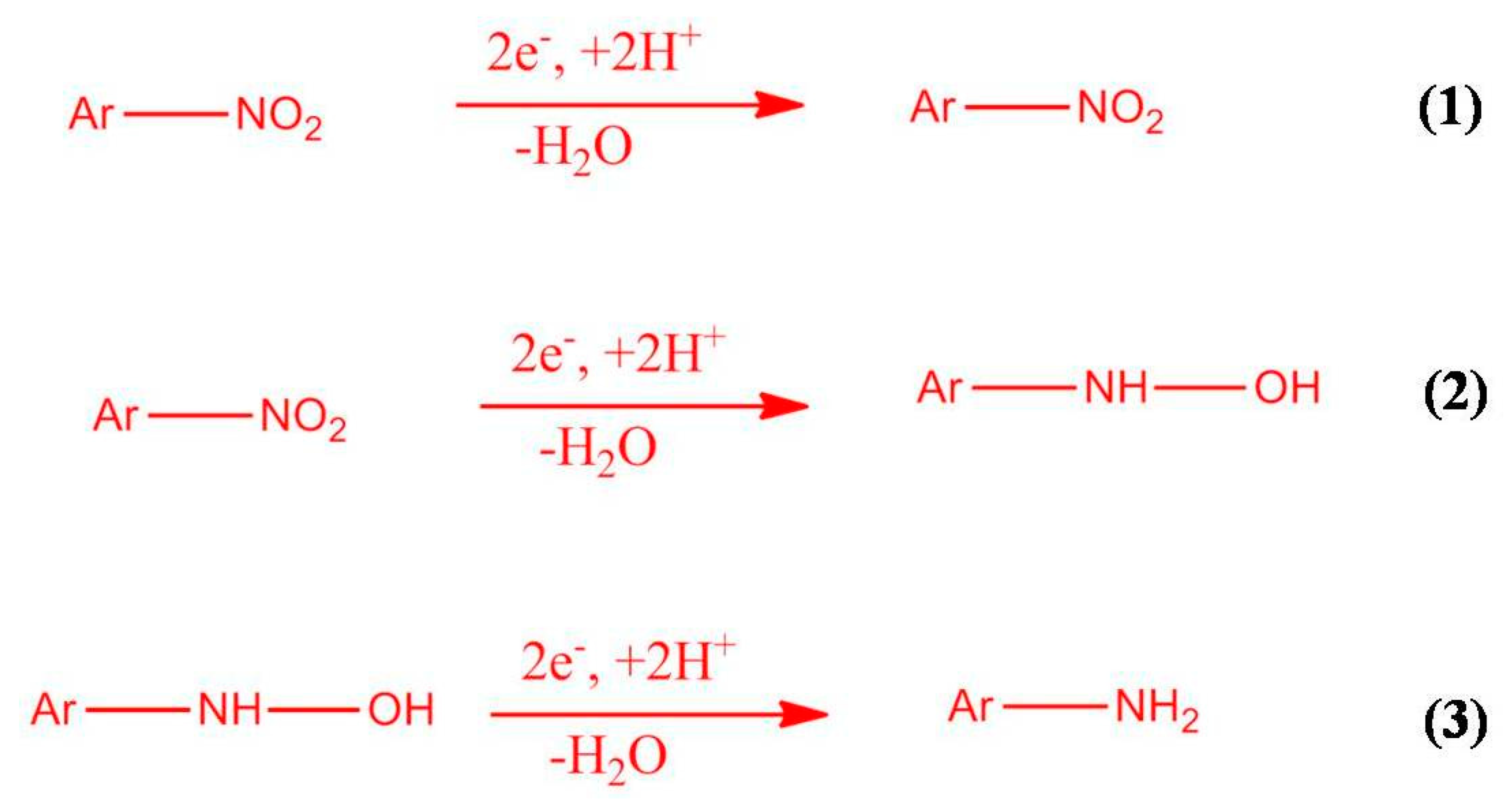
3.3. The Electrocatalytic Reduction and Detection of 2,4-Dinitrotoluene

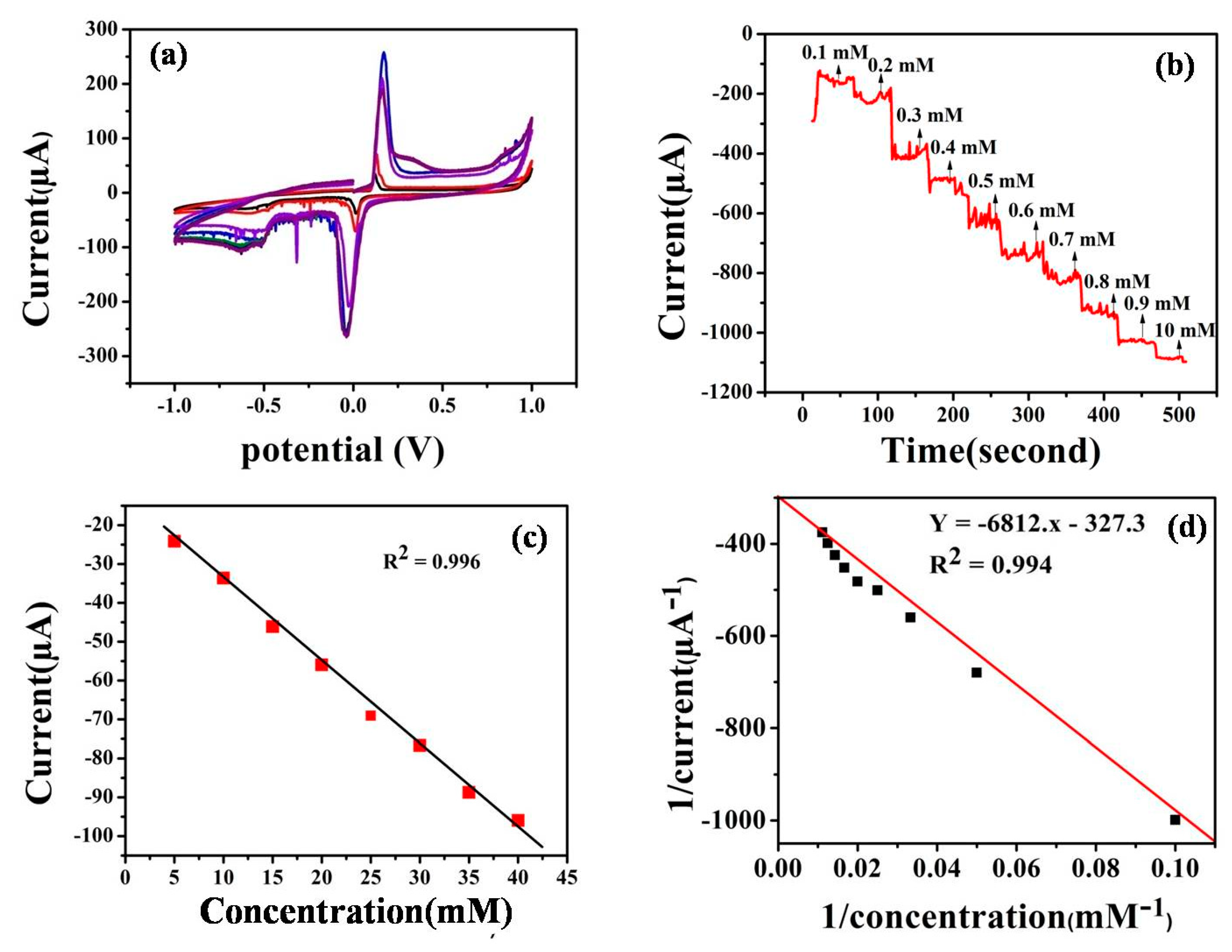
3.3.1. Cyclic Voltammetry

Cyclic voltammograms were recorded to estimate the electrocatalytic activity of the synthesized Zn–MOF-8@Ag/Au modified electrode towards the reduction of 2,4 DNT. In Figure 5b, CV was performed in the presence and absence modified electrode in presence of the 2,4 DNT. The dynamic active surface area of the modified gold electrode was calculated by using Randles–Sevcik equation; utilizing cyclic voltammetric technique, 5 mM K3Fe (CN)6 as a test solution and 0.1 M KCl as the supporting electrolyte, at different sweep rates. For a reversible process the equation is as follows:
Ip = 2.69 × 10⁵ × n3/2 A × D0½ × ν1/2 × C0
where Ip = peak current (µA), A = surface area of the electrode (cm2), C0= concentration of electroactive species (mol cm−3), n = number of electrons transferred, D0 = diffusion coefficient (cm2 s−1) and ν = scan rate (V s−1). The value of the diffusion coefficient can be obtained, by plotting Ipa vs. ν1/2 (for 0.05 mol cm−2 of K3[Fe(CN)6]3/4). Here, C0 = 0.05, n = 1, D0 = 7.6 × 10⁻⁶ cm2 s−1. On substituting these values, the active surface area was calculated to be 0.056 cm2 on a modified Au electrode [42]. The CV response was recorded for a modified Au electrode in 0.25 mM 2,4-DNT in 0.1 M, PBS (pH = 7.4), respectively, at a scan rate of 0.0050 mV/s. Figure 5b shows the CV curve of a Zn–MOF-8@Ag modified gold electrode in the absence gave no response (black line) and in presence afforded two reduction peaks (red line). The results indicated that the modified Au electrode showed negligible response in the absence of 2,4-DNT but gave two major reduction peaks in presence of 2,4-DNT. These reduction peak appeared at −0.50 V and −0.62 V which confirmed that the Zn–MOF-8@Ag modified Au electrode is much reactive towards 2,4-DNT and established that synthesized Zn–MOF-8@Ag is efficient for the reduction of 2,4-DNT. Further, the effect of scan rate of modified electrode was studied in detail by applying different scan rate dependent experiments. The scan rate experiments were performed (0.25 mM, 2,4 DNT, 0.1 M PBS) in scan rates ranging from 5 mA/s to 500 mA/s and are depicted in Figure 5c. The CV responses of modified electrodes increased with increase in the scan rate which confirmed that the reduction is diffusion controlled. The calibration curve of cathodic peak current and square root of scan rate is plotted in Figure 5d.
Further, we have endorsed the effect of different concentrations of 2,4-DNT against the modified gold electrode; CV response showed that electrocatalytic current increased with increase in concentration (0.2 to 0.25 mM) of 2,4-DNT in PBS at pH 7.4 against the modified Au electrode (Figure 6a). The results reveal that with the increment in the concentration of the 2,4-DNT, the reduction peak (R1 & R2) also increased, which confirmed that the modified electrode is sensitive towards 2,4 DNT. The irreversible electrochemical response shows that there is no anodic current observed in the reverse sweep, thus implying that Zn–MOF-8@AgQDs is an effective mediator for the reduction of 2,4-DNT.

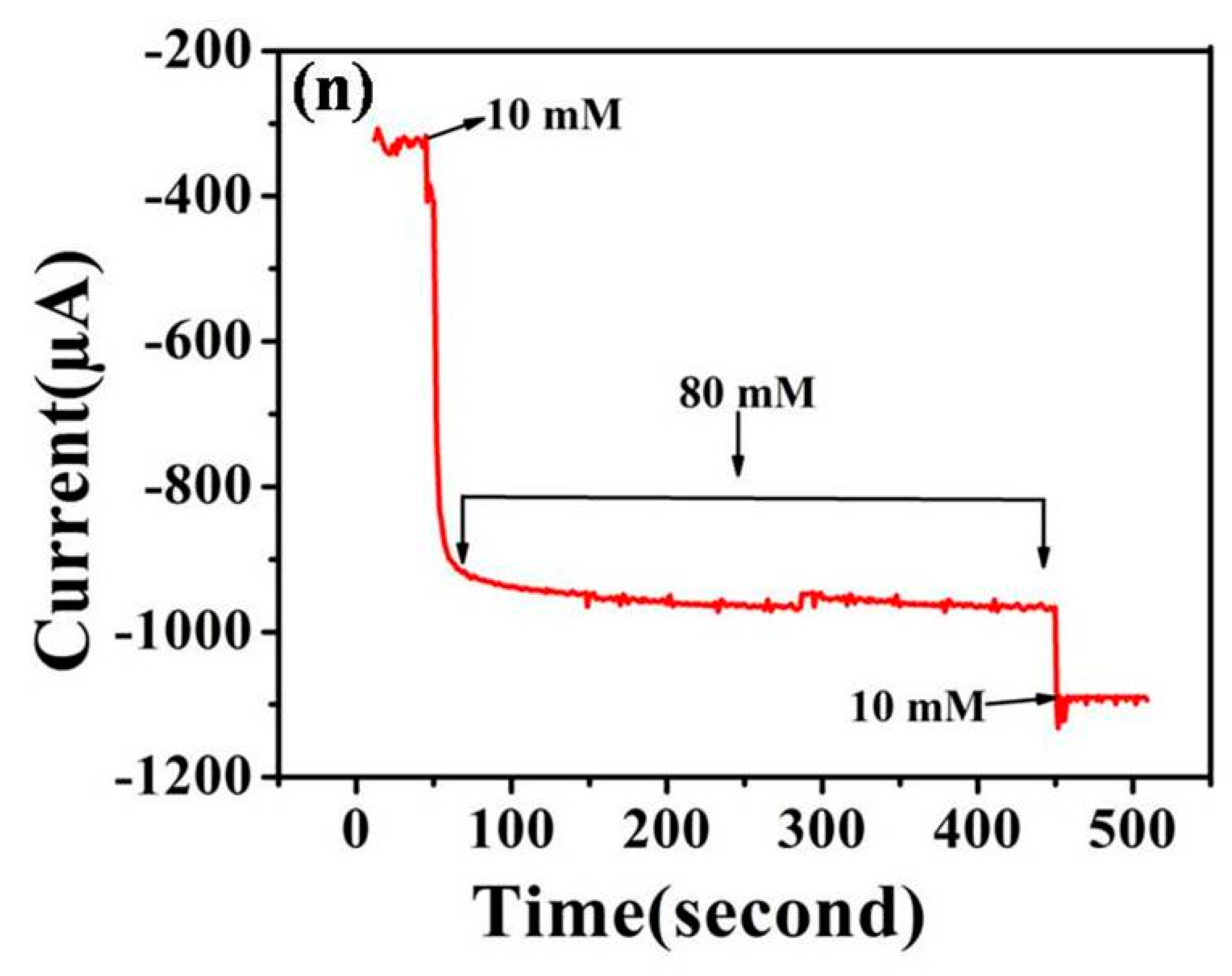
3.3.2. Amperometric Determination of 2,4-DNT

In order to evaluate sensor responses, such as sensitivity, detection limit and correlation coefficient, amperometric measurements were performed using Zn–MOF-8@AgQDs modified Au electrode for sensing of 2,4-DNT; Figure 6b shows typical responses for Zn–MOF-8@Ag/AuE at different concentration ranges from 0.10 mM to 10 mM of 2,4-DNT in phosphate buffer solution (PBS) at constant applied potential of 0.05 V. This low reduction potential (0.05 V) is more beneficial for the sensing of 2,4-DNT. A sharp and well-defined amperometric response was obtained during addition of, 4-DNT, which confirmed that the Zn–MOF-8@Ag modified electrode is efficient for the reduction of 2,4 DNT. Figure 6c, depicts a linear relationship for both cathodic peak current against different concentrations of 2,4-DNT; response current increased linearly at 2,4-DNT concentrations ranging from 0.100 mM to 10 mM.
Figure 6d shows the plot for 1/current vs. 1/concentration represents a linear relationship between constant current and 2,4 DNT concentration. The correlation coefficient of the fabricated sensor was calculated to be R2 = 0.995 and sensitivity was calculated (sensitivity = slope/area of the electrode) from the slope of the standard curve in Figure 6d; it was found to −0.238 µA µM−1 cm−2. The detection limit was calculated as 0.041 µM (LOD = 3σ/S, where σ denotes standard error and S denotes slope of standard curve) and response time was <2 s.

3.3.3. Comparative Study of the Previously Reported Literature

Many earlier studies have been reported sensing of 2,4-DNT using different nanocomposites and they are shown in Table 1. However, the prepared sensor in this study exhibited improved analytical presentation in term of sensitivity and limit of detection (LOD) as compared to previously reported sensors.

3.3.4. Interference Study

Good selectivity is very vital and challenging for electrochemical sensors. The selectivity of the modified sensor was evaluated using chronoamperometry as shown in Figure 7.
The selectivity of the modified electrode was assessed towards potential interfering species like hydrazine, thiourea, glucose, ascorbic acid, ethanol, urea, ammonia and KI in the presence of 0.01 mM 2,4-DNT. It was found that no amperometry response was obtained when 0.1 mM solution of each ion was introduced into 0.01 M PBS. However, the amperometric response was observed when 0.01 mM of 2,4-DNT was added to the solution which indicated good selectivity of the Zn–MOF-8@Ag modified Au electrode towards 2,4-DNT in practical applications.

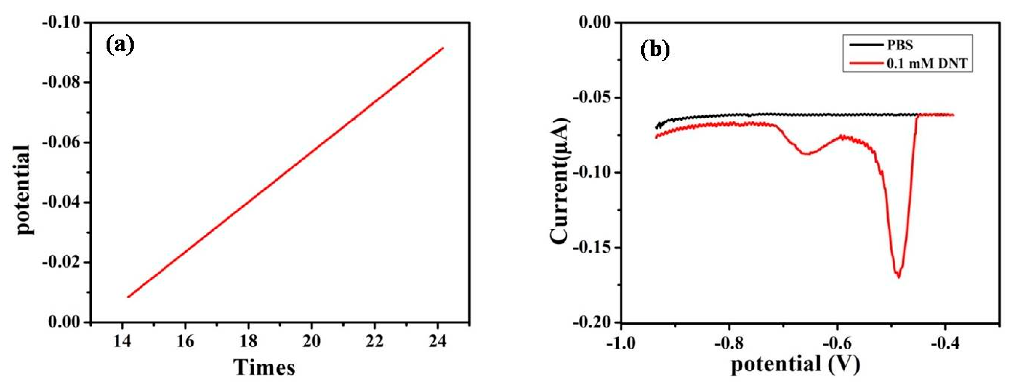
3.3.5. Linear Sweep Voltammetry

The electrochemical behavior of the modified electrode towards reduction of 2,4-DNT was investigated by linear sweep voltammetry (LSV) where current is measured at the working electrode. LSV response of the modified/Au electrode shows a linear relation between the concentrations of 2,4-DNT vs. time examining the reduction current (Figure 8a); the modified Au electrode was assessed by LSV for the detection of 0.1 mM 2,4-DNT in PBS (pH 7.4) with the applied electrode potential range between −0.2 to −0.8 V and a slow scan rate of 50 mV/s. The modified electrode in PBS without 2,4-DNT observed no reduction peak in LSV whereas for 2,4-DNT it detected two stepwise reduction peaks around −0.49 V and −0.68 V in LSV (Figure 8b). The LSV response shows the number of nitro group reduction peaks in the aromatic compound.
Consequently, the number of nitroaromatic groups can be easily detected with the help of modified electrode thus showing its prowess and high efficiency for the electrochemical detection of 2,4-DNT. The two-step reaction requires six electrons and six protons for the reduction of the nitro group. The initial reduction of the nitro group, via 2 e and 2 H+ exchange (removal of water molecules) to a nitroso derivative (Reaction 1) (Figure 9). The nitroso compound was further reduced to a hydroxylamine via exchange of 2 e and 2 H+ (Reaction 2) and finally to the aromatic amine via elimination of water molecules (Reaction 3). The procedure requires in total 12 e and 12 H+ for the reduction of the two nitro groups into corresponding amines.
The modified electrode showed good results towards detection of 2,4-DNT which proved that Zn–MOF-8@AgQDs composites efficiently worked for sensing. The reproducibility of the Zn–MOF@AgQDs modified Au electrode was evaluated by using CV in presence of 0.2 µM 2,4-DNT for 3 times using the same electrode and no significant variation was observed in terms of over potential and current response; the response time of the developed sensor being <2 s.
The schematic representation for the synthesis of Zn–MOF-8@AgQDs and the proposed redox reaction mechanism during the sensing of 2,4-DNT is given in Figure 10. Based on these results, the Zn–MOF-8@Ag/AuE shows a good potential for the detection of 2,4-DNT.

4. Conclusions

This work reports a new Zn–MOF-8@AgQDs composite material for the detection of 2,4-DNT. The Zn–MOF-8@AgQDs modified Au electrode possessed outstanding adsorption and electrochemical sensing of 2,4-DNT via cyclic voltameteric and amperometric methods. Under optimal conditions, the fabricated Zn–MOF-8@Ag modified Au electrode possessed higher sensitivity, and no interference was discerned from other explosive compounds including hydrazine, thiourea, glucose, ascorbic acid, ethanol, urea, ammonium and KI. The Zn–MOF-8@Ag modified Au electrode provides a low detection limit of 0.041 µM, high sensitivity −0.238 (µA−1µM−1cm−2) and shows good repeatability. The detection limit and sensitivity of Zn–MOF-8@AgQDs sensor was superior to other reported 2,4-DNT sensors.

Author Contributions

Conceptualization, N.D., S.R.; methodology, S.R. and B.S.; validation, S.R., B.S. and S.K.; formal analysis, investigation, S.R.; data curation, writing, R.M.; review and editing, R.S.V. All authors have provided critical feedback and assistance in the conducted research, analysis and finalization of the manuscript.

Funding

This research was funded by Sushma Rani, Council of Scientific and Industrial Research (CSIR) sanction latter no/file no 09/752(0057)/2014/EMR-1, New Delhi.and the research work was funded by Life Science Research Board- Defence Research and Development Organization. RSV acknowledges the support by the Operational Programme Research, Development and Education - European Regional Development Fund, project no. CZ.02.1.01/0.0/0.0/16_019/0000754 of the Ministry of Education, Youth and Sports of the Czech Republic.

Acknowledgments

Sushma Rani is thankful to CSIR, New Delhi, for providing financial assistance in the form of a Junior Research Fellowship. The authors thank LSRB-DRDO for providing research funding and MHRD-RUSA, DST-PURSE grant for providing funding for purchase of equipment and infrastructure. Authors are thankful to SAIF, AIIMS, Delhi for the TEM facility.

Conflicts of Interest

Authors declare no conflict of interest.

References

  1. Caygill, J.S.; Collyer, S.D.; Holmes, J.L.; Davis, F.; Higson, S.P. Disposable screen-printed sensors for the electrochemical detection of TNT and DNT. Analyst 2013, 138, 346–352. [Google Scholar] [CrossRef] [PubMed]
  2. Kumar, D.; Jha, P.; Chouksey, A.; Tandon, R.P.; Chaudhury, P.K.; Rawat, J.S. Flexible single walled nanotube based chemical sensor for 2, 4-dinitrotoluene sensing. J. Mater. Sci. Mater. Electron. 2018, 29, 6200–6205. [Google Scholar] [CrossRef]
  3. Sun, X.; Wang, Y.; Lei, Y. Fluorescence based explosive detection: From mechanisms to sensory materials. Chem. Soc. Rev. 2015, 44, 8019–8061. [Google Scholar] [CrossRef] [PubMed]
  4. Kannan, O.K.; Bhalla, R.; Kapoor, J.C.; Nimal, A.T.; Mittal, U.; Yadava, R.D.S. Detection of landmine signature using SAW-based polymer-coated chemical sensor. Def. Sci. J. 2004, 54, 309–315. [Google Scholar] [CrossRef]
  5. Krausa, M.; Massong, H.; Rabenecker, P.; Ziegler, H. Chemical methods for the detection of mines and explosives, Chap 1. In Detection of Explosives and Landmines: Methods and Field Experience; Schubert, H., Kuznetsov, A., Eds.; Springer: Dordrecht, The Netherlands, 2012; Volume 66, pp. 1–19. [Google Scholar]
  6. Marshall, M.; Oxley, J.C. (Eds.) Chap. 2 Explosives: The threats and the materials. In Aspects of Explosives Detection; Elsevier: Amsterdam, The Netherlands, 2011; pp. 11–26. [Google Scholar]
  7. Kovalev, I.S.; Taniya, O.S.; Slovesnova, N.V.; Kim, G.A.; Santra, S.; Zyryanov, G.V.; Chupakhin, O.N. Fluorescent detection of 2, 4-DNT and 2, 4, 6-TNT in aqueous media by using simple water-soluble pyrene derivatives. Chem. Asian J. 2016, 11, 775–781. [Google Scholar] [CrossRef]
  8. Stringer, R.C.; Gangopadhyay, S.; Grant, S.A. Detection of nitroaromatic explosives using a fluorescent-labeled imprinted polymer. Anal. Chem. 2010, 82, 4015–4019. [Google Scholar] [CrossRef]
  9. Bratin, K.; Kissinger, P.T.; Briner, R.C.; Bruntlett, C.S. Determination of nitro aromatic, nitramine, and nitrate ester explosive compounds in explosive mixtures and gunshot residue by liquid chromatography and reductive electrochemical detection. Anal. Chim. Acta 1981, 130, 295–311. [Google Scholar] [CrossRef]
  10. Li, X.J.; Ye, C.W.; Huo, X.L.; Zeng, Z. Solid-phase microextraction using a diglycidyloxycalix[4]arene coated fiber combined with gas chromatography: Very simple, rapid and sensitive method for the determination of chlorobenzenes in water. Microchim. Acta 2010, 168, 161–167. [Google Scholar] [CrossRef]
  11. Forbes, T.P.; Sisco, E. Mass spectrometry detection and imaging of inorganic and organic explosive device signatures using desorption electro-flow focusing ionization. Anal. Chem. 2014, 86, 7788–7797. [Google Scholar] [CrossRef]
  12. Peng, L.; Hua, L.; Wang, W.; Zhou, Q.; Li, H. On-site rapid detection of trace non-volatile inorganic explosives by stand-alone ion mobility spectrometry via acid-enhanced evaporization. Sci. Rep. 2014, 4, 6631. [Google Scholar] [CrossRef]
  13. Zhou, Q.; Peng, L.; Jiang, D.; Wang, X.; Wang, H.; Li, H. Detection of nitro-based and peroxide-based explosives by fast polarity-switchable ion mobility spectrometer with ion focusing in vicinity of Faraday detector. Sci. Rep. 2015, 5, 10659. [Google Scholar] [CrossRef] [PubMed]
  14. Roscioli, K.M.; Davis, E.; Siems, W.F.; Mariano, A.; Su, W.; Guharay, S.K.; Hill, H.H. Modular ion mobility spectrometer for explosives detection using corona ionization. Anal. Chem. 2011, 83, 5965–5971. [Google Scholar] [CrossRef] [PubMed]
  15. Chen, Y.; Liu, H.; Deng, Y.; Veksler, D.B.; Shur, M.S.; Zhang, X.C.; Schauki, D.; Fitch, M.J.; Osiander, R.; Dodson, C.; et al. Spectroscopic characterization of explosives in the far-infrared region. In Proceedings of the Terahertz for Military and Security Applications II, Orlando, FL, USA, 12–16 April 2004; Volume 5411, pp. 1–9. [Google Scholar]
  16. Collin, O.L.; Niegel, C.; DeRhodes, K.E.; McCord, B.R.; Jackson, G.P. Fast Gas Chromatography of Explosive Compounds Using a Pulsed-Discharge Electron Capture Detector. J. Forensic Sci. 2006, 51, 815–818. [Google Scholar] [CrossRef] [PubMed]
  17. O’Mahony, A.M.; Wang, J. Nanomaterial-based electrochemical detection of explosives: A review of recent developments. Anal. Methods 2013, 5, 4296–4309. [Google Scholar] [CrossRef]
  18. Gardner, J.W. Review of conventional electronic noses and their possible application to the detection of explosives, Chap.1. In Electronic Noses & Sensors for the Detection of Explosives; Gardner, J.W., Yinon, J., Eds.; Kluwer Academic Publishers: Dordrecht, The Netherlands, 2004; pp. 1–28. [Google Scholar]
  19. Chua, C.K.; Pumera, M.; Rulisek, L. Reduction pathways of 2, 4, 6-trinitrotoluene: An electrochemical and theoretical study. J. Phys. Chem. C 2012, 116, 4243–4251. [Google Scholar] [CrossRef]
  20. Chua, C.K.; Pumera, M. Influence of Methyl Substituent Position on Redox Properties of Nitroaromatics Related to 2, 4, 6-Trinitrotoluene. Electroanalysis 2011, 23, 2350–2356. [Google Scholar] [CrossRef]
  21. Pourmortazavi, S.M.; Marashianpour, Z.; Karimi, M.S.; Mohammad-Zadeh, M. Electrochemical synthesis and characterization of zinc carbonate and zinc oxide Nanoparticles. J. Mol. Struct. 2015, 1099, 232–238. [Google Scholar] [CrossRef]
  22. Pourmortazavi, S.M.; Taghdiri, M.; Makari, V.; Nasrabadi, M.R. Procedure optimization for green synthesis of silver nanoparticles by aqueous extract of Eucalyptus oleosa. Spectrochim. Acta A 2015, 136, 1249–1254. [Google Scholar] [CrossRef]
  23. Shahidzadeh, M.; Shabihi, P.; Pourmortazavi, S.M. Sonochemical preparation of copper (II) chromite nanocatalysts and particle size optimization via taguchi method. J. Inorg. Organomet. Polym. Mater. 2015, 25, 986–994. [Google Scholar] [CrossRef]
  24. Shamsipur, M.; Pourmortazavi, S.M.; Roushani, M.; Hajimirsadeghi, S.S. Electrochemical preparation and thermal characterization of copper sulfide Nanoparticles. Synth. React. Inorg. Met. Org. Nano-Met. Chem. 2014, 44, 951–958. [Google Scholar] [CrossRef]
  25. Choi, H.H.; Lee, J.; Dong, K.Y.; Ju, B.K.; Lee, W. Gas sensing performance of composite materials using conducting polymer/single-walled carbon nanotubes. Macromol. Res. 2012, 20, 143–146. [Google Scholar] [CrossRef]
  26. Sagar, M.; Mudhalwadkar, R.; Sonar, G. Metal oxide semiconductor based thin film sensor for nitro aromatic explosive detection. In Proceedings of the International Conference for convergence for Technology, Pune, India, 6–8 April 2014; pp. 1–4. [Google Scholar]
  27. Chen, T.W.; Sheng, Z.H.; Wang, K.; Wang, F.B.; Xia, X.H. Determination of explosives using electrochemically reduced graphene. Chem. Asian J. 2011, 6, 1210–1216. [Google Scholar] [CrossRef] [PubMed]
  28. Yew, Y.T.; Ambrosi, A.; Pumera, M. Nitroaromatic explosives detection using electrochemically exfoliated graphene. Sci. Rep. 2016, 6, 33276. [Google Scholar] [CrossRef] [PubMed]
  29. Ahmad, K.; Mohammad, A.; Mathur, P.; Mobin, S.M. Preparation of SrTiO3 perovskite decorated rGO and electrochemical detection of nitroaromatics. Electrochim. Acta 2016, 215, 435–446. [Google Scholar] [CrossRef]
  30. Koudehi, M.F.; Pourmortazavi, S.M. Synthesis and application of carbowax/polypyrrole nanocomposite for fabrication of electrochemical sensor to detect 2, 4-DNT vapor. Mater. Res. Express 2017, 4, 086303–086312. [Google Scholar] [CrossRef]
  31. Yaghi, O.M. Metal–organic frameworks: A tale of two entanglements. Nat. Mater. 2007, 6, 92–93. [Google Scholar] [CrossRef]
  32. Pan, L.; Parker, B.; Huang, X.; Olson, D.H.; Lee, J.; Li, J. Zn (tbip)(H2tbip= 5-tert-butyl isophthalic acid): A highly stable guest-free microporous metal organic framework with unique gas separation capability. J. Am. Chem. Soc. 2006, 128, 4180–4181. [Google Scholar] [CrossRef]
  33. Pan, L.; Sander, M.B.; Huang, X.; Li, J.; Smith, M.; Bittner, E.; Bockrath, B.; Johnson, J.K. Microporous metal organic materials: Promising candidates as sorbents for hydrogen storage. J. Am. Chem. Soc. 2004, 126, 1308–1309. [Google Scholar] [CrossRef]
  34. Gao, X.; Wang, Y.; Ji, G.; Cui, R.; Liu, Z. One-pot synthesis of hierarchical-pore metal–organic frameworks for drug delivery and fluorescent imaging. CrystaEngComm 2018, 20, 1087–1093. [Google Scholar] [CrossRef]
  35. Gao, S.; Zhao, L.; Han, L.; Zhang, Z.; Zhao, H. Synthesis, structure and characterization of two solvatochromic metal–organic frameworks for chemical-sensing applications. Cryst. Eng. Comm. 2018, 20, 2237–2240. [Google Scholar] [CrossRef]
  36. Rojas-Buzo, S.; Garcia-Garcia, P.; Corma, A. Catalytic Transfer Hydrogenation of Biomass-Derived Carbonyls over Hafnium-Based Metal–Organic Frameworks. ChemSusChem 2018, 11, 432–438. [Google Scholar] [CrossRef] [PubMed]
  37. Fang, X.; Zong, B.; Mao, S. Metal–organic framework-based sensors for environmental contaminant sensing. Nano-Micro Lett. 2018, 10, 64. [Google Scholar] [CrossRef] [PubMed] [Green Version]
  38. Jiang, H.L.; Akita, T.; Ishida, T.; Haruta, M.; Xu, Q. Synergistic catalysis of Au@Ag core−shell nanoparticles stabilized on metal−organic framework. J. Am. Chem. Soc. 2011, 133, 1304–1306. [Google Scholar] [CrossRef] [PubMed]
  39. Devi, R.N.; Edgar, M.; Gonzalez, J.; Slawin, A.M.; Tunstall, D.P.; Grewal, P.; Wright, P.A. Structural studies and computer simulation of the inclusion of aromatic hydrocarbons in a zinc 2, 6-naphthalene dicarboxylate framework compound. J. Phys. Chem. B 2004, 108, 535–543. [Google Scholar] [CrossRef]
  40. Pilloni, M.; Kumar, V.B.; Ennas, G.; Porat, Z.E.; Scano, A.; Cabras, V.; Gedanken, A. Formation of metallic silver and copper in non-aqueous media by ultrasonic radiation. Ultrason. Sonochem. 2018, 47, 108–113. [Google Scholar] [CrossRef]
  41. Arul, P.; John, S.A. Silver nanoparticles built-in zinc metal organic framework modified electrode for the selective non-enzymatic determination of H2O2. Electrochim. Acta 2017, 235, 680–689. [Google Scholar] [CrossRef]
  42. Shetti, N.P.; Nayak, D.S.; Malode, S.J.; Kulkarni, R.M. An electrochemical sensor for clozapine at ruthenium doped TiO2 nanoparticles modified electrode. Sens. Actuators B: Chem. 2017, 247, 858–867. [Google Scholar] [CrossRef]
Figure 1. FTIR spectra of (a) Zn–MOF-8, (b) Zn–MOF-8@AgQDs (c) Powder- XRD of Zn–MOF-8 and (d) Zn–MOF-8@AgQDs.
Applsci 09 04952 g001
Figure 2. TGA curves of (a) Zn–MOF-8 and, (b) Zn–MOF-8@Ag.
Applsci 09 04952 g002
Figure 3. TEM image of (a) Ag QDs particles, (b) Zn–MOF-8, (c) Zn–MOF-8@AgQDs.
Applsci 09 04952 g003
Figure 4. EDX analysis of (a) Zn–MOF-8, (b) Zn–MOF-8@AgQDs.
Applsci 09 04952 g004
Figure 5. (a) Nyquist plot of (a) bare and (b) Zn–MOF-8@Ag/AuE in PBS (7.4). (b) CV of bare and modified in PBS containing 0.25 mM of 2,4-DNT at scan rate of 0.005 mV/s. (c) CV sweep curve for the Zn–MOF-8@Ag modified AuE at scan rate range (5 to 500 mV/s) in 0.1 M PBS containing 0.25 mM 2-4-DNT, and (d) Plot between cathodic peak current and square root of scan rate.
Applsci 09 04952 g005
Figure 6. (a) CV sweep curve of the Zn–MOF-8@AgQDs modified gold electrode with subsequent addition of 2-4-DNT and plot between current vs. 2-4-DNT concentrations and, (b) typical amperometric response of Zn–MOF-8@Ag modified Au electrode by successive addition of 2-4-DNT (0.10 mM to 10 mM) in 0.1 M phosphate buffer saline (PBS) at constant potential of 0.05 V, (c) display of the linear relationship between constant current and 2-4-DNT concentration, (d) plot of 1/current vs. 1/concentration represents a linear relationship at constant current in different concentrations of 2-4-DNT.
Applsci 09 04952 g006
Figure 7. The amperometric anti-interference ability of a Zn–MOF-8@AgQDs modified Au electrode by addition of 10 mM 2,4-DNT and 80 mM of each interfering reagent (hydrazine, thiourea, glucose, ascorbic acid, ethanol, urea, ammonium, KI) followed by addition of 10 mM 2,4-DNT in PBS at 0.05 V applied potential.
Applsci 09 04952 g007
Figure 8. (a) LSV of modified Au electrode potential is swept linearly with respect to time and, (b) LSV of modified Au electrode in 0.1 M PBS (pH 7.4) in the presence of DNT and absence of DNT at the slow scan rate (50 mV/s).
Applsci 09 04952 g008
Figure 9. Reaction mechanism involved in the reduction of the aromatic (Ar) nitro group (1) reduction of nitro group into nitroso derivative, (2) conversion of nitroso group into hydroxylamine and (3) finally the conversion of hydroxylamine into aromatic amine.
Applsci 09 04952 g009
Figure 10. Schematic representation of the synthesis of Zn–MOF-8, its composite form, and its fabrication on a gold electrode; CV response depicted for reduction of 2-4-DNT.
Applsci 09 04952 g010
Table 1. Summary of linear range, detection limit and sensitivity to 2,4-DNT in previously reported literature.
Type of ElectrodeLinear Range (µM)Sensitivity (µA−1 µM−1 cm−2)Detection Limit (µM)References
GC/G-Li-CO30–0.20−0.5520.035[28]
GCE/rGO/SrTiO30.5–0.871.660.128 [29]
GE/Zn-MOF-8@Ag0.002–0.9−0.2380.041Our work

