Article

Influence of Gemini Surfactants on Biochemical Profile and Ultrastructure of Aspergillus brasiliensis

1
Institute of Fermentation Technology and Microbiology, Faculty of Biotechnology and Food Science, Lodz University of Technology, Wólczańska 171/173, 90-924 Lodz, Poland
2
Laboratory of Microscopic Imaging and Specialized Biological Techniques, Faculty of Biology and Environmental Protection, University of Lodz, Banacha 12/16, 90-237 Lodz, Poland
*
Author to whom correspondence should be addressed.
Appl. Sci. 2019, 9(2), 245; https://doi.org/10.3390/app9020245
Submission received: 2 December 2018 / Revised: 6 January 2019 / Accepted: 7 January 2019 / Published: 10 January 2019
(This article belongs to the Section Applied Biosciences and Bioengineering)

Abstract

:
In this study, we investigated the activities of hexamethylene-1,6-bis-(N,N-dimethyl-N-dodecylammonium bromide) (C6), pentamethylene-1,5-bis-(N,N-dimethyl-N-dodecylammonium bromide) (C5), and their two neutral analogues: hexamethylene-1,6-bis-(N-methyl-N-dodecylamine) (A6) and pentamethylene-1,5-bis-(N-methyl-N-dodecylamine) (A5) at concentrations of ½ MIC, MIC, and 2 MIC (minimal inhibitory concentration) against hyphal forms of Aspergillus brasiliensis ATCC 16404. Enzymatic profiles were determined using the API-ZYM system. Extracellular proteins were extracted from the mycelia and analyzed using sodium dodecyl sulfate polyacrylamide gel electrophoresis (SDS-PAGE). The ultrastructure was evaluated using a transmission electron microscope (TEM). Both groups of surfactants caused changes in the enzyme profiles. Larger changes in the number and concentration of enzymes were noted after the action of non-ionic gemini surfactants, which may have been due to the 100× higher concentration of neutral compounds. Larger differences between the protein profiles of the control sample and the biocide samples were observed following the use of cationic compounds. On the basis of TEM analyses, we found that, with increasing concentrations of compound C6, the mycelium cells gradually degraded. After treatment at 2 MIC, only membranous structures, multiform bodies, and dense electron pellets remained. Based on these results, we concluded that cationic gemini surfactants, in comparison with their non-ionic analogues, could have a wide range of practical applications as active compounds.

1. Introduction

Filamentous fungi are pervasive in the natural environment. They can cause the decomposition of different food stuffs of vegetable or animal origin [1]. They also contribute to the biodegradation of synthetic and natural materials, such as wood, leather, paper, or silk [2,3]. Often, these microorganisms are harmful to humans. They play a significant role in allergies, mycosis, and pulmonary infections, especially in immunocompromised patients. Mycotoxins—the secondary metabolites of molds—are also dangerous as they can accumulate over the years and contribute to the development of cancers in humans and animals [4,5]. Filamentous fungi have been used for decades as natural producers of enzymes, acids, and antibiotics [6,7]. One of the most common and important molds is the genus Aspergillus, and especially the species Aspergillus niger. Even though citric acid and many enzymes produced by these molds are Generally Recognized as Safe (GRAS status) [8], A. niger often causes food spoilage and biodegradation of building materials [9,10].
Various kinds of biocide are used to destroy microorganisms, including molds. Some fungicides have been prohibited by the European Chemical Agency, including carbendazim, which was used previously both as a pesticide [11] and as a preservative film in buildings [12]. New compounds are therefore being sought that are safer for both humans and the environment. Only a few articles have described the mechanisms of action of these new antifungal agents at the cellular and molecular levels, especially in molds. Biocides can act in several places on the cells of these microorganisms, such as the cell wall, cell membrane, and the entire cell interior, affecting the DNA, mitochondria, and cellular reactions [13]. Destruction of molds is difficult, as molds occur in two basic morphological forms: mycelia and spores. Many of the previously -used antifungal agents are becoming less effective due to spontaneous mutations or phenotypic adaptation of molds to harmful environmental conditions.
Gemini surfactants are a group of cationic and neutral compounds, described as antifungal agents. However, the antifungal activity of these substances has only been investigated in yeast [14,15]. No studies have been conducted on molds that are common in various places. In previous studies [16] on the effect of these substances on Aspergillus brasiliensis ATCC 16404 (formerly A. niger ATCC 16404), the authors found ionic gemini surfactants were characterized by high antimicrobial efficacy. Dry weight and ergosterol were reduced and the mycelium formed a conglomerated structure. Cationic gemini surfactants also had better affinity to ergosterol than their neutral analogues. The aim of our current research was to further examine the changes in mycelium cells of A. brasiliensis caused by the activity of hexamethylene-1,6-bis-(N,N-dimethyl-N-dodecylammonium bromide) (C6) and pentamethylene-1,5-bis-(N,N-dimethyl-N-dodecylammonium bromide) (C5), in comparison to their neutral analogues: hexamethylene-1,6-bis-(N-methyl-N-dodecylamine) (A6) and pentamethylene-1,5-bis-(N-methyl-N-dodecylamine) (A5). We investigated the protein and enzyme profiles as well as the ultrastructure of Aspergillus brasiliensis ATCC 16404. The results of our study provide an analysis of several potential areas of action of gemini surfactants in mycelial cells and prove the high effectiveness of these compounds.

2. Materials and Methods

2.1. Microorganisms

Experiments were performed with Aspergillus brasiliensis ATCC 16404 (recommended as a candidate quality control microorganism for antifungal susceptibility test EN 1650). This strain was obtained from the American Type Culture Collection and was isolated form blueberry in North Carolina, U.S. The biological material was stored in Malt Extract Agar (MEA, MERCK, Darmstadt, Germany) at 4 °C. Prior to each experiment, the strains were subcultured in MEA medium at 28 °C for 4–5 days. Spore suspensions were prepared by washing the conidia from the agar slants using deionized sterilized water with 0.1% Tween 80 and stirring. Spore concentrations were measured using a Thoma (Paul Marienfeld GmbH&Co.KG, Lauda-Königshofen, Germany) chamber and the concentrations were adjusted to 1.0–2.0 × 10⁷ conidia/mL.

2.2. Antimicrobial Agents

Two neutral dimeric alkylammonium salts, hexamethylene-1,6-bis-(N-methyl-N-dodecylamine) (A6) and pentamethylene-1,5-bis-(N-methyl-N-dodecylamine) (A5), together with their two cationic analogues, hexamethylene-1,6-bis-(N,N-dimethyl-N-dodecylammonium bromide) (C6) and pentamethylene-1,5-bis-(N,N-dimethyl-N-dodecylammonium bromide) (C5), were used as antimicrobial agents. The gemini surfactants were synthesized following the reaction described previously by Koziróg et al. [16].

2.3. Enzymatic Profiles of Mycelia after Surfactant Treatments

The fungal strain was cultured in 16 mL of Malt Extract Broth (MEB) medium (MERCK, Darmstadt, Germany), inoculated with 2 mL conidia and incubated for 48 h at 28 °C. After incubation, the samples were supplemented with 2 mL of the cationic gemini surfactants C5 or C6 at concentrations of ½ MIC = 0.155, MIC = 0.31, or 2 MIC = 0.62 mM. Following the same procedure, the samples were supplemented with neutral gemini surfactants at concentrations of 15, 30, or 60 mM A5, and 12.5, 25, or 50 mM A6. There were also samples without biocides that were treated as a control. All samples were incubated for another 48 h. The total incubation time of the test samples and control samples without biocides was 96 h. The API-ZYM test (bioMérieux, Marcy l‘Etoile, France) was used to determine the enzymatic profiles of the samples in suspension. The activities of 19 enzymes were investigated: alkaline phosphatase, esterase (C4), esterase lipase (C8), lipase (C14), leucine arylamidase, valine arylamidase, cystine arylamidase, trypsin, α-chymotrypsin, acid phosphatase, naphthol-AS-BI-phosphohydrolase, α-galactosidase, β-galactosidase, β-glucuronidase, α-glucosidase, β-glucosidase, N-acetyl-β-glucosaminidase, α-mannosidase, and α-fucosidase. To each of the 20 cells contained in one reaction strip, 65 μL of MEB medium after 96 h of incubation were added. The analyses were performed in triplicate. The semi-quantitative analysis was completed based on the increase in the color intensity of the samples (0–5 scale). The approximated number of free nM hydrolyzed substrate may be estimated from the color strength: 0—no activity; 1—liberation of 5 nM; 2—10 nM; 3—20 nM; 4—30 nM and 5 ≥40 nM [17,18].

2.4. Preparation of Extracellular Protein Extracts

Prior to extracellular protein extraction, A. brasiliensis ATCC 16404 was cultured in MEB (MERCK, Darmstadt, Germany), using the same procedure as for enzymatic analysis. The culture was filtered using a cellulose membrane (0.45 µm). The supernatants were gently mixed with 5 µL 0.1 M phenylmethylsulfonyl fluoride (PMSF, Sigma-Aldrich, St. Louis, MO, USA), 5 µL 10% 2-mercaptoethanol (Sigma-Aldrich, St. Louis, MO, USA), and 5 µL 1.0 M ethylenediaminetetraacetic acid (EDTA, Sigma-Aldrich, St. Louis, MO, USA). Then, 50% trichloroacetic acid (TCA, POCH, Gliwice, Poland) was added. The samples were incubated at 0 °C for 45 min and centrifuged (12,000 rpm, 15 min). The pellets were washed twice with distilled water, re-suspended in a loading buffer (0.06 M Tris-HCl pH 6.8; 10% glycerol; 2% sodium dodecyl sulphate; 5% 2-mercaptoethanol; 0.025% bromophenol blue) and heated at 100 °C for 5 min. The protein quality was determined using a NanoPhotometer™ Pearl ultraviolet-visible (UV-Vis) spectrophotometer (Implen GmbH, München, Germany ), according to the manufacturer’s protocol.

2.5. SDS-PAGE Analysis of Extracellular Proteins

The extracellular proteins of A. brasiliensis ATCC 16404 were analyzed using sodium dodecyl sulphate polyacrylamide gel electrophoresis (SDS-PAGE). Electrophoresis was performed in 5% stacking gel and 10% separating gel in a discontinuous buffer system (Tris-Glycine-SDS buffer), according to the procedure described by Laemmli [19]. The gel was run at a constant voltage of 20 V/cm using the PROTEAN System (Bio Rad Laboratories Inc., Hercules, CA, USA). After electrophoresis, the gel was fixed in 50% trichloroacetic acid T(CA, POCH, Gliwice, Poland) and stained with Coomassie Brilliant Blue R-250 (0.25% Coomassie Brilliant Blue R-250, 10% acetic acid, 45% methanol; Sigma-Aldrich, St. Louis, MO, USA). Electrophoresis was conducted in triplicate to ensure consistency. The analysis of SDS-PAGE gels and determination of molecular weight of the extracellular proteins were performed with SigmaGel software (Jandel Scientific, San Rafael, CA, USA). PageRuler™ Unstained Broad Range Protein Ladder (Thermo Fisher Scientific, Waltham, MA, USA) was used as a size standard for protein electrophoresis. The average similarities between protein profiles of untreated A. brasiliensis and treatments with gemini surfactants were calculated using the Dice coefficient [20]:
S (%) = 2X × 100/(a + b)
where a is the total number of patterns in protein profile of untreated A. brasiliensis (control sample), b is the total number of patterns for A. brasiliensis after treatment with gemini surfactants, and X is the total number of similar patterns in both compared lanes.

2.6. TEM Analysis

Prior to studies using a transmission microscope, the cultures were prepared using the same method as for enzymatic studies. After 48 h of treatment with hexamethylene-1,6-bis-(N,N-dimethyl-N-dodecylammonium bromide) (C6) at concentrations of ½ MIC, MIC, or 2 MIC, the mycelia were separated from the medium by filtration. A mycelium that had not been treated with biocide was used as a control sample.
Prior to electron microscopic observations, the control and the treated material were fixed in 2.5% glutaraldehyde in 0.2 M phosphate buffer, pH 6.2, at 0–4 °C for 3 h. After washing in buffer and post-fixation with 1% osmium tetraoxide, the samples were dehydrated in an ethanol series (10%, 30%, 50%, 70%, 80%, 90%, and 100%) with propylene oxide, then infiltrated with mixtures of Epon-Spurr (company, city, country) and propylene oxide and embedded in Epon-Spurr resin. After staining with uranyl acetate and lead citrate [21], ultra-thin sections were obtained using an ultramicrotome Ultracut E (Reichert-Jung, Munich, Germany). These were examined using a transmission electron microscope JEM 1010 (JEOL, Tokyo, Japan) transmission electron microscope operating at 80 kV.

3. Results

In the presented work four gemini surfactants were used. Their values of minimal inhibitory concentrations (MIC) for mycelium A. brasiliensis ATCC 16404 were determined by the authors in earlier studies [16]. The results of MIC values are as follows: for cationic gemini surfactants C5 and C6 0.31 mM, for non-ionic gemini surfactants: A5 = 30 mM and A6 = 25 mM, respectively.

3.1. Influence of Gemini Surfactants on Enzymatic Profile of Mycelia

In the first stage of the analysis, we studied the effects of the gemini surfactants on the enzymatic profile of A. brasiliensis ATCC 16404. The results are shown in Table 1.
The lipolytic enzymes alkaline phosphatase (2), esterase (3), esterase lipase (4), acid phosphatase (11), naphthol-AS-BI-phosphohydrolase (12), and the amylolytic enzymes α-galactosidase (13), α-glucosidase (16), and N-acetyl-β-glucosaminidase (18) were detected in the control sample. When surfactants were added to the cultures, several enzymes were suppressed. The amylases activity observed in control sample was almost completely inhibited in the samples with cationic and neutral gemini surfactants. The exceptions were α-glalactosidase (13) and N-acetyl-β-glucosaminidase (18), for which negligible activity (5 nmol) was observed for cationic gemini surfactant C5 across the entire concentration range, and for C6 at ½ MIC and MIC. During detailed analysis of the influence of the gemini surfactants on the overall levels of enzymatic activity of A. brasiliensis mycelia, we observed interesting results in the cases of acid phosphatase (11) and naphthol-AS-BI-phosphohydrolase (12). The highest activity of both lipases, exceeding 40 nmol, was noted in the control sample. As shown in Table 1, cationic gemini surfactants C5 and C6 had no influence on the levels of lipolytic activity by either enzyme. At ½ MIC, MIC, and 2 MIC (0.155 mM, 0.31 mM, and 0.62 mM, respectively), these biocides released the same amounts of substrate as the control sample. An exception was compound C5 at 2 MIC, for which the concentration of naphthol-AS-BI-phosphohydrolase was only 30 nM.
A relationship was observed between the concentrations of non-ionic gemini surfactants A5 and A6 at ½ MIC, MIC, and 2 MIC (15 mM, 30 mM, and 60 mM, respectively) and the activities of acid phosphatase (11) and naphthol-AS-BI-phosphohydrolase (12). As the surfactant concentration increased, the amounts of released substrate decreased: from 40 nM to 5 nM for acid phosphatase (11) and from 30 nM to 5 nM for naphthol-AS-BI-phosphohydrolase (12) for both biocides, A5 and A6. None of the samples treated with cationic or neutral gemini surfactants showed activity for the lipolytic enzymes alkaline phosphatase (2), esterase (3), or esterase lipase (4). Based on the effect of these compounds on the metabolism of A. brasiliensis mycelia, we concluded that neutral gemini surfactants A5 and A6 were better inhibitors of enzyme biosynthesis than cationic analogues, possibly due to the higher concentrations that were used.

3.2. Extracellular Protein Profiles after Treatment with Gemini Surfactants

The extracellular protein profile of untreated A. brasiliensis (control sample) contained six protein fractions with molecular weights ranging from 25 kDA to 114 kDA. In the entire range of molecular weights, the highest intensity was observed for protein bands corresponding to 114 kDa (Figure 1A, Figure S1). Analysis of extracellular proteins extracts of A. brasiliensis using SDS-PAGE revealed changes compared with the control sample after treatment with neutral gemini surfactants A5 and A6. There were no 76 kDa or 56 kDa proteins in the protein profile, whereas an additional 82 kDa protein was noted that was missing in the profile of the control strain (Figure 1A). This protein was also detected in the profiles of the samples treated with cationic gemini surfactants C5 and C6. The only band identified exclusively after C5 and C6 treatment was a protein with a molecular weight of about 91 kDa. Two proteins with low molecular weights of 46 kDa and 31 kDa were also detected in the case of the C5 compound (Figure 1).
We examined the influence of cationic gemini surfactants C5 and C6 at ½ MIC and 2MIC on the extracellular protein profiles of A. brasiliensis (Figure 1B,C). Changes in the protein profiles of the strain are related to the concentrations of the gemini surfactant. No significant differences were observed in the electrophoretic profiles after treatment with either C5 or C6 at ½ MIC in comparison with the control strain. Only one extra protein with a molecular weight of approximately 15 kDa was identified. The level of protein profiles similarity was 92.3% (Table 2). Significant changes were noted after treatment with the compounds at 2 MIC. The protein profiles did not show protein patterns with molecular weights below 65 kDa and the Dice coefficient was 54.5% (Table 2). Moreover, we found that the protein profile intensity decreased as the MICs of the gemini surfactants C5 and C6 increased. The highest intensity in the entire molecular weight was reported at ½ MIC for both C5 and C6 (Figure 1B,C). This may indicate a relationship between the concentration of gemini surfactants and changes in the extracellular protein profiles of the tested strains.

3.3. TEM Analysis

Changes in the mycelium ultrastructure were observed in the samples supplemented with hexamethylene-1,6-bis-(N,N-dimethyl-N-dodecylammonium bromide) C6. In an earlier study [16], the authors observed large variations in the morphology of A. brasiliensis mycelia treated with this compound compared with other gemini surfactants (C5, A5, and A6).
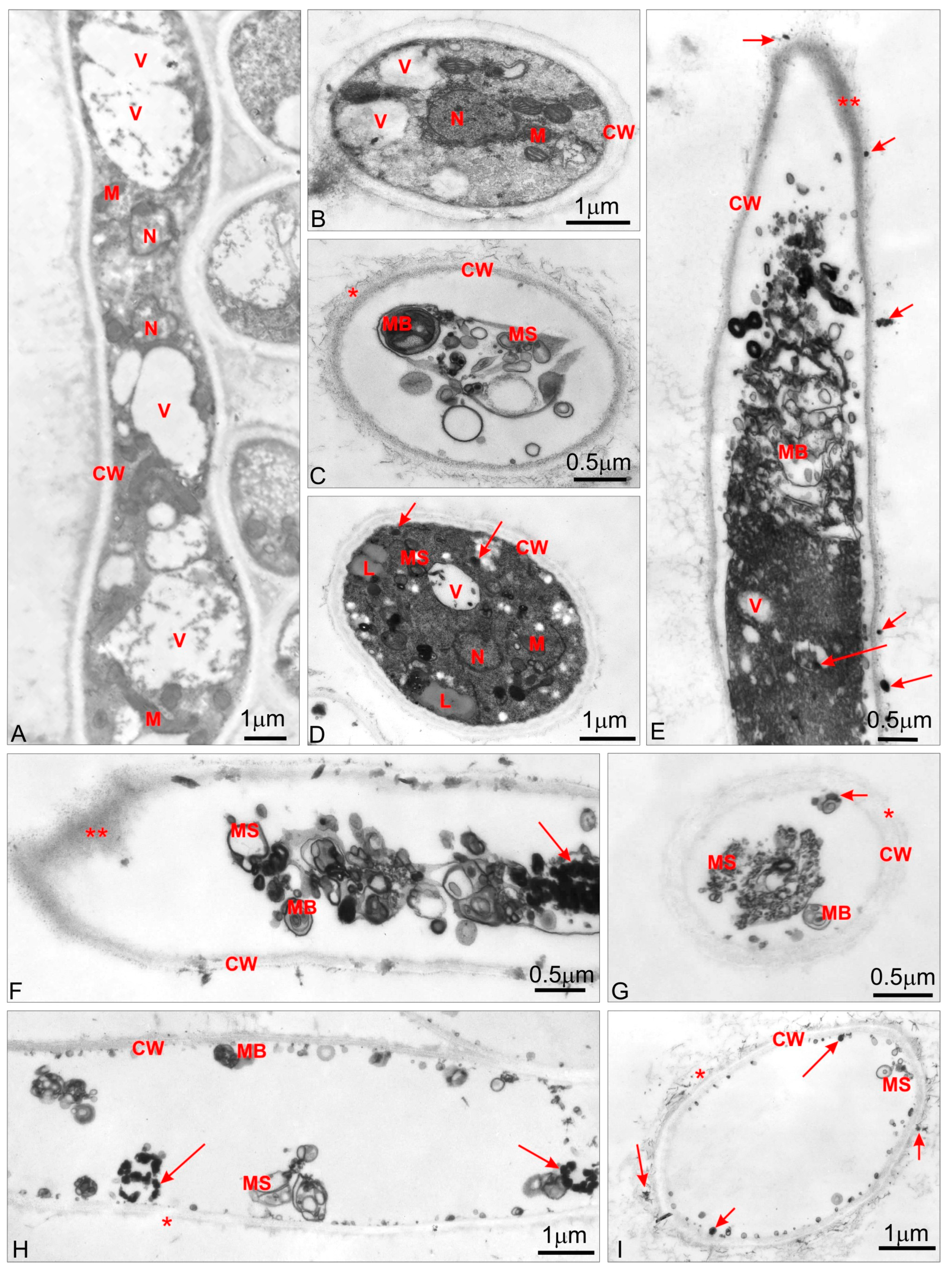
Transmission electron microscopy (TEM) analysis revealed that the control cells of A. brasiliensis were well organized (Figure 2A,B). In cells surrounded by well-defined cell walls (CW), regular nucleoli (N), mitochondria (M) with numerous cristae and electron-dense matrix, and vacuoles (V) with fibrillal material (Figure 2A) or electron transparent vacuoles (Figure 2A,B) could be distinguished. The hyphae in A. brasiliensis treated with hexamethylene-1,6-bis-(N,N-dimethyl-N-dodecylammonium bromide) at 0.5 MIC exhibited different degrees of disorganization (Figure 2C–E). Some cells showed severe ultrastructural changes, with only remnants of organelles in the form of membranous structures (MS) and multivesicular bodies (MB). Even the cell walls exhibited loose structures (* Figure 2C). However, after treatment at 0.5 MIC, some cells were much less affected (Figure 2D). Organelles, such as nucleoli, mitochondria, and vacuoles, did not show disorganization but the cytoplasm was condensed and symptoms of membrane destruction were visible in the form of membranous structures and large lipid droplets (L). Some electron-dense material, similar to that which stuck to the cell walls in the form of more or less large deposits (arrow Figure 2E), was also visible in the small vacuoles (arrow Figure 2D,E). More severe effects of cationic gemini surfactant C6 were observed in the growing part of the hyphae where the cell wall structure was distended (** Figure 2E). Higher concentrations of hexamethylene-1,6-bis-(N,N-dimethyl-N-dodecylammonium bromide),at MIC (Figure 2F,G) and 2 MIC (Figure 2H,I) caused the death of A. brasiliensis hyphae. Inside the cells, only membranous structures, multivesicular bodies, and electron-dense granules were observed (arrows). All three were numerous in cells treated at MIC (Figure 2F,G) and random in cells treated at 2 MIC (Figure 2H,I). Regardless of the concentration of cationic gemini surfactant C6, the cell wall maintained its integrity, although it was distended (* Figure 2G), especially at the hyphae tips (** Figure 2F).

4. Discussion

Molds secrete large amounts of enzymes into the environment. These substances are used extensively in various fields, such as the food, beverages, and pharmaceutical industries [5,22]. However, extracellular enzymes enable the growth of molds under various conditions and contribute to the biodegradation of materials such as paper or textiles [10]. This can lead to substantial material and economic losses.
The gemini surfactants used in this study inhibited the growth of molds. All of the compounds, both neutral and cationic, decreased the concentration of enzymes produced by A. brasiliensis strain ATCC 16404. This contradicts results reported by Pietrzak et al. [23], who investigated the influence of colloidal silver nanoparticles (Ag NPs) on the activity of extracellular enzyme. Nanosilver at 22.5 ppm caused the activity of all the enzymes to increase. In our work, after the addition of gemini surfactants, some enzymes, namely alkaline phosphatase, esterase, esterase lipase, and α-glucosidase, were not detected in the mycelium. Minor changes were noted for acid phosphatase and naphthol-AS-BI-phosphohydrolase. The former is found in the mycelium cell wall [24], which was not destroyed by the action of gemini surfactants. This may explain why the concentration of this enzyme was the same as in the untreated sample. Acid phosphatase and naphthol-AS-BI-phosphohydrolase are often detected in microscopic fungi that cause infections, such as Candida spp. [25] or Malassezia spp. [26]. They are also found in elevated concentrations in the mycelia of Aspergillus niger, Cladosporium cladosporoides, or Stachybotrys chartarum inhabiting building materials such as wallpaper, gypsum, and carton-gypsum board [10,27]. Since extracellular enzymes play a key role in biodegradation and infections, it is important to limit their synthesis.
Like most filamentous fungi, strains of A. niger have the ability to secrete high levels of both proteins and metabolites [28]. The intra- and extracellular proteome of Aspergillus spp. has been analyzed extensively using different approaches such as SDS-PAGE, two-dimensional (2D)-PAGE gel electrophoresis, and MALDI-ToF (Matrix-Assisted Laser Desorption/ Ionization- Time of Flight) [29,30,31]. However, little is known about the influence of gemini surfactants on fungal proteins, and there have been no studies addressing this issue. The neutral and cationic gemini surfactants tested in our research caused several changes in the extracellular protein profiles of A. brasiliensis ATCC 16404. No significant differences were observed between the types of biocide and the changes in the protein patterns. This is in agreement with results reported by Rajkowska et al. [18], who observed that the number of patterns in the protein profiles of Candida albicans were noticeably lower after treatment with essential oils (peppermint and clove oils). Changes in electrophoretic profiles depend on the concentrations of gemini surfactants used. Surfactants produce larger changes in the protein fraction profiles at 2 MIC than at MIC. Pi et al. [32] suggested that proteins can interact with surfactants both in vitro and in vivo. Ionic surfactants can lead to denaturation and conformational changes in proteins [33].
The changes that occurred after treatment with gemini surfactants were clearly visible using an electron microscope (TEM). These observations supported those described previously using scanning microscopy (SEM) [16]. There are only a few publications describing the impact of antifungal biocides (not antibiotics) on the ultrastructure of mold cells. The action of quaternary ammonium compounds (QAC) on yeast Sacchcaromyces cerevisiae was described by Obłąk et al. [14,34]. Monomeric surfactants, especially n-dodecyl-N,N-dimethylalaninum methyl bromide (DMALM-12), were found to cause cell wall and cell membrane deformation. In such cases, the cell interior fills with electronically dense granules while single lipid drops form in the cytoplasm. Increased lipid production has also been observed in the case of N-(dodecyloxycarboxymethyl)-N,N,N-trimethyl ammonium chloride in Saccharomyces cerevisiae cells [28].
Research by Shirai et al. [15], describing yeast treated with gemini surfactant 3,3′-(2,7-dioxaoctane)-bis(1-decylpyridinium bromide) (3DOBP-4,10) at a concentration of 6 μM, showed that the internal cell organelles were destroyed but the cell wall structure was preserved. In our study, TEM studies revealed that the cell walls of mycelia treated with C6 at a concentration of 0.155 mM had partly loose structures, especially in the apical part. Deformations in the apical parts of hypha are due to the nature of mold growth. Hyphal growth is apical, with continuous increases and extensions in the cell wall and membrane at the hyphal tip, where the cell wall is thin and therefore more sensitive to external factors. After treatment with cationic gemini surfactant at higher concentrations, the cell wall was distended but preserved its integrity. Similar results were reported by Razzaghi et al. [35], who studied the effect of the biocide Akacid®plus (Ch. 1007; POC, Vienna, Austria); composed of a mixture of two different polymeric guanidine compounds on Aspergillus parasiticus. No damage was observed to the cell wall, but similar to treatment with compound C6, destruction of the plasma membrane was noted. Changes in the endo-membrane system (plasma membrane and membranous organelles) were noted by Tolouee et al. [36] in their study of the effects of wild chamomile flower oil on the ultrastructure of A. niger. However, these authors also observed extensive changes in the cell wall, which was not broken down by the plant oil but did lose its integrity.
Li et al. conducted research on A. brasiliensis strain ATCC 16404 [4]. In their work regarding the antifungal activity of citronella oil, they observed slightly different changes in the mycelium ultrastructure compared with those reported here following treatment with gemini surfactants. These differences mainly concerned the cell wall, which was destroyed after the action of citronella oil. Microscopic analysis showed extensive vacuolation with vacuole fusion in the hyphae. If we compare the actions of these antifungal agents—chamomile and citronella oil, Akacid®plus and gemini surfactant C6—it can be concluded on the basis of TEM analysis that gemini surfactants at concentrations of 0.155–0.62 mM produce larger cell defects. Only membranous structures, multivesicular bodies, and electron dense granules are present in the cells.
Many authors indicated that the way in which disinfectants work especially on molds have not yet been fully investigated; therefore, these phenomena are not fully understood and explained [37,38]. The activity of modern antifungal biocides has been the subject of few works. Determining the effectiveness of gemini surfactants and understanding the areas of their action in the cell, revealing what causes its destruction is an important issue. In order to use them as antifungal agents, the research proposed in this study, as well as in a previous article [16], are needed. The revised results suggest that cationic gemini surfactants could have a wide range of practical applications as active compounds in disinfection.

5. Conclusions

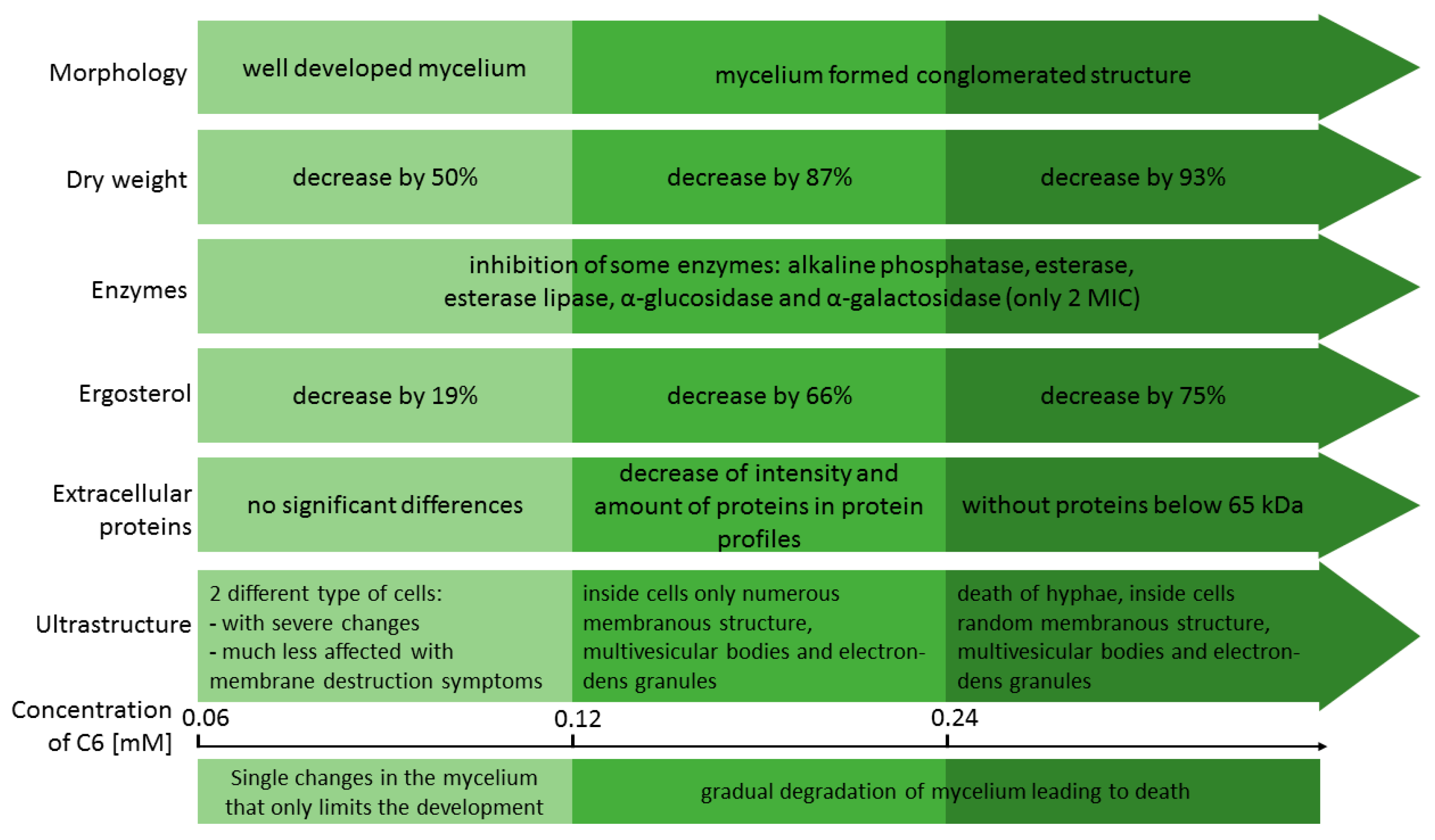
Based on the results presented in this and a previous study [16], we concluded that cationic gemini surfactants, in comparison with their non-ionic analogues, could have a wide range of practical applications as active compounds. As the concentration of hexamethylene-1,6-bis-(N,N-dimethyl-N-dodecylammonium bromide) (C6) increases, mycelia of A. brasiliensis gradually degrade, leading to death. The changes caused by treatment with cationic gemini surfactant C6 are presented in Figure 3.

Supplementary Materials

The following are available online at https://www.mdpi.com/2076-3417/9/2/245/s1, Figure S1: SDS-PAGE analysis of extracellular protein profiles of untreated A. brasiliensis ATCC 16404 (K – control samples) and treated with neutral (A5 and A6) and cationic (C5 and C6) gemini surfactants. M – molecular weight standard (PageRuler™ Unstained Broad Range Protein Ladder).

Author Contributions

A.K. conceived and designed the experiments; A.K., A.O., M.G., and S.M. performed the experiments; A.K. and A.O. prepared the original draft manuscript, M.G. prepared results of TEM analysis; A.K. reviewed and edited the work; A.K. project administration.

Funding

This study was supported by KBN (Scientific Research Committee in Poland) grant “Fungicidal properties of new gemini surfactants” N N401 027736.

Conflicts of Interest

The authors declare no conflict of interest.

References

  1. Filtenborg, O.; Frisved, J.C.; Thrane, U. Moulds in food spoilage. Int. J. Food Microbiol. 1996, 33, 85–102. [Google Scholar] [CrossRef]
  2. Gutarowska, B. Moulds in biodeterioration of technical materials. Foilia Biologica et Oecologica 2014, 10, 27–39. [Google Scholar] [CrossRef] [Green Version]
  3. Orlita, A. Microbial biodeterioration of leather and its control: A review. Int. Biodeter. Biodegr. 2004, 53, 157–163. [Google Scholar] [CrossRef]
  4. Li, W.-R.; Shi, Q.-S.; Ouyang, Y.-S.; Chen, Y.-B.; Duan, S.-S. Antifungal effects of citronella oil against Aspergillus niger ATCC 16404. Appl. Microbiol. Biotechnol. 2013, 97, 7483–7492. [Google Scholar] [CrossRef] [PubMed]
  5. Sharma, C.; Chowdhary, A. Molecular bases of antifungal resistance in filamentous fungi. Int. J. Antimicrob. Agents 2017, 50, 607–616. [Google Scholar] [CrossRef] [PubMed]
  6. Abrashev, R.; Stoitsova, S.; Krumova, E.; Pashova, S.; Paunova-Krasteva, T.; Vassilev, S.; Dolashka-Angelova, P. Temperatire- stress tolerance of the fungal strain Aspergillus niger 26: Physiological and ultrastructural changes. World J. Microbiol. Biotechnol. 2014, 30, 1661–1668. [Google Scholar] [CrossRef] [PubMed]
  7. Wakai, S.; Arazoe, T.; Ogino, C.; Kondo, A. Future insights in fungal metabolic engineering. Bioresour. Technol. 2017, 245, 1314–1326. [Google Scholar] [CrossRef] [PubMed]
  8. Schuster, E.; Dunn-Coleman, N.; Frisvad, J.C.; van Dijick, P.W.M. On the safety of Aspergillus niger—A review. Appl. Microbiol. Biot. 2002, 59, 426–435. [Google Scholar]
  9. Anderson, J.G.; Rowan, N.J.; MacGregor, S.J.; Fouracre, R.A.; Farish, O. Inactivation of food-borne enteropathogenic bacteria and spoilage fungi using pulsed-light. IEEE Trans. Plasma Sci. 2000, 28, 83–88. [Google Scholar] [CrossRef]
  10. Gutarowska, B. Metabolic activity of moulds as a factor of building materials biodegradation. Pol. J. Microbiol. 2010, 59, 119–124. [Google Scholar]
  11. Nemeth-Konda, L.; Füleky, G.; Morovjana, G.; Csokana, P. Sorption behaviour of acetochlor, atrazine, carbendazim, diazinon, imidacloprid and isoproturon on Hungarian agricultural soil. Chemosphere 2002, 48, 545–552. [Google Scholar] [CrossRef]
  12. Bollmann, U.E.; Vollertsen, J.; Carmeliet, J.; Bester, K. Dynamics of biocide emissions from buildings in a suburban stormwater catchment- concentrations, mass loads and emisiion processes. Water Res. 2014, 56, 66–76. [Google Scholar] [CrossRef] [PubMed]
  13. Mazu, T.K.; Bricker, B.A.; Flores-Rozas, H.; Ablordeppey, S.Y. The mechanistic targets of antifungal agents: An overview. Mini Rev. Med. Chem. 2016, 16, 555–578. [Google Scholar] [CrossRef] [PubMed]
  14. Obłąk, E.; Adamski, R.; Lachowicz, T.M. pH-dependent influence of a quaternary ammonium salt and an aminoester on the yeast Saccharomyces cerevisiae ultrastructure. Cell. Mol. Biol. Lett. 2003, 8, 105–110. [Google Scholar] [PubMed]
  15. Shirai, A.; Sumitomo, T.; Kurimoto, M.; Maeda, H.; Kourai, H. The mode of the antifungal activity of gemini-pyridinium salt against yeast. Biocontrol Sci. 2009, 14, 13–20. [Google Scholar] [CrossRef] [PubMed]
  16. Koziróg, A.; Brycki, B.; Pielech-Przybylska, K. Impact of cationic and neutral gemini surfactants on conidia and hyphal forms of Aspergillus brasiliensis. Int. J. Mol. Sci. 2018, 19, 873. [Google Scholar] [CrossRef] [PubMed]
  17. Papamanoli, E.; Tzanetakis, N.; Litopoulou-Tzanetaki, E.; Kotzekidou, P. Characterization of lactic acid bacteria isolated from a Greek, dry-fermented sausage inrespect of their technological and probiotic properties. Meat Sci. 2003, 65, 859–867. [Google Scholar] [CrossRef]
  18. Rajkowska, K.; Otlewska, A.; Kunicka-Styczyńska, A.; Krajewska, A. Candida albicans impairments induced by peppermint and clove oils at sub-inhibitory concentrations. Int. J. Mol. Sci. 2017, 18, 1307. [Google Scholar] [CrossRef]
  19. Laemmli, U.K. Cleavage of structural proteins during assembly of head of bacteriophage T4. Nature 1970, 227, 680–685. [Google Scholar] [CrossRef]
  20. Dice, L.R. Measures of the amount of ecologic association between species. Ecology 1945, 26, 297–302. [Google Scholar] [CrossRef]
  21. Reynolds, E.S. The use of lead citrate at high pH as an electron-opaque stain in electron microscopy. J. Cell Biol. 1963, 17, 208–212. [Google Scholar] [CrossRef] [PubMed]
  22. Gurung, N.; Ray, S.; Bose, S.; Rai, V. Abroader view: Microbial enzymes and their relevance in industries, medicine and beyond. BioMed. Res. Int. 2013, 2013, 329121. [Google Scholar] [CrossRef] [PubMed]
  23. Pietrzak, K.; Twarużek, M.; Czyżowska, A.; Kosicki, R.; Gutarowska, B. Influence of silver nanoparticles on metabolism and toxicity of moulds. Acta Biochim. Pol. 2015, 62, 851–857. [Google Scholar] [CrossRef]
  24. Pawar, V.C.; Thaker, V.S. Acid phosphatase and invertase activities of Aspergillus niger. Mycosciences 2009, 50, 323–330. [Google Scholar] [CrossRef]
  25. Kurnatowski, P.; Wójcik, A.; Błaszkowska, J.; Góralska, K. The hydrolytic enzymes produced by fungi strains isolated from the sand and soil of recreation areas. Ann. Parasitol. 2016, 62, 201–208. [Google Scholar] [CrossRef] [PubMed]
  26. Mancianti, F.; Rum, A.; Nardoni, S.; Corazza, M. Extracellular enzymatic activity of Malassezia spp. isolates. Mycopathologia 2000, 149, 131–135. [Google Scholar] [CrossRef]
  27. Nabrdalik, M. Harmfulness of mould fungi occurring in the human environment. Prot. Corros. 2008, 1, 28–33. (In Polish) [Google Scholar]
  28. Weenink, X. Protein Secretion in the Filamentous Fungus Aspergillus niger. Ph.D. Thesis, Universiteit Leiden, Leiden, The Netherlands, 2008. [Google Scholar]
  29. Carberry, S.; Neville, C.M.; Kavanagh, K.A.; Doyle, S. Analysis of major intracellular proteins of Aspergillus fumigatus by MALDI mass spectrometry: Identification and characterisation of an elongation factor 1B protein with glutathione transferase activity. Biochem. Biophys. Res. Commun. 2006, 341, 1096–1104. [Google Scholar] [CrossRef]
  30. Oda, K.; Kakizono, D.; Yamada, O.; Iefuji, H.; Akita, O.; Iwashita, K. Proteomic analysis of extracellular proteins from Aspergillus oryzae grown under submerged and solid-state culture conditions. Appl. Environ. Microbiol. 2006, 72, 3448–3457. [Google Scholar] [CrossRef]
  31. Lu, X.; Sun, J.; Nimtz, M.; Wissing, J.; Zeng, A.-P.; Rinas, U. The intra- and extracellular proteome of Aspergillus niger growing on defined medium with xylose or maltose as carbon substrate. Microb. Cell Fact. 2010, 9, 23. [Google Scholar] [CrossRef]
  32. Pi, Y.; Shang, Y.; Peng, C.; Liu, H.; Hu, Y.; Jiang, J. Interactions between bovine serum albumin and gemini surfactant alkanediyl-α, ω-bis(dimethyldodecyl-ammonium bromide). Biopolymers 2006, 83, 243–249. [Google Scholar] [CrossRef] [PubMed]
  33. Wu, D.; Xu, G.; Sun, Y.; Zhang, H.; Mao, H.; Feng, Y. Interaction between proteins and cationic gemini surfactant. Biomacromolecules 2007, 8, 708–712. [Google Scholar] [CrossRef] [PubMed]
  34. Obłąk, E.; Gamian, A. The biological activity of quaternary ammonium salts (QASs). Postepy Hig. Med. Dosw. 2010, 64, 201–211. (In Polish) [Google Scholar]
  35. Razzaghi-Abyaneh, M.; Shams-Ghahfarokhi, M.; Kawachi, M.; Eslamifar, A.; Schmidt, O.J.; Schmidt, A.; Allameh, A.; Yoshinari, T. Ultrastructural evidences of growth inhibitory effects of a novel biocide, Akacid®plus, on an aflatoxigenic Aspergillus parasiticus. Toxicon 2006, 48, 1075–1082. [Google Scholar] [CrossRef] [PubMed]
  36. Tolouee, M.; Alinezhad, S.; Saberi, R.; Eslamifar, A.; Zad, S.J.; Jaimand, K.; Taeb, J.; Razaee, M.-B.; Kawachi, M.; Shams-Ghahfarokhi, M.; et al. Effect of Matricaria chamomilla L. flower essential oil on the growth and ultrastructure of Aspergillus niger van Tieghem. Int. J. Food Microbiol. 2010, 139, 127–133. [Google Scholar] [CrossRef] [PubMed]
  37. Russell, A.D. Similarities and differences in the responses of microorganisms to biocides. J. Antimicrob. Chemother. 2003, 52, 750–763. [Google Scholar] [CrossRef] [PubMed] [Green Version]
  38. Tortorano, A.M.; Viviani, M.A.; Biraghi, E.; Rigoni, A.L.; Prigitano, A.; Grillot, R. In vitrotesting of fungicidal activity of biocides against Aspergillus fumigatus. J. Med. Microbiol. 2005, 54, 955–957. [Google Scholar] [CrossRef] [PubMed]
Figure 1. (A) Effect of neutral (A5 and A6) and cationic (C5 and C6) gemini surfactants on extracellular protein profiles of A. brasiliensis ATCC 16404 at MIC; changes in protein profiles after (B) C5 and (C) C6 treatment at ½ MIC, MIC, and 2 MIC in comparison with the sample without biocide (K). The sizes of the bubbles are related to pixel intensities in the protein profile patterns.
Applsci 09 00245 g001
Figure 2. (A,E,F,H) TEM micrographs of longitudinal-sections and (BD,G,I) cross-sections of Aspergillus brasiliensis: (A,B) control hyphae; (CE) hyphae treated with hexamethylene-1,6-bis-(N,N-dimethyl-N-dodecylammonium bromide) C6 for 48 h at 0.5 MIC; (F,G) hyphae treated with C6 for 48 h at MIC and (H,I) hyphae treated with C6 for 48 h at 2 MIC. CW—cell wall; L—lipid; M—mitochondrion; MB—multivesicular body; MS—membranous structure; N—nucleus; V—vacuole; arrows-electron-dense deposit.
Applsci 09 00245 g002
Figure 3. Representation the stepwise antifungal action of hexamethylene-1,6-bis-(N,N-dimethyl-N-dodecylammonium bromide) C6 against mycelium of Aspergillus brasiliensis ATCC 164040 in comparison to mycelium without gemini surfactant.
Applsci 09 00245 g003
Table 1. Effect of cationic C6 or C5 and neutral A5 or A6 surfactants at ½ MIC, MIC, and 2 MIC on the enzymatic profile of Aspergillus brasiliensis mycelia.
Type of SampleBiocide ConcentrationSymbol of Enzyme in API-ZYM Test
2341112131618
K0
C5½ MIC
MIC
2 MIC
C6½ MIC
MIC
2 MIC
A5½ MIC
MIC
2 MIC
A6½ MIC
MIC
2 MIC
2–18 refer to the enzyme numbers in the API -ZYM test: 2—alkaline phosphatase, 3—esterase (C4), 4—esterase lipase (C8), 11—acid phosphatase, 12—naphthol-AS-BI-phosphohydrolase, 13—α-galactosidase, 16—α-glucosidase and 18—N-acetyl-β-glucosaminidase, respectively. Concentration of hydrolyzed substrate: light grey—5 nM, grey—10 nM, orange—20 nM, yellow—30 nM, green—≥40 nM.
Table 2. Similarity of protein profiles of A. brasiliensis ATCC 16404 after treatment with gemini surfactants.
Type of SampleBiocide ConcentrationDice Index of Similarity (%)
C5½ MIC92.3
MIC62.5
2 MIC54.5
C6½ MIC92.3
MIC66.7
2 MIC54.5
A5MIC72.7
A6MIC72.7