Article

Three-Dimensional Numerical Analysis of Periciliary Liquid Layer: Ciliary Abnormalities in Respiratory Diseases


by
Shayan M. Vanaki
1,*,
David Holmes
1,*,
Pahala Gedara Jayathilake
2 and
Richard Brown
1
1
School of Chemistry, Physics and Mechanical Engineering, Queensland University of Technology (QUT), Brisbane 4000, Queensland, Australia
2
Department of Oncology, University of Oxford, Oxford OX1 2JD, UK
*
Authors to whom correspondence should be addressed.
Appl. Sci. 2019, 9(19), 4033; https://doi.org/10.3390/app9194033
Submission received: 27 August 2019 / Revised: 23 September 2019 / Accepted: 24 September 2019 / Published: 26 September 2019

Abstract

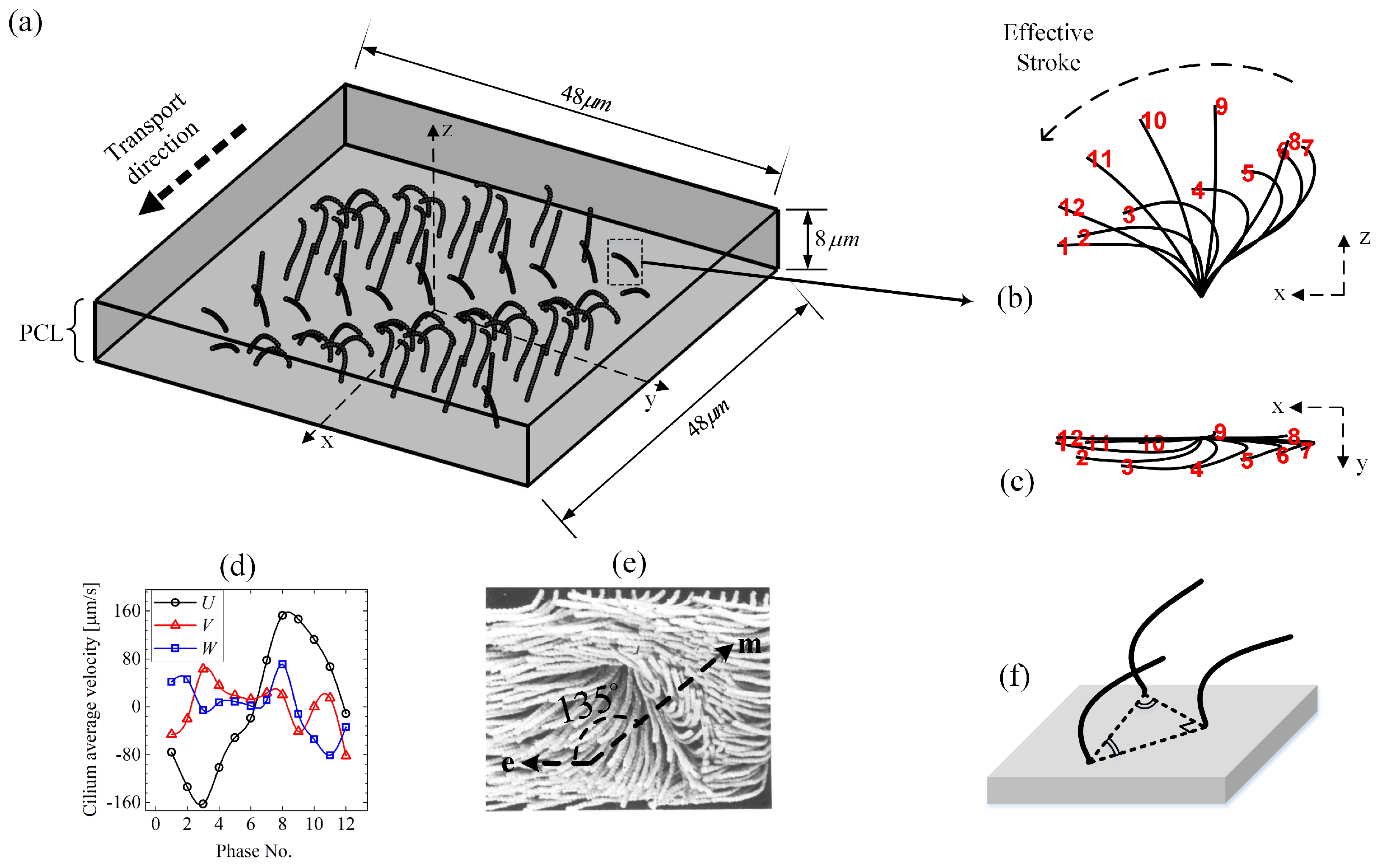
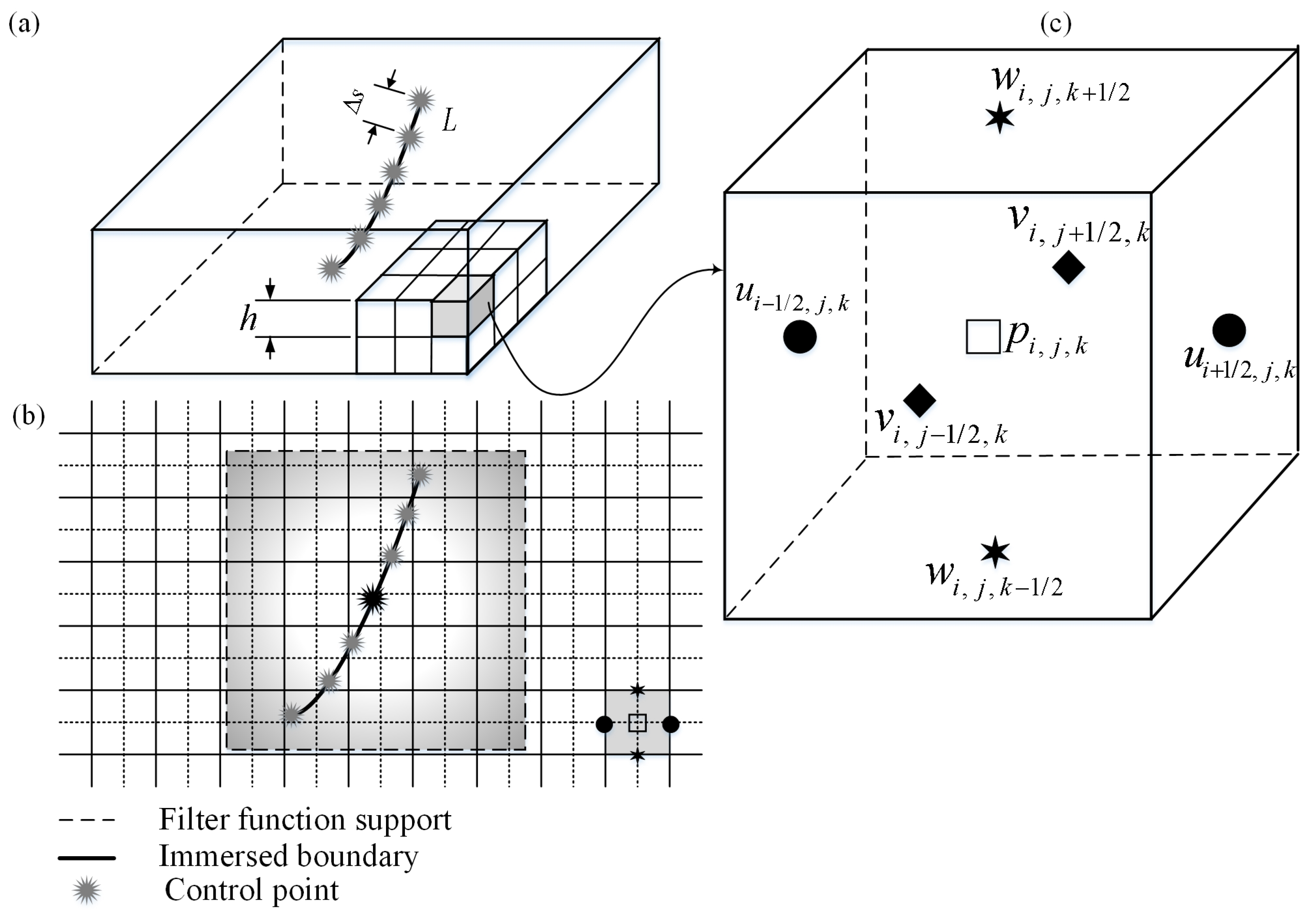
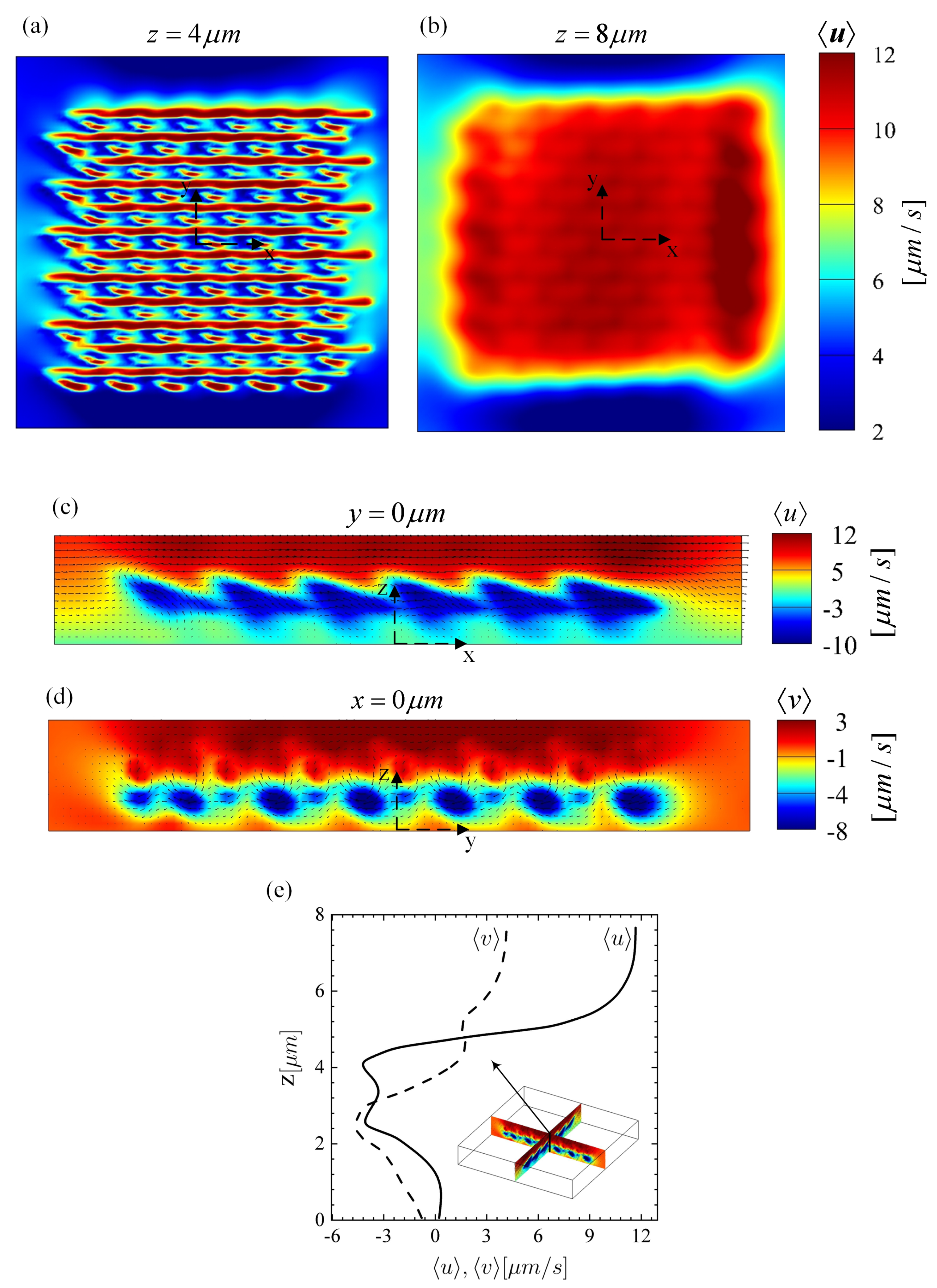
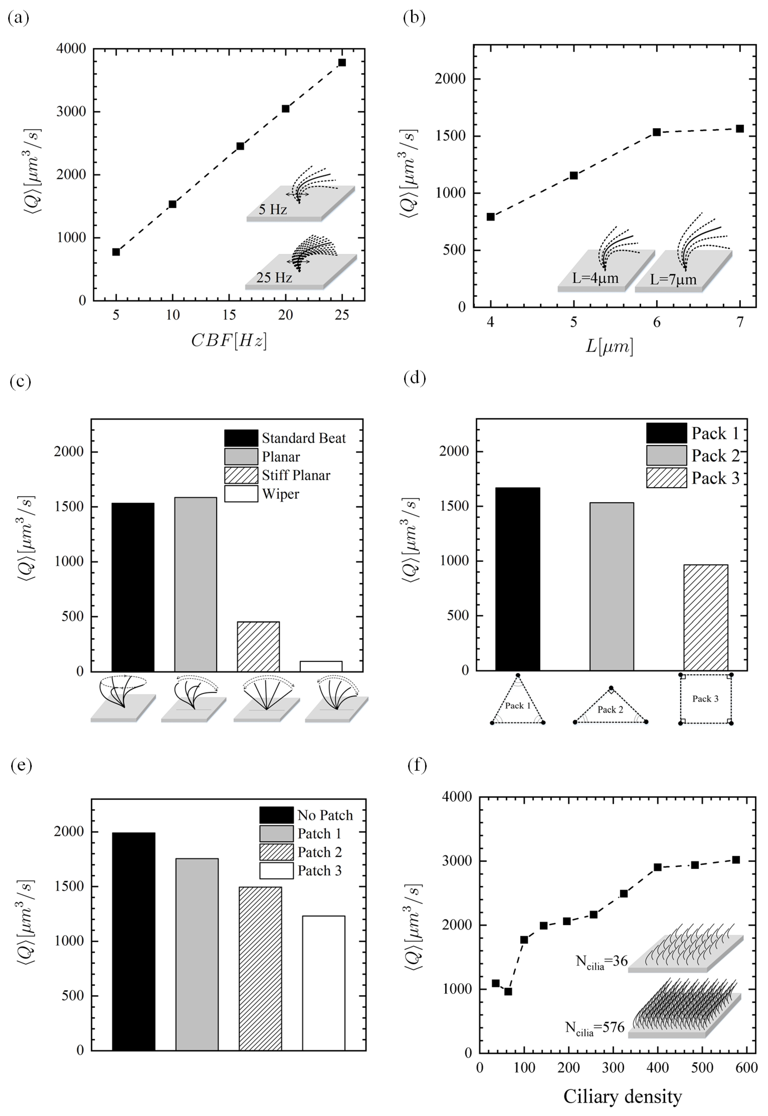
Human pulmonary epithelial cells are protected by two layers of fluid—the outer watery periciliary liquid layer (PCL) and the uppermost non-Newtonian mucus layer (ML). Aerosols and inhaled toxic particles are trapped by the ML which must then be removed swiftly to avoid adverse health implications. Epithelial cells are covered with cilia that beat rapidly within the PCL. Such ciliary motion drives the mucus transport. Although cilia can penetrate slightly inside the mucus to assist mucus movement, the motion of the underlying PCL layer within the airway surface liquid (ASL) is significant in mucus and pathogens transport. As such, a detailed parametric study of the influence of different abnormal cilia characteristics, such as low beating frequency, short length, abnormal beating pattern, reduced ciliary density, and epithelium patchiness due to missing cilia on the PCL transport, is carried out numerically. Such abnormalities are found in various chronic respiratory diseases. In addition, the shear stress at the epithelium is assessed due to the importance of shear stress on the epithelial function. Using the immersed boundary (IB) method combined with the finite-difference projection method, we found that the PCL, under standard healthy conditions, has net forward motion but that different diseased conditions decrease the forward motion of the PCL, as is expected based on clinical understanding.
Keywords: computational fluid dynamics; immersed boundary method; cilia-driven fluid; periciliary liquid layer; mucus; respiratory diseases computational fluid dynamics; immersed boundary method; cilia-driven fluid; periciliary liquid layer; mucus; respiratory diseases