Article

Three-Dimensional High-Energy Electron Radiography Method for Static Mesoscale Samples Diagnostics

1
Institute of Modern Physics, Chinese Academy of Sciences, Lanzhou 730000, China
2
School of Nuclear Science and Technology, University of Chinese Academy of Sciences, Beijing 100049, China
*
Authors to whom correspondence should be addressed.
Appl. Sci. 2019, 9(18), 3764; https://doi.org/10.3390/app9183764
Submission received: 1 July 2019 / Revised: 8 August 2019 / Accepted: 19 August 2019 / Published: 9 September 2019
(This article belongs to the Section Applied Physics General)

Abstract

:
In this paper, we propose a new method for static mesoscale sample diagnosis using three-dimensional radiography with high-energy electron radiography (HEER). The principle of three-dimensional high-energy electron radiography (TDHEER) is elucidated, and the feasibility of this method is confirmed by start-to-end simulation results. TDHEER is realized by combining HEER with the three-dimensional reconstruction method, by which more information about the samples can be attained, especially regarding the samples’ internal structures. With our study, the internal structures and the three-dimensional positions of the spherical sample are determined with a ~3 μm resolution. We believe that this new method enhances the HEER diagnostic capability and extends its application potential in mesoscale sciences.

1. Introduction

High-energy electron radiography (HEER) was proposed as a high spatial and temporal resolution probe tool for high-energy-density physics (HEDP) and inertial confinement fusion (ICF) experimental diagnostic studies [1,2]. In recent years, HEER technology was well developed through both simulations and experiments [3,4,5,6,7,8]. Radiography can be performed with a ps pulse-width electron beam, achieving a spatial resolution close to 1 μm in an experiment with a large magnification imaging lens [9]. HEER takes advantage of a high-energy electron beam with powerful penetration, which can be used for thicker samples (tens of microns to millimeters) diagnostics, short period (ps–ns) bunch train time structures for ultra-fast dynamic process diagnostics and point to point imaging with a magnetic lens to achieve high spatial resolution. The high-energy electron beam for HEER is generated by a high-energy electron linear accelerator based on a photocathode injector.
Concerning the mesoscale sciences, the spatial scale of the samples ranges from tens of μm to mm and the time scale from ps to ns, which is the bridge between microscale and macroscale. Currently, in mesoscale sciences, HEDP and ICF research are important topics and the focus of intense research efforts. The diagnostic method for mesoscale sciences, especially for material studies, was figured out to be the most important part and will present a substantial challenge in the future [10]. Some research has shown that the radiography capability and characteristics of HEER are suitable candidates for mesoscale sciences diagnostic studies. However, with normal HEER, we can only get a two-dimensional transverse projection of the samples and it is very difficult to get the exact details of the internal structures of samples.
Here, the three-dimensional high-energy electron radiography (TDHEER) method is put forward for the detailed diagnosis of static mesoscale samples. To our knowledge, this is the first time that the TDHEER method has been proposed, and its validity and usage in static mesoscale samples fully three-dimensional diagnostics is confirmed by start-to-end simulation studies. Detailed studies are shown below. In Section 2, the TDHEER method is introduced and its principle is explained. In Section 3, we present detailed start-to-end simulation studies of TDHEER with dedicated samples to evaluate its feasibility and capability. In Section 4, we report the conclusions and also present a future plan for TDHEER development, which would provide a powerful diagnostic tool for future mesoscale sciences.

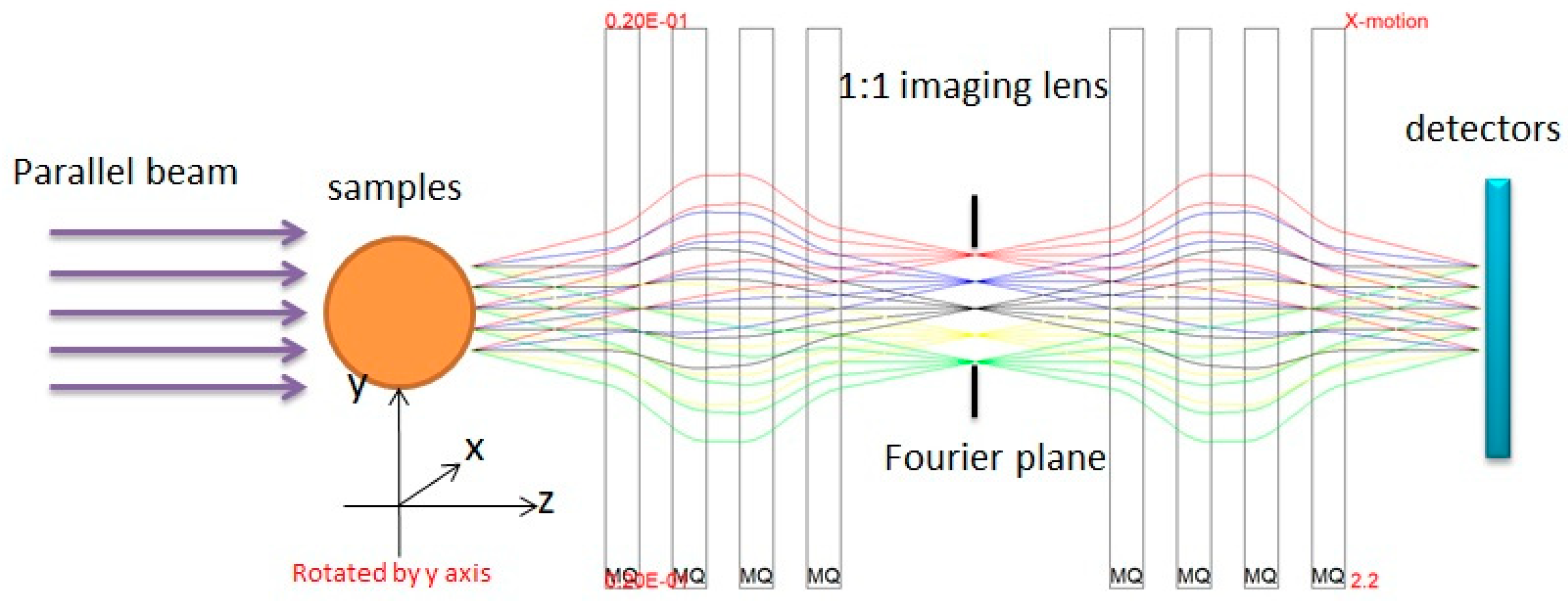
2. The TDHEER Method and General Layout

HEER benefits from the magnetic lens optics, which improves the spatial resolution dramatically compared to projection radiography. The principle of HEER is shown in Figure 1: The electron beams pass through the target and then are imaged point to point from the target plane to the image plane by the imaging lens. There are two primary requirements of the imaging lens system. First, the lens must provide point-to-point focus from the object to the image. Second, it must form a Fourier plane, where particles are radially sorted by the magnitude of the scattering within the object. With this correlation, particles scattered to large angles by multiple Coulomb scattering can be removed through collimation at the Fourier plane. The remaining parameters of a particular imaging lens system design are determined by the radiographic applications. The beam energy must be high enough to penetrate the areal density of the object to be radiographed, and the aperture of the lens system must be chosen to provide sufficient angular acceptance throughout the required field of view. An additional strong design requirement is the resolution of the radiography system. This resolution is typically dominated by chromatic aberrations due to the energy spread of the injected beam in combination with the spread of energy loss through the object due to areal density variations of the object.
The TDHEER is combined with HEER and a tomography reconstruction method. Its principle is also shown in Figure 1. The sample is placed on a rotatable platform, which can be rotated around the y-axis (±90 degrees) with a step of 1 degree and imaged with HEER at the detectors for each rotation angle. Afterward, a total of 180 images of the sample are collected with different rotation angles. Then we use the three-dimensional reconstruction algorithms [11] to get the sample’s three-dimensional slice images of each axis. From these slices, we can get the detailed internal structures of the sample. The method of TDHEER is studied and confirmed by start-to-end simulation. The electron beam parameters used in the simulation are the following: Beam energy 50 MeV, energy spread 0.1%, normalized emittance 1.0 mm mrad, bunch charge 1 nC, bunch length rms 10 ps, and parallel beam on the samples, as shown in Figure 1.

3. The Start-to-End Simulation Studies of TDHEER Method

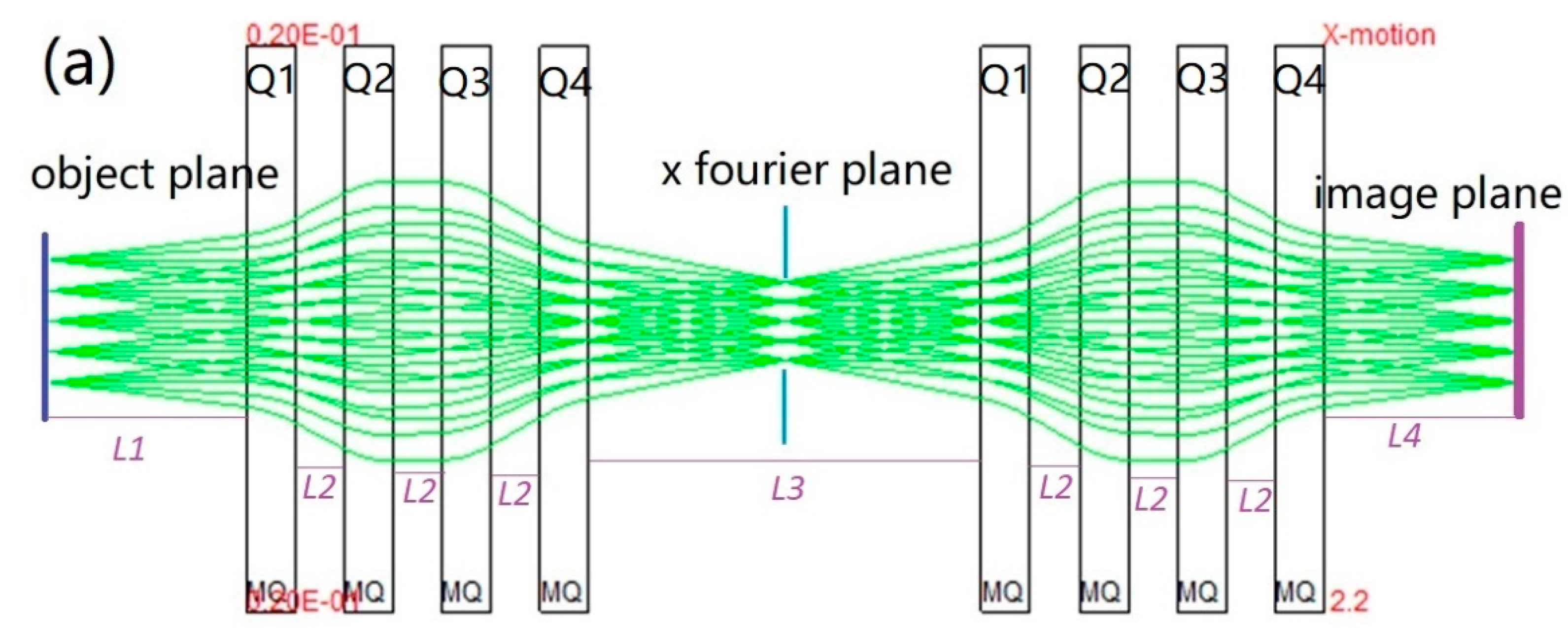
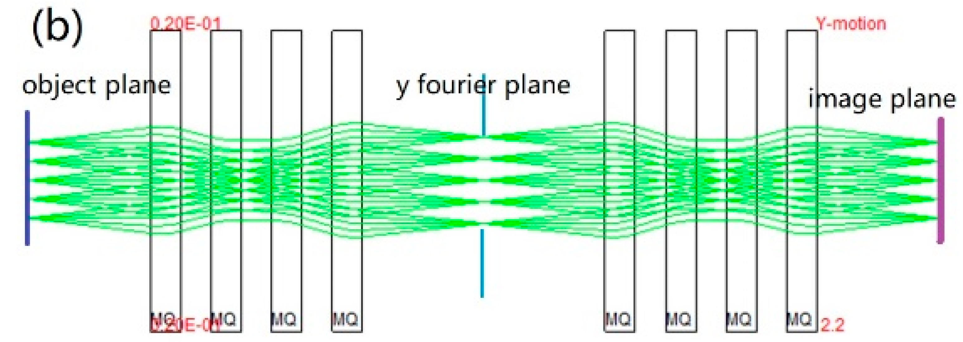
3.1. The Imaging Lens Design

The imaging lens is designed with a magnification of 1 using eight quadrupoles. The designed maximum field gradient of the quadrupoles is 12 T/m, the inner diameter is 20 mm, and the length is 10 cm. The imaging lens structure is optimized using COSY INFINITY9.1 [12] by tuning the quadrupoles field to achieve a magnification of 1 (R11 = R33 = −1) and point to point imaging (R12 = R34 = 0). The designed imaging lens parameters are shown in Table 1. All quadrupoles have the same structural design for manufacturing convenience. The electron beam trajectory is shown in Figure 2 with optimized quadrupoles field and magnification factor R11 = R33 = −1, also showing a good point-to-point imaging performance both in the x Figure 2a and y Figure 2b plane. The chromatic lengths in the x-plane and y-plane are 3.05 and 3.24 m, respectively. With this imaging lens, the T116 and T336 are zero; therefore, the matched beam should be parallel, shown in Figure 1. More details of the beam matching requirement for HEER are referred to in [5].

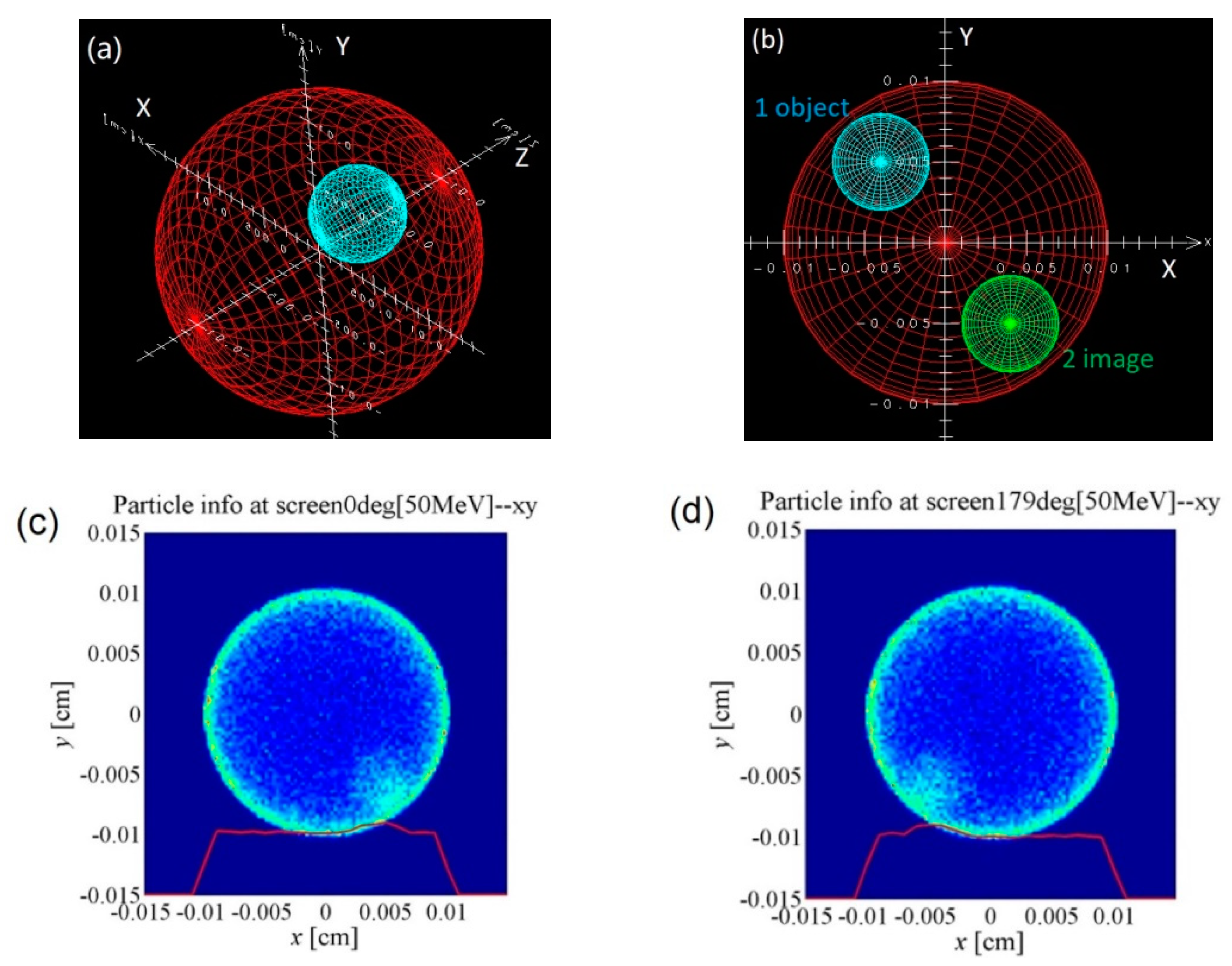
3.2. The HEER Simulation Studies with Rotating Sample

The important interaction processes between electrons and samples are multiple Coulomb scattering (MCS), ionization energy loss, radiative energy loss, and bremsstrahlung. The MCS, ionization energy loss, and straggling processes in electron radiography are very similar to those in proton radiography, an analogous process and well-understood radiography technique used around the world for thick object imaging. However, bremsstrahlung interactions are dominant in the formation of electron radiography when the electron beam energy is above the critical energy. The scattering angle distributions and the energy spectrum of the electron beam after passing through the target are important considerations for HEER [7]. Here we use the electron gamma shower (EGS) code [13] for the electron beam and sample interaction studies and PARMELA code [14] for particles tracking. EGS is a general-purpose package for the Monte Carlo simulation of the coupled transport of electrons and photons in an arbitrary geometry for particles with energies from a few keV up to several hundreds of GeV.
The sample used in the simulation is specially designed for internal structures diagnostics. The sample is a spherical aluminum ball with radius 0.01 cm and a small spherical hollow ball with radius 0.003 cm placed inner at a defined position. The sample parameters are shown in Table 2 and the structures are shown in Figure 2a, the red one is aluminum and the blue one is hollow. With HEER, the samples would be reverse imaged both in the x and y plane on the detectors, which is shown in Figure 2b, where the green ball (two images) is the reverse image of the blue object ball (one object).

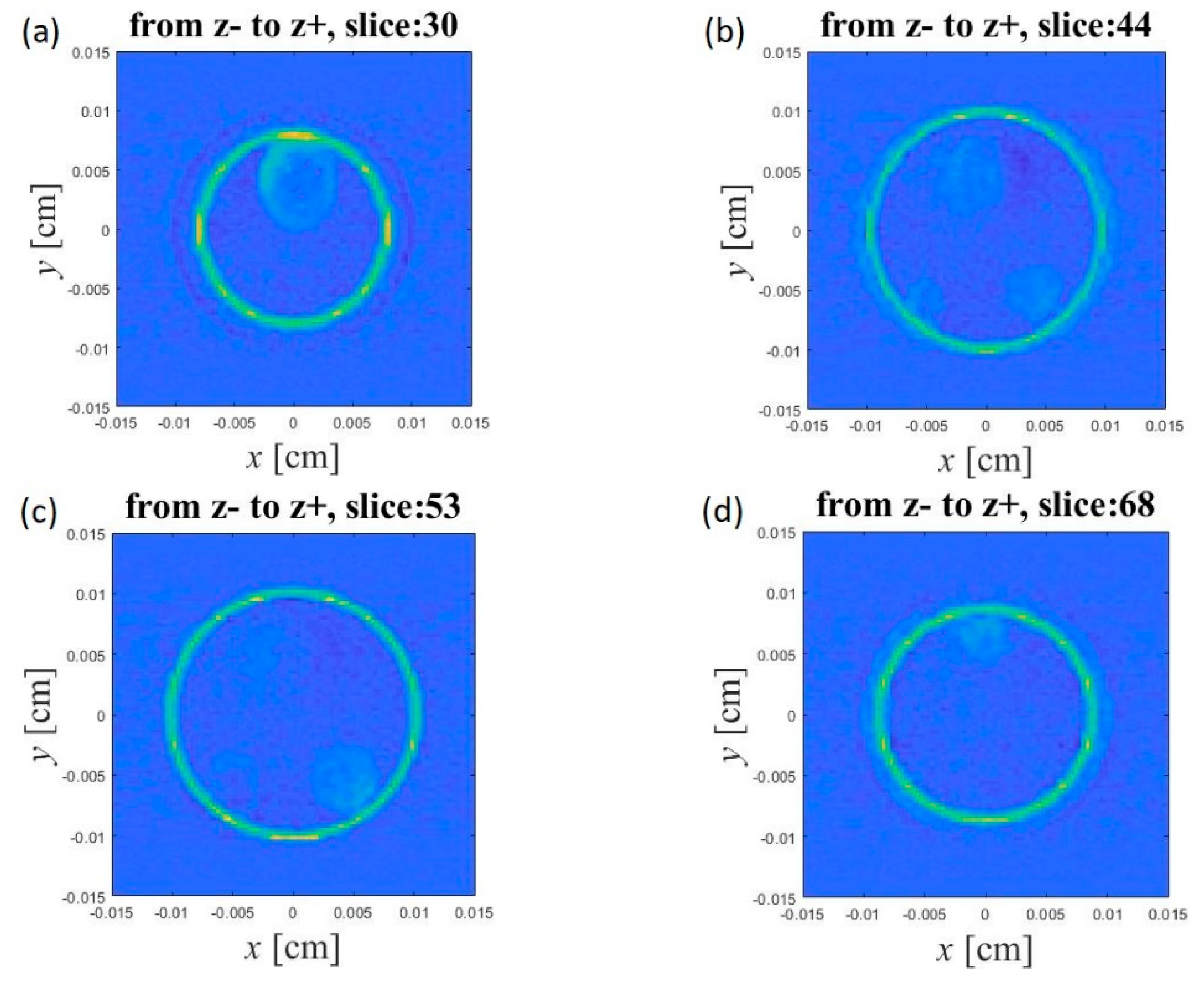
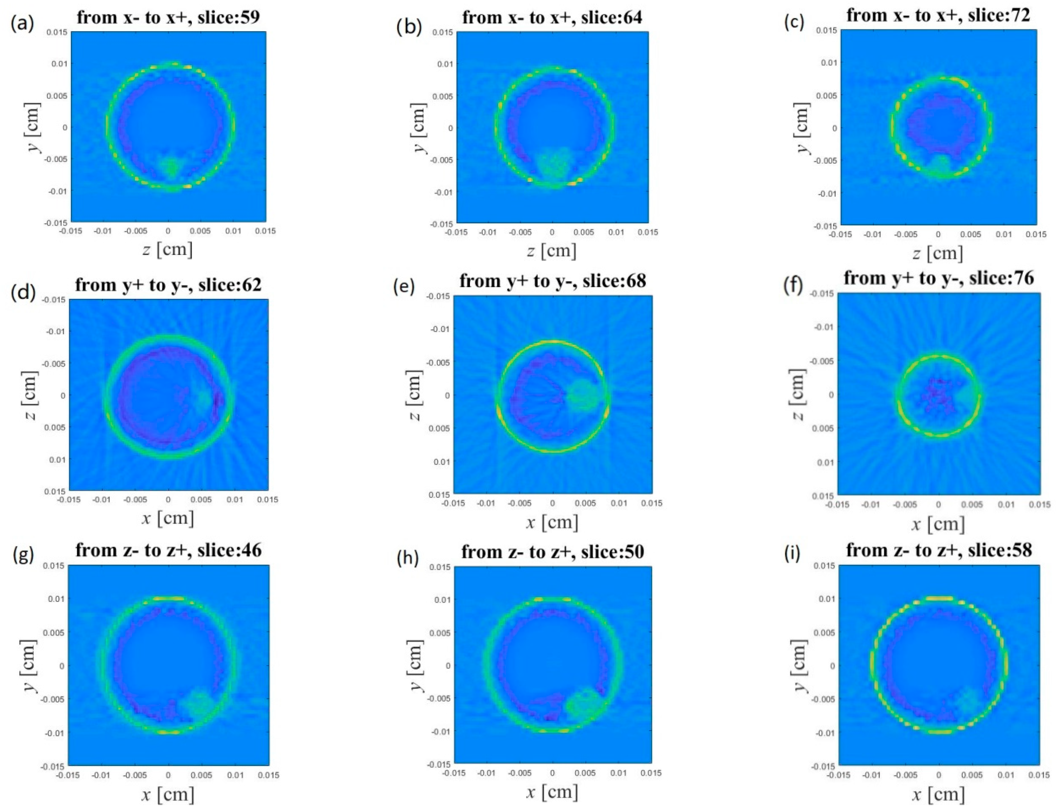
3.3. Three-Dimensional Reconstruction and Results Analysis

The three-dimensional reconstruction (TDR) method has been well developed in recent decades. Many algorithms, such as FBP (filter back projection) [15], ART (algebraic reconstruction technique), SIRT (simultaneous iterative reconstruction technique), SART (simultaneous algebraic reconstruction technique) [11], MEM (maximum entropy method) [16], have been developed and applied in many fields for solving different problems. The TDR of the mass density of an object from its two-dimensional line projection is the core of electron tomography [17,18]. Therefore, electron tomography can be seen as a problem of inverting projection transformation to recover the distribution of the mass density of the original object. Various algorithms have been proposed to cope with the practical difficulties of this inversion problem and they differ widely in terms of their robustness with respect to noise in the data, completeness of the collected projection data set, errors in projections’ orientation parameters, and the ability to efficiently handle large data sets. Our TDHEER method is similar to electron tomography but with an inverse image due to the imaging lens, so the TDR algorithm used in the electron tomography is also suitable for TDHEER. The FBP algorithm is selected in our TDHEER studies due to its computational timesaving and ease of implementation. The FBP is an analytic reconstruction algorithm designed to overcome the limitations of conventional back-projection. It applies a convolution filter to remove blurring. Here we use “Ram-Lak” filter in the TDHEER reconstruction [19].
The sample used for TDHEER studies is shown in Figure 3a. As in the HEER imaging process shown in Figure 1, the sample radiography images are collected of each rotation angle from 0 to 179 degree. They were used for 3D reconstruction studies. The reconstruction results with the FBP algorithm are shown in Figure 4, where (a–c) are the reconstructed slice images along the axis x from x− to x+; (d–f) are the reconstructed slice images along the axis y from y+ to y−; (g–i) are the reconstructed slice images along the axis z from z− to z+. Depending on the slices’ numbers and directions, the x, y, and z positions of the inside ball can be attained. For each of the three axes, the thickness and the total number of slices are 0.03 cm and 100, respectively. Therefore, the thickness of each slice is 3 μm. The inner ball in each slice image along the three axes is shown from invisibility to visibility, and then to invisibility again, by which the inner ball center position of each axis is determined, shown in Table 3. The errors of the inner ball center positions are due to the slice number uncertainty of the first and last slice images with the inner ball. The reconstruction results are consistent with the original settings in the simulation, and they confirm the validation of the TDHEER, from which the internal structure of the samples can be exactly determined.

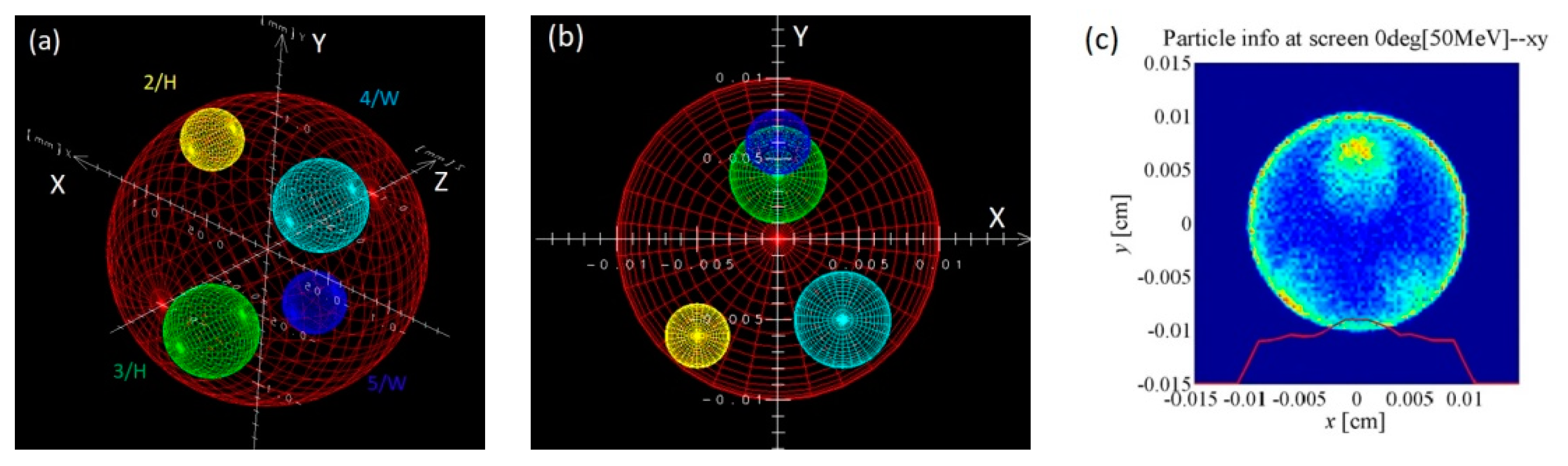
3.4. TDHEER Studies for Complicated Samples

In order to further investigate the capability of the TDHEER, a more complicated spherical sample has four small balls of different materials inside, which is shown in Figure 5a. The positions and materials of each ball are shown in Table 4. In Figure 4b, the sample reverse projection both in x and y plane is shown, which is equivalent to the HEER image with 0-degree rotation angle. It is consistent with the HEER imaging with 0-degree rotation angle, shown in Figure 5c. From Figure 5c, there is an obvious problem, where the inner small balls number 3 and 5 are overlapping and it is difficult to distinguish from the HEER image.
The TDR algorithm FBP was also used for the complicated sample to get the three-dimensional radiography. We also tried to determine the inner small balls exact three-dimensional positions. The reconstruction slice images along the z-axis are shown in Figure 6a–d. We can distinguish the inner four small balls’ z positions and solve the normal HEER problem, of which the small inner balls number 3 and 5 are overlapping as shown in Figure 5c. We also estimated the inner four small balls positions along the x, y, and z-axis and get the same results as shown in Table 3, which is not shown here. Therefore, the TDHEER capability is confirmed from complicated sample radiography studies.

4. Conclusions and Discussions

A new three-dimensional high-energy electron radiography method was proposed for mesoscale sciences diagnostics. The feasibility and validation of TDHEER are testified by start-to-end simulation studies. The TDHEER method is a combination of HEER and a three-dimensional reconstruction algorithm. With the TDHEER method, the diagnostics capability of HEER is enhanced greatly. The simulation results show that the internal structures of the sample can be attained from slice to slice along each axis. The inner flaw or structure of the sample can be exactly determined, and the spatial resolution can reach μm, which is very useful for mesoscale sciences studies.
For further TDHEER development, some possible methods were investigated. First of all, TDHEER experimental studies will be undertaken in the near future, using the same process as shown in the simulation studies. The TDHEER experiment has been scheduled and will be performed at the Lanzhou HEER experimental platform [20]. Secondly, the imaging lens used in this paper has a magnification factor of 1, which restrains the HEER spatial resolution to several microns. For further studies, a high-magnification imaging lens should be used, such as 10 to 100, which will help and improve the spatial resolution to sub μm [9] accordingly, improving the TDHEER resolution. Thirdly, here we used the FBP reconstruction algorithm with a total of 180 HEER images, in which data recording and images analysis took a long time. Therefore, we suggest that other three-dimensional reconstruction algorithms are used to achieve the three-dimensional reconstruction with fewer HEER images in less time, for example, with a 5 or 10 degree step. Fourth, in this paper, we considered the static mesoscale samples. We also considered TDHEER for the samples’ dynamic process diagnostics, for which we cannot use rotating sample technology. However, we proposed three-orthogonal directions HEER with three radiography electron beam lines from one accelerator [2,8]. Here, we propose the reconstruction of the sample’s three-dimensional structures with even fewer HEER images and the development of a new reconstruction algorithm [21] in the future. Furthermore, the electron beam energy can be selected from tens of MeV to GeV to achieve high spatial resolution and image contrast depending on the sample characteristics. In summary, we propose and demonstrate a new three-dimensional radiography method with HEER, which is more powerful and suitable for mesoscale sciences diagnostic studies.

Author Contributions

Q.Z. and Y.M. have equal contributions, conceptualization, Q.Z. and Z.Z.; methodology, Y.M.; software, Y.M, Y.Z. and Z.R.; validation, Q.Z., Y.M., and J.X.; writing—original draft preparation, Q.Z. and Y.M.; writing—review and editing, S.C. and X.S.; project administration, Z.Z.; funding acquisition, Z.Z.

Funding

This research was funded by the National Natural Science Foundation of China, grant number 11435015, the Ministry of Science and Technology of China 2016YFE0104900 and the Chinese Academy of Sciences 28Y740010.

Conflicts of Interest

The authors declare no conflict of interest.

References

  1. Gai, W.; Qiu, J.; Jing, C. Electron imaging system for untrafast diagnostics of HEDLP. In Proceedings of the SPIE 9211, Target Diagnostics Physics and Engineering for Inertial Confinement Fusion III, San Diego, CA, USA, 10 September 2014; p. 921104. [Google Scholar]
  2. Zhao, Y.; Zhang, Z.; Gai, W.; Du, Y.; Cao, S.; Qiu, J.; Zhao, Q.; Cheng, R.; Zhou, X.; Ren, J.; et al. High energy electron radiography scheme with high spatial and temporal resolution in three dimensions based on a e-LINAC. Laser Part. Beams 2016, 34, 338–342. [Google Scholar] [CrossRef]
  3. Merrill, F.; Harmon, F.; Hunt, A.; Mariam, F.; Morley, K.; Morris, C.; Saunders, A.; Schwartz, C. Electron radiography. Nucl. Instrum. Methods Phys. Res. B 2007, 26, 382–386. [Google Scholar] [CrossRef]
  4. Zhao, Q.; Cao, S.; Cheng, R.; Shen, X.; Zhao, Z.Z.Y.; Gai, W.; Du, Y. High Energy Electron Radiography Experiment Research Based on Picosencond Pulse Width Bunch. In Proceedings of the LINAC2014, Geneva, Switzerland, 31 August–5 September 2014; pp. 76–79. [Google Scholar]
  5. Zhao, Q.; Cao, S.C.; Liu, M.; Shen, X.K.; Wang, Y.R.; Zong, Y.; Zhang, X.M.; Jing, Y.; Cheng, R.; Zhao, Y.T.; et al. High energy electron radiography system design and simulation study of beam angle-position correlation and aperture effect on the images. Nucl. Instrum. Methods Phys. Res. A 2016, 832, 144–151. [Google Scholar] [CrossRef]
  6. Zhou, Z.; Du, Y.; Cao, S.; Zhang, Z.; Huang, W.; Chen, H.; Cheng, R.; Chi, Z.; Liu, M.; Su, X.; et al. Experiments on bright field and dark field high energy electron imaging with thick target material. Phys. Rev. Accel. Beams 2018, 21, 074701. [Google Scholar] [CrossRef]
  7. Zhao, Q.T.; Cao, S.C.; Cheng, R.; Du, Y.C.; Shen, X.K.; Wang, Y.R.; Xiao, J.H.; Zong, Y.; Zhu, Y.L.; Zhou, Y.W.; et al. Generation of uniform transverse beam distributions for high-energy electron radiography. Laser Part. Beams 2018, 36, 313–322. [Google Scholar] [CrossRef]
  8. Zhao, Q.T.; Cao, S.C.; Shen, X.K.; Wang, Y.R.; Zong, Y.; Xiao, J.H.; Zhu, Y.L.; Zhou, Y.W.; Liu, M.; Cheng, R.; et al. Design and simulation study of ultra-fast beam bunches split for three orthogonal planes high-energy electron dynamic radiography. Laser Part. Beams 2017, 35, 579–586. [Google Scholar] [CrossRef]
  9. Zhou, Z.; Fang, Y.; Chen, H.; Wu, Y.; Du, Y.; Yan, L.; Tang, C.; Huang, W. Demonstration of Single-Shot High-Quality Cascaded High-Energy-Electron Radiography using Compact Imaging Lenses Based on Permanent-Magnet Quadrupoles. Phys. Rev. Appl. 2019, 11, 034068. [Google Scholar] [CrossRef]
  10. Hemminger, J.; Crabtree, G.; Sarrao, J. From Quanta to the Continuum: Opportunities for Mesoscale Science. In A Report from the Basic Energy Sciences Advisory Committee; U.S. Department of Energy: Washington, DC, USA, 2012. [Google Scholar]
  11. Kak, A.C.; Slaney, M. Principles of Computerized Tomographic Imaging; IEEE Press: New York, NY, USA, 1999. [Google Scholar]
  12. Berz, M.; Makino, K. COSY INFINITY 9.1 Beam Physics Manual; MSU Report MSUHEP 060804-Rev; MSU: East Lansing, MI, USA, 2013. [Google Scholar]
  13. EGS (Electron Gamma Shower). Available online: http://rcwww.kek.jp/research/egs/ (accessed on 22 August 2019).
  14. Young, L.M. Parmela; LA-UR-96-1835; Los Alamos National Laboratory: Los Alamos, NM, USA, 23 May 2005.
  15. Penczek, P.A. Fundamentals of three-dimensional reconstruction from projections. Methods Enzymol. 2010, 482, 1–33. [Google Scholar] [CrossRef] [PubMed]
  16. Skilling, J.; Bryan, R.K. Maximum entropy image reconstruction: General algorithm. Mon. Not. R. Astron. Soc. 1984, 211, 111–124. [Google Scholar] [CrossRef]
  17. Frank, J. Electron Tomography-Methods for Three-Dimensional Visualization of Structures in the Cell, 2nd ed.; Springer: Berlin/Heidelberg, Germany, 2005. [Google Scholar]
  18. Miao, J.; Ercius, P.; Billinge, S.J. Atomic electron tomography: 3D structures without crystals. Science 2016, 353, aaf2157. [Google Scholar] [CrossRef] [PubMed]
  19. Zeng, G.L. Revisit of the Ramp Filter. IEEE Trans. Nucl. Sci. 2015, 62, 131–136. [Google Scholar] [CrossRef] [PubMed]
  20. Zhu, Y.; Yuan, P.; Cao, S.; Liu, M.; Zong, Y.; Zhao, Q.; Zhang, J.; Shen, X.; Zhang, Z. Design and simulation of a LINAC for high energy electron radiography Research. Nucl. Instrum. Methods Phys. Res. A 2018, 911, 74–78. [Google Scholar] [CrossRef]
  21. Volegov, P.L.; Danly, C.R.; Merrill, F.E.; Simpson, R.; Wilde, C.H. On three-dimensional reconstruction of a neutron/x-ray source from very few two dimensional Projections. J. Appl. Phys. 2015, 118, 205903. [Google Scholar] [CrossRef]
Figure 1. The sketch map of high-energy electron radiography (HEER) and three-dimensional high-energy electron radiography (TDHEER) with a parallel beam.
Applsci 09 03764 g001
Figure 2. Electron beam trajectories from the object plane to image plane through the imaging lens in the (a) x plane and (b) y plane.
Applsci 09 03764 g002aApplsci 09 03764 g002b
Figure 3. (a) The sample structures model, aluminum ball (big and red), and inner hollow ball (small and blue), (b) the conceptual projection showing the inner hollow small ball reverse image in x-y plane, the HEER image of the sample with (c) 0-degree rotation and (d) 179-degree rotation in simulation.
Applsci 09 03764 g003
Figure 4. The TDHEER reconstruction results from simulations, (ac) different slice images along the x-axis, (df) different slice image along the y-axis, and (gi) different slice images along the z-axis. The slice numbers and directions are shown in each picture.
Applsci 09 03764 g004
Figure 5. The complicated sample used for TDHEER simulation studies, (a) the sample structures with four small balls inside, (b) the sample structures with four small balls inside the reverse projection in the x-y plane, and (c) the HEER image of the sample with 0-degree rotation.
Applsci 09 03764 g005
Figure 6. Three-dimensional reconstruction slice images along the z-axis of the complicated spherical sample with four small balls inside. The slice numbers and the directions are shown in each picture.
Applsci 09 03764 g006
Table 1. The optimized parameters for imaging lens structure.
QuadsFlux Density at Pole Tip B (T)Transport Matrix ElementOptimized Transport Matrix Element ValuesDriftDrift Distance (m)
Q10.036593R11 = R33−1.0L10.4
Q2−0.03382R12 = R34 [m/rad]0L20.1
Q3−0.03382T116, T126 [m/rad]0, 3.05279L30.8
Q40.036593T336, T346 [m/rad]0, 3.24576L40.4
Table 2. The spherical sample parameters used in simulation.
Sample/MaterialX (cm)Y (cm)Z (cm)R (cm)
Red and big/aluminum0000.01
Blue and small/hollow−0.0040.00500.003
Table 3. The reconstructed inner ball x, y, and z positions compared with original settings.
Inner Ball/MaterialX (cm)Y (cm)Z (cm)
Hollow (original settings)−0.0040.0050
Hollow (reconstruction results)0.0042 ± 0.0003−0.0054 ± 0.00030 ± 0.0003
Table 4. The positions and material parameters of the complicated spherical sample with four small balls inside.
Sample Number/MaterialX (cm)Y (cm)Z (cm)R (cm)
1/aluminum0000.01
2/hollow0.0050.00600.002
3/hollow0−0.004−0.0050.003
4/tungsten−0.0040.00500.003
5/tungsten0−0.0060.0050.002

