Sex Differences in the Footprint Analysis During the Entire Gait Cycle in a Functional Equinus Condition: Novel Cross Sectional Research

 , , ,
, , ,  ,
,  ,
,  and
and
Abstract
Featured Application
Abstract
1. Introduction
2. Materials and Methods
2.1. Subjects
2.2. Procedures
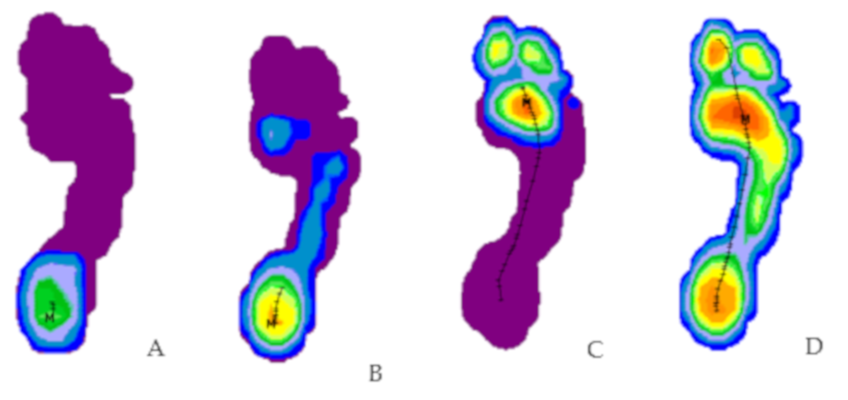
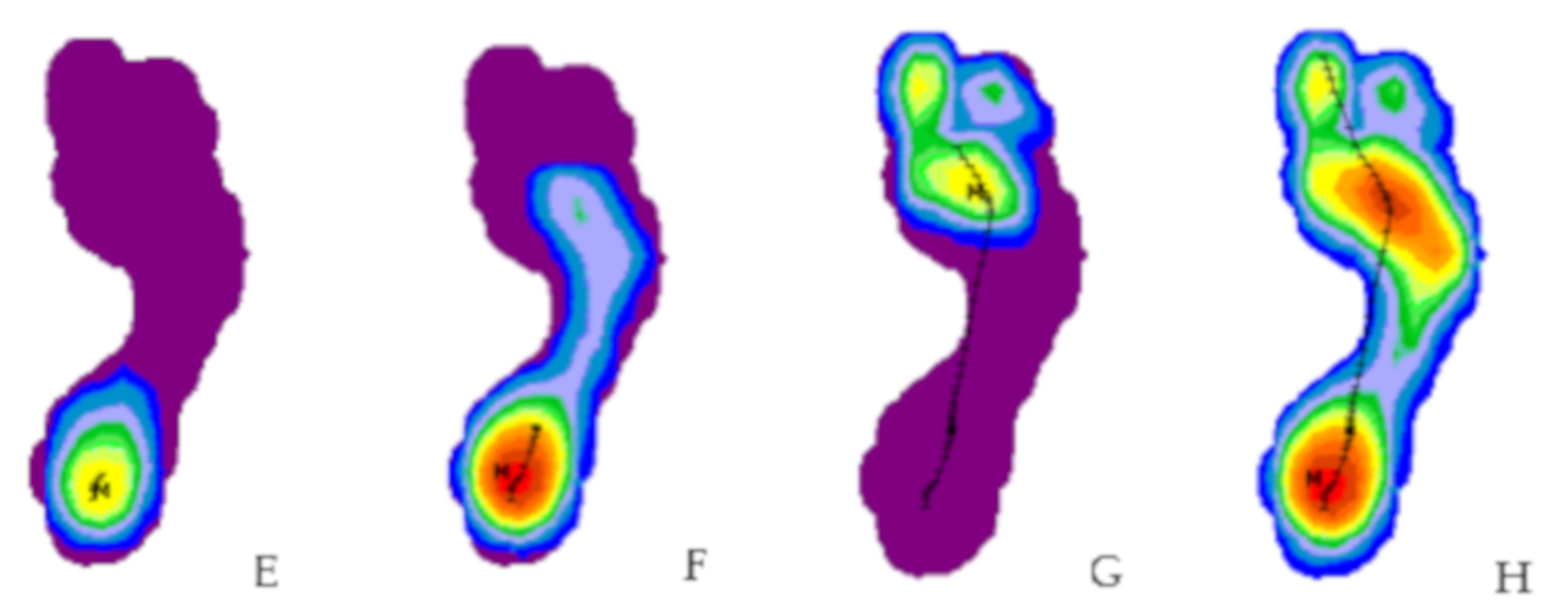
2.3. Variables
3. Results
3.1. Characteristics of the Sample
3.2. Reliability
3.3. Comparisons between Women and Men
4. Discussion
5. Conclusions
Author Contributions
Funding
Conflicts of Interest
References
- Domjanic, J.; Fieder, M.; Seidler, H.; Mitteroecker, P. Geometric morphometric footprint analysis of young women. J. Foot Ankle Res. 2013, 6, 27. [Google Scholar] [CrossRef] [PubMed]
- Ashizawa, K.; Kumakura, C.; Kusumoto, A.; Narasaki, S. Relative foot size and shape to general body size in Javanese, Filipinas and Japanese with special reference to habitual footwear types. Ann. Hum. Biol. 1997, 24, 117–129. [Google Scholar] [CrossRef] [PubMed]
- Mauch, M.; Grau, S.; Krauss, I.; Maiwald, C.; Horstmann, T. A new approach to children’s footwear based on foot type classification. Ergonomics 2009, 52, 999–1008. [Google Scholar] [CrossRef]
- Smith, S.L. Attribution of foot bones to sex and population groups. J. Forensic Sci. 1997, 42, 186–195. [Google Scholar] [CrossRef] [PubMed]
- Stankovi, K.; Booth, B.G.; Danckaers, F.; Burg, F.; Vermaelen, P.; Duerinck, S.; Sijbers, J.; Huysmans, T. Three-dimensional quantitative analysis of healthy foot shape: A proof of concept study. J. Foot Ankle Res. 2018, 11, 8. [Google Scholar] [CrossRef] [PubMed]
- Quatman, C.E.; Ford, K.R.; Myer, G.D.; Paterno, M.V.; Hewett, T.E. The effects of gender and pubertal status on generalized joint laxity in young athletes. J. Sci. Med. Sport 2008, 11, 257–263. [Google Scholar] [CrossRef] [PubMed]
- Hass, C.; Schick, E.; Chow, J. Lower extremity biomechanics differ in prepubescent and postpubescent female athletes during stride jump landings. J. Appl. Biomech. 2003, 19, 139–152. [Google Scholar] [CrossRef]
- Putti, A.B.; Arnold, G.P.; Abboud, R.J. Foot pressure differences in men and women. Foot Ankle Surg. 2010, 16, 21–24. [Google Scholar] [CrossRef] [PubMed]
- Kandil, O.; Aboelazm, S.; Mabrouk, M.S. Foot Biometrics Gender Differences in Plantar Pressure Distribution in Standing Position. Am. J. Biomed. Eng. 2014, 4, 1–9. [Google Scholar] [CrossRef]
- Higbie, E.J.; Contractor, B.S.; Tis, L.L.; Johnson, B.F. Foot Structure and in Shoe Plantar Pressure Differences Between Males and Females. Med. Sci. Sports Exerc. 1999, 31, S129. [Google Scholar] [CrossRef]
- Hills, A.; Hennig, E.; McDonald, M.; Bar-Or, O. Plantar pressure differences between obese and non-obese adults: A biomechanical analysis. Int. J. Obes. 2001, 25, 1674–1679. [Google Scholar] [CrossRef] [PubMed]
- Jung, J.-W.; Sato, T.; Bien, Z. Dynamic footprint-based person recognition method using a hidden markov model and a neural network. Int. J. Int. Syst. 2004, 19, 1127–1141. [Google Scholar] [CrossRef]
- Meyers-Rice, B.; Sugars, L.; McPoil, T.; Cornwall, M.W. Comparison of three methods for obtaining plantar pressures in nonpathologic subjects. J. Am. Podiatr. Med. Assoc. 1994, 84, 499–504. [Google Scholar] [CrossRef] [PubMed]
- Digiovanni, C.W.; Kuo, R.; Tejwani, N.; Price, R.; Hansen, S.T., Jr.; Cziernecki, J.; Sangeorzan, B.J. Isolated gastrocnemius tightness. J. Bone Jt. Surg. Am. 2002, 84, 962. [Google Scholar] [CrossRef] [PubMed]
- Downey, M.S.; Banks, A.S. Gastrocnemius recession in the treatment of nonspastic ankle equinus: A retrospective study. J. Am. Podiatr. Med. Assoc. 1989, 79, 159. [Google Scholar] [CrossRef] [PubMed]
- Hill, R.S. Ankle equinus. Prevalence and linkage to common foot pathology. J. Am. Podiatr. Med. Assoc. 1995, 85, 259–300. [Google Scholar] [CrossRef] [PubMed]
- Lamm, B.M.; Paley, D.; Herzenberg, J.E. Gastrocnemius soleus recession: A simpler, more limited approach. J. Am. Podiatr. Med. Assoc. 2005, 95, 18. [Google Scholar] [CrossRef] [PubMed]
- Williams, A. Footwear assessment and management. Podiatry Manag. 2007, 165–176. [Google Scholar]
- Gatt, A.; Chockalingam, N. Clinical assessment of ankle joint dorsiflexion: A review of measurement techniques. J. Am. Podiatr. Med. Assoc. 2011, 101, 59–69. [Google Scholar] [CrossRef]
- Johnsone, C.H.; Christensen, J.C. Biomechanics of the first ray part V: The effect of equinus deformity: A 3-dimensional kinematic study on a cadaver model. J. Foot Ankle Surg. 2005, 44, 114–120. [Google Scholar] [CrossRef]
- Becerro de Bengoa-Vallejo, R.; Losa-Iglesias, M.E.; Rodríguez.Sanz, D.; Prados Frutos, J.C.; Salvadores Fuentes, P.; López-Chicharro, J. Plantar Pressures in Children With and Without Sever’s Disease. J. Am. Podiatr. Med. Assoc. 2011, 10, 17–24. [Google Scholar] [CrossRef]
- Downey, M.S. Ankle Equinus. In Comprehensive Textbook of Foot Surgery, 2nd ed.; McGlamry, E.D., Banks, A.S., Downey, M.S., Eds.; Williams & Wilkins: Baltimore, MD, USA, 1992; Volume 1, pp. 687–698. [Google Scholar]
- Bryant, A.; Singer, K.; Tinley, P. Comparison of the Reliability of Plantar Pressure Measurements Using the Two-step and Midgait Methods of Data Collection. Foot Ankle Int. 1999, 20, 646–650. [Google Scholar] [CrossRef] [PubMed]
- McPoil, T.; Cornwall, M.; Dupuis, L.; Cornwell, M. Variability of plantar pressure data. A comparison of the two-step and midgait methods. J. Am. Podiatr. Med. Assoc. 1999, 89, 495–501. [Google Scholar] [CrossRef] [PubMed]
- Roca-Dols, A.; Losa-Iglesias, M.E.; Sánchez-Gómez, R.; Becerro-de-Bengoa-Vallejo, R.; López-López, D.; Rodríguez-Sanz, D.; Martínez-Jiménez, E.M.; Calvo-Lobo, C. Effect of the cushioning running shoes in ground contact time of phases of gait. J. Mech. Behav. Biomed. Mater. 2018, 88, 196–200. [Google Scholar] [CrossRef] [PubMed]
- Harrison, A.J.; Folland, J.P. Investigation of gait protocols for plantar pressure measurement of non-pathological subjects using a dynamic pedobarograph. Gait Posture 1997, 6, 50–55. [Google Scholar] [CrossRef]
- Rodriguez-Sanz, D.; Losa-Iglesias, M.E.; Becerro de Bengoa-Vallejo, R.; Palomo-Lopez, P.; Beltran-Alacreu, H.; Calvo-Lobo, C.; Navarro-Flores, E.; Lopez-Lopez, D. Skin temperature in youth soccer players with functional equinus and non-equinus condition after running. J. Eur. Acad. Dermatol. Venereol. 2018, 32, 2020–2024. [Google Scholar] [CrossRef] [PubMed]
- Zifchock, R.A.; Davis, I.; Hillstrom, H.; Song, J. The Effect of Gender, Age, and Lateral Dominance on Arch Height and Arch Stiffness. Foot Ankle Int. 2006, 2, 367–372. [Google Scholar] [CrossRef] [PubMed]
- Peters, E.J.G.; Urukalo, A.; Fleischli, J.G.; Lavery, L.A. Reproducibility of gait analysis variables: One-step versus three-step method of data acquisition. J. Foot Ankle Surg. 2002, 41, 206–212. [Google Scholar] [CrossRef]
- Bus, S.A.; De Lange, A. A comparison of the 1-step, 2-step, and 3-step protocols for obtaining barefoot plantar pressure data in the diabetic neuropathic foot. Clin. Biomech. 2005, 20, 892–899. [Google Scholar] [CrossRef]
- Martínez-Jiménez, E.M.; Losa-Iglesias, M.E.; Díaz-Velázquez, J.I.; Becerrro-De- Bengoa-Vallejo, R.; Palomo-López, P.; Clavo-Lobo, C.; López-López, D.; Rodríguez-Sanz, D. Acute Effects of Intermittent Versus Continuous Bilateral Ankle Plantar Flexor Static Stretching on Postural Sway and Plantar Pressures: A Randomized Clinical Trial. Clin. Med. 2019, 8, 52. [Google Scholar] [CrossRef]
- Oladeji, O.; Stackhouse, C.; Gracely, E.; Orlin, M. Comparison of the two-step and midgait methods of plantar pressure measurement in children. J. Am. Podiatr. Med. Assoc. 2008, 98, 268–277. [Google Scholar] [PubMed]
- Rosenbaum, D.; Becker, H.P. Plantar Preassure distribution measurements. Technical background and clinical applications. Foot Ankle Surg. 1997, 3, 1–14. [Google Scholar] [CrossRef]
- Cornwall, M.; McPoil, T. Velocity of the center of pressure during walking. J. Am. Podiatr. Med. Assoc. 2000, 90, 334–338. [Google Scholar] [CrossRef] [PubMed]
- Bruton, A.; Conway, J.H.; Holgate, S.T. Reliability: What is it and how is measured? Physiotherapy 2000, 86, 94–99. [Google Scholar] [CrossRef]
- Landis, J.R.; Koch, G.G. The measurement of observer agreement for categorical data. Biometrics 1977, 33, 159–174. [Google Scholar] [CrossRef] [PubMed]
- Wunderlich, R.E.; Cavanagh, P.R. Gender differences in adult foot shape: Implications for shoe design. Med. Sci. Sports Exerc. 2001, 33, 605–611. Available online: http://www.ncbi.nlm.nih.gov/pubmed/11283437 (accessed on 1 April 2018). [CrossRef]
- Murphy, D.F.; Beynnon, B.D.; Michelson, J.D.; Vacek, P.M. Efficacy of Plantar Loading Parameters During Gait in Terms of Reliability, Variability, Effect of Gender and Relationship Between Contact Area and Plantar Pressure. Foot Ankle Int. 2005, 26, 171–179. [Google Scholar] [CrossRef]
- Wilkerson, R.D.; Mason, M.A. Differences in men’s and women’s mean ankle ligamentous laxity. Iowa Orthop. J. 2000, 20, 46–48. [Google Scholar]
- Taylor, A.J.; Menz, H.B.; Keenan, A.-M. The influence of walking speed on plantar pressure measurements using the two-step gait initiation protocol. Foot 2004, 14, 49–55. [Google Scholar] [CrossRef]
- Michaud, T. Foot Orthoses and Other Forms of Conservative Foot Care, 2nd ed.; Williams and Wilkins: Baltimore, MD, USA, 1993. [Google Scholar]
- Aguelo-Mendoza, A.I.; Briñez Santamaria, T.J.; Guarín-Urrego, J.P.; Zapata-Garcia, M.C. Marcha: Descripcion, métodos, herramientas de evaluación y parámetros de normalidad reportados en la literatura. Gait: Decription, methods, assesment tolos and normality parameters reported in the literatura. Rev. Ces. Mov. Salud 2013, 1, 201. [Google Scholar]
- Crenna, P.; Frigo, C. A motor programme for the initiation of forward-oriented movements in humans. J. Physiol. 1991, 437, 635–653. [Google Scholar] [CrossRef] [PubMed]


| Total Group n = 119 (100%) Mean (SD) | Female Group n = 59 (49.6%) Mean (SD) | Male Group n = 60 (50.4%) Mean (SD) | p-Value * | |
|---|---|---|---|---|
| Age (years) | 30.50 (3.65) | 29.76 (5.15) | 31.22 (6.06) | p = 0.161 |
| Weight (kg) | 67.28 (11.92) | 58.74 (6.66) | 75.67 (9.81) | p = 0.001 |
| Height (cm) | 170.43 (8.94) | 163.65 (5.58) | 177.10 (6.16) | p = 0.001 |
| Shoe size (Eu) | 40.67 (2.68) | 38.46 (1.21) | 42.85 (1.78) | p = 0.001 |
| Protocol, Foot, and Biomechanical Phase | FIRST WEEK | SECOND WEEK | BOTH WEEKS | |||
|---|---|---|---|---|---|---|
| 95% CI | 95% CI | 95% CI | ||||
| ICC (Lower Limit–Upper Limit) | SEM | ICC (Lower Limit–Upper Limit) | SEM | ICC (Lower Limit–Upper Limit) | SEM | |
| First step, Left foot, 20% | 0.600(0.489–0.708) | 8.18 | 0.674(0.573–0.767) | 7.30 | 0.809(0.698–0.882) | 5.34 |
| First step, Right foot, 20% | 0.555(0.440–0.671) | 8.58 | 0.507(0.389–0.629) | 7.50 | 0.724(0.575–0.826) | 5.76 |
| First step, Left foot, 35% | 0.691(0.594–0.781) | 6.79 | 0.737(0.649–0.816) | 6.29 | 0.888(0.819–0.932) | 3.98 |
| First step, Right foot, 35% | 0.653(0.549–0.751) | 6.72 | 0.725(0.633–0.807) | 6.01 | 0.875(0.798–0.924) | 3.91 |
| First step, Left foot, 92% | 0.357(0.239–0.492) | 5.29 | 0.472(0.352–0.598) | 4.96 | 0.611(0.423–0.749) | 3.76 |
| First step, Right foot, 92% | 0.400(0.280–0.532) | 5.58 | 0.365(0.246–0.499) | 4.78 | 0.611(0.422–0.749) | 3.71 |
| Second step, Left foot, 20% | 0.655(0.552–0.753) | 7.53 | 0.629(0.521–0.732) | 6.99 | 0.797(0.681–0.874) | 5.19 |
| Second step, Right foot, 20% | 0.629(0.522–0.732) | 7.75 | 0.601(0.490–0.709) | 7.49 | 0.783(0.660–0.865) | 5.41 |
| Second step, Left foot, 35% | 0.769(0.688–0.840) | 5.45 | 0.663(0.560–0.759) | 5.68 | 0.879(0.805–0.927) | 3.57 |
| Second step, Right foot, 35% | 0.752(0.667–0.827) | 5.35 | 0.716(0.623–0.800) | 5.72 | 0.848(0.757–0.907) | 4.03 |
| Second step, Left foot, 92% | 0.224(0.117–0.357) | 4.93 | 0.337(0.220–0.472) | 4.96 | 0.653(0.478–0.778) | 3.13 |
| Second step, Right foot, 92% | 0.324(0.207–0.461) | 5.38 | 0.392(0.273–0.525) | 4.79 | 0.681(0.514–0.798) | 3.23 |
| Third step, Left foot, 20% | 0.655(0.551–0.753) | 6.63 | 0.692(0.594–0.781) | 6.63 | 0.791(0.672–0.870) | 5.02 |
| Third step, Right foot, 20% | 0.599(0.488–0.708) | 7.90 | 0.739(0.651–0.818) | 6.38 | 0.726(0.578–0.827) | 6.07 |
| Third step, Left foot, 35% | 0.742(0.655–0.820) | 5.84 | 0.765(0.683–0.837) | 5.74 | 0.885(0.814–0.930) | 3.84 |
| Third step, Right foot, 35% | 0.762(0.679–0.835) | 5.66 | 0.741(0.653–0.819) | 5.55 | 0.870(0.790–0.920) | 3.93 |
| Third step, Left foot, 92% | 0.267(0.156–0.402) | 4.67 | 0.351(0.234–0.486) | 4.96 | 0.678(0.512–0.795) | 3.02 |
| Third step, Right foot, 92% | 0.304(0.189–0.440) | 4.79 | 0.180(0.079–0.310) | 4.67 | 0.478(0.255–0.653) | 3.53 |
| Protocol, Foot, and Biomechanical Phase | FIRST WEEK | SECOND WEEK | BOTH WEEKS | |||
|---|---|---|---|---|---|---|
| 95% CI | 95% CI | 95% CI | ||||
| ICC (Lower Limit–Upper Limit) | SEM | ICC (Lower Limit–Upper Limit) | SEM | ICC (Lower Limit–Upper Limit) | SEM | |
| First step, Left foot, 20% | 0.656(0.553–0.753) | 9.15 | 0.593(0.482–0.70)1 | 9.59 | 0.860(0.777–0.914) | 5.53 |
| First step, Right foot, 20% | 0.648(0.544–0.746) | 9.60 | 0.658(0.556–0.754) | 9.01 | 0.798(0.684–0.874) | 6.73 |
| First step, Left foot, 35% | 0.665(0.563–0.760) | 8.73 | 0.811(0.741–0.870) | 6.42 | 0.891(0.824–0.934) | 4.79 |
| First step, Right foot, 35% | 0.704(0.610–0.791) | 7.36 | (0.793(0.719–0.857) | 6.55 | 0.890(0.823–0.933) | 4.50 |
| First step, Left foot, 92% | 0.542(0.427–0.659) | 5.65 | 0.509(0.392–0.630) | 5.62 | 0.795(0.679–0.872) | 3.51 |
| First step, Right foot, 92% | 0.437(0.318–0.566) | 6.00 | 0.473(0.355–0.599) | 5.77 | 0.779(0.655–0.862) | 3.54 |
| Second step, Left foot, 20% | 0.654(0.551–0.751) | 8.76 | 0.778(0.700–0.846) | 7.82 | 0.898(0.834–0.938) | 4.91 |
| Second step, Right foot, 20% | 0.678(0.578–0.770) | 8.46 | 0.734(0.645–0.813) | 7.31 | 0.861(0.777–0.914) | 5.23 |
| Second step, Left foot, 35% | 0.782(0.704–0.849) | 6.29 | 0.779(0.701–0.847) | 6.48 | 0.914(0.860–0.948) | 3.91 |
| Second step, Right foot, 35% | 0.802(0.730–0.864) | 5.94 | 0.804(0.733–0.865) | 5.26 | 0.932(0.888–0.959) | 3.24 |
| Second step, Left foot, 92% | 0.417(0.299–0.548) | 5.15 | 0.420(0.301–0.550) | 5.57 | 0.651(0.478–0.776) | 3.78 |
| Second step, Right foot, 92% | 0.482(0.364–0.607) | 5.84 | 0.410(0.291–0.541) | 5.77 | 0.750(0.613–0.842) | 3.66 |
| Third step, Left foot, 20% | 0.734(0.645–0.813) | 7.74 | 0.747(0.661–0.823) | 7.55 | 0.858(0.773–0.913) | 5.45 |
| Third step, Right foot, 20% | 0.647(0.543–0.746) | 8.95 | 0.719(0.627–0.802) | 7.48 | 0.864(0.783–0.917) | 5.20 |
| Third step, Left foot, 35% | 0.827(0.761–0.882) | 5.52 | 0.812(0.742–0.871) | 5.94 | 0.954(0.925–0.972) | 2.86 |
| Third step, Right foot, 35% | 0.781(0.702–0.849) | 5.92 | 0.834(0.771–0.887) | 5.26 | 0.920(0.869–0.951) | 3.55 |
| Third step, Left foot, 92% | 0.456(0.337–0.583) | 5.81 | 0.397(0.279–0.529) | 5.42 | 0.706(0.552–0.813) | 3.73 |
| Third step, Right foot, 92% | 0.480(0.361–0.604) | 6.04 | 0.456(0.336–0.584) | 5.65 | 0.58(0.385–0.727) | 4.93 |
| Biomechanical Intraphase | Total n = 119 (100%) Mean SD cm2 | Women n = 59 (49.6%) Mean SD cm2 | Men n = 60 (50.4%) Mean SD cm2 | Difference between Women and Men p-Value | ||
|---|---|---|---|---|---|---|
| First week | Left foot | 20% | 65.73 (15.46) | 59.79 (12.93) | 71.57 (15.60) | p = 0.001 * |
| 35% | 95.08 (15.99) | 86.76 (12.22) | 103.27 (15.09) | p = 0.001 * | ||
| 92% | 49.18 (8.22) | 45.81 (6.60) | 52.49 (8.35) | p = 0.001 * | ||
| Global | 148.12 (20.19) | 136.39 (13.79) | 159.65 (18.86) | p = 0.001 * | ||
| Right foot | 20% | 64.21 (15.29) | 59.56 (12.86) | 68.79 (16.18) | p = 0.001 * | |
| 35% | 95.14 (14.60) | 87.51 (11.40) | 102.64 (13.53) | p = 0.001 * | ||
| 92% | 49.72 (8.48) | 45.89 (7.20) | 53.48 (8.00) | p = 0.001 * | ||
| Global | 147.22 (19.04) | 135.69 (14.48) | 158.55 (15.99) | p = 0.001 * | ||
| Second week | Left foot | 20% | 64.95 (15.24) | 58.68 (12.79) | 71.13 (15.03) | p = 0.001 * |
| 35% | 93.58 (15.59) | 85.80 (12.27) | 101.23 (14.76) | p = 0.001 * | ||
| 92% | 48.82 (7.93) | 46.02 (6.83) | 51.57 (8.02) | p = 0.001 * | ||
| Global | 147.73 (20.99) | 135.21 (814.94) | 159.84 (18.85) | p = 0.001 * | ||
| Right foot | 20% | 64.82 (14.13) | 59.79 (10.68) | 69.76 (15.40) | p = 0.001 * | |
| 35% | 94.63 (15.17) | 86.73 (11.46) | 102.39 (14.40) | p = 0.001 * | ||
| 92% | 48.92 (7.80) | 45.52 (6.00) | 52.28 (7.95) | p = 0.001 * | ||
| Global | 146.94 (18.78) | 135.19 (14.07) | 158.69 (15.24) | p = 0.001 * | ||
| Both weeks | Left foot | 20% | 65.34 (14.82) | 59.24 (12.23) | 71.35 (14.77) | p = 0.001 * |
| 35% | 94.33 (15.46) | 86.28 (11.90) | 102.25 (14.51) | p = 0.001 * | ||
| 92% | 49.00 (7.57) | 45.92 (6.03) | 52.03 (7.75) | p = 0.001 * | ||
| Global | 147.97 (20.35) | 135.78 (13.99) | 159.75 (18.57) | p = 0.001 * | ||
| Right foot | 20% | 64.52 (13.95) | 59.68 (10.97) | 69.28 (14.98) | p = 0.001 * | |
| 35% | 94.88 (14.57) | 87.12 (11.07) | 102.52 (13.58) | p = 0.001 * | ||
| 92% | 49.32 (7.66) | 45.71 (5.95) | 52.88 (7.52) | p = 0.001 * | ||
| Global | 146.89 (18.54) | 135.44 (14.04) | 158.33 (15.17) | p = 0.001 * | ||
| Biomechanical Intraphase | Total n = 119 (100%) Mean SD cm2 | Women n = 59 (49.6%) Mean SD cm2 | Men n = 60 (50.4%) Mean SD cm2 | Difference between Women and Men p-Value | ||
|---|---|---|---|---|---|---|
| First week | Left foot | 20% | 72.36 (15.14) | 66.22 (12.82) | 78.40 (14.90) | p = 0.001 * |
| 35% | 96.24 (14.61) | 88.50 (11.34) | 103.85 (13.48) | p = 0.001 * | ||
| 92% | 48.12 (6.72) | 45.48 (5.60) | 50.71 (6.75) | p = 0.001 * | ||
| Global | 148.09 (19.67) | 136.85 (13.90) | 159.13 (18.27) | p = 0.001 * | ||
| Right foot | 20% | 71.13 (15.32) | 64.46 (12.72) | 77.69 (14.90) | p = 0.001 * | |
| 35% | 96.24 (14.28) | 88.57 (10.75) | 103.77 (13.34) | p = 0.001 * | ||
| 92% | 48.29 (7.81) | 45.61 (6.54) | 50.88 (8.11) | p = 0.001 * | ||
| Global | 148.13 (19.23) | 135.76 (14.09) | 160.29 (15.52) | p = 0.001 * | ||
| Second week | Left foot | 20% | 74.47 (15.60) | 68.05 (11.47) | 80.78 (16.60) | p = 0.001 * |
| 35% | 96.65 (14.08) | 89.13 (9.79) | 104.04 (13.78) | p = 0.001 * | ||
| 92% | 48.13 (7.39) | 45.18 (6.09) | 51.38 (7.32) | p = 0.001 * | ||
| Global | 149.22 (19.89) | 137.29 (13.21) | 160.95 (18.37) | p = 0.001 * | ||
| Right foot | 20% | 72.73 (14.62) | 66.04 (11.85) | 79.32 (14.17) | p = 0.001 * | |
| 35% | 96.22 (13.78) | 88.27 (10.73) | 104.03 (11.89) | p = 0.001 * | ||
| 92% | 47.84 (7.84) | 43.99 (6.14) | 51.62 (7.51) | p = 0.001 * | ||
| Global | 148.39 (19.73) | 135.75 (13.16) | 160.83 (17.07) | p = 0.001 * | ||
| Both weeks | Left foot | 20% | 73.42 (14.92) | 67.14 (11.53) | 79.59 (15.37) | p = 0.001 * |
| 35% | 96.45 (14.09) | 88.82 (10.27) | 103.95 (13.34) | p = 0.001 * | ||
| 92% | 48.21 (6.53) | 45.33 (5.32) | 51.04 (6.40) | p = 0.001 * | ||
| Global | 148.65 (19.38) | 137.07 (13.26) | 160.04 (17.65) | p = 0.001 * | ||
| Right foot | 20% | 71.93 (14.45) | 65.24 (11.61) | 78.51 (14.02) | p = 0.001 * | |
| 35% | 96.23 (13.78) | 88.42 (10.33) | 103.90 (12.42) | p = 0.001 * | ||
| 92% | 48.14 (7.28) | 44.92 (5.72) | 51.25 (7.31) | p = 0.001 * | ||
| Global | 148.26 (19.21) | 135.75 (13.37) | 160.56 (15.86) | p = 0.001 * | ||
| Biomechanical Intraphase | Total n = 119 (100%) Mean SD cm2 | Women n = 59 (49.6%) Mean SD cm2 | Men n = 60 (50.4%) Mean SD cm2 | Difference between Women and Men p-Value | ||
|---|---|---|---|---|---|---|
| First week | Left foot | 20% | 75.05 (14.75) | 68.50 (11.29) | 81.48 (15.01) | p = 0.001 * |
| 35% | 97.54 (14.59) | 89.78 (11.59) | 105.18 (13.27) | p = 0.001 * | ||
| 92% | 48.06 (7.39) | 45.06 (5.45) | 51.01 (7.88) | p = 0.001 * | ||
| Global | 148.64 (19.32) | 136.69 (12.71) | 160.38 (17.45) | p = 0.001 * | ||
| Right foot | 20% | 74.19 (15.92) | 66.18 (12.47) | 82.06 (15.06) | p = 0.001 * | |
| 35% | 96.69 (14.37) | 88.96 (11.60) | 104.42 (12.66) | p = 0.001 * | ||
| 92% | 48.06 (7.61) | 45.48 (5.74) | 50.60 (8.38) | p = 0.001 * | ||
| Global | 147.61 (19.53) | 134.94 (13.90) | 160.07 (17.95) | p = 0.001 * | ||
| Second week | Left foot | 20% | 76.42 (15.58) | 68.63 (11.94) | 84.07 (15.02) | p = 0.001 * |
| 35% | 97.99 (15.25) | 89.59 (11.84) | 106.25 (13.69) | p = 0.001 * | ||
| 92% | 48.01 (7.41) | 44.54 (6.16) | 51.42 (6.98) | p = 0.001 * | ||
| Global | 148.36 (18.94) | 136.49 (14.03) | 160.03 (15.65) | p = 0.001 * | ||
| Right foot | 20% | 75.10 (15.22) | 67.63 (12.49) | 82.45 (14.12) | p = 0.001 * | |
| 35% | 97.36 (14.64) | 88.81 (10.91) | 105.77 (12.91) | p = 0.001 * | ||
| 92% | 47.92 (7.06) | 45.19 (5.16) | 50.66 (7.66) | p = 0.001 * | ||
| Global | 148.69 (19.93) | 136.19 (13.37) | 160.97 (17.58) | p = 0.001 * | ||
| Both weeks | Left foot | 20% | 75.73 (14.66) | 68.57 (10.99) | 82.77 (14.47) | p = 0.001 * |
| 35% | 97.76 (14.72) | 89.68 (11.33) | 105.71 (13.33) | p = 0.001 * | ||
| 92% | 48.03 (6.92) | 44.80 (5.33) | 51.21 (6.87) | p = 0.001 * | ||
| Global | 148.29 (18.77) | 136.46 (13.05) | 159.91 (16.12) | p = 0.001 * | ||
| Right foot | 20% | 74.65 (14.99) | 66.91 (11.60) | 82.25 (14.09) | p = 0.001 * | |
| 35% | 96.96 (14.24) | 88.88 (10.89) | 105.03 (12.56) | p = 0.001 * | ||
| 92% | 48.03 (6.92) | 45.33 (4.89) | 50.73 (7.61) | p = 0.001 * | ||
| Global | 148.15 (19.43) | 135.56 (13.06) | 160.52 (16.54) | p = 0.001 * | ||
© 2019 by the authors. Licensee MDPI, Basel, Switzerland. This article is an open access article distributed under the terms and conditions of the Creative Commons Attribution (CC BY) license (http://creativecommons.org/licenses/by/4.0/).
Share and Cite
Martínez-Jiménez, E.M.; Becerro de Bengoa-Vallejo, R.; Losa-Iglesias, M.E.; Díaz-Velázquez, J.I.; Casado-Hernández, I.; Calvo-Lobo, C.; López-López, D.; Rodríguez-Sanz, D. Sex Differences in the Footprint Analysis During the Entire Gait Cycle in a Functional Equinus Condition: Novel Cross Sectional Research. Appl. Sci. 2019, 9, 3611. https://doi.org/10.3390/app9173611
Martínez-Jiménez EM, Becerro de Bengoa-Vallejo R, Losa-Iglesias ME, Díaz-Velázquez JI, Casado-Hernández I, Calvo-Lobo C, López-López D, Rodríguez-Sanz D. Sex Differences in the Footprint Analysis During the Entire Gait Cycle in a Functional Equinus Condition: Novel Cross Sectional Research. Applied Sciences. 2019; 9(17):3611. https://doi.org/10.3390/app9173611
Chicago/Turabian StyleMartínez-Jiménez, Eva María, Ricardo Becerro de Bengoa-Vallejo, Marta Elena Losa-Iglesias, José Ignacio Díaz-Velázquez, Israel Casado-Hernández, Cesar Calvo-Lobo, Daniel López-López, and David Rodríguez-Sanz. 2019. "Sex Differences in the Footprint Analysis During the Entire Gait Cycle in a Functional Equinus Condition: Novel Cross Sectional Research" Applied Sciences 9, no. 17: 3611. https://doi.org/10.3390/app9173611
APA StyleMartínez-Jiménez, E. M., Becerro de Bengoa-Vallejo, R., Losa-Iglesias, M. E., Díaz-Velázquez, J. I., Casado-Hernández, I., Calvo-Lobo, C., López-López, D., & Rodríguez-Sanz, D. (2019). Sex Differences in the Footprint Analysis During the Entire Gait Cycle in a Functional Equinus Condition: Novel Cross Sectional Research. Applied Sciences, 9(17), 3611. https://doi.org/10.3390/app9173611