Article

In Vitro Targeting and Imaging of Neurogenic Differentiation in Mouse Bone-Marrow Derived Mesenchymal Stem Cells with Superparamagnetic Iron Oxide Nanoparticles

1
Department of Orthopaedics, Chonnam National University Medical School and Hospital, Gwangju 61469, Korea
2
Department of Materials Science and Engineering, Chonnam National University, Gwangju 61186, Korea
*
Authors to whom correspondence should be addressed.
These authors contributed equally to this work.
Appl. Sci. 2019, 9(16), 3259; https://doi.org/10.3390/app9163259
Submission received: 15 July 2019 / Revised: 5 August 2019 / Accepted: 6 August 2019 / Published: 9 August 2019
(This article belongs to the Special Issue Nanoparticles for Diagnosis and Therapies)

Abstract

:
Spinal cord injuries (SCI) are well thought to be a crucial issue that roots various side effects for a patient during their entire lifetime. Although therapeutical methods to resolve the SCI are limited, stem cell therapy is determined to be a resolving factor since it possesses the ability to induce the neurogenic differentiation and the paracrine effect. However, stem cells are difficult to inject directly into the lesion, so they must be carefully guided through the spinal canal. Therefore, superparamagnetic iron oxide nanoparticles (SPIONs) are introduced as an instigator that makes the cells respond to the applied magnetic field. This study intends to report the synthesis strategy to develop SPIONs that could be used to treat the injury site by an applied magnetic field. SPION-internalized D1 Mesenchymal stem cells (MSCs) are observed consistently using a confocal fluorescence microscope to analyze the toxicity, maintenance, and monitoring points of intracellular SPIONs. The prepared SPIONs are much anticipated to increase the migration efficiency using magnetism, which was not cytotoxic. Hence, the prepared SPIONs can adeptly target the damaged neural tissue to promote tissue regeneration and treat nervous system disorders. This primary study stands as a focal point to solve SCI by stem cell migration effectively.

Graphical Abstract

1. Introduction

Spinal cord injury (SCI), which involves dyssynergia and serious damage to motor and sensory functions in the human body, is often caused by trauma, dysfunction, or degenerative disease. Despite decades of therapeutic research, current treatments are limited to drug treatment in the acute stage or surgical methods [1,2]. Additionally, the initial degree of impairment affects patients throughout life. Stem cell therapy is a promising strategy developed to treat SCI [3,4,5]. The transplanted stem cells promote neural and neural cord regeneration by providing a paracrine effect and a scaffold for replacing damaged neurons [6]. Recently, stem cell treatment has been suggested for radical SCI. Particularly, D1 Mesenchymal stem cells (MSCs) can cause auto-regeneration and differentiation of other mesenchymal systems and are used to restore cartilage in the orthopedic area [7]. Mesenchymal stem cells (MSCs) have recently been evaluated for use in cellular repair after central nervous system injury [8,9]. MSCs are derived from the bone marrow and can self-renew and differentiate into several distinct mesenchymal lineages [10,11,12]. Through morphological and phenotypic changes, MSCs differentiate into neuron-like cells, making these cells a good candidate for use in tissue regeneration and treatment of nervous system injuries [13,14].
Super-paramagnetic iron-oxide nanoparticles (SPIONs) are composed of magnetite, which is an iron-oxide, and are thought to be next-generation nano-drugs. These particles are coated with a biocompatible polymer to enhance their stability and cell transfer effects [15,16]. SPION-based drug delivery, nano-biomaterial, bio-imaging, and other methods are studied to determine their practical applications, and animal studies have been conducted to determine their potential in SCI therapy by using stem-cell-fusion to increase migration [17,18]. However, further studies are needed before SPIONs can be applied clinically and to demonstrate the migration, stability, and safety by magnetic induction of stem cell fusions [19,20]. Recent studies have focused, not only on improving biocompatibility and advancing magnetic materials, but also remotely controlling the magnetic field to cause SPIONs fusion [21]. Some research groups have reported risks such as cellular stress, gene expression changes, impacts on cell proliferation, and nanoparticle-protein interactions. Therefore, the cytotoxicity with nanoparticles should be further examined. Nano-materials could be improved to enable the safe use of SPIONs for developing new targeted therapies [22].
In this study, we differentiated our work from the previous study using dopamine coating. Polydopamine is a mussel-inspired adhesive coating, which has become attractive in the biomaterials field due to its ability to form strong adhesive interactions with materials and with functional biomolecules that contain amine groups. The introduction of Polydopamine in SPION does not initiate any phase transfer in the bulk SPIONs since the Polydopamine polymerization influences the formation of the carbon layer on the surface rather than the bulk [23]. Herein, we report neuronal differentiation of MSCs and evaluate whether SPION-labeled MSCs could be magnetically targeted at the injury site to keeping the nature of MSCs under in vitro conditions [24].

2. Materials and Methods

2.1. Osteogenic, Chondrogenic, and Neurogenic Induction of D1 MSCs

In this study, MSCs were induced to form osteocytes, cartilage cells, and nerve cells after incubating D1 mouse bone marrow-derived MSCs (D1 MSCs) in high-glucose Dulbecco’s Modified Eagle Medium (DMEM) containing 10% fetal bovine serum in a 6-well plate to evaluate the differentiation potential of the D1 MSCs.
For osteocyte and chondrocyte differentiation induction, the media containing stem cells were replaced with differentiation medium when the cells reached 70% confluence. The cells were cultured for three weeks, and Alizarin red staining was conducted to determine the osteogenic accumulation of differentiated cells. Calcium and Alcian blue staining were used to detect differentiated cartilage cells. After washing the differentiated cells twice with phosphate-buffered saline, the cells were fixed in paraformaldehyde solution at room temperature for 10 min. The fixative was removed, and the cells were washed twice with distilled water. Next, 2% Alizarin red solution and 1% Alcian blue solution was added and incubated for 30 min, followed by washing with distilled water. To induce nerve cell differentiation, after stabilizing the stem cells, the cells were incubated for 24 h in DMEM containing 10% fetal bovine serum medium and 5 ng/mL basic fibroblast growth factor. After replacing the medium with neuronal differentiation medium on the following day, the phenotype of neurons was observed with an optical microscope after 6 and 24 h.

2.2. Synthesis and Characterization of SPIONs

The synthesis of SPIONs has been described previously [19]. Briefly, iron (III) acetylacetonate (1.06 g), oleylamine (2.98 mL), oleic acid (2.85 mL), and 1,2-hexadecanediol (3.88 g) were added and stirred under nitrogen flow for approximately 10 min in 30 mL phenyl ether at room temperature. After removing the stir bar, the mixture was heated for 30 min to 200 °C under nitrogen gas flow, and then the temperature was increased to 265 °C for another 30 min. A black precipitate was formed, which was cooled and dissolved in hexanes in the presence of oleylamine and oleic acid and centrifuged at 6000 rpm for 10 min to remove any undispersed residue. Finally, 100-nm Fe3O4 nanoparticles was obtained by precipitation in ethanol. The SPION is prepared in a similar method and can be seen by XRD and SEM image. (Figure 1, Figure 2 and Figure 3).

2.3. Surface Modification with Dopamine and 675-nm N-hydroxysuccinimide (NHS)

Oleic acid SPIONs were modified to have a dopamine coat to render them hydrophilic and amine-functionalized [20]. Briefly, 100 mg of oleic acid-coated SPIONs dissolved in 5 mL of chloroform was mixed with 100 mg of dopamine dissolved in 100 mL DMSO. After the mixture formed a homogenous solution, stirring was continued, and then dopamine-coated SPIONs were separated by centrifugation and washed with water. To obtain the SPIONs, 675-nm light NHS (Flamma® 675 NHS ester, BioActs, Incheon, Korea) was added to the dopamine SPIONs in distilled water at a 1:0.01 ratio and stirred for 24 h.

2.4. Cell Viability and Magnetic Migration of SPION-Labeled D1 MSCs

The cytotoxicity of SPIONs was determined by the 3-(3,4-dimethylthiazol-2-yl)-2,5-diphenyltetrazolium bromide (MTT) assay, as described previously [25]. D1 mouse bone marrow stromal cells (1 × 10³ per well) were seeded into 96-well plates in 100 µL culture medium at 37 °C with 5% CO2. After overnight incubation, the supernatant was removed, and fresh medium containing varying concentrations of SPIONs (0.1, 1, 5, 10, 50, 100 µg/mL) was added. Twenty-four hours later, the surviving cells were stained with MTT and quantified by measuring the absorbance at 540 nm. The MTT assay results were plotted as the mean ± S.D. of three experiments.
To analyze the internalization of SPIONs, D1 cells were fixed and observed with a Bio-TEM. The fluorescence of SPION-internalized D1 MSCs was measured with a FOBI (Fluorescence-labeled Organism Bioimaging Instrument). Our SPIONs (100 nm) and commercial SPIONs (NEO-LIVETM Magnoxide675, Biterials, Seoul, Korea) were prepared, and the migration efficiency was measured at different times after dispersing the cells by using a 1.5 T magnetic bar. Additionally, SPION-internalized D1 MSCs were evaluated on days 1, 3, 5, and 7 with a confocal fluorescence microscope and toxicity, maintenance, and monitoring points of intracellular SPIONs were analyzed.

3. Results

3.1. Differentiation Ability Potential of D1 MSCs

When undergoing neurogenic differentiation, D1 MSCs were observed to form spindle-shaped cells with a typical glial and neuronal shape after 6 h; the cell body was condensed, and oligodendrocytes were detected. Some cells partially lost contact with the culture plate, while others were completely detached. However, most nerve cells adhered well and showed good morphology after 24 h. Osteogenic and chondrogenic differentiation ability was confirmed with Alizarin red (osteogenic) and Alcian blue (chondrogenic) staining, respectively, at day 21 (Figure 1).

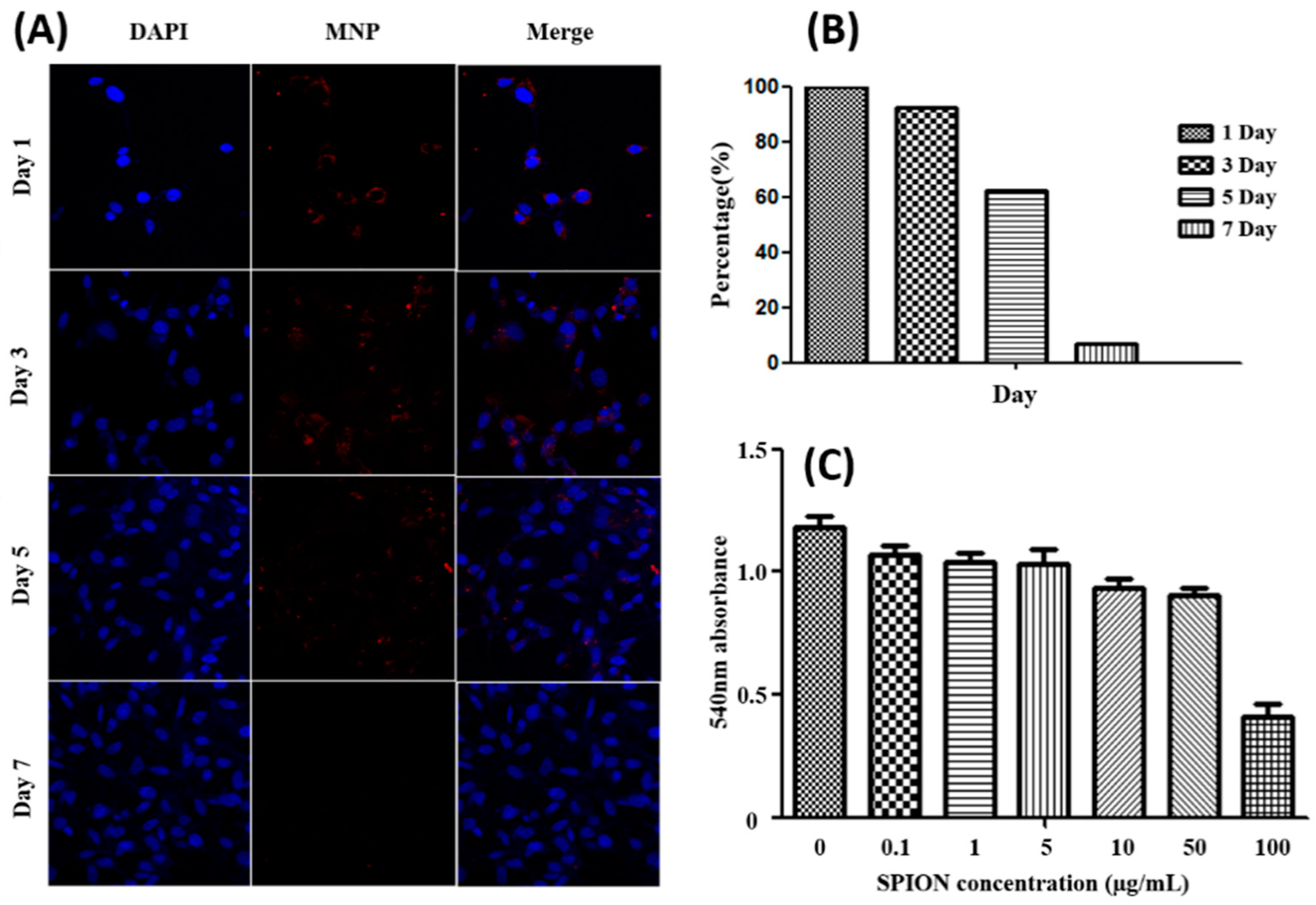
3.2. Cytotoxicity of SPIONs

SPIONs have been associated with toxic effects that have deleterious cellular consequences, eventually leading to cell death [15]. Labeling agents should be biocompatible and nontoxic and should not affect differentiation. The MTT assay showed that cell viability was not significantly affected by up to 50 µg/mL SPIONs after 24 h of treatment (>96% viability compared to the control sample) (Figure 2). SPION-internalized D1 MSCs were viable from day 1 to 7 and did not undergo apoptosis. However, the fluorescence signal gradually decreased over time, showing only approximately 10% of the initial value on day 7. This is because of the limited number of SPIONs and also because some cells did not contain SPIONs.

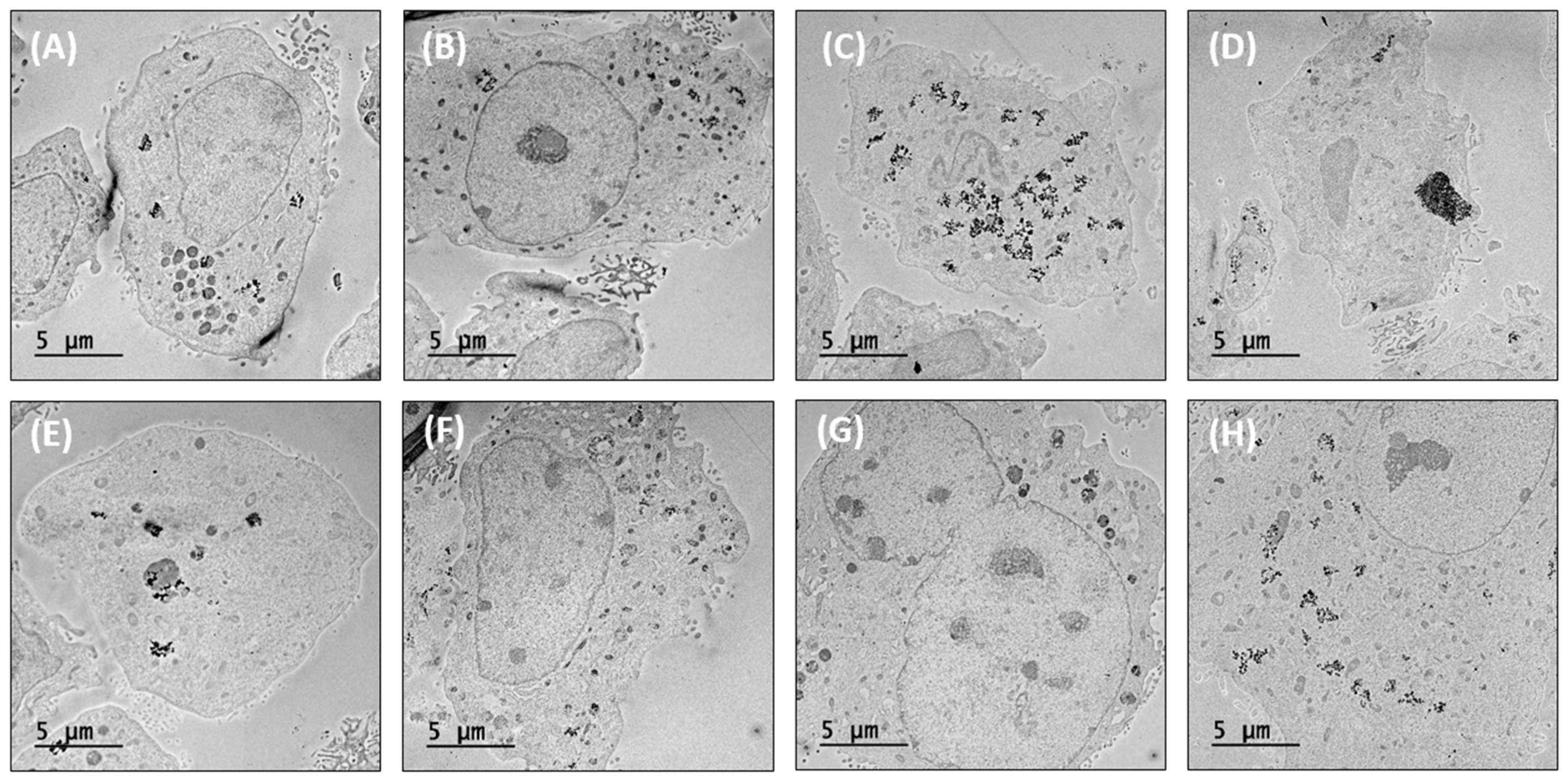
3.3. SPIONs Synthesis and Labeling with D1 MSCs

The SPIONs were found to have a spherical core-shell-type shape. The size of particles measured with 675-nm fluorescent dye and dopamine coating was estimated to be approximately 100 nm. Transmission electron microscopy analysis showed that SPIONs were in the cytoplasm of D1 MSCs. Cellular uptake of SPIONs was mediated by endocytosis. In most cells, the particles were near the cell membrane and nuclear membrane as SPION-containing vesicles. However, some SPION particles were detected in the nucleus (Figure 3).
Most cells contained different numbers of SPIONs inside internal vesicles, likely endosomes, which were taken up during nuclear division (Figure 4G). In some cells, we found that large numbers of SPIONs accumulated in enlarged endosomes (Figure 4D).

3.4. Magnetic Targeting and Migration of SPION-Labeled D1 MSCs

To investigate the magnetic targeting efficiency, SPION-labeled D1 MSCs were further labeled with a 675-nm fluorescent marker using 1 × 10⁴–2 × 10⁶ cells. Fluorescence signals were detected for 2 × 10⁶ cells, revealing high migration at the 1.5 T magnetic field. The commercial SPIONs prior to cell labeling only resulted in 50% migration even after 72 h in the magnetic field (Figure S4), while our cells showed a migration value of over 95% at 30 min (Figure 5). Fluorescence values increased consistently over time for different numbers of cells, demonstrating that SPIONs were uniformly distributed in each cell. We also confirmed that SPIONs were not lost after differentiation (Figure 6).

4. Discussion

Spinal cord injury continues to be a devastating injury to affected individuals and their families and creates enormous financial, psychological, and emotional costs [26]. Damaged nerve cells cannot regenerate or show limited regeneration, and thus, initial treatment can have lifelong effects. Despite years of research, current treatment is limited to early administration of high-dose steroids and acute surgical intervention to minimize cord edema and the subsequent cascade of secondary delayed injury [1,27]. Paralysis remains incurable. MSCs can also differentiate into nerve cells. These morphological and phenotypic changes make MSCs good candidates for clinical applications for tissue regeneration and neurological treatment. To effectively regenerate tissues and functions after SCI, stem cells capable of differentiating into nerve cells or glial cells and that can be directly injected into the lesion or administered by an intravascular transplant method are needed. However, direct injection may cause secondary neural injury of the spinal cord, and the poor environment into which stem cells are transplanted may decrease the cell survival, migration rate, and preventing tissue regeneration. To overcome these limitations, SPIONs could be used to label MSCs to increase stem cell migration under magnetic induction. Studies have shown that this system promotes the induction of tissue regeneration [28].
SPIONs have been applied as contrast agents in magnetic resonance imaging [16] and have been suggested to be useful in orthopedic clinical application of stem-cell-based therapies for the tagging, tracking, and activation of stem cells [29,30]. After targeting SPIONs to MSCs, through inducing the magnetic field, it will stimulate the differentiation of neurons and release more neurotrophic factors to increase the therapeutic effect [31]. In recent animal studies of SCI regeneration using SPIONs, stem cells were injected into the spinal canal of the L5 lumbar spine and were induced using a magnetic field. Analysis of the stem cell distribution revealed that the stem cells aggregated effectively in the target area. However, no studies have examined the stability of SPIONs and differentiation ability of SPION-internalized stem cells [32]. Therefore, we confirmed cell stability by producing SPION-internalized MSCs using fluorescence-labeled markers and analyzed stem cell migration induced by a magnetic field at the cellular level. D1 mouse MSCs could undergo osteogenic, chondrogenic, and neurogenic differentiation, which were labeled with dopamine-coated SPIONs [33]. The existence of the carbon layer in the polydopamine-coated SPION surface could effectively increase the stability and bioactivity of the sample. It also makes easy conjugation with the fluorescence probe, as well as, viable detection both in vitro and in vivo. Similarly, through the 675 nm-NHS coating the surface gets further modified to become a theranostic agent and is diagnosed roentgenology with CT or MRI [34]. Hence, both dopamine and 675-nm NHS act on the surface of the SPION and do not influence any phase transfer through the SPIONs.
MTT assays weer used to analyze the cytotoxicity of various concentrations of SPIONs, which was highest at 100 µg/mL. Additionally, 50 µg/mL SPIONs showed the highest migration efficiency without causing toxicity. SPIONs in the cytoplasm of cells were detected by Bio-TEM. Nearly all the SPIONs in the cells were in vesicles in the cytoplasm and nuclear membrane, indicating that absorption occurred through endocytosis. Some SPIONs particles were detected around the nucleus. This may be because, during nuclear collapses, SPIONs in the cytoplasm were near the nucleus. Some cells contained numerous SPIONs in the endosome, but this likely resulted from the aggregation between SPIONs because of the dopamine coating. To determine whether SPIONs internalized by the cells affected cell stability, we evaluated the optimal concentration of 50 µg/mL SPION-treated D1 stem cells from days 1 to 7. The results confirmed that proliferation occurred without cell apoptosis. However, as the cells proliferated, the number of SPIONs in each cell reduced, and fluorescence could not be measured in some cells. In vitro, fluorescence could be detected over seven days. While the cells containing SPIONs were cultured in a plate in the laboratory, the cells actually multiplied in all three dimensions in vitro. The fluorescence signal of SPIONs was retained without dispersion even during cell proliferation. In this study, cytotoxicity was confirmed before magnetic induction experiments, and cytotoxicity test is required after magnetic field in-vivo experiments. This could be studied and applied to the animal experiments in the future.
To analyze the migration efficiency and to monitor the recovering effects in vitro, commercial SPIONs and dopamine-coated SPIONs were used as fluorescent materials. However, the commercial SPION could only manage 50% of the magnetic induction, even though it was not introduced into the cell. The optimal fluorescence signal that could be analyzed in vitro was 2 × 10⁶ cells with 50 µg/mL internalized SPIONs, and the migration efficiency was 95% or more within 30 min under a neodymium magnet at a 1.5 T magnetic force. While measuring the migration rate, the tube was laid horizontally, in order to avoid the effect of gravity as much as possible. Also, the viscosity of the medium does not get affected because there are no additives in the medium. Analysis using a Fluorescence-labeled Organism Bioimaging Instrument (FOBI) showed that the fluorescence value increased steadily as the number of cells increased, indicating that SPIONs were uniformly present in stem cells. Additionally, the comparison of commercial 50-nm SPIONs and our 100-nm SPIONs confirmed that our SPIONs had a higher migration efficiency and were not cytotoxic, even when using a larger amount than the recommended concentration (up to 50 µg/mL). In contrast to existing commercialized SPIONs, our developed SPIONs could be used at high concentrations to increase the efficiency of migration using magnetism which was not cytotoxic.

5. Conclusions

In order to target lesions, SPIONs were labeled with a fluorescent substance, and dopamine was coated to inject to the cells. The MTT assay was used to confirm the cytotoxicity of the SPIONs and the appropriate concentration of SPIONs. Fluorescence materials labeled SPION helped in confirming the magnetic migration and confirmed that it had migrated remarkably. This result indicates that damaged neural tissue could be targeted to promote tissue regeneration and treat nervous system disorders. This primary study anticipates as a pivotal strategy to initiate future studies in treating SCI in vivo.

Supplementary Materials

The following are available online at https://www.mdpi.com/2076-3417/9/16/3259/s1, Figure S1: XRD result of iron oxide nanoparticles, Figure S2: SEM images of iron oxide nanoparticles according to the synthesis method, Figure S3: SEM images and size distribution of iron oxide nanoparticles, Figure S4: FOBI image of the magnetic induction of commercial SPIONs labeled MSCs.

Author Contributions

Conceptualization—U.S. and S.-K.K.; Methodology—H.-J.L.; Validation—U.S. and S.-K.K.; Formal Analysis—D.-K.L.; Investigation—H.-J.L.; Resources—U.S.; Data Curation—U.S.; Writing-Original Draft Preparation—H.-J.L.; Writing—Review & Editing—D.-K.L., U.S., and S.-K.K.; Visualization—D.-K.L.; Supervision and Project Administration—U.S. and S.-K.K.

Funding

This research was supported by the Basic Science Research Program through the National Research Foundation of Korea (NRF) funded by the Ministry of Education (NRF-2018R1D1A1A02086142) and a grant (BCRI 19026) of the Chonnam National University Hospital Biomedical Research Institute. This research was also supported by the Human Resources Development program (No.: 20194030202470) of the Korea Institute of Energy Technology Evaluation and Planning (KETEP) grant funded by the Korea government Ministry of Trade, Industry & Energy (MOTIE) of the Republic of Korea.

Conflicts of Interest

The authors declare no conflicts of interest.

References

  1. Vawda, R.; G Fehlings, M. Mesenchymal cells in the treatment of spinal cord injury: Current & future perspectives. Curr. Stem Cell Res. Ther. 2013, 8, 25–38. [Google Scholar] [PubMed]
  2. Shende, P.; Subedi, M. Pathophysiology, mechanisms and applications of mesenchymal stem cells for the treatment of spinal cord injury. Biomed. Pharmacother. 2017, 91, 693–706. [Google Scholar] [CrossRef]
  3. Teng, Y.D.; Lavik, E.B.; Qu, X.; Park, K.I.; Ourednik, J.; Zurakowski, D.; Langer, R.; Snyder, E.Y. Functional recovery following traumatic spinal cord injury mediated by a unique polymer scaffold seeded with neural stem cells. Proc. Natl. Acad. Sci. USA 2002, 99, 3024–3029. [Google Scholar] [CrossRef] [PubMed] [Green Version]
  4. Lu, P.; Jones, L.; Snyder, E.; Tuszynski, M. Neural stem cells constitutively secrete neurotrophic factors and promote extensive host axonal growth after spinal cord injury. Exp. Neurol. 2003, 181, 115–129. [Google Scholar] [CrossRef]
  5. Ronaghi, M.; Erceg, S.; Moreno-Manzano, V.; Stojkovic, M. Challenges of stem cell therapy for spinal cord injury: Human embryonic stem cells, endogenous neural stem cells, or induced pluripotent stem cells? Stem Cells 2010, 28, 93–99. [Google Scholar] [CrossRef]
  6. Pearse, D.D.; Bunge, M.B. Designing cell-and gene-based regeneration strategies to repair the injured spinal cord. J. Neurotrauma 2006, 23, 437–452. [Google Scholar] [CrossRef] [PubMed]
  7. Jones, I.A.; Chen, X.; Evseenko, D.; Vangsness, C.T., Jr. Nomenclature inconsistency and selective outcome reporting hinder understanding of stem cell therapy for the knee. JBJS 2019, 101, 186–195. [Google Scholar] [CrossRef]
  8. Li, J.; Lepski, G. Cell transplantation for spinal cord injury: A systematic review. BioMed Res. Int. 2013, 2013, 786475. [Google Scholar] [CrossRef]
  9. Grade, S.; Götz, M. Neuronal replacement therapy: Previous achievements and challenges ahead. NPJ Regen. Med. 2017, 2, 29. [Google Scholar] [CrossRef]
  10. Caplan, A. Why are MSCs therapeutic? New data: New insight. J. Pathol. A J. Pathol. Soc. Great Br. Irel. 2009, 217, 318–324. [Google Scholar] [CrossRef]
  11. Prockop, D.J. Marrow stromal cells as stem cells for nonhematopoietic tissues. Science 1997, 276, 71–74. [Google Scholar] [CrossRef] [PubMed]
  12. Pittenger, M.F.; Mackay, A.M.; Beck, S.C.; Jaiswal, R.K.; Douglas, R.; Mosca, J.D.; Moorman, M.A.; Simonetti, D.W.; Craig, S.; Marshak, D.R. Multilineage potential of adult human mesenchymal stem cells. Science 1999, 284, 143–147. [Google Scholar] [CrossRef] [PubMed]
  13. Hofstetter, C.; Schwarz, E.; Hess, D.; Widenfalk, J.; El Manira, A.; Prockop, D.J.; Olson, L. Marrow stromal cells form guiding strands in the injured spinal cord and promote recovery. Proc. Natl. Acad. Sci. USA 2002, 99, 2199–2204. [Google Scholar] [CrossRef] [PubMed] [Green Version]
  14. Pittenger, M.F.; Martin, B.J. Mesenchymal stem cells and their potential as cardiac therapeutics. Circ. Res. 2004, 95, 9–20. [Google Scholar] [CrossRef] [PubMed]
  15. Singh, N.; Jenkins, G.J.; Asadi, R.; Doak, S.H. Potential toxicity of superparamagnetic iron oxide nanoparticles (SPION). Nano Rev. 2010, 1, 5358. [Google Scholar] [CrossRef] [PubMed]
  16. Hofmann-Amtenbrink, M.; Hofmann, H.; Montet, X. Superparamagnetic nanoparticles—A tool for early diagnostics. Swiss Med. Wkly. 2010, 140. [Google Scholar] [CrossRef] [PubMed]
  17. Kojima, K.; Takahashi, S.; Saito, S.; Endo, Y.; Nittami, T.; Nozaki, T.; Sobti, R.; Watanabe, M. Combined Effects of Fe3O4 Nanoparticles and Chemotherapeutic Agents on Prostate Cancer Cells In Vitro. Appl. Sci. 2018, 8, 134. [Google Scholar] [CrossRef]
  18. McDermott, S.; Guimaraes, A.R. Magnetic nanoparticles in the imaging of tumor angiogenesis. Appl. Sci. 2012, 2, 525–534. [Google Scholar] [CrossRef]
  19. Lee, S.J.; Muthiah, M.; Lee, H.J.; Lee, H.-J.; Moon, M.-J.; Che, H.-L.; Heo, S.U.; Lee, H.-C.; Jeong, Y.Y.; Park, I.-K. Synthesis and characterization of magnetic nanoparticle-embedded multi-functional polymeric micelles for MRI-guided gene delivery. Macromol. Res. 2012, 20, 188–196. [Google Scholar] [CrossRef]
  20. Xie, J.; Chen, K.; Huang, J.; Lee, S.; Wang, J.; Gao, J.; Li, X.; Chen, X. PET/NIRF/MRI triple functional iron oxide nanoparticles. Biomaterials 2010, 31, 3016–3022. [Google Scholar] [CrossRef] [Green Version]
  21. Kozissnik, B.; Dobson, J. Biomedical applications of mesoscale magnetic particles. MRS Bull. 2013, 38, 927–932. [Google Scholar] [CrossRef]
  22. Huang, D.-M.; Hsiao, J.-K.; Chen, Y.-C.; Chien, L.-Y.; Yao, M.; Chen, Y.-K.; Ko, B.-S.; Hsu, S.-C.; Tai, L.-A.; Cheng, H.-Y. The promotion of human mesenchymal stem cell proliferation by superparamagnetic iron oxide nanoparticles. Biomaterials 2009, 30, 3645–3651. [Google Scholar] [CrossRef] [PubMed]
  23. An, T.-Y.; Surendran, S.; Kim, H.; Choe, W.-S.; Kim, J.K.; Sim, U. A polydopamine-mediated biomimetic facile synthesis of molybdenum carbide-phosphide nanodots encapsulated in carbon shell for electrochemical hydrogen evolution reaction with long-term durability. Compos. Part B Eng. 2019, 175, 107071. [Google Scholar] [CrossRef]
  24. Yun, W.; Choi, J.; Ju, H.; Kim, M.; Choi, S.; Oh, E.; Seo, Y.; Key, J. Enhanced homing technique of mesenchymal stem cells using iron oxide nanoparticles by magnetic attraction in olfactory-injured mouse models. Int. J. Mol. Sci. 2018, 19, 1376. [Google Scholar] [CrossRef] [PubMed]
  25. Van Meerloo, J.; Kaspers, G.J.; Cloos, J. Cell sensitivity assays: The MTT assay. In Cancer Cell Culture; Springer: Berlin, Germany, 2011; pp. 237–245. [Google Scholar]
  26. Dasari, V.R.; Veeravalli, K.K.; Dinh, D.H. Mesenchymal stem cells in the treatment of spinal cord injuries: A review. World J. Stem Cells 2014, 6, 120. [Google Scholar] [CrossRef] [PubMed]
  27. Tator, C.H. Update on the pathophysiology and pathology of acute spinal cord injury. Brain Pathol. 1995, 5, 407–413. [Google Scholar] [CrossRef] [PubMed]
  28. Gupta, A.K.; Gupta, M. Cytotoxicity suppression and cellular uptake enhancement of surface modified magnetic nanoparticles. Biomaterials 2005, 26, 1565–1573. [Google Scholar] [CrossRef]
  29. Bulte, J.W. In vivo MRI cell tracking: Clinical studies. Am. J. Roentgenol. 2009, 193, 314–325. [Google Scholar] [CrossRef]
  30. Van Buul, G.; Farrell, E.; Kops, N.; Van Tiel, S.; Bos, P.; Weinans, H.; Krestin, G.; Van Osch, G.; Bernsen, M. Ferumoxides–protamine sulfate is more effective than ferucarbotran for cell labeling: Implications for clinically applicable cell tracking using MRI. Contrast Media Mol. Imaging 2009, 4, 230–236. [Google Scholar] [CrossRef]
  31. Prasad, A.; Teh, D.B.L.; Blasiak, A.; Chai, C.; Wu, Y.; Gharibani, P.M.; Yang, I.H.; Phan, T.T.; Lim, K.L.; Yang, H. Static magnetic field stimulation enhances oligodendrocyte differentiation and secretion of neurotrophic factors. Sci. Rep. 2017, 7, 6743. [Google Scholar] [CrossRef]
  32. Tukmachev, D.; Lunov, O.; Zablotskii, V.; Dejneka, A.; Babic, M.; Syková, E.; Kubinová, Š. An effective strategy of magnetic stem cell delivery for spinal cord injury therapy. Nanoscale 2015, 7, 3954–3958. [Google Scholar] [CrossRef] [PubMed]
  33. Garate, A.; Ciriza, J.S.; Casado, J.G.; Blazquez, R.; Pedraz, J.L.; Orive, G.; Hernandez, R.M. Assessment of the behavior of mesenchymal stem cells immobilized in biomimetic alginate microcapsules. Mol. Pharm. 2015, 12, 3953–3962. [Google Scholar] [CrossRef] [PubMed]
  34. Thomas, R.; Park, I.-K.; Jeong, Y. Magnetic iron oxide nanoparticles for multimodal imaging and therapy of cancer. Int. J. Mol. Sci. 2013, 14, 15910–15930. [Google Scholar] [CrossRef] [PubMed]
Figure 1. Morphological changes of D1 Mesenchymal stem cells (MSCs) following neurogenic, osteogenic, and chondrogenic differentiation. (A) The zero phase of D1 cells before differentiation exhibiting MSC morphology. (B) After neurogenic induction, neuronal morphology was observed at 6 h after replacement the neurogenic media. (C) At 24 h later, most D1 cells had differentiated into neurocytes. However, some cells recovered. (D) Osteogenic differentiation and (E) chondrogenic differentiation induction for 21 days.
Applsci 09 03259 g001
Figure 2. (A,B) Superparamagnetic iron oxide nanoparticles (SPIONs) fluorescence was measured with a fluorescence microscope from day 1 to 7. (C) Cell viability was determined by a modified 3-(4,5-Dimethylthiazol-2-yl)-2,5-diphenyltetrazolium bromide, a tetrazole (MTT) assay. After 24 h during which up to 100 µg/mL SPIONs were used to label D1 MSCs, the cells were incubated with 0.5 mg/mL MTT solution for 2 h. Absorbance values were measured at 540 nm.
Applsci 09 03259 g002
Figure 3. (A) Size and shape of SPIONs after interaction with and uptake into D1 MSCs. A final concentration of 50 µg/mL SPIONs was used to label D1 MSCs by incubation for 24 h. After fixation, samples were analyzed by electron microscopy. (B) Most SPIONs were in the cell membrane and nuclear membrane. (C, D) Some SPIONs were observed in the cell nucleus.
Applsci 09 03259 g003
Figure 4. (AH) Electron microscopy study of SPIONs location in D1 MSCs.
Applsci 09 03259 g004
Figure 5. FOBI of SPION-labeled D1 MSCs. (A,B) Fluorescence value of 1 × 10⁴–2 × 10⁶ SPION-labeled cell number. (C,D) Migration change by magnetic field up to 30 min.
Applsci 09 03259 g005
Figure 6. Confocal fluorescence microscopy analysis of SPION-labeled D1 MSCs. (A) The final concentration of 50 µg/mL SPION-labeled D1 MSCs after incubation for 24 h. (B) SPIONs were retained after neurogenic differentiation.
Applsci 09 03259 g006

