Article

Lavandula Luisieri and Lavandula Viridis Essential Oils as Upcoming Anti-Protozoal Agents: A Key Focus on Leishmaniasis

1
Instituto de Investigação e Formação Avançada em Ciências e Tecnologias da Saúde (CESPU), Rua Central de Gandra, 1317, 4585-116 Gandra PRD, Portugal
2
Centro de Investigação em Biodiversidade e Recursos Genéticos (CIBIO-UP), Universidade do Porto, 4485-661 Vairão, Portugal
3
Faculty of Medicine, University of Porto, 4200-319 Porto, Portugal
4
Institute for Research and Innovation in Health (I3S), University of Porto, 4200-135 Porto, Portugal
5
Faculty of Pharmacy, University of Coimbra, 3000-548 Coimbra, Portugal
6
Chemical Process Engineering and Forest Products Research Center, University of Coimbra, 3030-790 Coimbra, Portugal
7
Center for Neurosciences and Cell Biology (CNC), University of Coimbra, 3030-790 Coimbra, Portugal
*
Author to whom correspondence should be addressed.
Appl. Sci. 2019, 9(15), 3056; https://doi.org/10.3390/app9153056
Submission received: 5 June 2019 / Revised: 11 July 2019 / Accepted: 12 July 2019 / Published: 29 July 2019

Abstract

:
Background and objectives:Leishmania species is the causative agent of leishmaniasis, a broad-spectrum clinical condition that can even be life-threatening when neglected. Current therapeutic strategies, despite beings highly cost-effective, have been increasingly associated with the appearance of drug-resistant microorganisms. Thus, an increasing number of thorough studies are needed towards upcoming drug discovery. This study aims to reveal the anti-protozoa activity of Lavandula luisieri and Lavandula viridis essential oils (EO) and their main components (1,8-cineole, linalool, and borneol). Materials and Methods: L. luisieri and L. viridis EO and their main components’ leishmanicidal effects were tested in vitro against Leishmania infantum, Leishmania major, and Leishmania tropica strains. Cell viability effects were estimated by using the tetrazolium-dye (MTT) colorimetric method, morphological changes were assessed by scanning electron microscopy (SEM) and ultrastructural investigation by transmission electronic microscopy (TEM). Phosphatidylserine externalization, mitochondrial membrane potential (MMP), and cathepsin D activity assessment were also carried out. Finally, cytotoxic activity of the studied matrices was also determined in mammalian cells. Results: Plant-studied EO exhibited prominent anti-Leishmania effects (IC50 = 31–263 µg/mL), with L. luisieri being the most active one. At concentrations corresponding to IC50 values, EO-exposed L. infantum promastigotes suffered marked ultrastructural modifications. The presence of aberrant-shaped cells, mitochondrial and kinetoplast swelling, and autophagosomal structures were the most common evidenced changes. L. luisieri EO exerted its leishmanicidal activity through different mechanisms, but mainly through unleashing apoptosis. Phosphatidylserine externalization, mitochondrial membrane potential loss, and cell-cycle arrest at G(0)/G(1) phase were the most remarkable apoptosis-mediated aspects. Inhibition of cathepsin D activity was also observed. No toxic effects were found on macrophage cells. Conclusions: L. luisieri seems to be an upcoming source of bioactive molecules for leishmaniasis control and to find leading molecules for new drugs formulation against Leishmania infections.

1. Introduction

Leishmania species is the causative agent of leishmaniasis. This unicellular trypanosomatid induces a broad-spectrum disease, varying from localized, self-healing, and cutaneous lesions to disfiguring forms of mucocutaneous leishmaniasis, possibly even lethal visceral forms, when neglected [1,2,3]. Chemotherapy treatment is considered the most cost-effective intervention on parasitic diseases. Nevertheless, given the ineffectiveness of the current therapeutic approaches, the appearance of drug-resistant microorganisms, the rate of incidence of parasitic diseases, linked with the associated toxicity, bioavailability, and care costs, more intense studies are needed towards effective drugs discovery [4].
Plant extracts, characterized by their rich diversity and complexity of secondary metabolites, involved on the expression of evolutionary strategies to overcome competitive disadvantages of plants, comprise valuable collections of compounds to screen bioactive effects [5,6]. In some endemic foci of parasitism, plants and their corresponding extracts are the only readily available treatment forms, and therefore, this knowledge must be preserved and scientifically examined towards upcoming drugs formulation. Aqueous and alcoholic extractions are the most commonly selected extraction methods used to assess the antimicrobial activity of plant matrices; however, the use of purified essential oils (EO) of a plant may represent a plus. In fact, EO composes a pool of hydrophobic molecules that easily diffuse across cell membranes [7] and, consequently, gain advantage and interact with intracellular targets [8]. They can be conceived as a promisor approach on parasite infections control and even treatment.
Lavandula genus gives valuable EO for food, perfume, and cosmetic industries. Moreover, its EO has been reported to have prominent biological activities, namely sedative and antispasmodic, as well as, acaricidal, antibacterial, antifungal, and antioxidant effects [9]. Among Lavandula genus, Lavandula viridis L’He’r. and Lavandula luisieri Rivas-Martinez, both endemic aromatic shrubs from the Iberian Peninsula, have been widely used for medical purposes [10]. For example, it has been reported that L. luisieri EO possesses remarkable antifeedant, antimicrobial, and antioxidant effects, in addition of being a source of biologically active molecules against yeast, dermatophyte, and Aspergillus strains, responsible for food contamination and human infections [11]. However, there are only few reports available investigating EO effects on Leishmania species, and, specifically, no previous reports were stated on L. luisieri and L. viridis.
In recent years, our research group focused on EO antimicrobial effects evaluation; thus, based on previous promising results obtained to Lavandula species EO and its main chemotypes (1,8-cineole, 1,8-cineole/borneol, and 1,8-cineole/linalyl acetate/linalool) on different fungal strains [1,2,3,4], we also started evaluating their anti-protozoa activity, mainly against Giardia lamblia and Leishmania spp. [5,6,7,8,9,10].
In this sense, this study aimed to access the L. luisieri, L. viridis, and its major compounds’ (α-pinene, 1,8-cineole, linalool, and borneol) leishmanicidal effects against three Leishmania species, namely L. infantum, L. major, and L. tropica. Other assays were also undertaken to extend our knowledge on EO safety and to assess the effective modes of action underlining its biological potential.

2. Materials and Methods

2.1. Plant Material

2.1.1. Origin

Lavandula luisieri L. inflorescences were identified and collected in the centre of Portugal (Beira Alta), and a voucher specimen was prepared and deposited in the Faculty of Pharmacy from the University of Coimbra (FFUC) herbarium (accession number: Zuzarte 02010). Lavandula viridis L’Hér aerial parts were collected from field-growing flowering plant parts in the south of Portugal (Algarve), being voucher specimens deposited at FFUC herbarium (accession number: Zuzarte 0206).

2.1.2. Essential Oil Preparation

The essential oil from the L. luisieri and L. viridis aerial parts was isolated by water distillation for 3 h from air dried material, using a Clevenger-type apparatus, following the procedure described in the European Pharmacopoeia (Council of Europe, 1997). Please see the Supplementary Materials for the Composition of the essential oil of Lavandula viridis and Lavandula luisieri.

2.1.3. Essential Oils Analysis

Gas chromatography (GC) and gas chromatography–mass spectroscopy (GC/MS) were used to perform EO analysis. GC was carried out using a Hewlett-Packard 6890 (Agilent Technologies, Palo Alto, CA, USA) gas chromatograph with a HP GC ChemStation Rev. A.05.04 data handling system, as previously described by Machado et al. (2012). GC/MS was performed using a Hewlett-Packard 6890 gas chromatograph fitted with a HP1 fused silica column, interfaced with a Hewlett-Packard mass selective detector 5973 (Agilent Technologies) operated by HP Enhanced ChemStation software, version A.03.00 [9]. Acquired mass spectra were compared with reference spectra from Wiley/NIST database [11] and literature data [12,13]. Individual components were determined based on GC peak areas without flame ionization detection (FID) response factor correction.

2.2. Parasites and Cultures

L. infantum Nicolle (zymodeme MON-1), L. major BCN, and L. tropica (ATCC 50129) promastigotes were maintained at 26 °C by weekly transfers in HEPES (25 mM)-buffered RPMI 1640 medium, enriched with 10% inactivated fetal bovine serum (FBS). Then, cells were used to investigate EO effects on Leishmania strains growth.

2.3. Viability Assays

EO and its major compounds (α-pinene, 1,8-cineol, borneol, and linalool) were formerly diluted in 100 mg/mL dimethyl sulfoxide (DMSO; Sigma Chemical) and then in a culture medium to get concentrations ranging from 10–300 μg/mL.
L. infantum, L. major, and L. tropica (106 cells/mL) log phase promastigotes were incubated as previously described [9] in the presence of different EO concentrations and compounds or DMSO (vehicle control) at 26 °C. Then, viability effects were estimated by tetrazolium-dye (MTT) colorimetric method [14]. Concentration inhibiting 50% (IC50) cells viability was reached 24 h after for L. infantum and L. tropica, and for L. major at 48 h.

2.4. Transmission and Scanning Electron Microscopy

L. infantum promastigotes were exposed to essential oils at concentrations that inhibit viability by 50% (IC50) and the morphological alterations were investigated by electronic microscopy. For ultrastructural studies with transmission electronic microscopy, samples were treated as reported previously (Sousa et al., 2001). Briefly, the cell was fixed with glutaraldehyde in sodium cacodylate buffer, post fixed in osmium tetroxide and uranyl acetate, dehydrated in ethanol and in propylene oxide, and embedded in Epon 812 (TAAB 812 resin). Ultrathin sections were stained with lead citrate and uranyl acetate. For scanning electronic microscopy, the samples were fixed and post-fixed as described for transmission, dehydrated in ethanol, critical point dried using CO2, and sputter-coated with gold. The specimens were examined in JEOL JEM-100 SX transmission electron microscopy (TEM) at 80 kV and in JEOL JSM-5400 scanning electron microscope (SEM) at 15 kV.

2.5. Flow Cytometry

2.5.1. Cell Cycle Analysis

For flow cytometry analysis of DNA content, exponentially grown L. infantum promastigote cells (106) were treated with essential oil at IC50 concentrations for 24 h at 26 °C. Promastigote suspension was then fixed in 200 µL of 70% ethanol for 30 min at 4 °C. Next, cells were washed in PBS and resuspended in 500 µL of propidium iodide (PI) solution (PI/Rnase, Immunostep) for 15 min at room temperature (Darzynkiewicz et al., 2001). Cells were then analyzed by flow cytometry (Facs Calibur-Beckton-Dickinson). Results were treated using the ModFit LT V 2.0 program.

2.5.2. Phosphatidylserine Externalization Analysis

Double staining for annexin V–FITC and propidium iodide (PI) was performed as described previously (Vermes et al.,1995). Briefly, L. infantum promastigotes (106 cells) were exposed to essential oil at IC50 concentrations for 24 h at 26 °C. Cells were then washed with PBS and resuspended in binding buffer (10 mM HEPES-NaOH, pH 7.4, 140 NaC1, 2.5 mM CaCI2). To 100 µL of this suspension were added 5 µL of Annexin V–FITC and 5 µL of PI (AnnexinV-FITC Apoptosis detection Kit, Immmunostep). After 15 min incubation in the dark at room temperature, 400 µL binding buffer was added and cells were then analyzed by flow cytometry (Facs Calibur-Beckton-Dickinson). Data analysis was carried out using the program Paint-a-gate, and values are expressed as a percentage of positive cells for a given marker, relative to the number of cells analyzed.

2.5.3. Assessment of Mitochondrial Membrane Potential (MMP)

To assess mitochondrial membrane potential (ΔΨm), a cell-permeable cationic and lipophilic dye, JC-1 (5, 5′, 6, 6′-tetrachloro-1, 1′, 3, 3′-tetraethylbenzimidazolcarbocyanine iodide), was used as previously described (Cossarizza, 1993). This probe aggregates within mitochondria and fluoresces red (590 nm) at higher ΔΨm. However, at lower ΔΨm, JC-1 cannot accumulate within the mitochondria and instead remains in the cytosol as monomers, which fluoresce green (490 nm). Therefore, the ratio of red to green fluorescence gives a measure of the transmembrane electrochemical gradient. L. infantum promastigotes (106 cells) were exposed to essential oil IC50 concentrations for 24 h at 26 °C. Promastigotes were then incubated JC-1 (5 µg/mL) (Molecular Probes, Invitrogen) in the dark for 15 min at room temperature. Then, cells were washed in PBS, suspended in 400 µL of PBS, and analyzed by flow cytometry. Data analysis was carried out using the program Paint-a-gate.

2.6. Cathepsin D Activity Assay

To evaluate the effect on protozoa aspartic proteases activity, axenic cultures of protozoa parasites were used. The enzymatic activity of cathepsin D was determined using parasites suspensions and following the instructions kit (Cathepsin D Activity Assay Kit, Abcam). The cleavage of cathepsin D substrate was monitored in an ELISA plate reader using an excitation wavelength of 328 nm and an emission wavelength of 460 nm.

2.7. Cytotoxicity Evaluation in Mammalian Cells

For cytotoxicity assays, log phase of macrophages cells (ATCC, RAW 264.7 cell line) were trypsinized and incubated at 37 °C in 24-well tissue culture plates in RPMI 1640 medium supplemented with 10% FBS under microaerophilic condition. When the monolayers reached confluence, the medium was removed and the cells were incubated with fresh medium plus essential oil at 100 µg/mL for 14 h. Cell viability was evaluated by MTT test and by morphological observation by optical microscopy.

2.8. Statistical Analysis

Data were expressed as mean ± SEM. T-tests and ANOVA with a Dunnett’s post-test were used to compare means. Significance level was * p < 0.05, ** p < 0.01, and *** p < 0.001. All samples were analyzed in triplicate and repeated in three different occasions.

3. Results

3.1. Analysis of EO

Major compounds found in both plant EO and their relative proportions are shown in Table 1. The most abundant components in L. luisieri right after necrodane derivatives (5/36%) were α-pinene (2.3%), linalool (3.1%), 1,8-cineol (18.9%); from L. viridis, the main compounds were α-pinene (9.2%), camphene (2.7%), 1,8-cineol (29.7%), linalool (9.0%), camphor (10.0%), borneol (2.7%), among others.

3.2. Evaluation of Leishmanicidal Activity

All studied Leishmania strains were susceptible to L. luisieri EO, and we observed a more prominent effect on L. infantum (IC50/24 h = 63 μg/mL), followed by L. tropica (IC50/24 h = 38 μg/mL) and L. major (IC50/48 h = 31 μg/mL) viability. L. viridis essential oil displayed less activity against L. infantum (IC50/24 h = 263 μg/mL) (Table 2).
The main compounds, 1,8-cineole, borneol, and linalool did not reveal any effect on L. infantum promastigotes at tested concentrations, while α-pinene revealed some activity against L. infantum (IC50/24 h = 161 μg/mL).

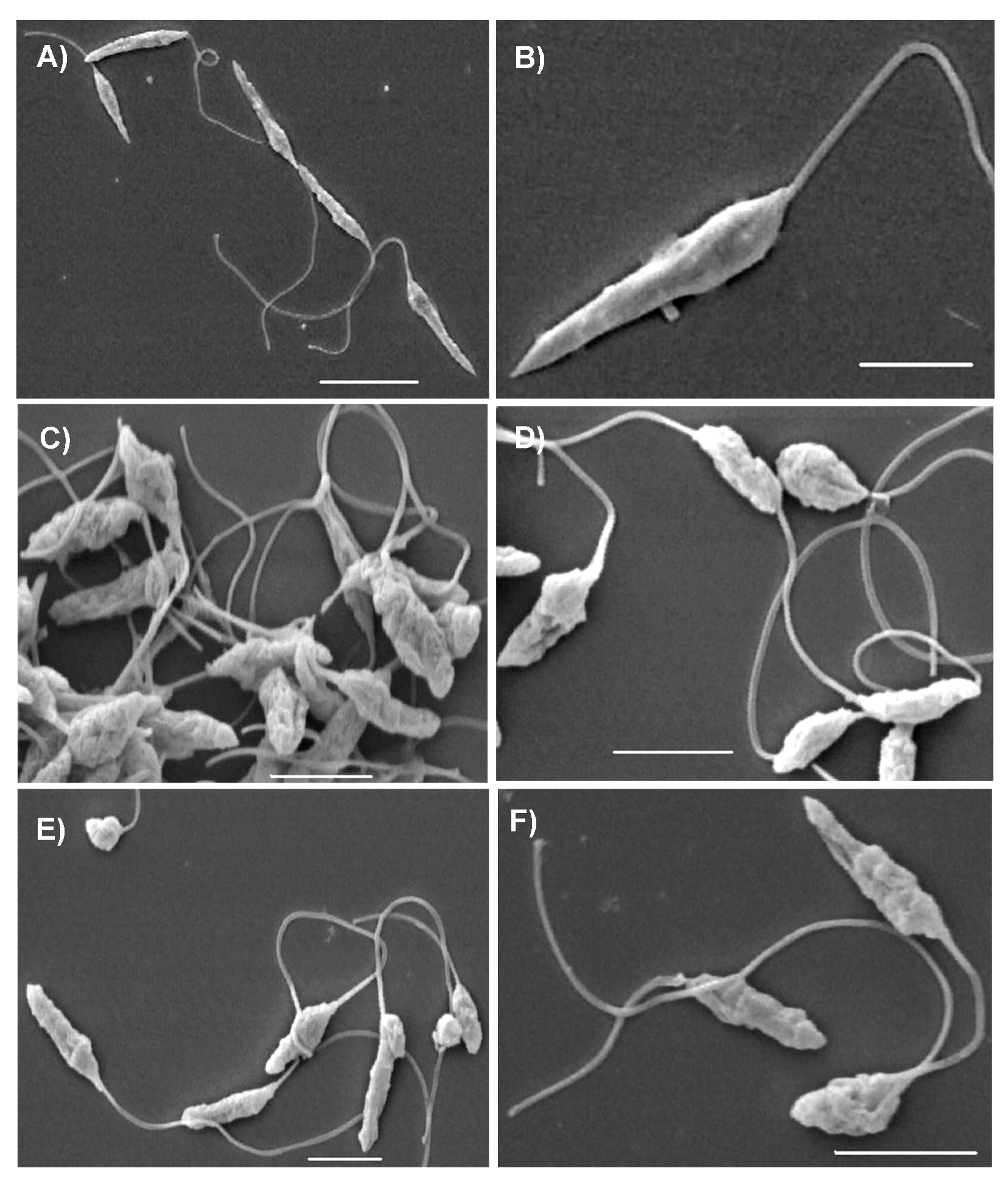
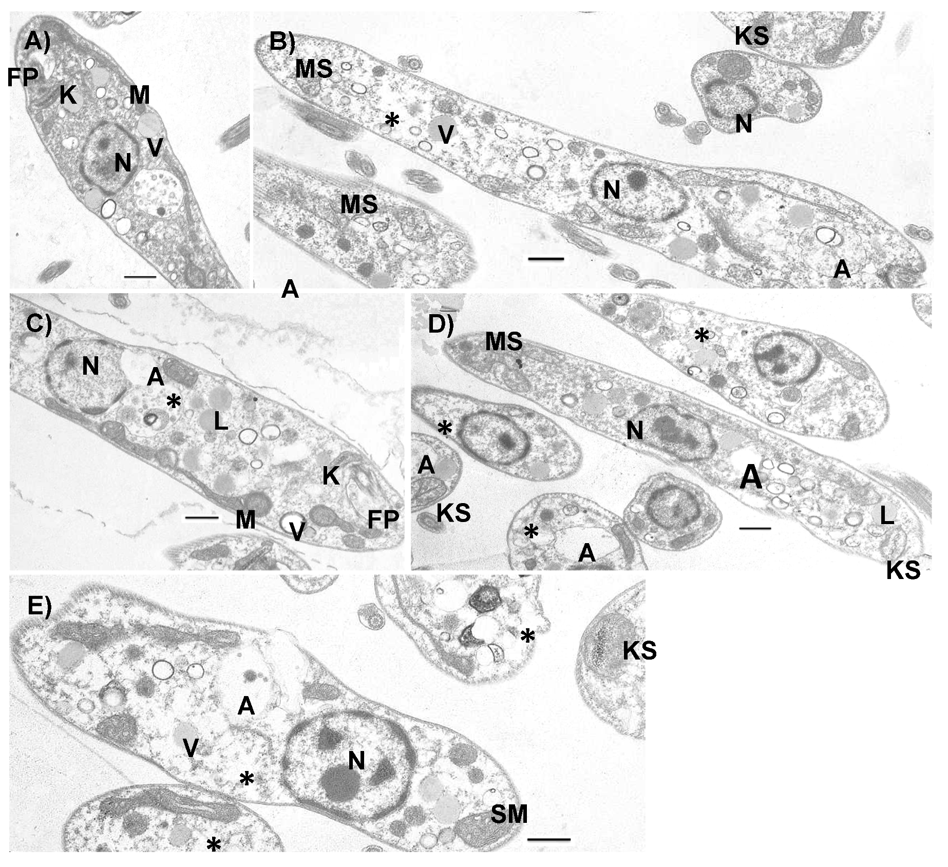
3.3. Ultrastructural Effects

SEM (Figure 1) and TEM (Figure 2) imaging were used to assess the occurrence of ultrastructural changes in L. infantum promastigotes after 7 h incubation in presence or absence of L. luisieri EO.
Regarding SEM findings, untreated cells (control) displayed elongated body shape and anterior flagella (Figure 1A,B), while EO-treated ones (Figure 1C–F) displayed round (Figure 1D,E) and aberrant forms (Figure 1C), with cell body septation (Figure 1 D). Note the uneven bubble-forming surface of all treated parasites. On the other hand, considering TEM imaging, control parasites evidenced normal nucleus, kinetoplast, mitochondria, and flagellar pocket (Figure 2A). Cytoplasmic organelles disorganization (Figure 2B,C,F), in addition, an increase in cytoplasmic clearing (Figure 2B,F) was the most prominent ultrastructural effect observed in L. luisieri-treated cells. A marked raise was also found in a number of autophagosomal structures, featured by intense cytoplasmic vacuolization (Figure 2F). EO-treated parasites also evidenced cell body (Figure 2F) and mitochondria (Figure 3B,D,F) swelling. Indeed, single and highly branched mitochondria swelling led to inner mitochondrial membrane disorganization, as shown by the presence of complex invaginations and concentric membranous structures formation (Figure 2B,D,F), and, lastly, mitochondria clearing (Figure 2B,D). Another common alteration was kinetoplast swelling and clearing (Figure 3B,D,E).

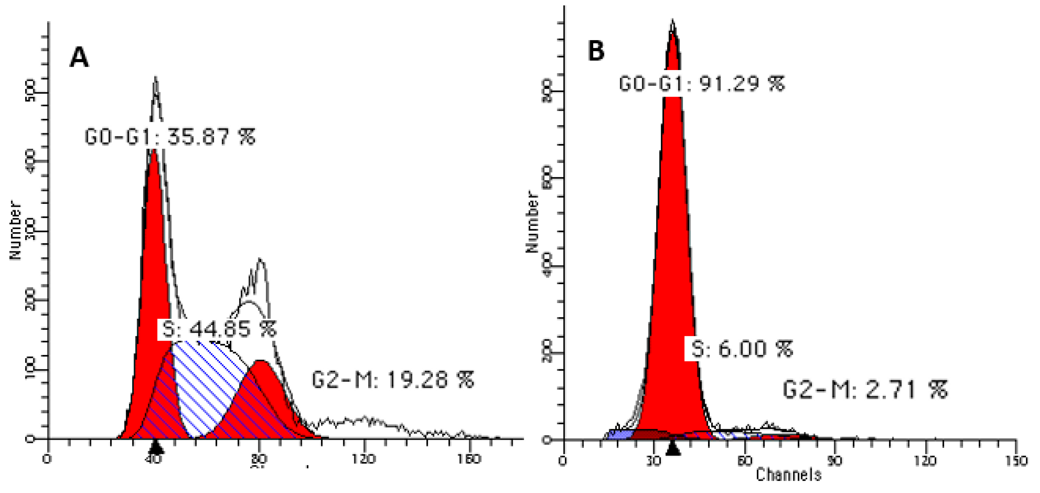
3.4. Cell-cycle Arrest at G(0)/G(1) Phase

Figure 3 shows cell DNA distribution through parasite cell cycle, in absence and presence of EO. After 24 h incubation, almost all treated cells were on cell cycle G0/G1 phase (91%) arrest, in opposite to what occurs in non-treated cells (36%).

3.5. Phosphatidylserine Externalization

As shown in Table 3, in non-treated cells, the annexin V lashing degree at 24 h was 3%. After L. luisieri EO-treatment, annexin V-positive cells percentage raised to 17.5%. PI-stained control cells percentage was 1.1%, while in presence of L. luisieri EO raised to 4%.

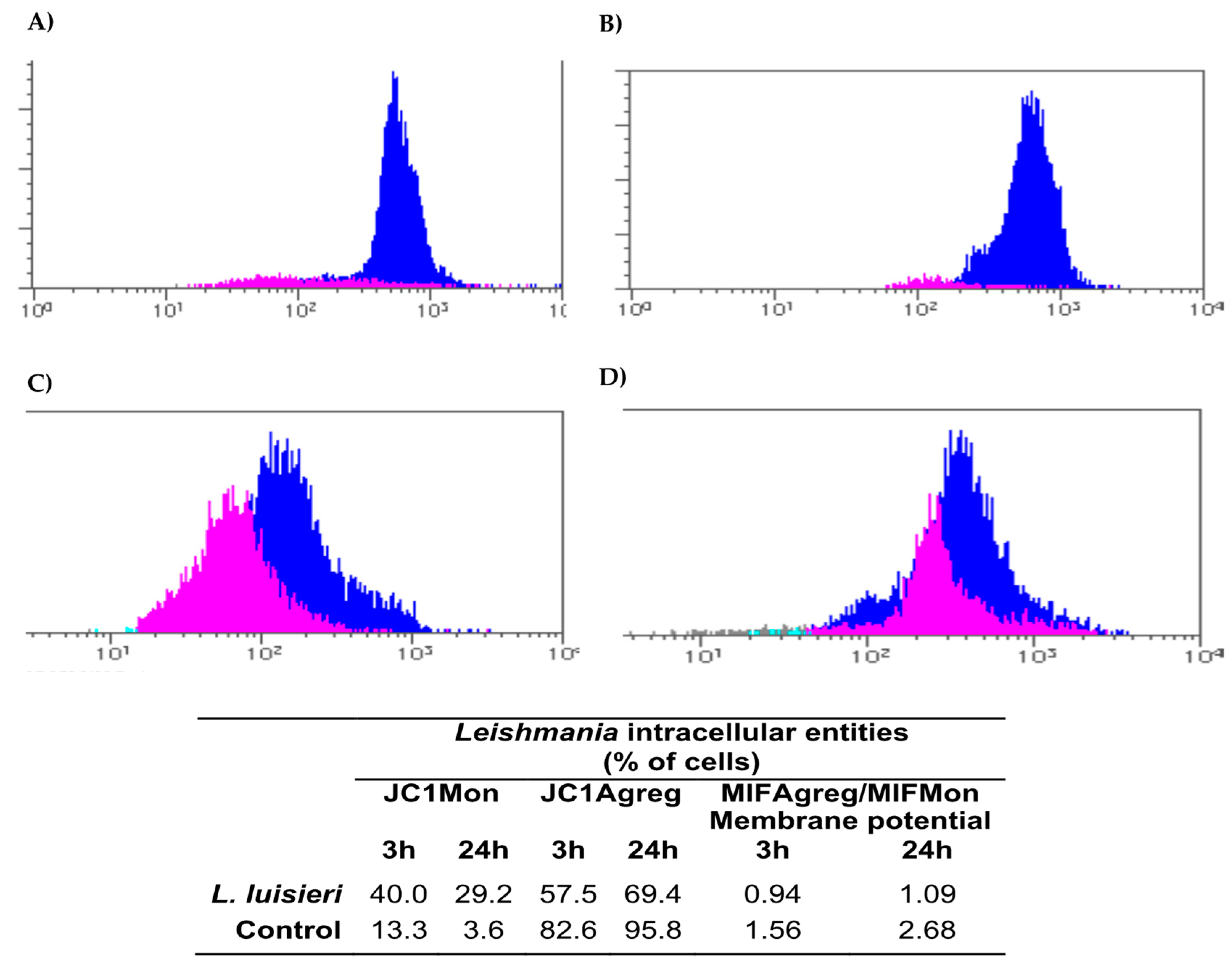
3.6. Mitochondrial Membrane Potential (MMP) Depolarization

MMP maintenance is crucial for cell survival, including for Leishmania species, as they have a single mitochondrion. L. luisieri EO induced a sustained decrease in ΔΨm (Figure 4). At first hour’s incubation (3 h), a significant difference was already observed between control and treated cells with low mitochondrial transmembrane potential, being, respectively, 13% and 40%. At 24 h L. luisieri incubation, 29.2% of cells displayed low ΔΨm when compared to control (3.6%).

3.7. Cathepsin D Activity Assay

The effect of L. luisieri essential oil on cathepsin D enzymatic activity was explored by applying a constant concentration (100 μg/mL) related to their IC50 value and evaluating changes over time (2.5, 4, and 7.5 h). Data obtained demonstrated any inhibition of cathepsin D enzymatic activity around 20% (data not shown).

3.8. Cytotoxicity Evaluation in Mammalian Cells

L. luisieri EO cytotoxic activity was assessed in macrophages cell lines using MTT test. Data obtained show that this EO did not induce macrophages toxicity at the tested dose (CC50 > 200 µg/mL; SI > 3.17).

4. Discussion

Currently, with the increasing rate of conventional therapy-resistant parasites, low drug efficacy, prominent adverse effects, and heavy costs, it is urgent to search for new anti-parasitic agents [15,16]. As previously highlighted, aromatic plant EO has low density and rapid diffusion across cell membranes, and this aspect may improve in targeting EO active components to endoparasites [17]. L. luisieri EO evidenced on its chemical composition oxygen containing monoterpenes (75.7%) in large amounts, with 1,8-cineole (18.9%) being the major compound and representing necrodane derivatives (36%). L. viridis presented α-pinene (9.2%), 1,8-cineole (29.7%), linalool (9.0%), and camphor (10.0%) as major compounds. The assessment of L. luisieri and L. viridis inhibitory concentration at 50% (IC50) showed promising anti-Leishmania activity, both against cutaneous and visceral strains, L. infantum, L. major, and L. tropica. A more prominent effect was observed in L. luisieri (IC5 = 31–63 μg/mL), while L. viridis showed a high IC50 value (263 μg/mL) as well as its compound, α-pinene (IC50 = 161 μg/mL). This fact might be related with the higher content on oxygen containing monoterpenes, and somehow, with necrodane derivatives. The other tested compounds did not reveal any activity. Therefore, it is feasible to suppose that the major compounds present in EO did not seem to be accountable for EO-stated effects.
L. luisieri EO revealed a promising potential, as it obtained data in accordance with that already described research by Simões [18]. In this study, L. luisieri EO also promoted phosphatidylserine exposure, MMP depolarization, and G0/G1 phase cell cycle arrest leading to cell death [18]. These features have been stated as playing a pivotal role in protists drug-induced death, such as Leishmania species [19]. Taking SEM and TEM imaging advantages, some of the above highlighted facts could be linked to L. luisieri EO-induced morphologic changes. Indeed, among treated cells, it was not possible to identify an intact kinetoplast (swelled), only an increment in cell and organelle volume, cytoplasm clearing, and disorganization was found, as already stated in other studies [20,21,22]. Further, low density and rapid diffusion across cell membranes can raise EO in targeting parasites organelles, as well as inducing membrane destabilization [23]. This also suggests the existence of autophagic process with the consequent formation of autophagosomes [24], which seems to be involved in abnormal membrane structures’ breakdown and recycling (supposing an intense intracellular organelles remodeling process, EO-irreversibly damaged). Similarly, other reports have already stated this change in drugs-treated kinetoplastids [25,26]. Mitochondrial changes were also stated, mainly on its matrix, arising complex structures, or otherwise, a less electrodense matrix, and mitochondrion swelling. Those alterations were also previously stated by Brenzan et al. [27] using L. brasiliensis, and by Rosa et al. [28] and Ueda-Nakamura et al. [29] using L. amazonensis. These data may be explained by the ability of inducing mitochondrial potential depolarization, which may promote apoptosis as corroborated by the anexin-V values reached [30]. Moreover, the occurrence of G0/G1 cycle phase cells arrest with the consequent reduction of cell numbers on S and G2/M phase, may be possibly explained by mitochondrial potential decrease that may trigger this event since it reduces energy available.
In addition, it is important to point out that cell toxicity risk due to EO ingestion or skin contact requires further investigation. In this sense, cytotoxic potential of the studied EO was assessed using macrophage cell lines, but no toxic effects were stated using L. luisieri EO. These findings are even more important as Leishmania amastigotes forms are intracellular. Therefore, L. luisieri essential oil might be effective against parasites without affecting host cell, as described before.

5. Conclusions

Overall, the present findings allow us to support the popularity of this plant as an upcoming approach to treat skin infections and, at the same time, the strengths they use on Leishmania infections, particularly in cutaneous forms, both in humans and animals. In any case, the upcoming use of EO for mammalian parasitic infections treatment remains unclear, although its potential for seeking new compounds is clear. Therefore, further studies are needed to expand our knowledge on this field, as well as to find the responsible phytochemicals by the observed effects.

Supplementary Materials

The following are available online at https://www.mdpi.com/2076-3417/9/15/3056/s1, Table S1. Composition of the essential oil of Lavandula viridis, Table S2. Composition of the essential oil of Lavandula luisieri (previously published by Videira, 2015).

Author Contributions

Conceptualization, M.M., C.C., M.C.S.; methodology, M.M., L.S., C.C., M.C.S.; formal analysis, M.M.; writing—original draft preparation, M.M.; writing—review and editing, N.M., M.C.S.; supervision, C.C., M.C.S.

Funding

This work was supported by the European Regional Development Fund (ERDF), through the Centro 2020 Regional Operational Programme under project CENTRO-01-0145-FEDER-000008:BrainHealth 2020, and through the COMPETE 2020—Operational Programme for Competitiveness and Internationalisation and Portuguese national funds via FCT—Fundação para a Ciência e a Tecnologia, I.P., under strategic project POCI-01-0145-FEDER-007440 (UID/NEU/04539/2013).

Acknowledgments

The authors are grateful to Jorge Paiva for helping in plant taxonomy; to José Correia da Costa from Centro de Imunologia e Biologia Parasitária, Instituto Nacional Ricardo Jorge, Porto for supplying L. infantum (zymodeme MON-1); to António Osuna, Departamento de Parasitología, Facultad de Ciencias, Instituto de Biotecnología, Universidad de Granada, for supplying L. major. FCT for Strategic project ref. UID/BIM/04293/2013, and “NORTE2020—Programa Operacional Regional do Norte” (NORTE-01-0145-FEDER-000012).

Conflicts of Interest

The authors declare no conflict of interest.

References

  1. Zuzarte, M.; Gonçalves, M.J.; Cavaleiro, C.; Dinis, A.M.; Canhoto, J.M.; Salgueiro, L.R. Chemical composition and antifungal activity of the essential oils of Lavandula viridis L’Hér. Chem. Biodivers 2009, 6, 1283–1292. [Google Scholar] [CrossRef] [PubMed]
  2. Zuzarte, M.; Gonçalves, M.J.; Cavaleiro, C.; Canhoto, J.; Vale-Silva, L.; Silva, M.J.; Pinto, E.; Salgueiro, L. Chemical composition and antifungal activity of the essential oils of Lavandula viridis L’Her. J. Med. Microbiol. 2011, 60, 612–618. [Google Scholar] [CrossRef]
  3. Zuzarte, M.; Vale-Silva, L.; Gonçalves, M.J.; Cavaleiro, C.; Vaz, S.; Canhoto, J.; Pinto, E.; Salgueiro, L. Antifungal activity of phenolic-rich Lavandula multifida L. Essential oil. Eur. J. Clin. Microbiol. Infect. Dis. 2012, 31, 1359–1366. [Google Scholar] [CrossRef] [PubMed]
  4. Zuzarte, M.; Gonçalves, M.; Cruz, M.T.; Cavaleiro, C.; Canhoto, J.; Vaz, S.; Pinto, E.; Salgueiro, L. Lavandula luisieri essential oil as a source of antifungal drugs. Food Chem. 2012, 135, 1505–1510. [Google Scholar] [CrossRef] [PubMed]
  5. Machado, M.; Sousa, M.D.C.; Salgueiro, L.; Cavaleiro, C. Effects of Essential Oils on the Growth of Giardia lamblia Trophozoites. Nat. Prod. Commun. 2010, 5, 137–141. [Google Scholar] [CrossRef] [PubMed]
  6. Machado, M.; Dinis, A.M.; Salgueiro, L.; Cavaleiro, C.; Custódio, J.B.A.; Sousa, M.D.C. Anti-Giardia activity of phenolic-rich essential oils: Effects of Thymbra capitata, Origanum virens, Thymus zygis subsp. sylvestris, and Lippia graveolens on trophozoites growth, viability, adherence, and ultrastructure. Parasitol. Res. 2010, 106, 1205–1215. [Google Scholar] [CrossRef] [PubMed]
  7. Machado, M.; Santoro, G.; Sousa, M.C.; Salgueiro, L.; Cavaleiro, C.; Sousa, M.D.C.R. Activity of essential oils on the growth ofLeishmania infantumpromastigotes. Flavour Fragr. J. 2010, 25, 156–160. [Google Scholar] [CrossRef]
  8. Machado, M.; Dinis, A.; Salgueiro, L.; Custódio, J.B.; Cavaleiro, C.; Sousa, M.; Sousa, M.D.C.R. Anti-Giardia activity of Syzygium aromaticum essential oil and eugenol: Effects on growth, viability, adherence and ultrastructure. Exp. Parasitol. 2011, 127, 732–739. [Google Scholar] [CrossRef]
  9. Machado, M.; Pires, P.; Dinis, A.M.; Santos-Rosa, M.; Alves, V.; Salgueiro, L.; Cavaleiro, C.; Sousa, M.C. Monoterpenic aldehydes as potential anti-Leishmania agents: Activity of Cymbopogon citratus and citral on L. infantum, L. tropica and L. major. Exp. Parasitol. 2012, 130, 223–231. [Google Scholar] [CrossRef]
  10. Machado, M.; Dinis, A.; Santos-Rosa, M.; Alves, V.; Salgueiro, L.; Cavaleiro, C.; Sousa, M.; Sousa, M.D.C.R. Activity of Thymus capitellatus volatile extract, 1,8-cineole and borneol against Leishmania species. Veter Parasitol. 2014, 200, 39–49. [Google Scholar] [CrossRef]
  11. Wiley, R. NIST 05 MS Spectra, 8th ed.; Revision 2005 D.06.00. s.l.; NIST Mass Spectral Library: Gaithersburg, MD, USA, 2007. [Google Scholar]
  12. Joulain, D.; Konig, W.A. The atlas of spectral data of sesquiterpene hydrocarbons. J. Nat. Prod. 1998, 62, 1212–1213. [Google Scholar] [CrossRef]
  13. Adams, R.P. Identification of Essential oil Components by Gas Chromatography/Mass Spectrometry; Allured Publishing Corporation: Carol Stream, IL, USA, 2007; pp. 1–804. [Google Scholar]
  14. Monzote, L.; Montalvo, A.M.; Scull, R.; Miranda, M.; Abreu, J. Combined effect of the essential oil from Chenopodium ambrosioides and antileishmanial drugs on promastigotes of Leishmania amazonensis. Rev. Inst. Med. Trop. São Paulo 2007, 49, 257–260. [Google Scholar] [CrossRef] [PubMed] [Green Version]
  15. Natera, S.; Machuca, C.; Padrón-Nieves, M.; Romero, A.; Díaz, E.; Ponte-Sucre, A. Leishmania spp.: Proficiency of drug-resistant parasites. Int. J. Antimicrob. Agents 2007, 29, 637–642. [Google Scholar] [CrossRef] [PubMed]
  16. Croft, S.L.; Sundar, S.; Fairlamb, A.H. Drug resistance in Leishmaniasis. Clin. Microbiol. Rev. 2006, 19, 111–126. [Google Scholar] [CrossRef] [PubMed]
  17. Bakkali, F.; Averbeck, S.; Averbeck, D.; Idaomar, M. Biological effects of essential oils–A review. Food Chem. Toxicol. 2008, 46, 446–475. [Google Scholar] [CrossRef] [PubMed]
  18. Simões, M.; Bennett, R.N.; Rosa, E.A.S. Understanding antimicrobial activities of phytochemicals against multidrug resistant bacteria and biofilms. Nat. Prod. Rep. 2009, 26, 746. [Google Scholar] [CrossRef] [PubMed]
  19. Sen, N.; Das, B.B.; Ganguly, A.; Mukherjee, T.; Tripathi, G.; Bandyopadhyay, S.; Rakshit, S.; Sen, T.; Majumder, H.K. Camptothecin induced mitochondrial dysfunction leading to programmed cell death in unicellular hemoflagellate Leishmania donovani. Cell Death Differ. 2004, 11, 924–936. [Google Scholar] [CrossRef]
  20. Pedroso, R.B.; Ueda-Nakamura, T.; Filho, B.P.D.; Cortez, D.A.G.; Cortez, L.E.R.; Morgado-Diaz, J.A.; Nakamura, C.V. Biological activities of essential oil obtained from Cymbopogon citratus on Crithidia deanei. Acta Protozool. 2006, 45, 231–240. [Google Scholar]
  21. Santoro, G.F.; Cardoso, M.G.; Guimarães, L.G.L.; Freire, J.M.; Soares, M.J.; Soares, M. Anti-proliferative effect of the essential oil of Cymbopogon citratus (DC) Stapf (lemongrass) on intracellular amastigotes, bloodstream trypomastigotes and culture epimastigotes of Trypanosoma cruzi (Protozoa: Kinetoplastida). Parasitology 2007, 134, 1649–1656. [Google Scholar] [CrossRef]
  22. Oliveira, V.C.S.; Moura, D.M.S.; Lopes, J.A.D.; De Andrade, P.P.; Da Silva, N.H.; Figueiredo, R.C.B.Q. Effects of essential oils from Cymbopogon citratus (DC) Stapf., Lippia sidoides Cham., and Ocimum gratissimum L. on growth and ultrastructure of Leishmania chagasi promastigotes. Parasitol. Res. 2009, 104, 1053–1059. [Google Scholar] [CrossRef]
  23. Lipinski, C.A.; Lombardo, F.; Dominy, B.W.; Feeney, P.J. Experimental and computational approaches to estimate solubility and permeability in drug discovery and development settings. Adv. Drug Deliv. Rev. 1997, 23, 3–25. [Google Scholar] [CrossRef]
  24. Rodrigues, J.C.F.; Attias, M.; Rodriguez, C.; Urbina, J.A.; De Souza, W.; Souza, W.D. Ultrastructural and Biochemical Alterations Induced by 22,26-Azasterol, a Δ24(25)-Sterol Methyltransferase Inhibitor, on Promastigote and Amastigote Forms of Leishmania amazonensis. Antimicrob. Agents Chemother. 2002, 46, 487–499. [Google Scholar] [CrossRef] [PubMed]
  25. Lazardi, K.; Urbina, J.A.; De Souza, W. Ultrastructural alterations induced by two ergosterol biosynthesis inhibitors, ketoconazole and terbinafine, on epimastigotes and amastigotes of Trypanosoma (Schizotrypanum) cruzi. Antimicrob. Agents Chemother. 1990, 34, 2097–2105. [Google Scholar] [CrossRef] [PubMed] [Green Version]
  26. Lorente, S.O.; Rodrigues, J.C.F.; Jimenez, C.J.; Joyce-Menekse, M.; Rodrigues, C.; Croft, S.L.; Yardley, V.; De Luca-Fradley, K.; Ruiz-Pérez, L.M.; Urbina, J.; et al. Novel Azasterols as Potential Agents for Treatment of Leishmaniasis and Trypanosomiasis. Antimicrob. Agents Chemother. 2004, 48, 2937–2950. [Google Scholar] [CrossRef] [PubMed]
  27. Brenzan, M.A.; Nakamura, C.V.; Filho, B.P.D.; Ueda-Nakamura, T.; Young, M.C.M.; Cortez, D.A.G.; Young, M.C.M. Antileishmanial activity of crude extract and coumarin from Calophyllum brasiliense leaves against Leishmania amazonensis. Parasitol. Res. 2007, 101, 715–722. [Google Scholar] [CrossRef]
  28. Rosa, M.D.S.S.; Bizzo, H.R.; Rodrigues, I.D.A.; Soares, R.M.A.; Souto-Padrón, T.; Alviano, C.S.; Lopes, A.H.C.S.; Mendonça-Filho, R.R. Antileishmanial Activity of a Linalool-Rich Essential Oil from Croton cajucara. Antimicrob. Agents Chemother. 2003, 47, 1895–1901. [Google Scholar] [CrossRef]
  29. Ueda-Nakamura, T.; Mendonça-Filho, R.R.; Morgado-Díaz, J.A.; Maza, P.K.; Filho, B.P.D.; Cortez, D.A.G.; Alviano, D.S.; Rosa, M.D.S.S.; Lopes, A.H.C.; Alviano, C.S.; et al. Antileishmanial activity of Eugenol-rich essential oil from Ocimum gratissimum. Parasitol. Int. 2006, 55, 99–105. [Google Scholar] [CrossRef]
  30. Arnoult, D.; Akarid, K.; Grodet, A.; Petit, P.X.; Estaquier, J.; Ameisen, J.C. On the evolution of programmed cell death: Apoptosis of the unicellular eukaryote Leishmania major involves cysteine proteinase activation and mitochondrion permeabilization. Cell Death Differ. 2002, 9, 65–81. [Google Scholar] [CrossRef]
Figure 1. Scanning electron micrographs of Leishmania infantum promastigotes exposed to Lavandula luisieri essential oil. (A,B) Untreated cells showing the typical elongated shape, parasite body and anterior flagella; (CF), Treated promastigotes. Note round forms and aberrant forms (CF) with cell body septation (D). Note the irregular surface. (AF) Bars = 5 µm.
Applsci 09 03056 g001
Figure 2. Transmission electron micrographs of Leishmania infantum promastigotes exposed to Lavandula luisieri essential oil. (A) Control parasites; (BE) parasites treated with essential oil. Note mitochondrial swelling (MS) (B,D,E) and kinetoplast swelling (B,D,E), gross alterations in the organization of cytoplasm (*) (BE). N, nucleus; K, kinetoplast; F, flagellum; FP, flagellar pocket; A, autophagic vesicles; V, cytoplasm vesicles. Bars, 2 µm.
Applsci 09 03056 g002
Figure 3. Cell cycle histograms of Leishmania infantum promastigotes exposed to Lavandula luisieri essential oil. L. infantum promastigotas were incubated at 26 °C for 24 h in the absence (A) or presence (B) of L. luisieri essential oil at IC50 concentrations. Propidium iodide staining was performed and samples were analyzed by flow cytometry.
Applsci 09 03056 g003
Figure 4. Representative dot plots showing JC-1 staining of Leishmania infantum promastigotes exposed to Lavandula luisieri essential oil. L. infantum promastigotas were incubated at 26 °C for 24 h in the absence ((A) 3 h; (B) 24 h) or presence of L. luisieri essential oil ((C) 3 h; (D) 24 h) at IC50 concentrations. JC-1 staining was performed and samples were analyzed by flow cytometry. J-aggregates (blue) reflect functioning mitochondria and monomers are indicative of compromised mitochondria (pink).
Applsci 09 03056 g004
Table 1. Major composition of Lavandula luisieri and Lavandula viridis essential oils.
CompoundsLavandula luisieri (%)Lavandula viridis (%)
Monoterpene hydrocarbons4.717.3
Oxygen containing monoterpenes75.758.3
Sesquiterpene hydrocarbons2.418.6
Oxygen containing sesquiterpenes3.0-
Others1.21.9
Total identified(49) 86.9(38) 96.1
Major compounds (>2.0%)α-Pinene (2.3)
1,8-cineole (18.9)
Linalool (3.1)
Necrodane derivatives (36.0)
Lavandulyl acetate (7.2)
α-Pinene (9.2)
Camphene (2.7)
1,8-cineole (29.7)
Linalool (9.0)
Camphor (10.0)
Borneol (2.7)
Z-α-bisabolene (6.3)
Selina-3,7(11)-diene (6.6)
Table 2. Lavandula luisieri and Lavandula viridis essential oils activity and major compounds on Leishmania promastigotes viability.
L. infantum
IC50 μg/mL (CI) *
L. tropica
IC50 μg/mL (CI) *
L. major
IC50 μg/mL (CI) *
L. luisieri63 (52–77)38 (33–45)31 (25–38)
L. viridis263 (248–279)>400>400
α-pinene161 (149–175)>400>400
1,8-cineole>400>400>400
Borneol>400>400>400
Linalool>400>400>400
Cultures of log-phase promastigotes (106) were incubated at 26 °C for 24 h (L. infantum, L. tropica) or 48 h (L. major), in function of essential oil concentration; * CI, Confidence Intervals; n.d., not determined.
Table 3. Flow cytometry analysis of Leishmania infantum promastigotes treated with Lavandula luisieri essential oil, showing the percentage of propidium iodide (PI) and annexin-V positive cells.
Leishmania Promastigotes (% of Cells) 24 h
AnexinePIAnexine/PI
L. luisieri17.5410
Control3.31.14.8

