Article

Biopurification of Oligosaccharides by Immobilized Kluyveromyces Lactis

1
Department of Biological Sciences, Korea Advanced Institute of Science and Technology, 291 Daehak-Ro, Yuseong-Gu, Daejeon 34141, Korea
2
Department of Food and Nutrition, Chungnam National University, 99 Daehak-Ro, Yuseong-Gu, Daejeon 34134, Korea
3
Graduate School of Analytical Science and Technology, Chungnam National University, 99 Daehak-Ro, Yuseong-Gu, Daejeon 34134, Korea
*
Author to whom correspondence should be addressed.
These authors contributed equally to this work.
Appl. Sci. 2019, 9(14), 2845; https://doi.org/10.3390/app9142845
Submission received: 24 June 2019 / Revised: 14 July 2019 / Accepted: 15 July 2019 / Published: 17 July 2019
(This article belongs to the Special Issue Poly- and Oligosaccharides from Biomass)

Abstract

:
Oligosaccharides with diverse and complex structures such as milk oligosaccharides have physiological functions including modulating intestinal microbiota or stimulating immune cell responses. However, milk carbohydrates include about 40–50% of lactose which requires a cost-effective method to separate. We developed a new method to purify the oligosaccharides from carbohydrate mixtures such as human milk oligosaccharides (HMOs) and galactooligosaccharides (GOSs) by exploiting immobilized Kluyveromyces lactis as microbial catalysts. Evaluation of media components exhibited no significant differences in the lactose removal efficiency when nutrient-rich media, minimal salt media, and distilled water without any media components were used. With the immobilization on alginate beads, the lactose removal efficiency was increased 3.4 fold compared to that of suspension culture. When the immobilized cells were reused to design a continuous process, 4 h of pre-activation enhanced the lactose eliminating performance 2.5 fold. Finally, immobilized K. lactis was used as microbial catalysts for the biopurification of HMOs and GOSs, and lactose was effectively removed without altering the overall distribution of oligosaccharides.

1. Introduction

Human milk oligosaccharides (HMOs) are active carbohydrates observed in human milk. With more than a hundred different structural isomers, HMOs play critical roles in maintaining and improving the intestinal microbiota of breast-fed infants [1]. Human milk uniquely contains such complex oligosaccharides in high concentrations up to 20 g/L. Bovine or porcine milk has oligosaccharides as well but only less than 1 g/L [2]. Since HMOs are hardly hydrolyzed in the human digestive system [3,4], they can reach the small and large intestines while retaining their intact structures. In the human gut, HMOs serve as prebiotics for promoting beneficial bacteria and soluble decoy receptors for preventing pathogen attachment to mucosal surfaces. In addition to this, they serve as critical factors in modulating the responses of epithelial and immune cells [5].
For the commercial use of HMOs, bovine milk oligosaccharides (BMOs) can be considered as an alternative to HMOs [6,7]. Since whey is one of the largest byproducts of dairy products, massive amounts of BMOs can be produced from it [8,9]. However, about 50 g/L of lactose has to be eliminated from bovine milk for practical application of BMOs. Hence, pilot-scale filtration or chromatographic manners have been studied for lactose removal [10,11,12,13,14]. A biological method using β-galactosidase (EC 3.2.1.23) was also studied [15].
2′-Fucosyllactose (2′-FL) is one of the simplest but most abundant oligosaccharides in human milk. It is currently produced at an industrial scale by the recombinant yeast or bacterial system [16,17,18,19] via two distinct pathways, the salvage pathway and de novo pathway. GDP-L-fucose is synthesized through the engineered metabolic pathway and then transferred to a lactose backbone, which results in the production of 2′-FL. In this process, purification of 2′-FL from the mixture of lactose and 2′-FL is crucial for maximizing the prebiotic activity of 2′-FL.
Galactooligosaccharides (GOSs) are also non-digestible oligosaccharides used as prebiotics [20]. They have been used for the health-promoting components of infant milk formulas to mimic the beneficial effects of HMOs [21]. Advantageous functions like anticarcinogenic effects and lowering serum cholesterol levels have been reported, as well [22,23]. In the enzymatic synthesis of GOSs, however, 50% of the lactose remains in its unreacted form [24,25]. This residual lactose makes it difficult to apply to people with lactose intolerance and decreases the valuable effects of GOSs [26].
Kluyveromyces lactis, the Crabtree-negative yeast, is widely used in the food industry because it is considered as “generally recognized as safe” (GRAS) [27]. It is extensively studied for the host system to produce recombinant proteins. The most successful utilization of K. lactis is as the production host for β-galactosidase. Additionally, K. lactis can assimilate lactose efficiently because it harbors the LAC4 (β-galactosidase) and LAC12 (lactose permease) genes.
In this work, we developed a biopurification system of oligosaccharides using immobilized K. lactis. From the mixture of lactose and oligosaccharides, K. lactis selectively consumed the lactose with high efficiency. To reduce the burden in the process, lactose removal in a nutrient-rich, minimal salt media or distilled water was examined. The immobilized cells with high density were applied to maximize the consumption of lactose. Then, biopurification of HMOs and GOSs was examined to evaluate the selective removal of lactose without altering the distribution of oligosaccharides.

2. Materials and Methods

2.1. Culture Conditions for the Growth of Kluyveromyces lactis

The YPD media (10 g/L of yeast extract, 20 g/L of peptone, 20 g/L of dextrose or other carbon sources in accordance with the purpose of experiments) and YNB media (6.7 g/L of yeast nitrogen base with or without amino acids, 20 g/L or 40 g/L of carbon sources) were used for evaluating substrate consumption and pre-activation of immobilized K. lactis. Cells were cultured at 30 °C with shaking at 200 rpm. The cell growth was determined spectrophotometrically at 600 nm.

2.2. Immobilization of K. lactis with Sodium Alginate

The K. lactis cells grown in 400 mL of YPD media for 12 h were used for immobilization. The media was centrifuged at 4500 rpm for 15 min at 4 °C. The pallets were washed three times by being resuspended with PBS and centrifuged at 4500 rpm for 15 min at 4 °C. The washed cells were resuspended again in 50 ml of distilled water and mixed well with the same volume of 2.5% sodium alginate beads. The mixtures were added drop-wise into 0.5 M CaCl2 and incubated 5–10 min to harden in the calcium chloride solution. The immobilized cells were kept at 4 °C in 0.5 M CaCl2 solution for further experiments. Before use, the immobilized cells were washed with distilled water.

2.3. The Extraction of Semi-Purified Human Milk Oligosaccharides (HMOs) from Human Milk

The HMOs were extracted from human milk as described by Gnoth et al. [28], with modifications. For lipids removal, the human milk was centrifuged at 5000 × g for 30 min at 4 °C. The middle layer was collected from the sample and four volumes of an extracting solvent (chloroform:methanol = 2:1) were added to the collected sample. The solution was centrifuged again at 5000 × g for 30 min at 4 °C. Three volumes of cold (−30 °C) ethanol were added to the supernatants, and the solution was placed overnight at 4 °C to precipitate proteins. The mixture was then centrifuged at 5000 × g for 30 min at 4 °C, and the precipitated protein was discarded. Semi-purified HMOs were acquired by drying the supernatants using centrifugal evaporation (SpeedVac EZ-2, Genevac Ltd., Ipswitch, England).

2.4. Analysis of Mono-, Di-, and Oligosaccharides

The concentrations of mono- and di-saccharides were quantified by high-performance liquid chromatography with a refractive index detector (Agilent Technologies, Santa Clara, CA, USA). The samples were separated by a Rezex ROA-Organic Acid H+ column (Phenomex, Torrance, CA, USA) with 5 mM H2SO4 aqueous solution at a flow rate of 0.6 ml/min.
HMOs and GOSs were readily purified and enriched by solid-phase extraction (SPE) using a porous graphitized carbon cartridge (PGC). Firstly, the samples were loaded onto the SPE cartridges and washed with pure water to remove any salt and buffer residues. The oligosaccharides bound to the cartridges were eluted through the sequential addition of 20% acetonitrile (v/v) (neutral fraction) followed by 40% acetonitrile/0.05% trifluoroacetic acid (v/v) (acidic fraction). The samples were dried under a vacuum prior to mass spectrometry (MS) analysis.
Enriched HMOs and GOSs were chromatographically separated and detected with an Agilent 6550 UHPLC/Q-TOF MS (Santa Clara, CA, USA). 10 fold diluted samples were injected into a Hypercarb column (Thermo Fisher Scientific, MA, USA). After injection, the oligosaccharides were separated with a gradient system consisting of 3.0% acetonitrile/0.1% formic acid (v/v) (A) and 90.0% acetonitrile/0.1% formic acid (v/v) (B) at a flow rate of 0.2 mL/min: 0.0–0.5 min, 97% A, 3% B; 0.5–20.0 min, 84% A, 16% B; 20.0–30.0 min, 60% A, 40% B; 30.0–32.0 min, 40% A, 60% B; 32.0–40.0 min, 10% A, 90% B; 40.0–55.0 min, 97% A, 3% B. The mass range of detection was m/z 300–2500. The data was collected in the positive mode. LC/MS results were exported using the Agilent’s MassHunter Qualitative Analysis software, and then HMOs and GOSs were identified by comparing with a compound library that includes retention time and accurate mass.

3. Results

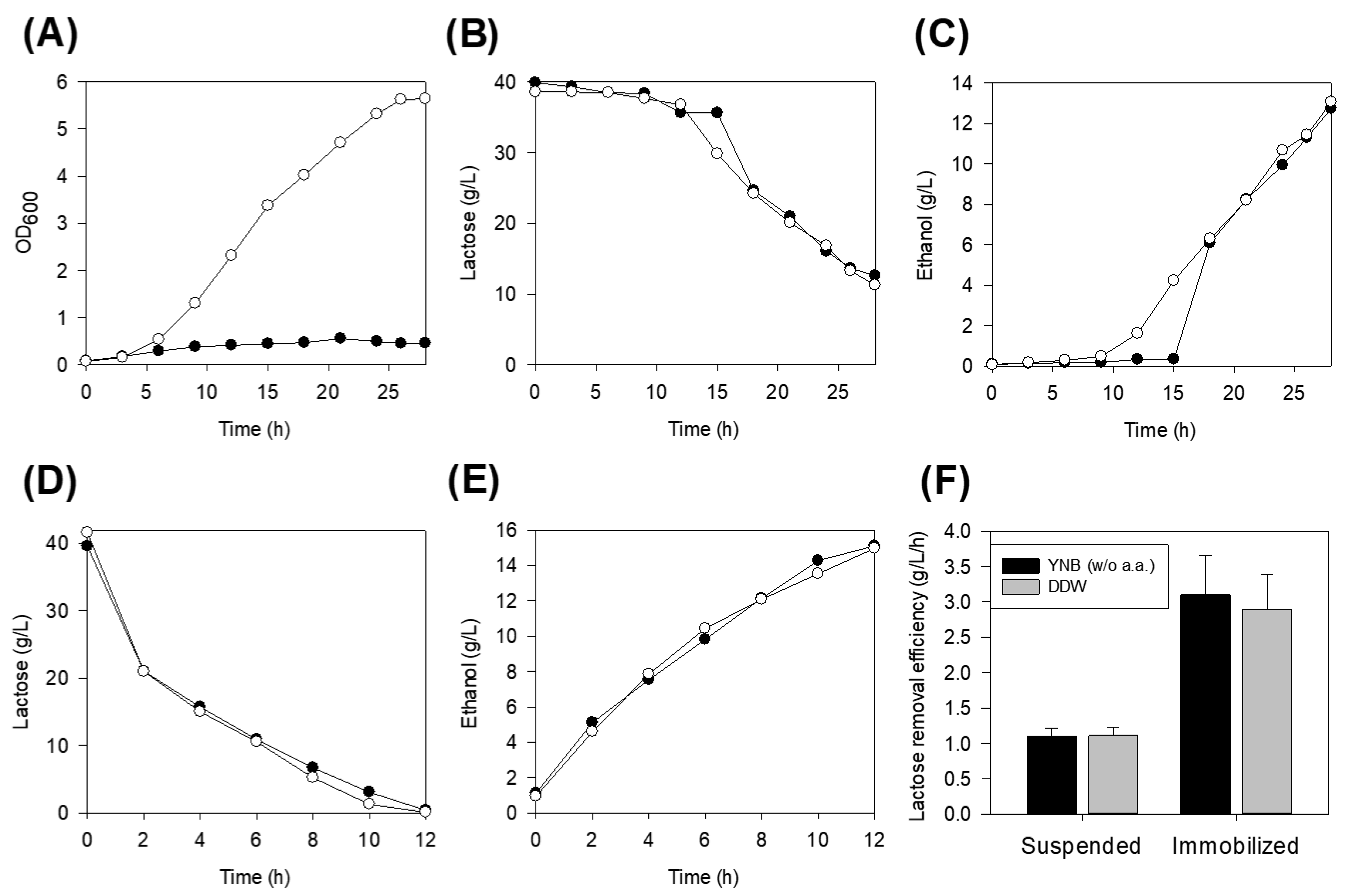
3.1. Sugar Utilization in the Nutrient-Rich and Defined Salt Media

To reduce burdens from the biological elimination of lactose, we tried to avoid or minimize the organic components, such as peptone, amino acids, or vitamins. Firstly, we examined the utilization of glucose, galactose, and lactose in the nutrient-rich (YPD) and defined salt media (YNB) by K. lactis.
As shown in Table 1, the highest optical density (OD600 = 7.9) and maximum specific growth rate (0.33 h−1) were achieved when glucose was used as a substrate for cell growth in YPD media. With the lactose as a substrate, the growth of K. lactis was maintained at a similar level as shown in glucose. Though maximum OD was lower (OD600 = 6.9) in YPD media with lactose, the maximum specific growth rate (0.31 h−1) and substrate consumption (20.0 g/L) were similar to those observed in glucose. In contrast, the substrate consumption and maximum specific growth rate were the lowest (11.0 g/L and 0.20 h−1, respectively) when cells were cultivated on galactose as a substrate.
In YNB media with the addition of amino acids, the maximum specific growth rate was around 0.31 h−1 when cells were utilizing glucose, lactose, or even galactose as a substrate. However, the maximum OD and substrate consumption, compared to YPD media, were decreased 26–38% and 5–38%. The lowest decrease in substrate consumption was lactose.
We further analyzed the lactose utilization by K. lactis in YNB without amino acids. Although the maximum specific growth rate and substrate consumption were decreased to 0.27 h−1 and 14 g/L, the maximum OD (OD600 = 5.0) and ethanol production (9.5 g/L) were similar in comparison with cells grown in YNB media with amino acids.
Considering the substrate consumption and ethanol production in the YPD and YNB media, we could conclude that K. lactis uses lactose efficiently as well as glucose. In addition, we could determine that the addition of amino acids to the YNB media had minimal effect on the viability and lactose consumption of K. lactis.

3.2. Lactose Removal on Defined Salt Media and Distilled Water

In order to simplify the purification media, the lactose removal activity was evaluated using the distilled water without the addition of any media components. The K. lactis could not grow well in distilled water compared to YNB without amino acids (Figure 1A). The lactose consumption and ethanol production, on the other hand, showed comparable patterns even though a lag period (15 h) existed when the cells were grown in distilled water (Figure 1B,C). From this, it is concluded that the distilled water instead of YNB media could be used for lactose removal media once the low cell growth was overcome.

3.3. Effect of Immobilization

In our preliminary data, the lactose consumption rate was proportional to the initial cell density (data not shown). Moreover, the low cell growth needed to be solved when distilled water was used as a simplified media. To approach these problems, we immobilized high concentrations of K. lactis so that lactose was rapidly consumed in the media. After grown in YPD media for 12 h, the cells at the exponential phase were immobilized with sodium alginate beads.
The YNB media without amino acids and distilled water without any media components were evaluated for the lactose elimination. After 12 h of fermentation using immobilized cells, 40 g/L of lactose was completely removed from both media (Figure 1D,E). Between distilled water and the YNB media without amino acids, we could not find any differences in the lactose removal (3.33 g/L/h) and ethanol production rates (1.25 g/L/h). As we immobilized K. lactis, whose density was already high, we could not find any lag period during lactose elimination in distilled water. More importantly, the efficiency of lactose elimination by immobilized K. lactis was 3.4 times higher than that of cell suspension (Figure 1F).

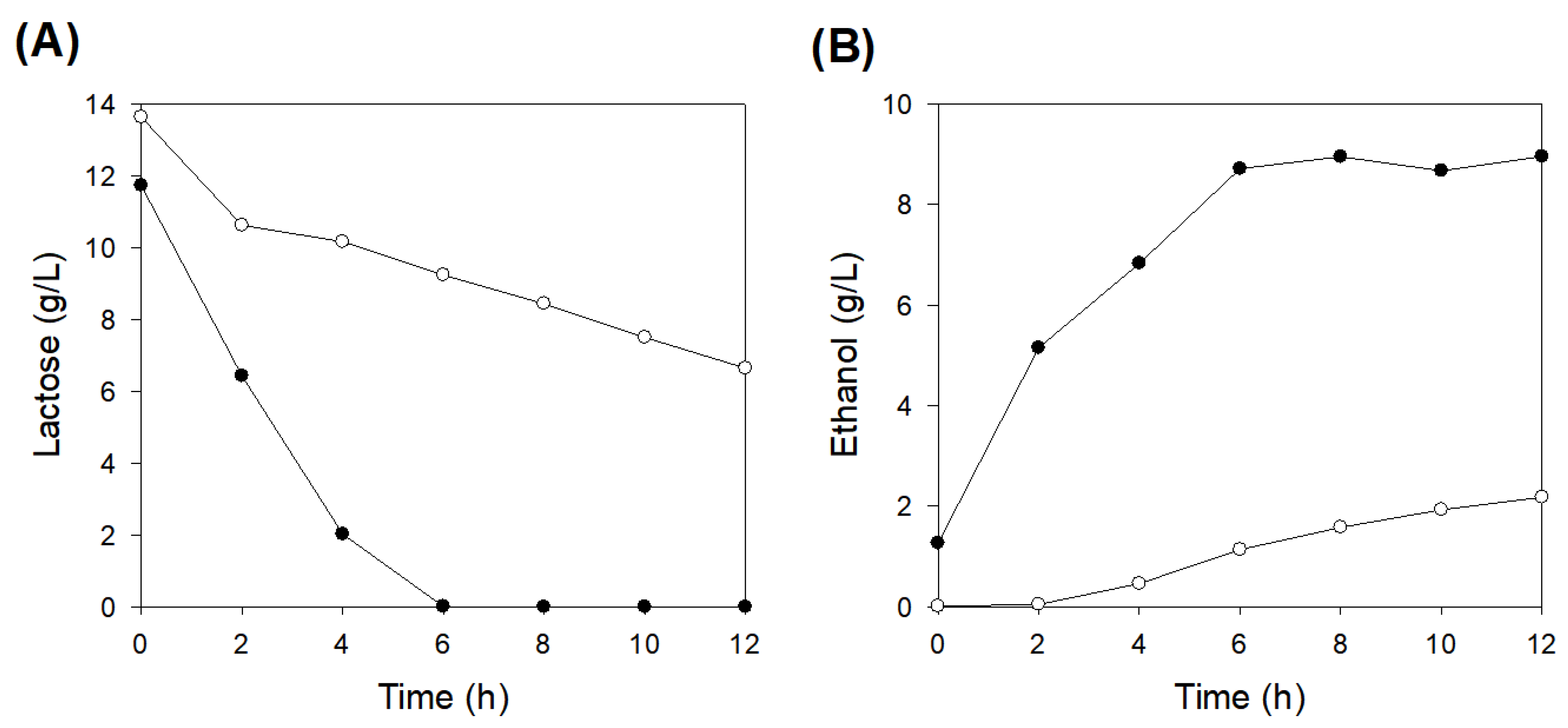
3.4. Impact of Pre-Activation of Immobilized Cells

With the immobilization of concentrated K. lactis, lactose could be removed rapidly in water without any lag period. To design a continuous process for further utilization, the reusability of immobilized cells was evaluated. After the first-round of biopurification, immobilized cells were stored at 4 °C for 48 h then reused for the next round of biopurification. One group of cells was used after pre-activation in YNB media with 20 g/L of lactose for 4 h, and another group was used without any pre-activation.
The immobilized cells with or without pre-activation were monitored to observe the lactose consumption and ethanol production (Figure 2). Repeated use of immobilized cells without pre-activation showed remarkable decreases in lactose consumption and ethanol production. Compared to the first-round process, 48 h of storage decreased the lactose removal activity 5.6 fold. Meanwhile, after the pre-activation of immobilized cells by 4 h of incubation restored the activity 2.5-fold higher than that of without pre-activation. It is concluded that at least 4 h of pre-activation is necessary for the recovery of lactose consumption efficiency for the repeated use of immobilized K. lactis.

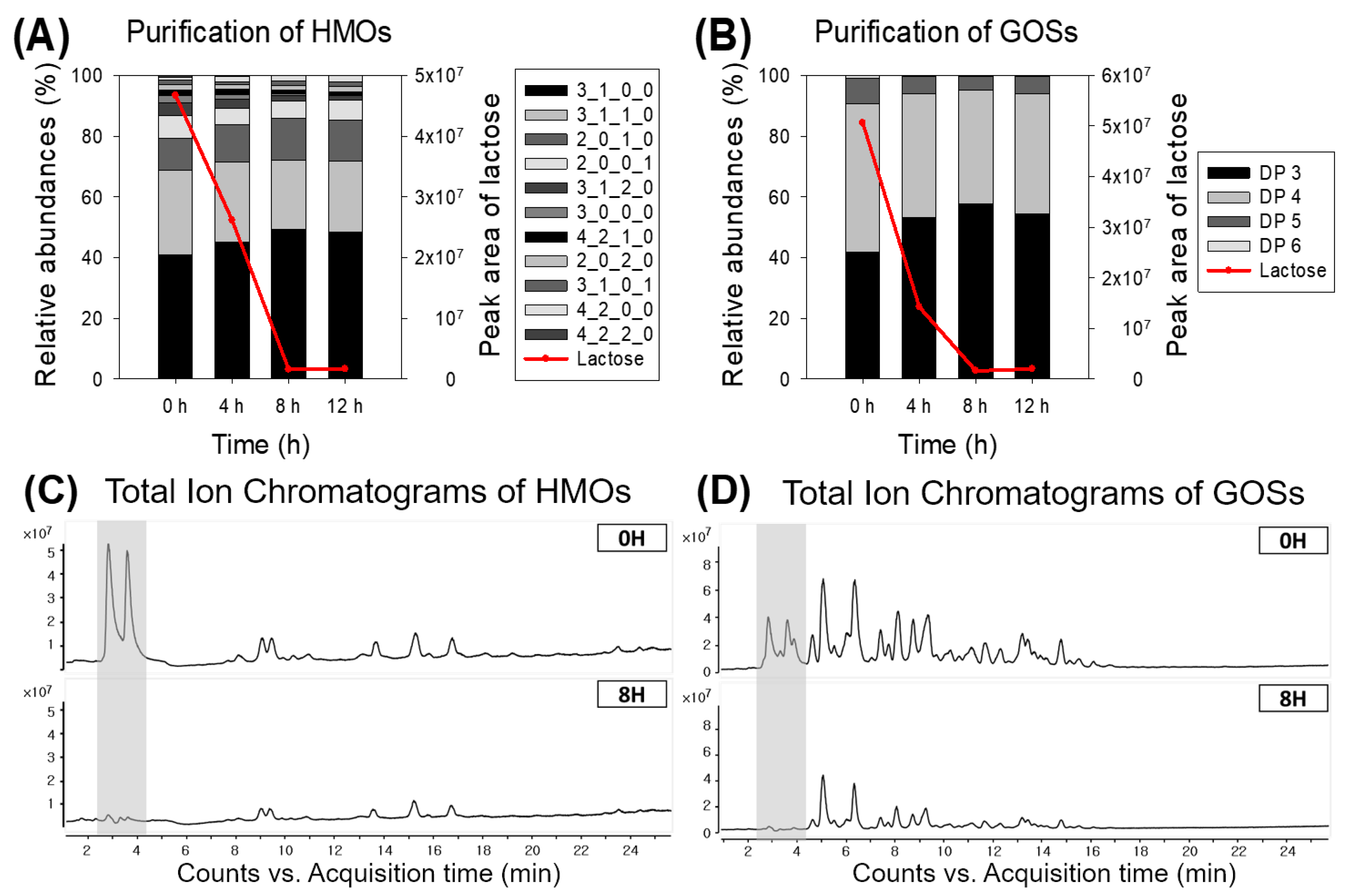
3.5. Lactose Removal of Human Milk Oligosaccharides (HMOs) and Galactooligosaccharides (GOSs)

Lastly, we evaluated the selective consumption of immobilized K. lactis from the semi-purified HMOs and GOSs. HMOs and GOSs were the oligosaccharides that exhibited biological functions such as prebiotic or bifidogenic activity. However, those beneficial effects can be maximized in the absence of lactose, which can be utilized by most bacteria. Therefore, it is crucial to eliminate the residual lactose from the mixture of oligosaccharides to guarantee their functionalities.
Semi-purified HMOs were prepared from human milk by the removal of lipids and proteins. Semi-purified HMOs and GOSs were dissolved in water to a final concentration of 20 g/L, and freshly-prepared immobilized K. lactis was added directly without the addition of any media components. Lactose concentrations in the semi-purified HMOs and GOSs were 12 g/L and 2.5 g/L, respectively.
LC/MS analysis was performed to observe the quantitative changes of the oligosaccharides. By considering retention time and accurate mass, biologically possible compositions of hexose (Hex), N-acetylhexosamine (HexNAc), fucose (Fuc), and N-acetylneuraminic acid (NeuAc) were determined. A total of 11 major HMOs and GOSs with a degree of polymerization (DP) of 3 to 6 including lactose were monitored during the biopurification (Figure 3).
Within 8 h, lactose in the solution of semi-purified HMOs or GOSs was rapidly consumed to a negligible amount. The overall distribution of HMOs was not changed during the lactose removal process. In particular, fucosyllactose (2_0_1_0, Hex2Fuc1) and sialyllactose (2_0_0_1, Hex2NeuAc1), which are the simplest structures of HMOs, were preserved during the process. In the biopurification of GOSs, the relative concentrations of DP 4 and DP 5 decreased slightly while DP 3 increased. After the elimination of lactose, however, the overall distribution of oligosaccharides was not changed, which indicated that the GOSs, except for lactose, were not consumed by immobilized K. lactis.

4. Discussion

K. lactis is well known for its distinguished activity for metabolizing lactose as a sole carbon source. In this report, we further analyzed its lactose-metabolizing activity and utilized it to remove lactose from the oligosaccharide mixtures, HMOs and GOSs.
We have shown that K. lactis could assimilate lactose as efficiently as glucose and maintain cells’ viability even without the addition of a nitrogen source or other media components such as peptone, yeast extract, or salts, which enable the biopurification to be simple. With the immobilization, the lactose removal efficiency was increased 3.4 fold compared to that of suspension culture. For the repeated use of immobilized cells, at least 4 h of pre-activation was necessary for the recovery of lactose consumption efficiency. Finally, lactose was effectively removed from HMOs or GOSs without altering the overall distribution of oligosaccharides by utilizing immobilized cells. As a result, we developed a biopurification system for oligosaccharide mixtures using immobilized K. lactis as biocatalysts.
Lactose removal from oligosaccharides mixtures like HMOs and GOSs is a critical step for maximizing the efficacy of oligosaccharides. Hence, pilot-scale filtration or chromatographic processes have been studied for their industrial application [10,11,12]. Small-scale separation methods were also studied for the identification of oligosaccharides [13,14]. However, these methods are expensive and require complicated procedures to operate. Biological removal with β-galactosidase [15] is an alternative to these, but the enzymatic degradation results in glucose and galactose that could reduce the benefits. By utilizing our novel approach, it is possible to readily purify oligosaccharides mixtures from lactose.

Author Contributions

Data Curation, I.-S.Y., Y.-J.Y., N.S., H.J.A., and J.-H.K.; Investigation, Y.-J.Y. and N.S.; Writing – Review & Editing, I.-S.Y., H.J.A., and J.-H.K.

Funding

This research was supported by the research grant from Chungnam National University.

Conflicts of Interest

The authors declare no conflict of interest.

References

  1. Smilowitz, J.T.; Lebrilla, C.B.; Mills, D.A.; German, J.B.; Freeman, S.L. Breast milk oligosaccharides: Structure-function relationships in the neonate. Annu. Rev. Nutr. 2014, 34, 143–169. [Google Scholar] [CrossRef] [PubMed]
  2. Fong, B.; Ma, K.; McJarrow, P. Quantification of bovine milk oligosaccharides using liquid chromatography-selected reaction monitoring-mass spectrometry. J. Agric. Food Chem. 2011, 59, 9788–9795. [Google Scholar] [CrossRef] [PubMed]
  3. Engfer, M.B.; Stahl, B.; Finke, B.; Sawatzki, G.; Daniel, H. Human milk oligosaccharides are resistant to enzymatic hydrolysis in the upper gastrointestinal tract. Am. J. Clin. Nutr. 2000, 71, 1589–1596. [Google Scholar] [CrossRef] [PubMed] [Green Version]
  4. Gnoth, M.J.; Kunz, C.; Kinne-Saffran, E.; Rudloff, S. Human milk oligosaccharides are minimally digested in vitro. J. Nutr. 2000, 130, 3014–3020. [Google Scholar] [CrossRef] [PubMed]
  5. Bode, L. Human milk oligosaccharides: Every baby needs a sugar mama. Glycobiology 2012, 22, 1147–1162. [Google Scholar] [CrossRef] [PubMed] [Green Version]
  6. Zivkovic, A.M.; Barile, D. Bovine milk as a source of functional oligosaccharides for improving human health. Adv. Nutr. 2011, 2, 284–289. [Google Scholar] [CrossRef] [PubMed]
  7. Mehra, R.; Barile, D.; Marotta, M.; Lebrilla, C.B.; Chu, C.; German, J.B. Novel high-molecular weight fucosylated milk oligosaccharides identified in dairy streams. PLoS ONE 2014, 9, e96040. [Google Scholar] [CrossRef] [PubMed]
  8. Smilowitz, J.T.; Lemay, D.G.; Kalanetra, K.M.; Chin, E.L.; Zivkovic, A.M.; Breck, M.A.; German, J.B.; Mills, D.A.; Slupsky, C.; Barile, D. Tolerability and safety of the intake of bovine milk oligosaccharides extracted from cheese whey in healthy human adults. J. Nutr. Sci. 2017, 6, e6. [Google Scholar] [CrossRef]
  9. Barile, D.; Meyrand, M.; Lebrilla, C.B.; German, J.B. Examining bioactive components of milk Sources of complex oligosaccharides (Part 2). Agro. Food Ind. Hi Tech. 2011, 22, 37–39. [Google Scholar]
  10. Morr, C.V.; Brandon, S.C. Membrane fractionation processes for removing 90% to 95% of the lactose and sodium from skim milk and for preparing lactose and sodium-reduced skim milk. J. Food Sci. 2008, 73, C639–C647. [Google Scholar] [CrossRef]
  11. Geisser, A.; Hendrich, T.; Boehm, G.; Stahl, B. Separation of lactose from human milk oligosaccharides with simulated moving bed chromatography. J. Chromatogr. A 2005, 1092, 17–23. [Google Scholar] [CrossRef] [PubMed]
  12. Vente, J.A.; Bosch, H.; de Haan, A.B.; Bussmann, P.J.T. Comparison of sorption isotherms of mono- and disaccharides relevant to oligosaccharide separations for Na, K, and Ca loaded cation exchange resins. Chem. Eng. Commun. 2005, 192, 23–33. [Google Scholar] [CrossRef]
  13. Strum, J.S.; Aldredge, D.; Barile, D.; Lebrilla, C.B. Coupling flash liquid chromatography with mass spectrometry for enrichment and isolation of milk oligosaccharides for functional studies. Anal. Biochem. 2012, 424, 87–96. [Google Scholar] [CrossRef] [Green Version]
  14. Robinson, R.C.; Colet, E.; Tian, T.; Poulsen, N.A.; Barile, D. An improved method for the purification of milk oligosaccharides by graphitised carbon-solid phase extraction. Int. Dairy J. 2018, 80, 62–68. [Google Scholar] [CrossRef] [PubMed] [Green Version]
  15. Santibanez, L.; Fernandez-Arrojo, L.; Guerrero, C.; Plou, F.J.; Illanes, A. Removal of lactose in crude galacto-oligosaccharides by beta-galactosidase from Kluyveromyces lactis. J. Mol. Catal. B-Enzym. 2016, 133, 85–91. [Google Scholar] [CrossRef]
  16. Yu, S.; Liu, J.J.; Yun, E.J.; Kwak, S.; Kim, K.H.; Jin, Y.S. Production of a human milk oligosaccharide 2’-fucosyllactose by metabolically engineered Saccharomyces cerevisiae. Microb. Cell. Fact. 2018, 17, 101. [Google Scholar] [CrossRef]
  17. Hollands, K.; Baron, C.M.; Gibson, K.J.; Kelly, K.J.; Krasley, E.A.; Laffend, L.A.; Lauchli, R.M.; Maggio-Hall, L.A.; Nelson, M.J.; Prasad, J.C.; et al. Engineering two species of yeast as cell factories for 2’-fucosyllactose. Metab. Eng. 2019, 52, 232–242. [Google Scholar] [CrossRef]
  18. Chin, Y.W.; Seo, N.; Kim, J.H.; Seo, J.H. Metabolic engineering of Escherichia coli to produce 2’-fucosyllactose via salvage pathway of guanosine 5’-diphosphate (GDP)-l-fucose. Biotechnol. Bioeng. 2016, 113, 2443–2452. [Google Scholar] [CrossRef]
  19. Chin, Y.W.; Kim, J.Y.; Kim, J.H.; Jung, S.M.; Seo, J.H. Improved production of 2’-fucosyllactose in engineered Escherichia coli by expressing putative alpha-1,2-fucosyltransferase, WcfB from Bacteroides fragilis. J. Biotechnol. 2017, 257, 192–198. [Google Scholar] [CrossRef]
  20. Rycroft, C.E.; Jones, M.R.; Gibson, G.R.; Rastall, R.A. A comparative in vitro evaluation of the fermentation properties of prebiotic oligosaccharides. J. Appl. Microbiol. 2001, 91, 878–887. [Google Scholar] [CrossRef] [Green Version]
  21. Lamsal, B.P. Production, health aspects and potential food uses of dairy prebiotic galactooligosaccharides. J. Sci. Food Agric. 2012, 92, 2020–2028. [Google Scholar] [CrossRef] [PubMed]
  22. Depeint, F.; Tzortzis, G.; Vulevic, J.; I’Anson, K.; Gibson, G.R. Prebiotic evaluation of a novel galactooligosaccharide mixture produced by the enzymatic activity of Bifidobacterium bifidum NCIMB 41171, in healthy humans: A randomized, double-blind, crossover, placebo-controlled intervention study. Am. J. Clin. Nutr. 2008, 87, 785–791. [Google Scholar] [CrossRef] [PubMed]
  23. Moreno, F.J.; Montilla, A.; Villamiel, M.; Corzo, N.; Olano, A. Analysis, structural characterization, and bioactivity of oligosaccharides derived from lactose. Electrophoresis 2014, 35, 1519–1534. [Google Scholar] [CrossRef] [Green Version]
  24. Urrutia, P.; Rodriguez-Colinas, B.; Fernandez-Arrojo, L.; Ballesteros, A.O.; Wilson, L.; Illanes, A.; Plou, F.J. Detailed analysis of galactooligosaccharides synthesis with beta-galactosidase from Aspergillus oryzae. J. Agric. Food Chem. 2013, 61, 1081–1087. [Google Scholar] [CrossRef] [PubMed]
  25. Crittenden, R.G.; Playne, M.J. Purification of food-grade oligosaccharides using immobilised cells of Zymomonas mobilis. Appl. Microbiol. Biotechnol. 2002, 58, 297–302. [Google Scholar] [CrossRef] [PubMed]
  26. Cheng, C.C.; Yu, M.C.; Cheng, T.C.; Sheu, D.C.; Duan, K.J.; Tai, W.L. Production of high-content galacto-oligosaccharide by enzyme catalysis and fermentation with Kluyveromyces marxianus. Biotechnol. Lett. 2006, 28, 793–797. [Google Scholar] [CrossRef] [PubMed]
  27. Spohner, S.C.; Schaum, V.; Quitmann, H.; Czermak, P. Kluyveromyces lactis: An emerging tool in biotechnology. J. Biotechnol. 2016, 222, 104–116. [Google Scholar] [CrossRef]
  28. Gnoth, M.J.; Rudloff, S.; Kunz, C.; Kinne, R.K. Investigations of the in vitro transport of human milk oligosaccharides by a Caco-2 monolayer using a novel high performance liquid chromatography-mass spectrometry technique. J. Biol. Chem. 2001, 276, 34363–34370. [Google Scholar] [CrossRef]
Figure 1. Lactose assimilation activity by Kluyveromyces lactis in YNB media without amino acid (open circle) and distilled water (filled circle). Cell growth, lactose, and ethanol production in suspension culture are shown in (A), (B), and (C). Lactose consumption and ethanol production by immobilized cells are shown in (D) and (E), respectively. The efficiency of biopurification is presented in (F).
Applsci 09 02845 g001
Figure 2. The impact of pre-activation of immobilized K. lactis. Lactose consumption (A) and ethanol production (B) differences of immobilized K. lactis cells with or without pre-activation. Filled circle, immobilized cells with pre-activation; open circle, immobilized cells without pre-activation.
Applsci 09 02845 g002
Figure 3. The change of the peak area of lactose and the relative abundances of oligosaccharides during the biopurification of (A) human milk oligosaccharides (HMOs) and (B) galactooligosaccharides (GOSs) by immobilized K. lactis. Moreover, the representative total ion chromatograms of (C) HMOs and (D) GOSs are shown. Ions in the shaded area were confirmed to be lactose. Due to the characteristics of the porous graphitized carbon cartridge (PGC) column, oligosaccharides with aldehyde form were separated into two peaks which indicated the α– and β– forms.
Applsci 09 02845 g003
Table 1. Growth characteristics of Kluyveromyces lactis in different media.
MediaSubstrateMaximum OD600 (AU)μmax (h−1)Substrate Consumption 1 (g/L)Ethanol Production 1 (g/L)
YPDGlucose7.90.3320.0 g/L10.0 g/L
Galactose5.70.2011.0 g/L4.2 g/L
Lactose6.90.3120.0 g/L12.0 g/L
YNB
(with amino acids)
Glucose5.40.3117.0 g/L7.3 g/L
Galactose3.50.296.8 g/L3.5 g/L
Lactose5.10.3119.0 g/L9.3 g/L
YNB (without amino acids)Lactose5.00.2714.0 g/L9.5 g/L
1 Substrate consumption and ethanol production were determined at 15 h fermentation.

