Article

Duration of the Symptoms and Brain Aging in Women with Fibromyalgia: A Cross-Sectional Study

by
Santos Villafaina
1,
Daniel Collado-Mateo
1,2,
Juan P. Fuentes-García
3,*,
Francisco J. Domínguez-Muñoz
1 and
Narcís Gusi
1,4
1
Physical Activity and Quality of Life Research Group (AFYCAV), Faculty of Sport Science, University of Extremadura, 10003 Cáceres, Spain
2
Facultad de Educación, Universidad Autónoma de Chile, Talca 1670, Chile
3
Faculty of Sport Science, University of Extremadura, 10003 Cáceres, Spain
4
CIBER de Fragilidad y Envejecimiento Saludable, 28029 Madrid, Spain
*
Author to whom correspondence should be addressed.
Appl. Sci. 2019, 9(10), 2106; https://doi.org/10.3390/app9102106
Submission received: 25 April 2019 / Revised: 13 May 2019 / Accepted: 20 May 2019 / Published: 23 May 2019

Abstract

:
Fibromyalgia is a chronic syndrome that is characterized by widespread pain and an altered brain dynamic. The aim of this study was to analyze the effect of the duration of the symptoms on the cortical activity of women with fibromyalgia using electroencephalogram power spectrum analyses in an eye-closed resting state. Twenty-nine women participated in this cross-sectional study (N: 29; age: 55.59 [9.50]). Theta, alpha, beta-1, beta-2, and beta-3 frequency bands were analyzed using EEGLAB. Theta power significantly correlated with the duration of the symptoms, but not with age. In addition, participants were divided into two groups according to number the years for which they were suffering from fibromyalgia. Participants who had a longer duration of symptoms obtained higher theta power in the frontal (Fp1, F4, F7, F8, and Fz), central (C3, C4, and Cz), and parietal (P3 and Pz) areas than those who had a shorter duration of symptoms, which may be related to brain aging. This exploratory study demonstrates for the first time that the frontal, central, and parietal areas may be influenced by the years in which they were suffering from the symptoms of fibromyalgia. This might indicate that the duration of these symptoms may have a higher impact on brain aging than the actual age of the patient.

1. Introduction

Fibromyalgia (FM) is a chronic syndrome characterized by widespread pain accompanied by several symptoms, such as fatigue, stiffness, sleep disturbance, and affective and cognitive problems [1]. These commonly cause a reduction in the ability to perform daily life activities [2], diminishing the quality of life of fibromyalgia patients [3].
Electroencephalogram (EEG) power has been commonly used to study brain dynamics, providing temporally resolved information [4,5]. In relation with that, previous neuroimaging and EEG studies have proposed that central nervous system hyperexcitability represents an important mechanism in the maintenance of the chronic pain observed in these patients [6,7]. In this regard, during pain processing, FM patients may experience abnormal activation in several areas related with the processing of pain, such as the thalamic nuclei, somatosensory cortex, anterior cingulate cortex, insular cortex, and prefrontal cortex [8,9,10]. This altered dynamic of the brain, even at rest, has been observed in fibromyalgia in comparison with pain-free controls [5,11,12].
Similar results have been obtained in neuropathic pain patients, observing an increment of power in theta and beta frequencies in pain-related areas (anterior cingulate, prefrontal, and somatosensory cortices) [4] using EEG in a resting state. Interestingly, the theta band (4–7 Hz) is considered of particular interest because of its relationship with higher cognitive functions and the induction of synaptic plasticity [13]. Previous studies have described increased theta activity and a decreased hippocampus volume in Alzheimer’s disease patients [14], which may also be related to brain aging in healthy people [15]. This relation was confirmed in previous correlation studies [16,17]. This decrease in hippocampus volume was also reported in chronic pain [18]. Therefore, these changes may be related to the learning and emotional deficits commonly observed in chronic pain patients [19].
Given the altered brain dynamics observed in women with fibromyalgia and the relevance of the theta band in brain aging processes, this study aims to evaluate, for the first time, the effect of age and the duration of the symptoms on the brain dynamics of women with fibromyalgia, by using EEG power spectrum analyses in an eye-closed resting state. The hypothesis for this aim is based on previous studies that have indicated the importance of symptoms’ duration in the prognosis of chronic pain patients, such as lower back pain patients [20]. Moreover, chronic back pain patients often have both altered brain activity and a decreased gray matter, which may be related to the duration of pain symptoms [21,22]. Thus, we hypothesized that both age and the duration of fibromyalgia symptoms would positively correlate with the EEG power spectrum in the theta band.

2. Methods

2.1. Participants

Twenty-nine women with fibromyalgia (mean age: 55.89 ± 9.50), who were diagnosed according to the American College of Rheumatology’s criteria [1], participated in the study.
Participants with neurological diseases, psychiatric diagnoses (i.e., major depression with suicidal ideation, schizophrenia, or substance abuse), and autoimmune or inflammatory diseases that cause pain were also excluded. However, two participants with diabetes, seven with hyperlipidemia, and seven with hypertension took part in the study.
Participants were informed verbally about the details of the study, and they gave written informed consent to participate in the study. All the procedures were approved by the research ethics committee of the University of Extremadura (approval number: 62/2017) and were carried out in accordance with the Declaration of Helsinki.

2.2. Questionnaires and Procedure

A semi-standardized interview was fulfilled by the participants, assessing when the fibromyalgia symptoms appeared and the medication intake. They also completed the Fibromyalgia Impact Questionnaire (FIQ) [23], which is a specific measure to assess the pain and disease impact in this population.

2.3. EEG Recording and Data Processing

The EEG signal was assessed during a one-minute resting period with eyes closed, using the Enobio device, which is a wireless electrode system (Neuroelectrics, Cambridge, MA, USA) [24]. The reliability of this instrument was demonstrated, even using dry electrodes [25]. The EEG was recorded from 19 scalp locations such as frontal (Fz, Fp1, Fp2, F3, F4, F7 and F8), central (Cz, C3, and C4), temporal (T3, T4, T5 and T6), parietal (Pz, P3, and P4), and occipital (O1 and O2), according to the International 10–20 system.
Electrodes placed in the mastoids served as references, and impedance was kept below 10 KΩ. EEG was recorded with a sampling rate of 500 Hz, using a 50-Hz notch filter, and bandpass filtering (1–40 Hz) was employed as well. EEGlab toolbox (MatLab) was utilized for pre-processing and data analysis. Rough artifacts were removed from EEG signals, and eye movement artifacts were corrected using independent component analysis (ICA) [26]. Participants were instructed to keep their eyes closed, and were seated on a chair in a calm room.
After pre-processing, using the power of the EEG, data was computed and banded into theta (4–7 Hz), alpha (8–12 Hz), beta-1 (13–18 Hz), beta-2 (19–21 Hz), and beta-3 (22–30) frequency bands. It was obtained using the function pop_spectopo.m from EEGlab.

2.4. Statistical Analysis

The SPSS statistical package (version 20.0; SPSS, Inc., Chicago, IL, USA) was used to analyze the data. Considering the results in the Shapiro–Wilk and Kolmogorov–Smirnov tests, non-parametric analyses were conducted.
Spearman Rho and Kendall’s Tau-b correlations were performed to examine the relationship between EEG frequency bands and both the duration of fibromyalgia symptoms and the age of the participants in the whole sample (N = 29). Given that significant correlations were only observed between the duration of symptoms and the theta band power spectrum, further analyses were performed to explore that relation. In this regard, we calculated the median of the years in which participants were suffering from fibromyalgia symptoms in order to divide the sample into two groups. The results indicated that participants had a median duration of 17 years of suffering from fibromyalgia symptoms, and this was the cutoff that was used to divide the sample into two groups: those with less than 17 years with fibromyalgia symptoms (shorter duration of symptoms group), and those with 17 or more years with fibromyalgia symptoms (longer duration of symptoms group). This strategy for splitting sub-groups of patients was previously performed for women with fibromyalgia using their pain symptom scores [27]. Neither chi-squared statistic nor the Mann–Whitney U-test revealed significant differences between groups in medication intake or age, respectively.
The Mann–Whitney U-test was conducted to examine differences between groups in the different frequency bands. The alpha-level of significance (set at 0.05) was adjusted by the Benjamini–Hochberg procedure in order to control the false discovery rate [28]. Significant differences were represented in a diagram chart where error bars represent the standard error of the mean.

3. Results

3.1. Sociodemographic Characteristics of the Whole Sample and Correlation between EEG Frequency Bands and Duration of Symptoms

Participants had a mean duration of symptoms of 20.69 (13.60) years and presented a FIQ score of 48.45 (15.21). Most of the participants had primary education (48.30%) or secondary education (41.40%) levels. Eleven participants (37.95%) were taking antidepressant medication and 10 (34.5%) were taking analgesic/relaxants medication. The complementary therapies that participants reported include massages (17.25%), or physiotherapy (27.6%) (see Table 1).
Spearman Rho and Kendall’s Tau-b revealed positive correlations between the years for which participants (N = 29) were suffering from fibromyalgia and the theta power in different scalp locations. These locations comprised the frontal (Fp1 and Fz), central (Cz), and parietal (P3 and Pz) areas (see Table 2). No other significant correlations were found between the duration of fibromyalgia symptoms and the remaining frequency bands (alpha, beta-1, beta-2, or beta-3) (see Table 2).
In addition, Spearman Rho and Kendall’s Tau-b did not report any significant correlation between EEG frequency bands and the age of the participants.

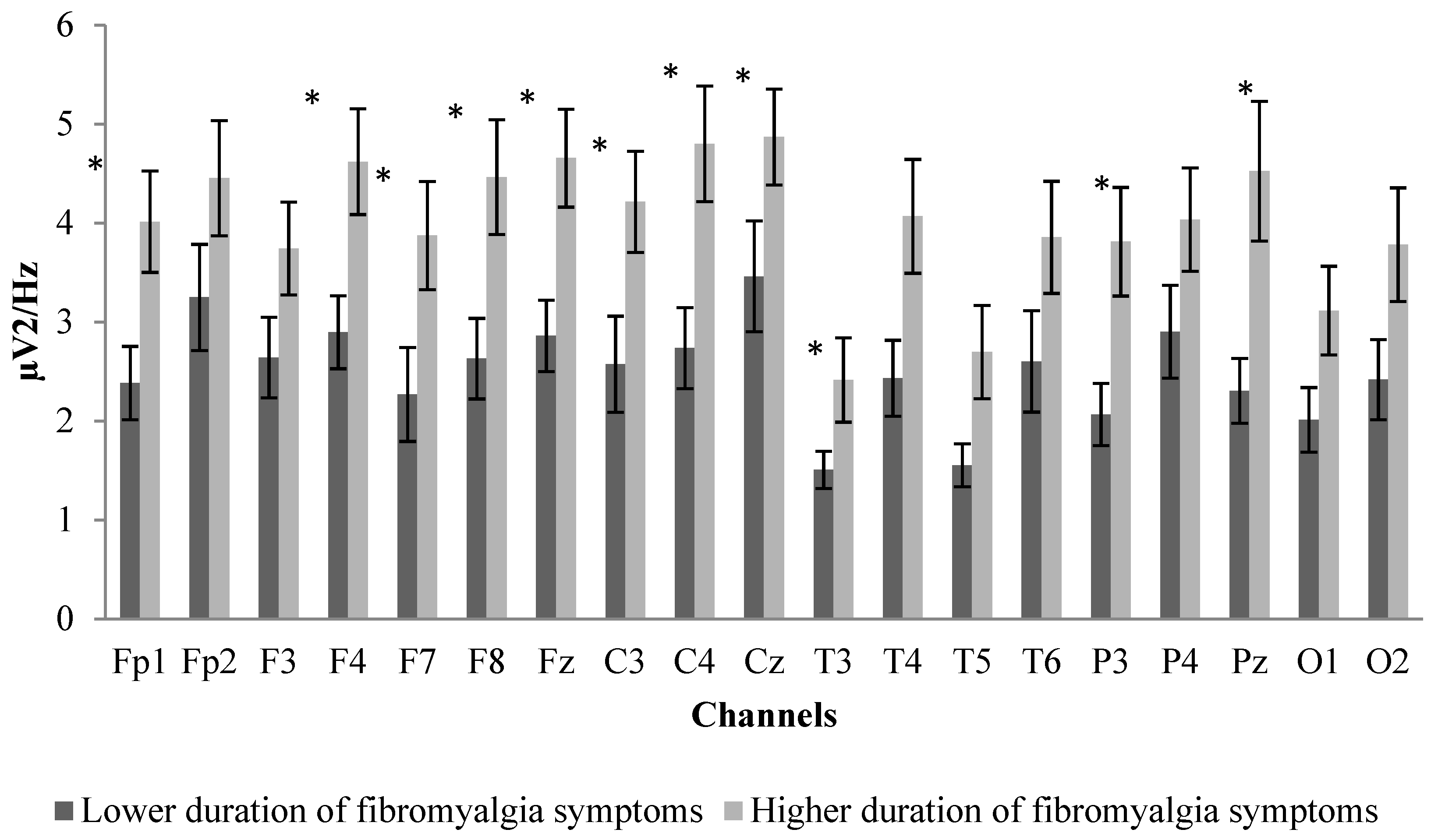
3.2. Between-Group Differences in Sociodemographic Characteristics, Age, Impact of Fibromyalgia, and EEG Power Spectrum

Participants were divided into two groups (longer and shorter duration of symptoms), taking into account the median duration of symptoms: 17 years. Table 3 summarizes the main sociodemographic characteristics of the two sub-groups. There were no differences in age, medication intake, or fibromyalgia impact, as assessed by FIQ, between the longer duration of symptoms group (N: 15; mean age: 52.07 ± 9.10) and the shorter duration of symptoms group (N: 14; mean age: 59.36 ± 8.69) (see Table 3) using Mann–Whitney U-test and Pearson’s chi-squared test.
The Mann–Whitney U-test was conducted to compare the group with less than 17 years with fibromyalgia symptoms (shorter duration of symptoms group) and the group with 17 or more years with fibromyalgia symptoms (longer duration of symptoms group) in the different EEG frequency bands (theta, alpha, beta-1, beta-2, and beta-3). Main significant differences were obtained in the theta power band. After applying the multiple comparison correction [28], the theta values in the frontal (Fp1, F4, Fz, F8, and F7), central (C3, Cz and C4), and parietal (Pz and P3) areas were significantly higher (p-value < 0.05) in the group that was suffering from fibromyalgia for more years (see Figure 1).
No other significant between-group differences were found in the rest of EEG frequency bands (see Figure 2 for the alpha power spectrum).

4. Discussion

The present study investigated the effect of age and symptom duration of women with fibromyalgia in EEG power spectrum frequency bands (theta, alpha, beta-1, beta-2, and beta-3). Analyzing the results, significant correlations showed that the impact of the years suffering from fibromyalgia symptoms on brain aging might be even higher than the impact of the actual age of the participants. This was also supported by significantly higher theta power being found in the group that was suffering from fibromyalgia for more years.
The long-term consequences of pain have traditionally been of great interest in neuroscience [29]. In this regard, this is the first study of fibromyalgia patients reporting an association between the duration of the symptoms and an altered brain dynamic. These findings support previous research focused on chronic back pain patients where altered brain activity and gray matter decreases correlated with the duration of the pain [21,22]. Thus, fibromyalgia symptoms might elicit similar long-term responses in the brain dynamic to those caused by pain alone, but further studies are needed to confirm this hypothesis.
Previous studies have shown altered brain dynamics between fibromyalgia and pain-free controls [5,11,12], and there is certain evidence about the seeming alteration of theta power in fibromyalgia patients [30]. In this sense, Gonzalez-Roldan, Cifre, Sitges, and Montoya [5] observed alterations in EEG coherence in fibromyalgia patients, showing greater EEG coherence over the left than over the right hemisphere in the centro-parietal area in a wide range of frequency bands (2 to 30 Hz). They discussed that these findings could suggest an over-activation of the pain network together with an imbalance of intra-hemispheric EEG coherence in fibromyalgia patients as compared with pain-free controls, even when no stimulation was applied. Furthermore, alterations in the theta band were also reported in the resting state in fronto-central brain regions, which are relevant in cognitive–attentional aspects of pain processing and pain inhibition [30,31]. In this line, the authors related these alterations to ongoing tonic pain and fatigue in fibromyalgia. However, according to our results, an altered theta power band may be not only associated with the ongoing pain and fatigue, but it could also be the result of years suffering from fibromyalgia symptoms.
Regarding alterations in theta power, previous studies have found them in Alzheimer’s disease [14,32], mild cognitive impairment [33,34], or even related with brain aging in healthy people [15]. The authors related these alterations with hippocampus atrophy, which is located in the frontal areas [17,33] and also in temporoparietal areas in Alzheimer’s disease patients [32], which is in line with the results from the current study. Furthermore, in fibromyalgia [35] and chronic pain [18,36], a significant reduction in important grey matter structures such as the hippocampus has been reported to be an indicator of hippocampus atrophy in fibromyalgia. In the current study, correlations between power spectrum and age were not significant in any of the channels. Therefore, the effect of age in the differences obtained in those channels is limited, whereas the duration of symptoms seems to be the key variable under those alterations. In that sense, another previous study showed that fibromyalgia symptoms could be partially explained by hippocampus symptoms [37]. Supporting that notion, microstructural changes in this brain area have been observed in patients with chronic pain [38] and also in fibromyalgia patients [39,40]. In addition, higher pain scores were correlated with increased fractional anisotropy values in the white matter of the right superior frontal gyrus [40].
Based on the studies mentioned above, our results might support the notion that suffering from fibromyalgia during a long period of time could lead to changes in the hippocampus. This could be an interesting topic for future studies in order to confirm this hypothesis with different design and techniques. In this regard, previous studies in chronic pain have reported how gray matter decreases with prolonged pain duration [41,42]. Also, with pain relief, reverse effects have been reported [43]. These results could indicate that gray matter decrease may be a consequence of prolonged pain duration. Interestingly, morphological changes derived from fibromyalgia, such as a reduction in hippocampus volume [35], would be considered as a form of premature brain aging. In this regard, a previous study comparing fibromyalgia and age-related healthy controls [39] reported that those who are suffering from fibromyalgia experience a yearly accelerated decline of grey matter that was more than three times larger than that of the age-matched controls. Given the well-known association between theta power and grey matter decreases in the hippocampus [15,32,33,34], differences between the two age-matched fibromyalgia groups regarding the different durations of the fibromyalgia symptoms reported in our study may confirm the premature aging of the brain in this population. However, future studies using magnetic resonance imaging (MRI) must confirm these results.
One potential limitation of our study was the possible effects of pharmacological treatment in the results. In this regard, participants of the two groups were taking medications, and chi-squared statistics revealed no differences between groups in this aspect. Second, although our results showed that the influence of age is limited in our findings of the duration of the symptoms, the impact of age cannot be discarded with a sample comprised by only fibromyalgia patients. In addition, our sample was only composed of women; thus, we cannot generalize the results to male fibromyalgia patients. Moreover, in future studies, cranial magnetic resonance imaging (MRI) may be included in order to correlate EEG (neurophysiologic) and MRI (anatomic) findings. Lastly, the relatively small sample size (N = 29) might have caused only large differences to have reached the statistical significance level. Future longitudinal studies focused on the evaluation of the evolution of brain dynamics and fibromyalgia symptoms are needed in order to confirm the findings.
To sum up, the duration of the symptoms significantly correlated with theta power, while the actual age of the patients did not. Interestingly, significantly higher theta power in the frontal and parietal areas was found in the group with a longer duration of fibromyalgia symptoms. These differences might be associated with hippocampus atrophy and the premature aging of the brain. However, further longitudinal studies using MRI and increasing the sample size are needed to confirm the results.

Author Contributions

J.P.F.-G., S.V., and D.C.-M. conceived the study. J.P.F.-G., S.V. and D.C.-M. collected the data. F.J.D.-M., N.G., J.P.F.-G. and S.V. analysed the data. J.P.F.-G., N.G. and D.C.-M. designed Figures and Tables. S.V., J.P.F.-G. and D.C.-M. wrote the manuscript. S.V., F.J.D.-M. and N.G. provided critical revisions on the successive drafts. All authors approved the manuscript in its final form.

Acknowledgments

In the framework of Spanish National R + D + i Plan, the current study has been co-funded by the Spanish Ministry of Economy and Competitiveness (MINECO) with the reference DEP2015-70356-R. This study was also funded by the Research Grant for Groups (GR18155) funded by Junta de Extremadura (Regional Government of Extremadura) and European Regional Development Fund (ERDF/FEDER) ‘a way of doing Europe’. Moreover, this study has been supported by the Biomedical Research Networking Center on Frailty and Healthy Aging (CIBERFES) and FEDER funds from the European Union (CB16/10/00477). Also, the author SV is supported by a grant from regional department of economy and infrastructure of the Government of Extremadura and European Social Fund (PD16008). The funders had no role in study design, data collection and analysis, decision to publish, or preparation of the manuscript. We also acknowledge the valuable comments on the manuscript of the neurophysiology team of the San Pedro de Alcántara Hospital in Cáceres, Spain. We are also grateful to the Extremadura Association of Fibromyalgia (AFIBROEX) in Cáceres for helping recruit the participants for this study.

Conflicts of Interest

The authors declare no competing interest.

Abbreviations

EEGElectroencephalogram
FIQFibromyalgia Impact Questionnaire
MRIMagnetic resonance imaging
SPSSStatistical Package for Social Sciences

References

  1. Wolfe, F.; Clauw, D.J.; Fitzcharles, M.A.; Goldenberg, D.L.; Katz, R.S.; Mease, P.; Russell, A.S.; Russell, I.J.; Winfield, J.B.; Yunus, M.B. The American College of Rheumatology Preliminary Diagnostic Criteria for Fibromyalgia and Measurement of Symptom Severity. Arthritis Care Res. 2010, 62, 600–610. [Google Scholar] [CrossRef] [Green Version]
  2. Huijnen, I.P.J.; Verbunt, J.A.; Meeus, M.; Smeets, R. Energy Expenditure during Functional Daily Life Performances in Patients with Fibromyalgia. Pain Pract. 2015, 15, 748–756. [Google Scholar] [CrossRef] [PubMed]
  3. Burckhardt, C.S.; Clark, S.R.; Bennett, R.M. Fibromyalgia and quality of life: A comparative analysis. J. Rheumatol. 1993, 20, 475–479. [Google Scholar] [PubMed]
  4. Stern, J.; Jeanmonod, D.; Sarnthein, J. Persistent EEG overactivation in the cortical pain matrix of neurogenic pain patients. Neuroimage 2006, 31, 721–731. [Google Scholar] [CrossRef] [PubMed]
  5. Gonzalez-Roldan, A.M.; Cifre, I.; Sitges, C.; Montoya, P. Altered Dynamic of EEG Oscillations in Fibromyalgia Patients at Rest. Pain Med. 2016, 17, 1058–1068. [Google Scholar] [CrossRef]
  6. Desmeules, J.A.; Cedraschi, C.; Rapiti, E.; Baumgartner, E.; Finckh, A.; Cohen, P.; Dayer, P.; Vischer, T.L. Neurophysiologic evidence for a central sensitization in patients with fibromyalgia. Arthritis Rheum. 2003, 48, 1420–1429. [Google Scholar] [CrossRef]
  7. Williams, D.A.; Gracely, R.H. Functional magnetic resonance imaging findings in fibromyalgia. Arthritis Res. Ther. 2006, 8. [Google Scholar] [CrossRef]
  8. Staud, R.; Craggs, J.G.; Perlstein, W.M.; Robinson, M.E.; Price, D.D. Brain activity associated with slow temporal summation of C-fiber evoked pain in fibromyalgia patients and healthy controls. Eur. J. Pain 2008, 12, 1078–1089. [Google Scholar] [CrossRef]
  9. Gracely, R.H.; Petzke, F.; Wolf, J.M.; Clauw, D.J. Functional magnetic resonance imaging evidence of augmented pain processing in fibromyalgia. Arthritis Rheum. 2002, 46, 1333–1343. [Google Scholar] [CrossRef]
  10. Burgmer, M.; Pogatzki-Zahn, E.; Gaubitz, M.; Wessoleck, E.; Heuft, G.; Pfleiderer, B. Altered brain activity during pain processing in fibromyalgia. Neuroimage 2009, 44, 502–508. [Google Scholar] [CrossRef]
  11. Hargrove, J.B.; Bennett, R.M.; Simons, D.G.; Smith, S.J.; Nagpal, S.; Deering, D.E. Quantitative Electroencephalographic Abnormalities in Fibromyalgia Patients. Clin. EEG Neurosci. 2010, 41, 132–139. [Google Scholar] [CrossRef]
  12. Villafaina, S.; Collado-Mateo, D.; Fuentes-García, J.P.; Cano-Plasencia, R.; Gusi, N. Impact of Fibromyalgia on Alpha-2 EEG Power Spectrum in the Resting Condition: A Descriptive Correlational Study. BioMed Res. Int. 2019, 2019. [Google Scholar] [CrossRef]
  13. Buzsáki, G. Rhythms of the Brain; Oxford University Press: New York, NY, USA, 2006. [Google Scholar]
  14. Schuff, N.; Woerner, N.; Boreta, L.; Kornfield, T.; Shaw, L.M.; Trojanowski, J.Q.; Thompson, P.M.; Jack, C.R.; Weiner, M.W.; the Alzheimer’s; et al. MRI of hippocampal volume loss in early Alzheimer’s disease in relation to ApoE genotype and biomarkers. Brain 2009, 132, 1067–1077. [Google Scholar] [CrossRef]
  15. Prichep, L.S.; John, E.R.; Ferris, S.H.; Reisberg, B.; Almas, M.; Alper, K.; Cancro, R. Quantitative EEG correlates of cognitive deterioration in the elderly. Neurobiol. Aging 1994, 15, 85–90. [Google Scholar] [CrossRef]
  16. Grunwald, M.; Busse, F.; Hensel, A.; Kruggel, T.; Riedel-Heller, S.; Wolf, M.; Arendt, T.; Gertz, H.J. Correlation between cortical theta activity and hippocampal volumes in health, mild cognitive impairment, and mild dementia. J. Clin. Neurophysiol. 2001, 18, 178–184. [Google Scholar] [CrossRef]
  17. Grunwald, M.; Hensel, A.; Wolf, H.; Weiss, T.; Gertz, H.J. Does the hippocampal atrophy correlate with the cortical theta power in elderly subjects with a range of cognitive impairment? J. Clin. Neurophysiol. 2007, 24, 22–26. [Google Scholar] [CrossRef]
  18. Mutso, A.A.; Radzicki, D.; Baliki, M.N.; Huang, L.; Banisadr, G.; Centeno, M.V.; Radulovic, J.; Martina, M.; Miller, R.J.; Apkarian, A.V. Abnormalities in Hippocampal Functioning with Persistent Pain. J. Neurosci. 2012, 32, 5747–5756. [Google Scholar] [CrossRef] [Green Version]
  19. Gormsen, L.; Rosenberg, R.; Flemming, W.; Troels, S. Depression, anxiety, health-related quality of life and pain in patients with chronic fibromyalgia and neuropathic pain. Eur. J. Pain 2010, 14, e121–e127.e8. [Google Scholar] [CrossRef]
  20. Dunn, K.M.; Croft, P.R. The importance of symptom duration in determining prognosis. Pain 2006, 121, 126–132. [Google Scholar] [CrossRef]
  21. Baliki, M.N.; Geha, P.Y.; Apkarian, A.V.; Chialvo, D.R. Beyond feeling: Chronic pain hurts the brain, disrupting the default-mode network dynamics. J. Neurosci. Off. J. Soc. Neurosci. 2008, 28, 1398–1403. [Google Scholar] [CrossRef]
  22. Apkarian, A.V.; Sosa, Y.; Sonty, S.; Levy, R.M.; Harden, R.N.; Parrish, T.B.; Gitelman, D.R. Chronic back pain is associated with decreased prefrontal and thalamic gray matter density. J. Neurosci. Off. J. Soc. Neurosci. 2004, 24, 10410–10415. [Google Scholar] [CrossRef]
  23. Bennett, R. The Fibromyalgia Impact Questionnaire (FIQ): A review of its development, current version, operaflng characteristics and uses. Clin. Exp. Rheumatol. 2005, 23, S154–S162. [Google Scholar] [PubMed]
  24. Ruffini, G.; Dunne, S.; Farres, E.; Cester, I.; Watts, P.C.P.; Silva, S.R.P.; Grau, C.; Fuentemilla, L.; Marco-Pallares, J.; Vandecasteele, B.; et al. ENOBIO dry electrophysiology electrode; first human trial plus wireless electrode system. In Proceedings of the 2007 29th Annual International Conference of the IEEE Engineering in Medicine and Biology Society, Lyon, France, 22–26 August 2007; pp. 6690–6694. [Google Scholar]
  25. Collado-Mateo, D.; Adsuar, J.C.; Olivares, P.R.; Cano-Plasencia, R.; Gusi, N. Using a dry electrode EEG device during balance tasks in healthy young-adult males: Test-retest reliability analysis. Somatosens. Mot. Res. 2015, 32, 219–226. [Google Scholar] [CrossRef] [PubMed]
  26. Jung, T.P.; Makeig, S.; Westerfield, M.; Townsend, J.; Courchesne, E.; Sejnowski, T.J. Removal of eye activity artifacts from visual event-related potentials in normal and clinical subjects. Clin. Neurophysiol. 2000, 111, 1745–1758. [Google Scholar] [CrossRef]
  27. Amris, K.; Luta, G.; Christensen, R.; Danneskiold-Samsoe, B.; Bliddal, H.; Waehrens, E.E. Predictors of improvement in observed functional ability in patients with fibromyalgia as an outcome of rehabilitation. J. Rehabil. Med. 2016, 48, 65–71. [Google Scholar] [CrossRef] [Green Version]
  28. Benjamini, Y.; Hochberg, Y. Controlling the false discovery rate: A practical and powerful approach to multiple testing. J. R. Stat. Soc. Ser. B 1995, 57, 289–300. [Google Scholar] [CrossRef]
  29. Fine, P.G. Long-term consequences of chronic pain: Mounting evidence for pain as a neurological disease and parallels with other chronic disease states. Pain Med. 2011, 12, 996–1004. [Google Scholar] [CrossRef]
  30. Fallon, N.; Chiu, Y.; Nurmikko, T.; Stancak, A. Altered theta oscillations in resting EEG of fibromyalgia syndrome patients. Eur. J. Pain 2017, 22, 49–57. [Google Scholar] [CrossRef] [Green Version]
  31. Choe, M.K.; Lim, M.; Kim, J.S.; Lee, D.S.; Chung, C.K. Disrupted Resting State Network of Fibromyalgia in Theta frequency. Sci. Rep. 2018, 8, 2064. [Google Scholar] [CrossRef]
  32. Fernandez, A.; Arrazola, J.; Maestu, F.; Amo, C.; Gil-Gregorio, P.; Wienbruch, C.; Ortiz, T. Correlations of hippocampal atrophy and focal low-frequency magnetic activity in Alzheimer disease: Volumetric MR imaging-magnetoencephalographic study. Am. J. Neuroradiol. 2003, 24, 481–487. [Google Scholar]
  33. Moretti, D.V.; Miniussi, C.; Frisoni, G.B.; Geroldi, C.; Zanetti, O.; Binetti, G.; Rossini, P.M. Hippocampal atrophy and EEG markers in subjects with mild cognitive impairment. Clin. Neurophysiol. 2007, 118, 2716–2729. [Google Scholar] [CrossRef] [PubMed] [Green Version]
  34. Moretti, D.V. Theta and alpha EEG frequency interplay in subjects with mild cognitive impairment: Evidence from EEG, MRI, and SPECT brain modifications. Front. Aging Neurosci. 2015, 7, 31. [Google Scholar] [CrossRef] [PubMed]
  35. McCrae, C.S.; O’Shea, A.M.; Boissoneault, J.; Vatthauer, K.E.; Robinson, M.E.; Staud, R.; Perlstein, W.M.; Craggs, J.G. Fibromyalgia patients have reduced hippocampal volume compared with healthy controls. J. Pain Res. 2015, 8, 47–52. [Google Scholar] [CrossRef] [Green Version]
  36. Rodriguez-Raecke, R.; Niemeier, A.; Ihle, K.; Ruether, W.; May, A. Structural Brain Changes in Chronic Pain Reflect Probably Neither Damage Nor Atrophy. PLoS ONE 2013, 8, e54475. [Google Scholar] [CrossRef] [PubMed]
  37. Emad, Y.; Ragab, Y.; Zeinhom, F.; El-Khouly, G.; Abou-Zeid, A.; Rasker, J.J. Hippocampus dysfunction may explain symptoms of fibromyalgia syndrome. A study with single-voxel magnetic resonance spectroscopy. J. Rheumatol. 2008, 35, 1371–1377. [Google Scholar]
  38. Schmidt-Wilcke, T.; Leinisch, E.; Straube, A.; Kampfe, N.; Draganski, B.; Diener, H.C.; Bogdahn, U.; May, A. Gray matter decrease in patients with chronic tension type headache. Neurology 2005, 65, 1483–1486. [Google Scholar] [CrossRef] [PubMed]
  39. Kuchinad, A.; Schweinhardt, P.; Seminowicz, D.A.; Wood, P.B.; Chizh, B.A.; Bushnell, M.C. Accelerated brain gray matter loss in fibromyalgia patients: Premature aging of the brain? J. Neurosci. 2007, 27, 4004–4007. [Google Scholar] [CrossRef]
  40. Lutz, J.; Jaeger, L.; de Quervain, D.; Krauseneck, T.; Padberg, F.; Wichnalek, M.; Beyer, A.; Stahl, R.; Zirngibl, B.; Morhard, D.; et al. White and Gray Matter Abnormalities in the Brain of Patients with Fibromyalgia A Diffusion-Tensor and Volumetric Imaging Study. Arthritis Rheum. 2008, 58, 3960–3969. [Google Scholar] [CrossRef]
  41. Wartolowska, K.; Hough, M.G.; Jenkinson, M.; Andersson, J.; Wordsworth, B.P.; Tracey, I. Structural changes of the brain in rheumatoid arthritis. Arthritis Rheum. 2012, 64, 371–379. [Google Scholar] [CrossRef]
  42. Jin, C.; Yuan, K.; Zhao, L.; Zhao, L.; Yu, D.; von Deneen, K.M.; Zhang, M.; Qin, W.; Sun, W.; Tian, J. Structural and functional abnormalities in migraine patients without aura. NMR Biomed. 2013, 26, 58–64. [Google Scholar] [CrossRef]
  43. Seminowicz, D.A.; Wideman, T.H.; Naso, L.; Hatami-Khoroushahi, Z.; Fallatah, S.; Ware, M.A.; Jarzem, P.; Bushnell, M.C.; Shir, Y.; Ouellet, J.A.; et al. Effective Treatment of Chronic Low Back Pain in Humans Reverses Abnormal Brain Anatomy and Function. J. Neurosci. 2011, 31, 7540–7550. [Google Scholar] [CrossRef] [PubMed] [Green Version]
Figure 1. Channel comparison between the two groups (shorter duration versus longer duration of symptom groups) in theta power. Error bars represent the standard error of the mean. *: Significant higher values (p < 0.05) between longer duration of fibromyalgia symptoms group versus shorter duration of fibromyalgia symptoms group.
Applsci 09 02106 g001
Figure 2. Channel comparison between the two groups (shorter duration versus longer duration of symptom groups) in alpha power. Error bars represent the standard error of the mean. Significant between-group differences were not found.
Applsci 09 02106 g002
Table 1. Sociodemographic characteristics of the whole sample.
VariableParticipants (N = 29)
Mean (SD)
Frequency (%)
Age (years)55.89 (9.50)
≤45 (frequency and %)4 (13.80%)
45–50 (frequency and %)8 (27.59%)
51–60 (frequency and %)8 (27.59%)
≥61 (frequency and %)9 (31.02%)
Duration of symptoms20.69 (13.60)
≤10 years 7 (24.14%)
11–20 years9 (31.02%)
21–30 years7 (24.14%)
≥31 years6 (20.70%)
FIQ-10048.45 (15.21)
Stiffness5.21 (2.72)
Sadness3.55 (2.87)
Pain6.00 (2.28)
Anxiety5.48 (2.63)
Education level (n of patients)
Able to write and read a 1 (3.40%)
Primary education14 (48.30%)
Secondary education12 (41.40%)
Higher education2 (6.90%)
Medication (n of patients)
Antidepressants11 (37.9%)
Analgesics/relaxants10 (34.5%)
Complementary therapies
Massages5 (17.2%)
Physiotherapy8 (27.6%)
Comorbidities
Type 2 diabetes2 (6.9%)
Hyperlipidemia7 (24.1%)
Hypertension7 (24.1%)
a Able to write and read but without formal education. FIQ: Fibromyalgia impact questionnaire; SD: Standard deviation.
Table 2. Correlation between theta EEG frequency power in the different scalp locations and the years suffering from fibromyalgia symptoms and the age of participants. All the participants (N = 29) were included in the analysis.
Years Suffering from Fibromyalgia SymptomsAge of the Participants
EEG Scalp Locations aSpearman Rho Correlationp-ValueKendall’s Tau-bp-ValueSpearman Rho Correlationp-ValueKendall’s Tau-bp-Value
Fp10.2640.046 *0.4010.031*0.0700.5980.1150.552
Fp20.1790.1760.2380.213−0.1010.452−0.1550.421
Fz0.2760.037 *0.4080.028 *0.0130.9250.0680.725
F30.1540.2440.2490.192−0.0850.522−0.0490.802
F40.2470.0630.3480.0650.0130.9250.0220.908
F70.1770.1820.3410.0710.1890.1580.2440.201
F80.2290.0840.3520.0610.0500.7070.0710.715
Cz0.2910.028 *0.4300.020 *0.0930.4860.1540.424
C40.2190.0980.3470.0650.1310.3280.2050.286
C30.2220.0940.3580.0570.0080.9550.0220.911
Pz0.2690.042 *0.3930.035 *0.0760.5720.1010.603
P30.2870.031 *0.4220.023 *0.1130.3970.1670.387
P40.0970.4640.1570.4150.2190.1020.3230.088
T30.1640.2150.2560.1800.0450.7350.0620.749
T40.1550.3870.1580.4140.1710.2000.2210.250
T50.1820.1700.1560.2230.1590.2360.2330.233
T60.1590.2290.2410.2080.1260.3470.1970.305
O10.2040.1230.3180.093–0.0450.735–0.0560.772
O20.2420.0680.3520.0610.3090.1170.2130.122
a The EEG scalp locations correspond to the theta frequency band (4–8 Hz). * p-value < 0.05.
Table 3. Descriptive measures of lower and longer duration of symptom groups.
VariableShorter Duration of Fibromyalgia Symptoms (N = 15)
Mean (SD)
Longer Duration of Fibromyalgia Symptoms (N = 14)
Mean (SD)
Age (years)52.07 (9.10)59.36 (8.69)
Year since initial diagnosis10.27 (4.92)31.86 (10.61)
FIQ-10051.65 (17.74)44.75 (12.87)
Stiffness6.13 (2.53)4.21 (2.64)
Sadness4.00 (2.75)3.07 (3.02)
Pain6.00 (2.39)6.00 (2.25)
Anxiety5.53 (2.56)5.43 (2.79)
Education level (n of patients)
Able to write and read a 0 (0%)2 (7.1%)
Primary education7 (46.70%)7 (50.00%)
Secondary education7 (46.70%)5 (35.7%)
Higher education1 (6.70%)1 (7.1%)
Medication (n of patients)
Antidepressants7 (46.7%)4 (28.6%)
Analgesics/relaxants7 (46.7%)3 (21.4%)
Complementary physical therapies
Massages2 (13.3%)3 (21.4%)
Physiotherapy5 (33.3%)3 (21.4%)
Comorbidities
Type 2 diabetes0 (0%)2 (14.3%)
Hyperlipidemia2 (13.3%)5 (35.7%)
Hypertension2 (13.3%)5 (35.7%)
a Able to write and read but without formal education. FIQ: Fibromyalgia impact questionnaire; SD: Standard deviation.

