Review

Cardiac Magnetic Resonance in Athletes: Advanced Techniques in Sports Cardiology

by
Simone Ungaro
1,2,
Amedeo De Antoni
1,
Matteo Pizzolato
1,3,
Francesco Antonini Canterin
3,
Domenico Corrado
1,
Alessandro Zorzi
1,3,* and
Francesca Graziano
1,4
1
Department of Cardiac, Thoracic and Vascular Sciences and Public Health, University of Padova, 35128 Padova, Italy
2
PhD Program in Translation Specialistic Medicine “G.B. Morgagni”, Curriculum “Cardiovascular Sciences”, University of Padova, 35128 Padova, Italy
3
Cardiology Unit, Ospedale Riabilitativo di Alta Specializzazione (ORAS), 31045 Motta Di Livenza, Italy
4
Department of Sports Medicine, Semmelweis University, 1085 Budapest, Hungary
*
Author to whom correspondence should be addressed.
Appl. Sci. 2026, 16(9), 4330; https://doi.org/10.3390/app16094330
Submission received: 16 March 2026 / Revised: 17 April 2026 / Accepted: 23 April 2026 / Published: 29 April 2026

Abstract

The growing participation in competitive and recreational sports has increased the clinical relevance of Sports Cardiology, particularly for athletes with borderline or ambiguous findings during cardiovascular screening. Advanced imaging is essential to differentiate physiological cardiac remodeling from early or subclinical cardiomyopathy. Cardiovascular magnetic resonance (CMR) has emerged as the reference standard for comprehensive assessment of cardiac morphology, function, and myocardial tissue characterization. Beyond conventional cine imaging and late gadolinium enhancement (LGE), parametric mapping techniques (including native T1, T2, and extracellular volume (ECV) quantification) enable quantitative and reproducible evaluation of diffuse fibrosis, edema, and low-grade inflammation that may precede overt structural disease. In athletes, physiological remodeling may overlap with pathologic phenotypes, posing diagnostic and prognostic challenges. Accurate interpretation requires integration of volumetric and functional measurements with the athlete’s training profile and electrocardiographic features, using population-specific reference values to avoid misclassification of adaptive changes as pathological. This narrative review provides a clinically oriented overview of CMR in athletes, focusing on typical findings, advanced tissue characterization, and emerging techniques such as myocardial strain analysis, right ventricular-focused imaging, and 4D flow. Technical challenges, standardization issues, and future directions are discussed. Multiparametric CMR is increasingly recognized as a key tool for improving diagnostic accuracy, refining risk stratification, and supporting clinical decision-making in athletes with suspected cardiomyopathy or ventricular arrhythmias.

1. Introduction

The progressive increase in competitive and recreational sports participation has paralleled the expansion of Sports Cardiology as a distinct subspecialty within Cardiology, integrating preventive medicine and cardiovascular imaging diagnostic [1,2,3,4]. Contemporary pre-participation screening programs increasingly confront athletes with borderline or ambiguous findings, underscoring the need for advanced diagnostic tools capable of discriminating physiological cardiac remodeling from early or subclinical cardiomyopathic processes. In this context, multimodality imaging and CMR have assumed a central role. CMR is currently regarded as the reference standard for the comprehensive assessment of cardiac structure and function, owing to its superior spatial resolution and reproducibility [2,5]. Beyond conventional morpho-functional evaluation, CMR uniquely enables in vivo myocardial tissue characterization. Historically, cine imaging and late gadolinium enhancement (LGE) sequences have defined macroscopic structural abnormalities and focal replacement fibrosis. However, LGE is intrinsically limited in detecting diffuse interstitial expansion and subtle myocardial abnormalities, particularly in early disease stages [1,2,6]. The introduction and progressive validation of parametric mapping techniques—including native T1 mapping, T2 mapping, and extracellular volume (ECV) quantification—have marked a paradigm shift toward quantitative, reproducible, and pathophysiologically grounded assessment of myocardial tissue composition. These techniques permit detection of diffuse fibrosis, edema, and low-grade inflammatory changes that may precede overt structural remodeling [1,2,6,7].
Such capabilities are particularly relevant in athletes, in whom the physiological adaptations collectively referred to as the “athlete’s heart” may overlap phenotypically with pathologic conditions [2,6,8,9,10]. Endurance training induces chamber enlargement, increased myocardial mass, and functional adaptations that can mimic early dilated or arrhythmogenic phenotypes. Accurate differentiation between adaptive remodeling and pathological substrates is therefore crucial, given the prognostic and eligibility implications in competitive sports [6,11,12].
Notably, the interpretation of CMR findings in athletes requires population-specific reference standards. Physiological remodeling may influence ventricular volumes, wall thickness, and parametric mapping values (including native T1, T2, and ECV), raising the risk of false-positive interpretations when general population cut-offs are applied. Emerging evidence supports the need for normative datasets derived from well-phenotyped athletic cohorts to enhance diagnostic accuracy and reduce inappropriate disqualification or psychological burden [7,12,13,14].
This narrative review provides a clinically oriented overview of the role of CMR in Sports Cardiology, with emphasis on advanced tissue characterization and the emerging concept of non-ischemic left ventricular scar (NILVS). Compared with previous reviews, it specifically focuses on advanced methodologies, including native T1 and T2 mapping, ECV quantification, and qualitative and quantitative assessment of LGE, while also addressing emerging techniques such as 4D flow and myocardial strain. We summarize current evidence, highlight unresolved questions, and discuss future perspectives to support the integration of CMR into the diagnostic and risk-stratification pathway of athletes.
Study selection was based on relevance and methodological quality, in line with the narrative scope of the review.

2. Indications for CMR in Athletes

In the clinical workflow of athlete evaluation, CMR is generally requested as a third-level investigation when abnormalities emerge during first-line assessment, consisting of personal and family history, physical examination, and resting electrocardiography, or when findings from second-level tests remain inconclusive or raise suspicion of underlying pathology [5,15]. Although transthoracic echocardiography remains the primary tool for initial structural and functional assessment, representing the cornerstone of second-line structural assessment, its intrinsic limitations in tissue characterization and acoustic window dependency justify the integration of CMR in selected clinical scenarios (Figure 1) [11,14].
In particular, CMR is often employed when transthoracic echocardiography fails to provide a definitive diagnosis due to suboptimal acoustic windows or when structural abnormalities are suspected but not clearly demonstrable. In other scenarios, echocardiography may identify borderline or potentially pathological findings, such as increased ventricular volumes or systolic function at the lower limits of normal, prompting further evaluation with CMR to confirm ventricular dimensions, clarify functional parameters, and better characterize myocardial tissue [16,17,18]. These aspects are particularly relevant for the right ventricle, whose complex geometry and anterior position frequently limit echocardiographic assessment, whereas CMR provides highly reproducible volumetric and functional analysis. CMR is therefore increasingly when second-level investigations such as ambulatory electrocardiographic monitoring or exercise testing in athletes reveal abnormal electrocardiographic patterns suggestive of cardiomyopathy, uncommon/complex/repetitive ventricular arrhythmias, unexplained decline in exercise performance, or ventricular remodeling that exceeds or deviates qualitatively from expected training-related adaptation [19,20,21,22,23,24]. In these contexts, the tomographic nature of CMR, its high spatial resolution, and its excellent reproducibility enable comprehensive biventricular volumetric quantification and detailed assessment of regional wall motion, thereby improving phenotypic characterization and diagnostic confidence.
Beyond ventricular morphology and function, CMR can provide additional anatomical information that may be clinically relevant in selected athletes. Dedicated sequences allow non-invasive visualization of the proximal coronary artery origins, which may be useful when congenital coronary anomalies are suspected and transthoracic imaging is unconclusive or ambiguous.
Similarly, CMR permits detailed evaluation of the aortic valve and proximal aorta. Phase-contrast velocity mapping provides quantitative assessment of valvular hemodynamics and can be used to quantify aortic regurgitation severity when echocardiographic findings are inconclusive [25]. This technique allows accurate measurement of forward and reverse flow across the aortic valve, enabling calculation of regurgitant volume and regurgitant fraction and therefore providing a reliable, non-invasive estimation of regurgitation severity. In addition, dedicated cine planes aligned with the left ventricular outflow tract and aortic root allow detailed assessment of valve morphology and leaflet motion, facilitating the identification of congenital abnormalities such as bicuspid aortic valve, which represents one of the most common valvular findings in young individuals, including competitive athletes [14,25]. In this setting, CMR also allows precise measurement of the aortic root and proximal ascending aorta, structures that may be involved in bicuspid-associated aortopathy [25]. Moreover, this third-level examination can be used for further diagnosis of other valvular heart diseases, such as mitral valve prolapse [26].
In addition, phase-contrast CMR enables quantification of the pulmonary-to-systemic flow ratio (Qp/Qs) in presence of suspected right-sided volume overload. In patients with right ventricular and atrial dilation, this approach helps identify left-to-right shunts and supports the differential diagnosis with primary right heart diseases, such as arrhythmogenic right ventricular cardiomyopathy. When uncertainty persists, dedicated CMR acquisitions may be performed to further evaluate potential sources of shunting, including the interatrial septum or pulmonary venous return [2,11,27].
Specific non-contrast angiographic sequences can also be employed to visualize major vascular structures when standard echocardiographic windows are inadequate, thereby providing complementary anatomical information without necessarily resorting to more invasive techniques such as transesophageal echocardiography, which may be less desirable in young athletic populations [25].
A key added value of CMR lies in its ability to detect myocardial scarring and subtle structural abnormalities that may escape conventional echocardiographic evaluation. Identification of such substrates is particularly relevant in athletes, in whom early or concealed cardiomyopathies may manifest with minimal morphological expression yet carry potential arrhythmic risk [2,6,18,28,29,30].
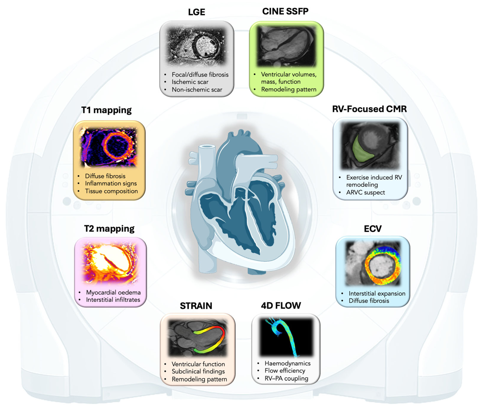
LGE imaging enables detection and localization of focal replacement fibrosis, while the overall multiparametric nature of the technique provides complementary structural and tissue-level information. In this diagnostic framework, advanced CMR techniques have assumed a progressively central role. Quantitative parametric mapping techniques (es. native T1 mapping, T2 mapping, and ECV quantification) provide objective, pixel-wise measurements of myocardial tissue properties. These methods allow the identification of diffuse fibrosis, myocardial edema, and low-grade inflammatory activity, thereby enabling detection of early or subclinical myocardial involvement that may precede overt structural remodeling [13].
Despite these advantages, the use of CMR in the routine evaluation of athletes must also consider several practical and methodological limitations. CMR is a relatively time-consuming examination, often not well tolerated by the patient, requires specialized equipment and expertise, and may not be universally available in all clinical settings. In addition, costs and limited accessibility may restrict its systematic use as a screening tool. These challenges are further compounded by inter-center variability in image acquisition, post-processing, and interpretation, which may affect reproducibility and comparability of findings across institutions. Moreover, athlete-specific factors, such as higher heart rates, respiratory variability, and increased cardiac volumes, may further impact image quality, particularly in gating-dependent sequences, thus requiring tailored acquisition protocols to ensure reliable and reproducible assessment. In line with these aspects, there is a growing need for standardization of CMR protocols, especially in the context of advanced tissue characterization techniques, to ensure consistency, improve diagnostic accuracy, and facilitate broader clinical implementation. For these reasons, current practice emphasizes a targeted application of CMR within a stepwise diagnostic algorithm, reserving it for cases in which prior clinical and instrumental evaluation raises suspicion of underlying disease or when additional tissue characterization and precise volumetric quantification are required [2,6,11].
In this context, CMR in the athletic population should not be conceptualized as a single-purpose examination aimed solely at excluding overt cardiomyopathy. Rather, plays a central and increasingly recognized role that extends beyond diagnostic refinement, encompassing both arrhythmic risk stratification and the complex clinical decision-making process surrounding return-to-play (RTP). CMR imaging should be considered as a multiparametric platform integrating morphological, functional, and tissue characterization data, including LGE and advanced quantitative mapping techniques, to define a comprehensive myocardial phenotype in order to distinguish athlete’s heart from pathological conditions (Figure 2). When interpreted within the clinical context of sport type, training load, and physiological remodeling, this integrated approach allows differentiation between adaptive and pathological findings, ultimately supporting safe and individualized RTP decisions [12,31].

3. Morphologic Findings and Ventricular Function in Athlete’s Heart

In athletes undergoing CMR, the expected imaging findings largely reflect the spectrum of physiological cardiac remodeling induced by chronic training, commonly referred to as the athlete’s heart [2,17,32,33]. On cine imaging, balanced steady-state free precession (bSSFP) sequences represent the cornerstone for morphological and functional assessment, allowing accurate quantification of ventricular volumes, myocardial mass, and systolic function through a complete stack of contiguous short-axis slices covering both ventricles from base to apex, complemented by standard long-axis views. In trained athletes, CMR typically demonstrates balanced enlargement of both ventricular cavities with preserved myocardial architecture and normal regional wall motion. Ventricular volumes are frequently increased compared with sedentary reference populations, reflecting exercise-induced volume loading and augmented diastolic filling. The magnitude of this remodeling follows a graded pattern influenced by several determinants, including cumulative training exposure (hours and years of training), type of sport, age, sex, and body size [12,34,35]. Endurance disciplines characterized by sustained dynamic exercise are generally associated with more pronounced increases in ventricular end-diastolic volumes and myocardial mass, whereas strength-oriented sports may produce more modest cavity enlargement [36,37,38]. Male athletes usually demonstrate greater absolute ventricular volumes and mass compared with female athletes, although indexing to body surface area attenuates these differences [12,39,40]. Younger athletes and those with high weekly training loads also tend to exhibit more marked physiological remodeling [35,41].
Despite the increase in cavity size, global systolic function assessed by left ventricular ejection fraction typically remains within the normal range, although values may occasionally lie at the lower end of normal due to increased end-diastolic volumes and augmented stroke volume at rest [2,17,32]. Importantly, regional wall motion abnormalities are absent, and myocardial thickening during systole remains homogeneous across ventricular segments [33,35,42].
A dedicated right ventricular-focused CMR assessment is another crucial element in athletes, particularly when, in the presence of arrhythmias, their morphology suggests a right ventricular origin or when exercise-induced RV remodelling is suspected. The right ventricle is uniquely susceptible to volume and pressure overload during intense endurance exercise, and distinguishing physiological RV enlargement from early arrhythmogenic right ventricular cardiomyopathy or exercise-induced RV dysfunction remains challenging [43,44,45]. CMR provides accurate quantification of RV volumes, mass, and function. Although tissue characterization of the right ventricle can be attempted by cardiac magnetic resonance, its assessment should be interpreted with caution because of the thin RV wall, which limits the sensitivity for detecting abnormalities on late gadolinium enhancement and T1-weighted imaging. For the same reason, parametric mapping techniques are generally not reliable for RV myocardial characterization [1,2,6,32]. Subtle RV dysfunction, regional akinesia or dyskinesia, and the presence of RV or LV insertion-point fibrosis may all contribute to arrhythmic risk stratification, particularly when integrated with clinical and electrophysiological findings [37].
Right ventricular volumes may also be significantly increased in highly trained individuals, sometimes approaching upper reference limits for the general population; therefore, interpretation must rely on athlete-specific reference ranges and on the overall pattern of symmetrical remodeling rather than isolated volumetric thresholds [17,27,46].
In addition to standard ventricular assessment, a comprehensive CMR protocol in athletes should include dedicated cine planes for the evaluation of atrial chambers, the proximal aorta, and the origins of the coronary arteries [17]. Atrial enlargement, particularly of the left atrium, is a frequent finding in endurance athletes and reflects chronic volume loading rather than intrinsic atrial disease when occurring in the absence of fibrosis or functional impairment [33,41,47]. Visualization of the aortic root and ascending aorta is important to exclude congenital or acquired abnormalities that may have clinical relevance in competitive sports participation [1,48]. Furthermore, additional targeted imaging planes oriented along the aortic root and proximal coronary segments are recommended to assess coronary artery origins, especially when transthoracic echocardiography provides suboptimal visualization [2,17,49]. These acquisitions can be complemented by dedicated non-contrast coronary sequences when needed, allowing reliable depiction of proximal coronary anatomy without exposure to contrast agents [1,6,17,31,50,51].
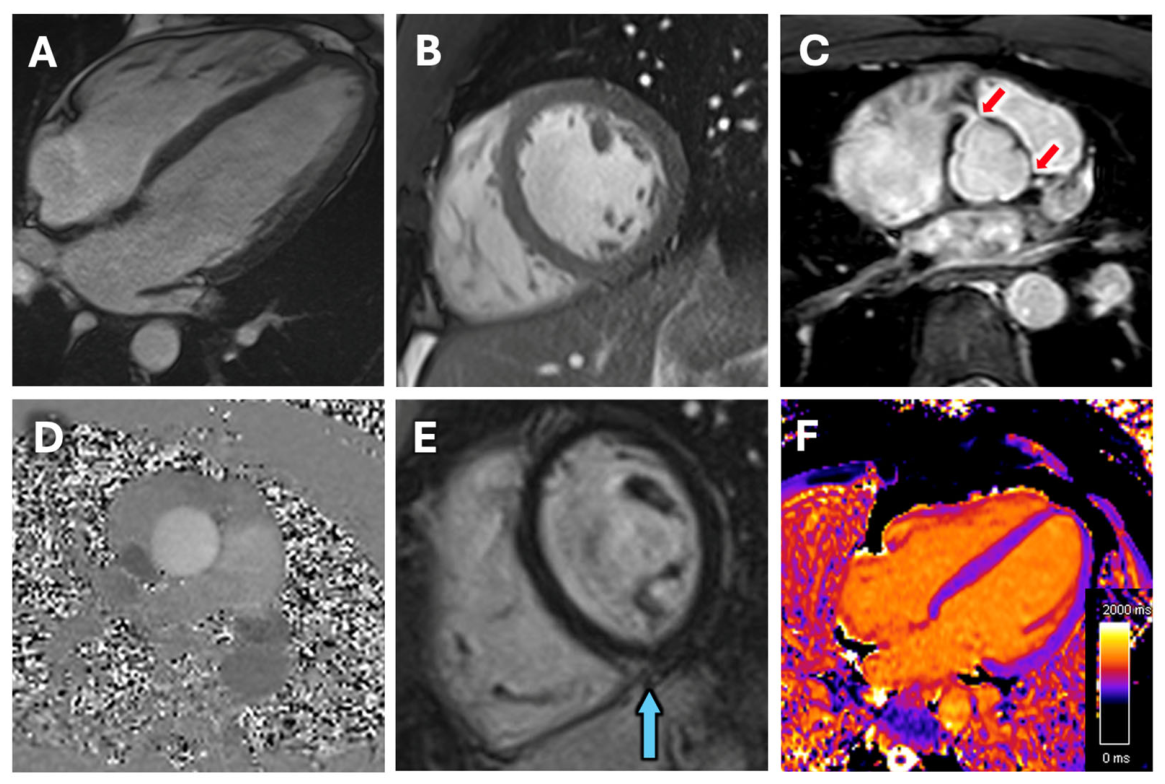
Overall, the interpretation of CMR findings in athletes requires integration of volumetric measurements, functional parameters, and the athlete’s training profile, including sport discipline and training load, in order to differentiate physiological remodeling from early manifestations of cardiomyopathy. Symmetrical chamber enlargement, preserved systolic function, absence of regional wall motion abnormalities, and lack of pathological tissue characterization findings together define the typical CMR phenotype of the healthy athlete’s heart (Figure 3) [6,11,17,33]. These findings should be carefully evaluated by experts and should be consistent with the expected degree of exercise-induced cardiac adaptation and interpreted in conjunction with the athlete’s electrocardiographic pattern, which commonly reflects training-related changes. Concordance between CMR features, type of activity, intensity of athletic training, and typical ECG characteristics of the athlete, supports the diagnosis of physiological remodeling, whereas discordant or disproportionate findings should prompt further evaluation for underlying cardiac disease (Table 1).

4. T1 Mapping and Extracellular Volume Quantification in Athletes

T1 mapping enables quantitative assessment of myocardial longitudinal relaxation time and constitutes a cornerstone technique for detecting diffuse and focal myocardial abnormalities either in general population or athletic individuals [13,52,53]. While each center defines its own reference ranges, standardized according to the specific scanners and acquisition protocols used, in healthy population, native T1 values range approximately from 950 to 1050 ms at 1.5 T and 1150–1300 ms at 3 T, largely overlapping with those of athletes [54,55].
Native T1 values are modestly prolonged in the presence of interstitial fibrosis, whereas more substantial elevations are typically observed in conditions characterized by marked extracellular expansion, such as acute myocardial edema (including active myocarditis) and infiltrative disorders [52,56]. Conversely, post-contrast T1 mapping permits calculation of ECV, allowing quantification of the relative distribution between intracellular and extracellular compartments [52]. ECV quantification represents a major advancement in CMR-based tissue characterization. By integrating native and post-contrast myocardial and blood T1 values, corrected for hematocrit, ECV provides a quantitative estimate of interstitial expansion. Unlike LGE, which detects focal replacement fibrosis based on relative signal differences, ECV enables assessment of diffuse interstitial fibrosis even when no visually apparent scar is present [55].
Contemporary inversion-recovery sequences—most commonly Modified Look-Locker Inversion Recovery (MOLLI) and Shortened MOLLI (ShMOLLI)—acquire multiple images within a single breath-hold, enabling pixel-wise quantification of native and post-contrast T1 values. Given the influence of sequence design, field strength (1.5 T vs. 3 T), heart rate, and motion artifacts, absolute T1 values must be interpreted in the context of acquisition protocol and locally validated reference ranges [13,57].
Native T1 mapping is particularly valuable in athletes when conventional cine imaging and LGE are non-diagnostic. Notably, mildly reduced native T1 values have been described in highly trained athletes and are thought to reflect increased cardiomyocyte volume secondary to physiological hypertrophy, with relative reduction in the interstitial fraction. This adaptive remodeling may lead to lower global T1 and ECV values compared with sedentary controls, necessitating cautious interpretation to avoid overdiagnosis of pathological conditions [55,58,59].
Isolated T1 abnormalities should not be interpreted in the absence of clinical and imaging correlates [52]. However, in athletes presenting with ventricular arrhythmias, segmental or regionally elevated native T1 values—particularly when concordant with electrocardiographic abnormalities—raise suspicion for occult myocardial disease even in the absence of overt LGE-defined scar. Post-contrast T1 mapping and ECV quantification provide complementary information by estimating diffuse fibrosis burden, thereby refining phenotypic characterization [29,60,61,62].
T1 mapping has demonstrated particular utility in differentiating physiological remodeling from dilated cardiomyopathy (DCM), as athletes typically exhibit lower native T1, ECV, and T2 values compared with DCM patients. Furthermore, in subjects with gray zone (12–15 mm) hypertrophy, T1 mapping has been proven to be useful in differentiating true HCM from athlete’s heart, as HCM patients seem to show prolonged T1 values [63,64,65].
From a methodological standpoint, ECV is less susceptible than native T1 alone to certain technical confounders and may therefore provide greater reproducibility for longitudinal follow-up and inter-individual comparison. Incorporating ECV into the evaluation of athletes enhances the sensitivity of CMR in detecting subtle but clinically meaningful myocardial abnormalities. When interpreted in conjunction with arrhythmic burden, electrocardiographic features, and functional parameters, ECV contributes to a more refined stratification of arrhythmogenic risk within a multiparametric framework [11,66].

5. LGE Quantification and Interpretation in Athletes

LGE reflects regional expansion of the myocardial extracellular space leading to delayed washout of gadolinium contrast. In Athletic populations, LGE does not always correspond to replacement fibrosis or pathological myocardial injury. Therefore, careful interpretation of its distribution, its quantification and context are essential to avoid overdiagnosis [67].
Accurate quantification of LGE is essential in the evaluation of athletes with suspected myocardial disease [5,11,14,16]. Overestimation of scar burden may lead to unnecessary restriction from competitive sports, increased psychological burden, and excessive follow-up, whereas underestimation may result in failure to identify individuals at increased arrhythmic risk [5,16,17]. Although visual assessment remains the cornerstone of routine clinical interpretation, quantitative LGE analysis has gained increasing interest as a strategy to enhance objectivity, reproducibility, and risk stratification. Several quantitative approaches have been proposed, most commonly based on signal intensity (SI) thresholding using a predefined number of standard deviations (SD) above reference myocardium, as well as the full width at half maximum (FWHM) technique. However, substantial variability exists among these methods, and no consensus has been established regarding the optimal strategy for LGE quantification, particularly in non-ischemic settings [68,69,70,71].
Athletes represent a particularly challenging subgroup for LGE analysis. In this population, myocardial enhancement is often subtle, patchy, and non-transmural, frequently involving thin myocardial segments. Under these conditions, low-threshold SD-based methods may overestimate fibrotic burden by incorporating noise and partial-volume effects, whereas higher thresholds may underestimate scar extent and fail to capture heterogeneous enhancement. Importantly, the clinical implications of these methodological differences in athletes remain poorly defined [70].
While previous studies have compared quantitative LGE techniques in ischemic and non-ischemic cardiomyopathies, data specifically focused on athletic populations are limited. In addition, few investigations have examined how different quantification methods relate to visual assessment and arrhythmic burden, two key determinants in clinical decision-making in sports cardiology [69,70,71].
LGE patterns in athletes requires crucial interpretation to differentiate physiological remodeling from underlying myocardial disease, due to their notable heterogeneity [14,17,72,73]. The most common finding in asymptomatic athletes is junctional LGE at the right ventricular (RV) insertion points within the interventricular septum. This pattern is generally considered benign, likely reflecting localized fibrosis due to repetitive mechanical stress during endurance training, and is not associated with adverse outcomes [60,61,72,74,75,76].
Outside this typical location, the interpretation becomes more clinically relevant. Subepicardial or mid-wall LGE in the lateral or inferolateral wall may suggest prior subclinical myocarditis or, in selected cases, an early substrate of arrhythmogenic cardiomyopathy, particularly when associated with arrhythmias or structural abnormalities [61,73,77,78,79]. Mid-septal LGE has historically raised concern for early dilated or hypertrophic cardiomyopathy [80,81,82]. However, growing evidence suggests that many small focal lesions in the mid-interventricular septum correspond to fibrosis along septal perforating coronary branches, originating from the left anterior descending artery or the posterior interventricular artery [83]. When isolated and limited in extent, this pattern is increasingly considered a vascular-related and potentially benign finding, rather than cardiomyopathic scar [83,84,85,86]. However, when uncertainty persists, coronary Computed Tomography (CT) angiography may help confirm the presence of the vessel. In addition, partial RV volume defects related to prominent right ventricular trabeculations, which are commonly observed in athletes, may represent another source of false-positive findings and should not be misinterpreted as pathological abnormalities [6,33].
Conversely, extensive septal fibrosis, inferior wall involvement, or RV free-wall LGE should prompt careful evaluation, as these patterns may be associated with structural cardiomyopathies or arrhythmogenic right ventricular cardiomyopathy [87,88,89,90,91].
A detailed understanding of the topographic distribution and morphological features of LGE is therefore fundamental for appropriate risk stratification, helping clinicians avoid both overdiagnosis of disease and under-recognition of clinically relevant myocardial pathology in athletes.

6. T2 Mapping in Athletes

T2 mapping quantifies myocardial transverse relaxation time and serves as a sensitive marker of increased tissue water content, thereby enabling detection of myocardial edema and active inflammation. Compared with conventional T2-weighted techniques (e.g., T2-STIR), which are prone to signal inhomogeneity and limited reproducibility, T2 mapping provides objective, quantitative assessment across the entire myocardium [92]. Native T2 values are typically around 45–55 ms at 1.5 T and do not differ significantly in athletic population in absence of edema or inflammation [54,55]. Therefore, interpretation should rely on site-specific reference ranges rather than absolute cut-offs.
Clinically, T2 mapping is particularly relevant in athletes with recent viral illness, acute symptom onset, or dynamic changes in arrhythmic burden. Elevated myocardial T2 values are indicative of active inflammation or residual edema, as observed in acute or subacute myocarditis—one of the principal causes of ventricular arrhythmias in athletic populations [6,29,93,94,95]. When combined with native T1 mapping and ECV quantification, T2 mapping increases diagnostic confidence for inflammatory cardiomyopathy and facilitates differentiation between active inflammatory processes and chronic fibrotic remodeling [92]. In athletes presenting with ventricular arrhythmias, detection of elevated T2 values may identify potentially reversible inflammatory substrates acting as transient arrhythmogenic triggers, with direct implications for temporary sports restriction and follow-up strategies [11,29,66].
Available data suggest that, after adjustment for indexed left ventricular mass, T2 values do not correlate significantly with training load. Although slightly lower T2 values have been described in athletes compared with sedentary controls, this finding likely reflects physiological myocardial remodeling rather than exercise-induced inflammation. Notably, there is no robust evidence supporting chronic exercise-related myocardial edema in healthy athletes, reinforcing the concept that isolated T2 alterations must be interpreted within the broader clinical and imaging context [12,55,60,94].

7. Advanced CMR Techniques Beyond Mapping: Strain and 4D Flow

In addition to parametric mapping, several advanced CMR techniques provide critical functional and haemodynamic information, particularly relevant in athletes with ventricular arrhythmias or suspected cardiomyopathies. Among these, myocardial strain analysis, right ventricular-focused CMR protocols, and four-dimensional flow CMR represent key components of a comprehensive, multiparametric evaluation [66].
CMR-derived myocardial strain, most commonly assessed using feature-tracking techniques applied to standard cine images, enables quantitative evaluation of myocardial deformation without the need for additional imaging sequences [96,97,98]. Global longitudinal strain (GLS) and regional strain parameters provide sensitive markers of subclinical systolic dysfunction, often preceding reductions in ejection fraction. In athletes, strain analysis is especially valuable for detecting early functional impairment in the presence of preserved global function and physiological remodelling [42,99]. In specific populations, especially athletes with high training loads, practising endurance or resistance disciplines, GLS is often slightly less negative than in sedentary peers, and its interpretation must account for training load, sport type, age, sex, and clinical context [34,42,100,101]. Reduced global or regional strain has been described in association with non-ischemic myocardial scar and cardiomyopathic processes, and regional strain abnormalities may co-localize with areas of fibrosis identified by LGE or abnormal mapping values [97,98]. Feature-tracking CMR is today used to assess atrial strain, provides a sensitive measure of atrial function and compliance. In athletes, it helps distinguish physiological remodeling from early atrial dysfunction, atrial fibrosis assessment, and may indicate increased arrhythmic risk before conventional parameters are abnormal [6,11]. In the context of ventricular arrhythmias, ventricular strain abnormalities may therefore reflect electrically unstable regions characterized by altered mechanical properties, supporting their role as indirect markers of arrhythmogenic substrate [66,99].
Four-dimensional (4D) flow CMR represents a rapidly evolving technique that extends CMR assessment beyond myocardial structure and function to include comprehensive characterization of intracardiac and great vessel haemodynamics. By enabling time-resolved, three-dimensional visualization and quantification of blood flow, 4D-flow CMR provides advanced metrics such as kinetic energy, vorticity, and flow efficiency [102,103]. In athletes, these parameters offer novel insights into the haemodynamic consequences of exercise-induced remodelling, particularly in the right ventricle and pulmonary circulation. Altered flow patterns or increased energetic inefficiency may reflect maladaptive remodelling or abnormal ventricular–arterial coupling. Although currently largely confined to research settings, 4D-flow CMR holds promise as a complementary tool for identifying functional and hemodynamic correlates of ventricular arrhythmias, especially in athletes with otherwise inconclusive structural imaging, and in athletes with aortic dilatation to assess flow direction, shear stress and flow patterns [2,66].
Together, myocardial strain analysis and advanced flow imaging expand the diagnostic scope of CMR beyond tissue characterization alone. When integrated with parametric mapping and LGE, these techniques enable a holistic evaluation of the athlete’s heart, potentially detecting early alterations or arrhythmogenic substrates that may be not be overt, incorporating myocardial composition, mechanics, and haemodynamics within a unified imaging framework [2].

8. Specific Applications in Athletes Populations

The application of advanced CMR techniques in athletes has grown rapidly in research over the past decade, allowing exploration of how intense training and physiological remodelling influence myocardial tissue characteristics, differentiating cardiac training adaptation to pathological remodelling (Table 1). Data from a recent cohort study of Olympic athletes demonstrated that native T1 values vary according to sport discipline and sex, with endurance and mixed-discipline athletes (who typically experience more pronounced left ventricular remodelling) exhibiting lower native T1 values than controls and other athlete categories, while T2 values remained unchanged. Additionally, male athletes with higher left ventricular mass index displayed lower T1 values compared with females, suggesting sex-specific tissue remodelling signatures [55]. These findings align with earlier observations that elite athletes tend to have lower native T1 compared with non-athletic counterparts, potentially reflecting increased cardiomyocyte volume and altered interstitial composition associated with physiological remodelling [12,55,58,59]. Such distinctions are critical because lower native T1 or altered ECV in athletes could, if misinterpreted, be mistaken for pathological states in the absence of athletic-specific reference ranges. Furthermore, emerging evidence suggests a relationship between cardiorespiratory fitness and mapping values, where greater overall fitness associates with larger cardiac volumes and lower T1/T2 values, implicating training load and physiological adaptation as modulators of myocardial tissue characteristics [12,33,38,104]. In addition, long-term exercise load seems to be linked to the presence of LGE, especially in male subjects [38,72,78,105]. This link reinforces the need for sport-specific interpretive frameworks and raises intriguing questions regarding tissue adaptation thresholds that may distinguish physiology from pathological remodelling.

9. Technical Challenges and Standardization

Advanced parametric mapping techniques, although highly promising, show methodological and technical constraints that currently limit their universal implementation in routine clinical practice. Foremost among these are intercenter and inter-vendor variability in acquisition protocols, as well as the dependence of absolute quantitative values on magnetic field strength and specific sequence parameters. Furthermore, the requirement for sophisticated motion correction algorithms to adequately compensate for respiratory and cardiac motion introduces additional complexity. Collectively, these factors hinder direct comparability of measurements across institutions and studies, thereby mandating stringent quality assurance and harmonization procedures [13,57,106].
Although ECV quantification demonstrates comparatively greater reproducibility, native T1 values remain susceptible to variability related to heart rate, altitude, humidity, temperature, specific implementations, and sequence selection. This variability reinforces the necessity of establishing robust, site-specific reference ranges and of contextualizing results according to sex and athletic discipline. Although ongoing initiatives focused on refining post-processing methodologies and establishing standardized reporting frameworks are anticipated to improve reproducibility and interoperability, a comprehensive methodological consensus has not yet been reached [3,12,13,58,59].

10. Future Directions of Advanced CMR Techniques in Athletes

Beyond parametric mapping, future developments in advanced CMR are increasingly focused on multiparametric phenotyping aimed at refining the early identification and characterization of subtle abnormalities that may suggest cardiomyopathies and/or arrhythmogenic substrates in athletes [14,31,42,107]. Ventricular arrhythmias in this population often arise from a complex interplay between myocardial tissue abnormalities, ventricular mechanics, and exercise-induced hemodynamic stress, underscoring the need for imaging approaches that extend beyond static tissue characterization [29,38]. Four-dimensional flow CMR (4D-flow) represents a particularly promising technique, enabling time-resolved, three-dimensional assessment of intracardiac and great vessel blood flow. Advanced flow-derived metrics, including wall shear stress, kinetic energy, vorticity, and flow efficiency, may provide novel insights into maladaptive hemodynamic patterns associated with ventricular remodelling, particularly within the right ventricle and pulmonary circulation, increasingly implicated in exercise-related arrhythmogenesis and in the early detection of cardiomyopathies [37,102,103].
Advances in myocardial deformation imaging are also likely to play an expanding role in the evaluation of the athlete’s population. Feature-tracking CMR strain, when integrated with mapping techniques and late gadolinium enhancement, allows simultaneous assessment of myocardial mechanics and tissue composition, facilitating detection of electromechanical heterogeneity that may predispose to ventricular arrhythmias despite preserved global systolic function, or initial diffuse or regional cardiomyopathy-related wall motion abnormalities [42,97,98]. More experimental approaches, such as cardiac diffusion tensor imaging (cDTI), offer the potential to characterize myocardial fiber architecture in vivo, providing insights into structural substrates that may influence conduction properties and arrhythmogenic mechanisms. CDTI enables the assessment of cardiomyocyte orientation and has shown promise in detecting myocardial disarray, a hallmark of hypertrophic cardiomyopathy that may precede overt morphological changes. This is especially relevant in athletes, where differentiating physiological left ventricular remodeling from early or subclinical cardiomyopathy remains challenging [108,109]. Although its clinical applicability is currently limited, cDTI represents a promising tool for early disease detection and improved discrimination between adaptive and pathological hypertrophy.
Equally transformative are developments in post-processing software and artificial intelligence-driven analytic pipelines. Automated segmentation, motion correction, and multiparametric integration reduce operator dependency and improve reproducibility, which is essential for longitudinal monitoring of athletes. Machine learning applied to comprehensive CMR datasets may enable identification of subtle imaging signatures, supporting earlier detection of concealed cardiomyopathies and more precise stratification of athletes, e.g., with apparently idiopathic ventricular arrhythmias [110].
Emerging advanced CMR techniques are involved in further expansion of the diagnostic potential of artificial intelligence (AI) even in sports cardiology. AI-based approaches are increasingly applied to image acquisition, reconstruction, and post-processing, enabling faster scan times, automated ventricular segmentation, and improved detection of subtle myocardial abnormalities that may be overlooked by conventional analysis [111,112]. Although these cutting-edge techniques are not yet routinely implemented in clinical practice, they hold significant promise for improving phenotyping, risk stratification, and longitudinal evaluation of athletes, particularly in complex or borderline cases.
Within this constantly evolving framework, the convergence of advanced tissue characterization, deformation imaging, flow analysis, and automated analytics is expected to further strengthen the role of CMR as a central tool in precision sports cardiology, enabling not only earlier detection but also a more tailored, pathology-oriented management of athletic individuals.

11. Conclusions

Advanced CMR techniques represent a cornerstone in the contemporary evaluation of athletes, providing non-invasive, quantitative insights into myocardial tissue composition, ventricular mechanics, and haemodynamic behaviour beyond conventional imaging. Parametric mapping enables the detection of diffuse fibrosis or inflammation that may act as an arrhythmogenic substrate even in the absence of overt structural disease, while deformation and flow imaging offer complementary information on electromechanical and haemodynamic features. Within a multiparametric framework, these techniques improve the differentiation between physiological adaptation and pathological remodelling, addressing a key challenge in sports cardiology. Despite current limitations related to standardization, availability, and interpretative complexity, ongoing technological advances are progressively enhancing the clinical value of CMR. Consequently, advanced CMR is increasingly emerging as a central tool for risk stratification and clinical decision-making in athletes with suspected cardiomyopathy or ventricular arrhythmias.

Author Contributions

Conceptualization, F.G. and A.Z.; writing—original draft preparation, S.U. and A.D.A.; writing—review and editing, S.U., A.D.A., A.Z., M.P., F.A.C., D.C. and F.G.; supervision, F.G. All authors have read and agreed to the published version of the manuscript.

Funding

This research received no external funding.

Institutional Review Board Statement

Not applicable.

Data Availability Statement

No new data were created.

Acknowledgments

During the preparation of this manuscript, the authors used ChatGPT (version 5.2) as support for checking the text for clarity, grammar, and language, and for generating schematic images.

Conflicts of Interest

The authors declare no conflicts of interest.

Abbreviations

The following abbreviations are used in this manuscript:
AIArtificial Intelligence
ARVCArrhythmogenic Right Ventricular Cardiomyopathy
bSSFPbalanced State Free Precession
CMRCardiac Magnetic Resonance
CTComputed Tomography
cDTICardiac Diffusion Tensor Imaging
ECVExtra-cellular Volume
DCMDilated Cardiomyopathy
EFEjection Fraction
FWHMFull Width Half Maximum
GLSGlobal Longitudinal Strain
HCMHypertrophic Cardiomyopathy
LGELate Gadolinium Enhancement
LVLeft Ventricle
LVEFLeft Ventricular Ejection Fraction
LVEDVLeft Ventricular End Diastolic Volume
MOLLIModified Look-Locker Inversion Recovery
NILVSNon-ischemic Left Ventricular Scar
PAPulmonary Artery
Qp/QsPulmonary-to-Systemic flow ratio
RTPReturn-to-play
RVRight Ventricle
RVEFRight Ventricular Ejection Fraction
RVEDVRight Ventricular End Diastolic Volume
SDStandard Deviation
ShMOLLIShortened Modified Look-Locker Inversion Recovery
SISignal Intensity
SSFPSteady State Free Precession
4DFour Dimensions

References

  1. Grech, N.; Abela, M. The Role of Cardiovascular Magnetic Resonance Imaging in Athletic Individuals—A Narrative Review. J. Clin. Med. 2025, 14, 3576. [Google Scholar] [CrossRef]
  2. Gati, S.; Sharma, S.; Pennell, D. The Role of Cardiovascular Magnetic Resonance Imaging in the Assessment of Highly Trained Athletes. JACC Cardiovasc. Imaging 2018, 11, 247–259. [Google Scholar] [CrossRef]
  3. Fogante, M.; Agliata, G.; Basile, M.C.; Compagnucci, P.; Volpato, G.; Falanga, U.; Stronati, G.; Guerra, F.; Vignale, D.; Esposito, A.; et al. Cardiac Imaging in Athlete’s Heart: The Role of the Radiologist. Medicina 2021, 57, 455. [Google Scholar] [CrossRef]
  4. Rajiah, P.S.; Kumar, V.; Domenech-Ximenos, B.; Francone, M.; Broncano, J.; Allison, T.G. Utility of MRI and CT in Sports Cardiology. RadioGraphics 2025, 45, e240045. [Google Scholar] [CrossRef]
  5. Pelliccia, A.; Sharma, S.; Gati, S.; Bäck, M.; Börjesson, M.; Caselli, S.; Collet, J.-P.; Corrado, D.; Drezner, J.A.; Halle, M.; et al. 2020 ESC Guidelines on sports cardiology and exercise in patients with cardiovascular disease. Eur. Heart J. 2021, 42, 17–96. [Google Scholar] [CrossRef]
  6. Szabo, L.; Brunetti, G.; Cipriani, A.; Juhasz, V.; Graziano, F.; Hirschberg, K.; Dohy, Z.; Balla, D.; Drobni, Z.; Marra, M.P.; et al. Certainties and Uncertainties of Cardiac Magnetic Resonance Imaging in Athletes. J. Cardiovasc. Dev. Dis. 2022, 9, 361. [Google Scholar] [CrossRef]
  7. Androulakis, E.; Mouselimis, D.; Tsarouchas, A.; Antonopoulos, A.; Bakogiannis, C.; Papagkikas, P.; Vlachopoulos, C. The Role of Cardiovascular Magnetic Resonance Imaging in the Assessment of Myocardial Fibrosis in Young and Veteran Athletes: Insights From a Meta-Analysis. Front. Cardiovasc. Med. 2021, 8, 784474. [Google Scholar] [CrossRef]
  8. Bakogiannis, C.; Mouselimis, D.; Tsarouchas, A.; Papatheodorou, E.; Vassilikos, V.P.; Androulakis, E. Hypertrophic cardiomyopathy or athlete’s heart? A systematic review of novel cardiovascular magnetic resonance imaging parameters. Eur. J. Sport Sci. 2021, 23, 143–154. [Google Scholar] [CrossRef]
  9. Zloić, S.K.; Hrabak-Paar, M. The Role of Cardiac Magnetic Resonance Imaging in Distinguishing the Athlete’s Heart from Hypertrophic Cardiomyopathy–A Brief Literature Review. Echocardiography 2024, 41, e70021. [Google Scholar] [CrossRef]
  10. Ommen, S.R.; Mital, S.; Burke, M.A.; Day, S.M.; Deswal, A.; Elliott, P.; Evanovich, L.L.; Hung, J.; Joglar, J.A. 2020 AHA/ACC Guideline for the Diagnosis and Treatment of Patients with Hypertrophic Cardiomyopathy: A Report of the American College of Cardiology/American Heart Association Joint Committee on Clinical Practice Guidelines. Circulation 2020, 142, e558–e631. [Google Scholar] [CrossRef]
  11. Maestrini, V.; Torlasco, C.; Hughes, R.; Moon, J.C. Cardiovascular Magnetic Resonance and Sport Cardiology: A Growing Role in Clinical Dilemmas. J. Cardiovasc. Transl. Res. 2020, 13, 296–305. [Google Scholar] [CrossRef]
  12. Balla, D.; Szabo, L.; Graziano, F.; Mesko, C.; Dohy, Z.; Juhasz, V.; Amirifard, D.; Sydo, N.; Csulak, E.; Petrov, I.; et al. The role of sex, training load, and sports type in athletic cardiac remodelling: Insights from T1 and T2 mapping via cardiac magnetic resonance. Int. J. Cardiol. 2025, 426, 133080. [Google Scholar] [CrossRef]
  13. Messroghli, D.R.; Moon, J.C.; Ferreira, V.M.; Grosse-Wortmann, L.; He, T.; Kellman, P.; Mascherbauer, J.; Nezafat, R.; Salerno, M.; Schelbert, E.B.; et al. Clinical recommendations for cardiovascular magnetic resonance mapping of T1, T2, T2* and extracellular volume: A consensus statement by the Society for Cardiovascular Magnetic Resonance (SCMR) endorsed by the European Association for Cardiovascular Imaging (EACVI). J. Cardiovasc. Magn. Reson. 2016, 19, 75. [Google Scholar] [CrossRef]
  14. Maestrini, V.; Penza, M.; Monosilio, S.; Borrazzo, C.; Prosperi, S.; Filomena, D.; Birtolo, L.I.; Lemme, E.; Mango, R.; Di Gioia, G.; et al. The role of cardiac magnetic resonance in sports cardiology: Results from a large cohort of athletes. Clin. Res. Cardiol. 2024, 113, 781–789. [Google Scholar] [CrossRef]
  15. Kim, J.H.; Baggish, A.L.; Levine, B.D.; Ackerman, M.J.; Day, S.M.; Dineen, E.H.; Ii, J.S.G.; La Gerche, A.; Lampert, R.; Martinez, M.W.; et al. Clinical Considerations for Competitive Sports Participation for Athletes with Cardiovascular Abnormalities. J. Am. Coll. Cardiol. 2025, 85, 1059–1108. [Google Scholar] [CrossRef]
  16. Pelliccia, A.; Solberg, E.E.; Papadakis, M.; Adami, P.E.; Biffi, A.; Caselli, S.; La Gerche, A.; Niebauer, J.; Pressler, A.; Schmied, C.M.; et al. Recommendations for participation in competitive and leisure time sport in athletes with cardiomyopathies, myocarditis, and pericarditis: Position statement of the Sport Cardiology Section of the European Association of Preventive Cardiology (EAPC). Eur. Heart J. 2018, 40, 19–33. [Google Scholar] [CrossRef]
  17. Pelliccia, A.; Caselli, S.; Sharma, S.; Basso, C.; Bax, J.J.; Corrado, D.; D’aNdrea, A.; D’aScenzi, F.; Di Paolo, F.M.; Edvardsen, T.; et al. European Association of Preventive Cardiology (EAPC) and European Association of Cardiovascular Imaging (EACVI) joint position statement: Recommendations for the indication and interpretation of cardiovascular imaging in the evaluation of the athlete’s heart. Eur. Heart J. 2017, 39, 1949–1969. [Google Scholar] [CrossRef]
  18. Arbelo, E.; Protonotarios, A.; Gimeno, J.R.; Arbustini, E.; Barriales-Villa, R.; Basso, C.; Bezzina, C.R.; Biagini, E.; Blom, N.; de Boer, R.; et al. 2023 ESC Guidelines for the management of cardiomyopathies: Developed by the task force on the management of cardiomyopathies of the European So-ciety of Cardiology (ESC). Eur. Heart J. 2023, 44, 3503–3626. [Google Scholar] [CrossRef]
  19. Graziano, F.; Mastella, G.; Merkely, B.; Vago, H.; Corrado, D.; Zorzi, A. Ventricular arrhythmias recorded on 12-lead ambulatory electrocardiogram monitoring in healthy volunteer athletes and controls: What is common and what is not. Europace 2023, 25, euad255. [Google Scholar] [CrossRef]
  20. Graziano, F.; Tonelli, R.; Balla, D.; Pizzolato, M.; Bondarev, S.; Vago, H.; Corrado, D.; Zorzi, A. Modern Interpretation of Electrocardiogram and Premature Ventricular Beats in Athletes. Cardiol. Discov. 2025, 5, 225–236. [Google Scholar] [CrossRef]
  21. De Lazzari, M.; Zorzi, A.; Bettella, N.; Cipriani, A.; Pilichou, K.; Cason, M.; Vessella, T.; Sarto, P.; Gualea, M.R.; Chianura, F.; et al. Papillary Muscles Abnormalities in Athletes with Otherwise Unexplained T-Wave Inversion in the ECG Lateral Leads. J. Am. Heart Assoc. 2021, 10, e019239. [Google Scholar] [CrossRef]
  22. Graziano, F.; Genta, O.E.; Manfrin, L.; Corrado, D.; Brusamolin, L.; Giada, F.; Gerbino, L.; Compagno, S.; Zorzi, A. Prevalence and determinants of low QRS voltages and QRS fragmentation in children and adolescents undergoing sports pre-participation screening. Eur. J. Prev. Cardiol. 2024, 31, 1535–1542. [Google Scholar] [CrossRef]
  23. Graziano, F.; Balla, D.; Juhasz, V.; Sydo, N.; Kiss, O.; Csulak, E.; Babity, M.; Zamodics, M.; Mesko, C.; Dohy, Z.; et al. Cardiopulmonary physical fitness and tissue characterization through T1 and T2 mapping: New insights on athlete’s heart. Eur. J. Prev. Cardiol. 2025, zwaf616. [Google Scholar] [CrossRef]
  24. Zorzi, A.; D’AScenzi, F.; Andreini, D.; Castelletti, S.; Casella, M.; Cavarretta, E.; Cipriani, A.; Compagnucci, P.; Delise, P.; Russo, A.D.; et al. Interpretation and management of premature ventricular beats in athletes: An expert opinion document of the Italian Society of Sports Cardiology (SICSPORT). Int. J. Cardiol. 2023, 391, 131220. [Google Scholar] [CrossRef]
  25. Myerson, S.G. CMR in Evaluating Valvular Heart Disease. JACC Cardiovasc. Imaging 2021, 14, 2020–2032. [Google Scholar] [CrossRef]
  26. Marra, M.P.; Basso, C.; De Lazzari, M.; Rizzo, S.; Cipriani, A.; Giorgi, B.; Lacognata, C.; Rigato, I.; Migliore, F.; Pilichou, K.; et al. Morphofunctional Abnormalities of Mitral Annulus and Arrhythmic Mitral Valve Prolapse. Circ. Cardiovasc. Imaging 2016, 9, e005030. [Google Scholar] [CrossRef]
  27. Monosilio, S.; Prosperi, S.; Ciuffreda, A.; Lemme, E.; Di Gioia, G.; Mango, R.; Tonti, G.; Pedrizzetti, G.; Gualdi, G.; Pelliccia, A.; et al. Right ventricle adaptation to high intensity training assessed by cardiac magnetic resonance in olympic athletes. Eur. J. Prev. Cardiol. 2023, 30, zwad125.133. [Google Scholar] [CrossRef]
  28. Corrado, D.; Zorzi, A.; Cipriani, A.; Bauce, B.; Bariani, R.; Brunetti, G.; Graziano, F.; De Lazzari, M.; Mattesi, G.; Migliore, F.; et al. Scarring/arrhythmogenic cardiomyopathy. Eur. Heart J. Suppl. 2023, 25, C144–C154. [Google Scholar] [CrossRef]
  29. Brunetti, G.; Cipriani, A.; Marra, M.P.; De Lazzari, M.; Bauce, B.; Calore, C.; Rigato, I.; Graziano, F.; Vio, R.; Corrado, D.; et al. Role of Cardiac Magnetic Resonance Imaging in the Evaluation of Athletes with Premature Ventricular Beats. J. Clin. Med. 2022, 11, 426. [Google Scholar] [CrossRef]
  30. Zeppenfeld, K.; Tfelt-Hansen, J.; de Riva, M.; Winkel, B.G.; Behr, E.R.; Blom, N.; Charron, P.; Corrado, D.; Dagres, N.; de Chillou, C.; et al. 2022 ESC Guidelines for the management of patients with ventricular arrhythmias and the prevention of sudden cardiac death. Eur. Heart J. 2022, 43, 3997–4126. [Google Scholar] [CrossRef]
  31. Galderisi, M.; Cardim, N.; D’Andrea, A.; Bruder, O.; Cosyns, B.; Davin, L.; Donal, E.; Edvardsen, T.; Freitas, A.; Habib, G.; et al. The multi-modality cardiac imaging approach to the Athlete’s heart: An expert consensus of the European Association of Cardiovascular Imaging. Eur. Heart J. Cardiovasc. Imaging 2015, 16, 353–353r. [Google Scholar] [CrossRef]
  32. Gati, S.; Sharma, S. Determinants of the athlete’s heart: A cardiovascular magnetic resonance imaging study. Eur. J. Prev. Cardiol. 2019, 27, 536–539. [Google Scholar] [CrossRef]
  33. Balla, D.; Szabo, L.; Juhasz, V.; Mesko, C.S.; Dohy, Z.S.; Sydo, N.; Graziano, F.; Csulak, E.; Merkely, B.; Vago, H. Cardiac magnetic resonance characteristics of the athlete’s heart: Deeper insights into cardiac remodelling. Eur. Heart J. 2024, 45, ehae666.3004. [Google Scholar] [CrossRef]
  34. Monosilio, S.; Filomena, D.; Birtolo, L.; Penza, M.; Lemme, E.; Squeo, M.R.; Mango, R.; Tonti, G.; Pedrizzetti, G.; Fedele, F.; et al. Left ventricle myocardial deformation in olympic athletes assessed by cardiac magnetic resonance: Does the sex and discipline matter? Eur. J. Prev. Cardiol. 2022, 29, 328. [Google Scholar] [CrossRef]
  35. D’AScenzi, F.; Anselmi, F.; Piu, P.; Fiorentini, C.; Carbone, S.F.; Volterrani, L.; Focardi, M.; Bonifazi, M.; Mondillo, S. Cardiac Magnetic Resonance Normal Reference Values of Biventricular Size and Function in Male Athlete’s Heart. JACC Cardiovasc. Imaging 2019, 12, 1755–1765. [Google Scholar] [CrossRef]
  36. Pujadas, S.; Doñate, M.; Li, C.-H.; Merchan, S.; Cabanillas, A.; Alomar, X.; Pons-Llado, G.; Serra-Grima, R.; Carreras, F. Myocardial remodelling and tissue characterisation by cardiovascular magnetic resonance (CMR) in endurance athletes. BMJ Open Sport Exerc. Med. 2018, 4, e000422. [Google Scholar] [CrossRef]
  37. La Gerche, A.; Burns, A.T.; Mooney, D.J.; Inder, W.J.; Taylor, A.J.; Bogaert, J.; MacIsaac, A.I.; Heidbüchel, H.; Prior, D.L. Exercise-induced right ventricular dysfunction and structural remodelling in endurance athletes. Eur. Heart J. 2011, 33, 998–1006. [Google Scholar] [CrossRef]
  38. Graziano, F.; Juhasz, V.; Brunetti, G.; Cipriani, A.; Szabo, L.; Merkely, B.; Corrado, D.; D’ascenzi, F.; Vago, H.; Zorzi, A. May Strenuous Endurance Sports Activity Damage the Cardiovascular System of Healthy Athletes? A Narrative Review. J. Cardiovasc. Dev. Dis. 2022, 9, 347. [Google Scholar] [CrossRef]
  39. van Hattum, J.C.; van Diepen, M.; Verwijs, S.M.; Boekholdt, S.M.; van Randen, A.; Groenink, M.; Planken, R.N.; Moen, M.H.; Daems, J.J.N.; Prakken, N.H.J.; et al. Extreme phenotypes of the female athlete’s heart: A sports-specific cardiac magnetic resonance imaging study. Eur. Heart J. Cardiovasc. Imaging 2025, 26, 1199–1207. [Google Scholar] [CrossRef]
  40. Bryde, R.; Applewhite, A.I.; Abu Dabrh, A.M.; Taylor, B.J.; Heckman, M.G.; Filmalter, S.E.; Pujalte, G.; Rojas, C.; Heckman, A.J.; Brigham, T.J.; et al. Cardiac structure and function in elite female athletes: A systematic review and meta-analysis. Physiol. Rep. 2021, 9, e15141. [Google Scholar] [CrossRef]
  41. Csecs, I.; Czimbalmos, C.; Toth, A.; Dohy, Z.; Suhai, I.F.; Szabo, L.; Kovacs, A.; Lakatos, B.; Sydo, N.; Kheirkhahan, M.; et al. The impact of sex, age and training on biventricular cardiac adaptation in healthy adult and adolescent athletes: Cardiac magnetic resonance imaging study. Eur. J. Prev. Cardiol. 2019, 27, 540–549. [Google Scholar] [CrossRef]
  42. Monosilio, S.; Prosperi, S.; Filomena, D.; Lemme, E.; Di Gioia, G.; Mango, R.; Netti, L.; Tonti, G.; Pedrizzetti, G.; Gualdi, G.; et al. Cardiac magnetic resonance feature tracking in Olympic athletes: A myocardial deformation analysis. Eur. J. Prev. Cardiol. 2025, zwaf042. [Google Scholar] [CrossRef]
  43. Moccia, E.; Papatheodorou, E.; Miles, C.J.; Merghani, A.; Malhotra, A.; Dhutia, H.; Bastiaenen, R.; Sheikh, N.; Zaidi, A.; Sanna, G.D.; et al. Arrhythmogenic cardiomyopathy and differential diagnosis with physiological right ventricular remodelling in athletes using cardiovascular magnetic resonance. Int. J. Cardiovasc. Imaging 2022, 38, 2723–2732. [Google Scholar] [CrossRef]
  44. Czimbalmos, C.; Csecs, I.; Dohy, Z.; Toth, A.; Suhai, F.I.; Müssigbrodt, A.; Kiss, O.; Geller, L.; Merkely, B.; Vago, H. Cardiac magnetic resonance based deformation imaging: Role of feature tracking in athletes with suspected arrhythmogenic right ventricular cardiomyopathy. Int. J. Cardiovasc. Imaging 2018, 35, 529–538. [Google Scholar] [CrossRef]
  45. Bohm, P.; Schneider, G.; Linneweber, L.; Rentzsch, A.; Krämer, N.; Abdul-Khaliq, H.; Kindermann, W.; Meyer, T.; Scharhag, J. Right and Left Ventricular Function and Mass in Male Elite Master Athletes. Circulation 2016, 133, 1927–1935. [Google Scholar] [CrossRef]
  46. Maceira, A.M.; Monmeneu, J.V.; López, M.P.; García, M.P.; Higueras, L.; Masiá, M.D.; Boraita, A. Reference ventricular dimensions and function parameters by cardiovascular magnetic resonance in highly trained Caucasian athletes. J. Cardiovasc. Magn. Reson. 2023, 25, 12. [Google Scholar] [CrossRef]
  47. Monosilio, S.; Ferrera, A.; Daniello, C.; Di Gioia, G.; Spinelli, A.; Serdoz, A.; Lemme, E.; Mango, R.; Pedrizzetti, G.; Tonti, G.; et al. Deeper insight into elite athletes cardiac remodelling: A CMR cluster analysis. Eur. J. Prev. Cardiol. 2025, 32, zwaf236.005. [Google Scholar] [CrossRef]
  48. Nussbaumer, C.; Bouchardy, J.; Blanche, C.; Piccini, D.; Pavon, A.-G.; Monney, P.; Stuber, M.; Schwitter, J.; Rutz, T. 2D cine vs. 3D self-navigated free-breathing high-resolution whole heart cardiovascular magnetic resonance for aortic root measurements in congenital heart disease. J. Cardiovasc. Magn. Reson. 2021, 23, 65. [Google Scholar] [CrossRef]
  49. Verwijs, S.; Van Hattum, J.; Stomilovic, N.; Daems, J.; Boekholdt, S.; Planken, J.; Groenink, M.; Van Randen, A.; Bakermans, A.; Nederveen, A.; et al. Aortic dilatation using cardiac magnetic resonance in asymptomatic ELITE athletes. Eur. J. Prev. Cardiol. 2022, 29, zwac056.270. [Google Scholar] [CrossRef]
  50. Androulakis, E.; Swoboda, P.P. The Role of Cardiovascular Magnetic Resonance in Sports Cardiology; Current Utility and Future Perspectives. Curr. Treat. Options Cardiovasc. Med. 2018, 20, 86. [Google Scholar] [CrossRef]
  51. Nazir, M.S.; Bustin, A.; Hajhosseiny, R.; Yazdani, M.; Ryan, M.; Vergani, V.; Neji, R.; Kunze, K.P.; Nicol, E.; Masci, P.G.; et al. High-resolution non-contrast free-breathing coronary cardiovascular magnetic resonance angiography for detection of coronary artery disease: Validation against invasive coronary angiography. J. Cardiovasc. Magn. Reson. 2022, 24, 26. [Google Scholar] [CrossRef]
  52. Haaf, P.; Garg, P.; Messroghli, D.R.; Broadbent, D.A.; Greenwood, J.P.; Plein, S. Cardiac T1 Mapping and Extracellular Volume (ECV) in clinical practice: A comprehensive review. J. Cardiovasc. Magn. Reson. 2016, 18, 89. [Google Scholar] [CrossRef]
  53. Moon, J.C.; Messroghli, D.R.; Kellman, P.; Piechnik, S.K.; Robson, M.D.; Ugander, M.; Gatehouse, P.D.; Arai, A.; Friedrich, M.G.; Neubauer, S.; et al. Myocardial T1 mapping and extracellular volume quantification: A Society for Cardiovascular Magnetic Resonance (SCMR) and CMR Working Group of the European Society of Cardiology consensus statement. J. Cardiovasc. Magn. Reson. 2013, 15, 92. [Google Scholar] [CrossRef]
  54. Roy, C.; Slimani, A.; de Meester, C.; Amzulescu, M.; Pasquet, A.; Vancraeynest, D.; Vanoverschelde, J.-L.; Pouleur, A.-C.; Gerber, B.L. Age and sex corrected normal reference values of T1, T2 T2* and ECV in healthy subjects at 3T CMR. J. Cardiovasc. Magn. Reson. 2016, 19, 72. [Google Scholar] [CrossRef]
  55. Prosperi, S.; Monosilio, S.; Lemme, E.; Filomena, D.; Penza, M.; Birtolo, L.I.; Mango, R.; Di Gioia, G.; Gualdi, G.; Squeo, M.R.; et al. CMR native T1 and T2 mapping in Olympic athletes: The influence of sports discipline and sex. Eur. Heart J. Cardiovasc. Imaging 2024, 26, 89–95. [Google Scholar] [CrossRef]
  56. Gaizauskiene, K.; Leketaite, K.; Glaveckaite, S.; Valeviciene, N. Diagnostic Value of Cardiovascular Magnetic Resonance T1 and T2 Mapping in Acute Myocarditis: A Systematic Literature Review. Medicina 2024, 60, 1162. [Google Scholar] [CrossRef]
  57. Ogier, A.C.; Bustin, A.; Cochet, H.; Schwitter, J.; van Heeswijk, R.B. The Road Toward Reproducibility of Parametric Mapping of the Heart: A Technical Review. Front. Cardiovasc. Med. 2022, 9, 876475. [Google Scholar] [CrossRef]
  58. Daems, J.J.N.; Verwijs, S.M.; van Diepen, M.; van Hattum, J.C.; van Luijk, R.D.; Moen, M.H.; Nelissen, J.L.; Nederveen, A.J.; Planken, R.N.; van Randen, A.; et al. Native T1 mapping times are strongly influenced by elite athlete status. Eur. Heart J. Cardiovasc. Imaging 2025, 26, 1208–1216. [Google Scholar] [CrossRef]
  59. Daems, J.J.N.; Verwijs, S.M.; Janssen, R.D.; Van Hattum, J.C.; Boekholdt, S.M.; Van Randen, A.; Planken, R.N.; Van Luijk-Snoeks, R.D.; Moen, M.; Groenink, M.; et al. Elite athlete status, gender and mitchell sports classification strongly influence native t1 mapping times. Eur. J. Prev. Cardiol. 2023, 30, zwad125.035. [Google Scholar] [CrossRef]
  60. Ragab, H.; Lund, G.K.; Breitsprecher, L.; Sinn, M.R.; Muellerleile, K.; Cavus, E.; Stehning, C.; Tahir, E.; Blankenberg, S.; Patten, M.; et al. Prevalence and pattern of focal and potential diffuse myocardial fibrosis in male and female marathon runners using contrast-enhanced cardiac magnetic resonance. Eur. Radiol. 2023, 33, 4648–4656. [Google Scholar] [CrossRef]
  61. Farooq, M.; Brown, L.A.E.; Fitzpatrick, A.; Broadbent, D.A.; Wahab, A.; Klassen, J.R.L.; Farley, J.; Saunderson, C.E.D.; Das, A.; Craven, T.; et al. Identification of non-ischaemic fibrosis in male veteran endurance athletes, mechanisms and association with premature ventricular beats. Sci. Rep. 2023, 13, 14640. [Google Scholar] [CrossRef]
  62. Javed, W.; Botis, I.; Goh, Z.M.; Shabi, M.; Brown, B.; Tomoaia, R.; Farooq, M.; Levelt, E.; Graham, L.; Gierula, J.; et al. Ventricular Arrhythmia and Cardiac Fibrosis in Endurance Experienced Athletes (VENTOUX). Circ. Cardiovasc. Imaging 2025, 18, e018470. [Google Scholar] [CrossRef]
  63. Klaeboe, L.G.; Lie, Ø.H.; Brekke, P.H.; Bosse, G.; Hopp, E.; Haugaa, K.H.; Edvardsen, T. Differentiation of Myocardial Properties in Physiological Athletic Cardiac Remodeling and Mild Hypertrophic Cardiomyopathy. Biomedicines 2024, 12, 420. [Google Scholar] [CrossRef]
  64. Javed, W.; Tomoaia, R.; Farooq, M.; Chambers, B.; Botis, I.; Goh, Z.M.; Brown, B.; Brown, L.; Farley, J.; Xue, H.; et al. Cardiovascular magnetic resonance to differentiate veteran athlete’s heart with cavity dilatation and mild dilated cardiomyopathy. Eur. Heart J. Cardiovasc. Imaging 2025, 26, 1762–1770. [Google Scholar] [CrossRef]
  65. Puntmann, V.O.; Voigt, T.; Chen, Z.; Mayr, M.; Karim, R.; Rhode, K.; Pastor, A.; Carr-White, G.; Razavi, R.; Schaeffter, T.; et al. Native T1 Mapping in Differentiation of Normal Myocardium From Diffuse Disease in Hypertrophic and Dilated Cardiomyopathy. JACC Cardiovasc. Imaging 2013, 6, 475–484. [Google Scholar] [CrossRef]
  66. Monaco, M.L.; Stankowski, K.; Figliozzi, S.; Nicoli, F.; Scialò, V.; Gad, A.; Lisi, C.; Marchini, F.; Dellino, C.M.; Mollace, R.; et al. Multiparametric Mapping via Cardiovascular Magnetic Resonance in the Risk Stratification of Ventricular Arrhythmias and Sudden Cardiac Death. Medicina 2024, 60, 691. [Google Scholar] [CrossRef]
  67. Zorzi, A.; Marra, M.P.; Rigato, I.; De Lazzari, M.; Susana, A.; Niero, A.; Pilichou, K.; Migliore, F.; Rizzo, S.; Giorgi, B.; et al. Nonischemic Left Ventricular Scar as a Substrate of Life-Threatening Ventricular Arrhythmias and Sudden Cardiac Death in Competitive Athletes. Circ. Arrhythmia Electrophysiol. 2016, 9, e004229. [Google Scholar] [CrossRef]
  68. Jada, L.; Holtackers, R.J.; Martens, B.; Nies, H.M.J.M.; Van De Heyning, C.M.; Botnar, R.M.; Wildberger, J.E.; Ismail, T.F.; Razavi, R.; Chiribiri, A. Quantification of myocardial scar of different etiology using dark- and bright-blood late gadolinium enhancement cardiovascular magnetic resonance. Sci. Rep. 2024, 14, 5395. [Google Scholar] [CrossRef]
  69. Gavrysh, J.; Reisdorf, P.; Hadler, T.; Mayr, T.; Ammann, C.; Gröschel, J.; Kuhnt, J.; von Knobelsdorff-Brenkenhoff, F.; Muehlberg, F.; Schwenke, C.; et al. Reproducibility of late gadolinium enhancement quantification techniques in ischemic and non-ischemic heart diseases (ReLate study). Front. Cardiovasc. Med. 2025, 12, 1621292. [Google Scholar] [CrossRef]
  70. Jensen, T.N.; Omara, S.; Nielsen, J.C.; Samuel, M.; van der Geest, R.J.; Kim, W.Y.; Zeppenfeld, K. Prediction of ventricular arrhythmias and sudden cardiac death by quantification and location of late gadolinium enhancement on cardiac magnetic resonance: A systematic review and meta-analysis. Europace 2025, 27, euaf214. [Google Scholar] [CrossRef]
  71. Jensen, T.N.; Omara, S.; Nielsen, J.C.; Zeppenfeld, K. Quantification of late-gadolinium enhancement magnetic resonance imaging to predict ventricular arrythmias and sudden cardiac death: A systematic review. Europace 2025, 27, euaf085-047. [Google Scholar] [CrossRef]
  72. Domenech-Ximenos, B.; la Garza, M.S.-D.; Prat-González, S.; Sepúlveda-Martínez, A.; Crispi, F.; Duran-Fernandez, K.; Perea, R.J.; Bijnens, B.; Sitges, M. Prevalence and pattern of cardiovascular magnetic resonance late gadolinium enhancement in highly trained endurance athletes. J. Cardiovasc. Magn. Reson. 2020, 22, 62–69. [Google Scholar] [CrossRef]
  73. Wilson, M.; O’Hanlon, R.; Prasad, S.; Deighan, A.; MacMillan, P.; Oxborough, D.; Godfrey, R.; Smith, G.; Maceira, A.; Sharma, S.; et al. Diverse patterns of myocardial fibrosis in lifelong, veteran endurance athletes. J. Appl. Physiol. 2011, 110, 1622–1626. [Google Scholar] [CrossRef]
  74. Grigoratos, C.; Pantano, A.; Meschisi, M.; Gaeta, R.; Ait-Ali, L.; Barison, A.; Todiere, G.; Festa, P.; Sinagra, G.; Aquaro, G.D. Clinical importance of late gadolinium enhancement at right ventricular insertion points in otherwise normal hearts. Int. J. Cardiovasc. Imaging 2020, 36, 913–920. [Google Scholar] [CrossRef]
  75. Verwijs, S.; Van Hattum, J.; Spies, J.; Boekholdt, S.; Planken, J.; Groenink, M.; Van Randen, A.; Van Luijk, R.; Berg-Faaij, A.V.D.; Bakermans, A.; et al. Late gadolinium enhancement of the hinge point is a common finding in asymptomatic ELITE athletes. Eur. J. Prev. Cardiol. 2022, 29, zwac056.268. [Google Scholar] [CrossRef]
  76. Claver, E.; Di Marco, A.; Brown, P.F.; Bradley, J.; Nucifora, G.; Ruiz-Majoral, A.; Dallaglio, P.D.; Rodriguez, M.; Comin-Colet, J.; Anguera, I.; et al. Prognostic impact of late gadolinium enhancement at the right ventricular insertion points in non-ischaemic dilated cardiomyopathy. Eur. Heart J. Cardiovasc. Imaging 2022, 24, 346–353. [Google Scholar] [CrossRef]
  77. Vermes, E.; Carbone, I.; Friedrich, M.G.; Merchant, N. Patterns of myocardial late enhancement: Typical and atypical features. Arch. Cardiovasc. Dis. 2012, 105, 300–308. [Google Scholar] [CrossRef]
  78. Zhang, C.-D.; Xu, S.-L.; Wang, X.-Y.; Tao, L.-Y.; Zhao, W.; Gao, W. Prevalence of Myocardial Fibrosis in Intensive Endurance Training Athletes: A Systematic Review and Meta-Analysis. Front. Cardiovasc. Med. 2020, 7, 585692. [Google Scholar] [CrossRef]
  79. Allwood, R.P.; Papadakis, M.; Androulakis, E. Myocardial Fibrosis in Young and Veteran Athletes: Evidence from a Systematic Review of the Current Literature. J. Clin. Med. 2024, 13, 4536. [Google Scholar] [CrossRef]
  80. Balaban, G.; Halliday, B.P.; Porter, B.; Bai, W.; Nygåard, S.; Owen, R.; Hatipoglu, S.; Ferreira, N.D.; Izgi, C.; Tayal, U.; et al. Late-Gadolinium Enhancement Interface Area and Electrophysiological Simulations Predict Arrhythmic Events in Patients with Nonischemic Dilated Cardiomyopathy. JACC Clin. Electrophysiol. 2021, 7, 238–249. [Google Scholar] [CrossRef]
  81. Augusto, J.B.; Eiros, R.; Nakou, E.; Moura-Ferreira, S.; Treibel, T.; Captur, G.; Akhtar, M.M.; Protonotarios, A.; Gossios, T.D.; Savvatis, K.; et al. Dilated cardiomyopathy and arrhythmogenic left ventricular cardiomyopathy: A comprehensive genotype-imaging phenotype study. Eur. Heart J. Cardiovasc. Imaging 2019, 21, 326–336. [Google Scholar] [CrossRef]
  82. Chan, R.H.; van der Wal, L.; Liberato, G.; Rowin, E.; Soslow, J.; Maskatia, S.; Chan, S.; Shah, A.; Fogel, M.; Hernandez, L.; et al. Myocardial Scarring and Sudden Cardiac Death in Young Patients with Hypertrophic Cardiomyopathy. JAMA Cardiol. 2024, 9, 2824. [Google Scholar] [CrossRef]
  83. Cipriani, A.; Fusaro, M.; De Conti, G.; Corrado, D.; Zorzi, A. Coronary artery branch misinterpreted as pathological septal late gadolinium enhancement: A common pitfall during evaluation of athletes with ventricular arrhythmias. Eur. Heart J. Cardiovasc. Imaging 2021, 23, e124. [Google Scholar] [CrossRef]
  84. Monosilio, S.; Squeo, M.R.; Casciani, E.; Pelliccia, A.; Maestrini, V. Perforating arteries at mid-interventricular septum: Another late gadolinium enhancement pitfall in athletes? J. Cardiovasc. Med. 2025, 26, 527–528. [Google Scholar] [CrossRef]
  85. Nakamura, M.; Kido, T.; Hirai, K.; Tabo, K.; Tanabe, Y.; Kawaguchi, N.; Kurata, A.; Kido, T.; Yamaguchi, O.; Mochizuki, T. What is the mid-wall linear high intensity “lesion” on cardiovascular magnetic resonance late gadolinium enhancement? J. Cardiovasc. Magn. Reson. 2020, 22, 66. [Google Scholar] [CrossRef]
  86. Ma, P.; Shang, Y.; Hu, Y.; Liu, J.; Zhou, X.; Wang, J. Linear late gadolinium enhancement in the basal anterior septum and lateral wall may represent the contrast enhancement of vessels: A CMR and CCTA comparison study. J. Cardiol. 2022, 79, 581–587. [Google Scholar] [CrossRef]
  87. Vio, R.; Zorzi, A.; Corrado, D. Myocarditis in the Athlete: Arrhythmogenic Substrates, Clinical Manifestations, Management, and Eligibility Decisions. J. Cardiovasc. Transl. Res. 2020, 13, 284–295. [Google Scholar] [CrossRef]
  88. Abou, R.; Prihadi, E.; Goedemans, L.; van der Geest, R.; El Mahdiui, M.; Schalij, M.J.; Marsan, N.A.; Bax, J.J.; Delgado, V. Left ventricular mechanical dispersion in ischaemic cardiomyopathy: Association with myocardial scar burden and prognostic implications. Eur. Heart J. Cardiovasc. Imaging 2020, 21, 1227–1234. [Google Scholar] [CrossRef]
  89. Parisi, V.; Graziosi, M.; Lopes, L.R.; De Luca, A.; Pasquale, F.; Tini, G.; Targetti, M.; Cueto, M.R.; Moura, A.R.; Ditaranto, R.; et al. Arrhythmic risk stratification in patients with left ventricular ring-like scar. Eur. J. Prev. Cardiol. 2024, 32, 1064–1074. [Google Scholar] [CrossRef]
  90. Corrado, D.; Basso, C. Arrhythmogenic left ventricular cardiomyopathy. Heart 2021, 108, 733–743. [Google Scholar] [CrossRef]
  91. Zorzi, A.; Graziano, F.; Vago, H.; Corrado, D. Sport activity and non-ischemic left ventricular scarring: A controversial relationship. Int. J. Cardiol. 2023, 395, 131555. [Google Scholar] [CrossRef]
  92. O’bRien, A.T.; Gil, K.E.; Varghese, J.; Simonetti, O.P.; Zareba, K.M. T2 mapping in myocardial disease: A comprehensive review. J. Cardiovasc. Magn. Reson. 2022, 24, 33. [Google Scholar] [CrossRef]
  93. Halle, M.; Binzenhöfer, L.; Mahrholdt, H.; Schindler, M.J.; Esefeld, K.; Tschöpe, C. Myocarditis in athletes: A clinical perspective. Eur. J. Prev. Cardiol. 2020, 28, 1050–1057. [Google Scholar] [CrossRef]
  94. Wassmuth, R.; Prothmann, M.; Utz, W.; Dieringer, M.; von Knobelsdorff-Brenkenhoff, F.; Greiser, A.; Schulz-Menger, J. Variability and homogeneity of cardiovascular magnetic resonance myocardial T2-mapping in volunteers compared to patients with edema. J. Cardiovasc. Magn. Reson. 2013, 15, 27. [Google Scholar] [CrossRef]
  95. Drazner, M.H.; Bozkurt, B.; Cooper, L.T.; Aggarwal, N.R.; Basso, C.; Bhave, N.M.; Caforio, A.L.; Ferreira, V.M.; Heidecker, B.; Kontorovich, A.R.; et al. 2024 ACC Expert Consensus Decision Pathway on Strategies and Criteria for the Diagnosis and Management of Myocarditis. J. Am. Coll. Cardiol. 2024, 85, 391–431. [Google Scholar] [CrossRef]
  96. Xu, J.; Yang, W.; Zhao, S.; Lu, M. State-of-the-art myocardial strain by CMR feature tracking: Clinical applications and future perspectives. Eur. Radiol. 2022, 32, 5424–5435. [Google Scholar] [CrossRef]
  97. Schuster, A.; Hor, K.N.; Kowallick, J.T.; Beerbaum, P.; Kutty, S. Cardiovascular Magnetic Resonance Myocardial Feature Tracking. Circ. Cardiovasc. Imaging 2016, 9, e004077. [Google Scholar] [CrossRef]
  98. Taylor, R.J.; Moody, W.E.; Umar, F.; Edwards, N.C.; Taylor, T.J.; Stegemann, B.; Townend, J.N.; Hor, K.N.; Steeds, R.P.; Mazur, W.; et al. Myocardial strain measurement with feature-tracking cardiovascular magnetic resonance: Normal values. Eur. Heart J. Cardiovasc. Imaging 2015, 16, 871–881. [Google Scholar] [CrossRef]
  99. Silva, C.; Marcos-Carrion, A.; Garcia-Lopez, M.P.; Lopez-Lereu, M.P.; Monmeneu, J.V.; Higueras, L.; Ferreira, A.M.; Maceira, A.M. Myocardial deformation in athletes measured with feature tracking cardiovascular magnetic resonance. Eur. Heart J. 2022, 43, ehac544-271. [Google Scholar] [CrossRef]
  100. Juhasz, V.; Szabo, L.; Dohy, Z.; Balla, D.; Toth, A.; Czimbalmos, C.; Suhai, I.F.; Graziano, F.; Hirschberg, K.; Merkely, B.; et al. Predictors of ventricular and atrial feature tracking-derived global longitudinal strain in healthy athletes and volunteers. Eur. Heart J. 2024, 45, ehae666.2998. [Google Scholar] [CrossRef]
  101. Bewarder, Y.; Kulenthiran, S.; Schaefer, O.; Lauder, L.; Ukena, C.; Marshall, R.; Hepp, P.; Laufs, U.; Stoebe, S.; Hagendorff, A.; et al. Left ventricular longitudinal strain in professional athletes, a useful tool to detect an athletes hearts? Eur. Heart J. 2020, 41, ehaa946-0101. [Google Scholar] [CrossRef]
  102. Bissell, M.M.; Raimondi, F.; Ali, L.A.; Allen, B.D.; Barker, A.J.; Bolger, A.; Burris, N.; Carhäll, C.-J.; Collins, J.D.; Ebbers, T.; et al. 4D Flow cardiovascular magnetic resonance consensus statement: 2023 update. J. Cardiovasc. Magn. Reson. 2023, 25, 40. [Google Scholar] [CrossRef]
  103. Azarine, A.; Garçon, P.; Stansal, A.; Canepa, N.; Angelopoulos, G.; Silvera, S.; Sidi, D.; Marteau, V.; Zins, M. Four-dimensional Flow MRI: Principles and Cardiovascular Applications. RadioGraphics 2019, 39, 632–648. [Google Scholar] [CrossRef]
  104. Vago, H.; Dohy, Z.; Szabo, L.; Czimbalmos, C.; Suhai, F.; Toth, A.; Sydo, N.; Kiss, O.; Csulak, E.; Juhasz, V.; et al. Tissue characteristics of the athlete’s heart: Differentiation of physiological and pathological hypertrophy using parametric T1 and T2 mapping. Eur. Heart J. Cardiovasc. Imaging 2021, 22, jeaa356.317. [Google Scholar] [CrossRef]
  105. Zorzi, A.; Ungaro, S.; Graziano, F.; De Antoni, A.; Pizzolato, M.; Cipriani, A.; Marra, M.P.; Bauce, B.; Basso, C.; Balla, D.; et al. Isolated Nonischemic Left Ventricular Scar in Asymptomatic Athletes. JACC Clin. Electrophysiol. 2026. [Google Scholar] [CrossRef]
  106. Nakou, E.; Patel, R.K.; Fontana, M.; Bucciarelli-Ducci, C. Cardiovascular Magnetic Resonance Parametric Mapping Techniques: Clinical Applications and Limitations. Curr. Cardiol. Rep. 2021, 23, 185. [Google Scholar] [CrossRef]
  107. Palermi, S.; Cavarretta, E.; D’aScenzi, F.; Castelletti, S.; Ricci, F.; Vecchiato, M.; Serio, A.; Cavigli, L.; Bossone, E.; Limongelli, G.; et al. Athlete’s Heart: A Cardiovascular Step-By-Step Multimodality Approach. Rev. Cardiovasc. Med. 2023, 24, 151. [Google Scholar] [CrossRef]
  108. Dall’Armellina, E.; Ennis, D.B.; Axel, L.; Croisille, P.; Ferreira, P.F.; Gotschy, A.; Lohr, D.; Moulin, K.; Nguyen, C.T.; Nielles-Vallespin, S.; et al. Cardiac diffusion-weighted and tensor imaging: A consensus statement from the special interest group of the Society for Cardiovascular Magnetic Resonance. J. Cardiovasc. Magn. Reson. 2025, 27, 101109. [Google Scholar] [CrossRef]
  109. Joy, G.; Kelly, C.I.; Webber, M.; Pierce, I.; Teh, I.; McGrath, L.; Velazquez, P.; Hughes, R.K.; Kotwal, H.; Das, A.; et al. Microstructural and Microvascular Phenotype of Sarcomere Mutation Carriers and Overt Hypertrophic Cardiomyopathy. Circulation 2023, 148, 808–818. [Google Scholar] [CrossRef]
  110. Aromiwura, A.A.; Cavalcante, J.L.; Kwong, R.Y.; Ghazipour, A.; Amini, A.; Bax, J.; Raman, S.; Pontone, G.; Kalra, D.K. The role of artificial intelligence in cardiovascular magnetic resonance imaging. Prog. Cardiovasc. Dis. 2024, 86, 13–25. [Google Scholar] [CrossRef]
  111. Palermi, S.; Vecchiato, M.; Saglietto, A.; Niederseer, D.; Oxborough, D.; Ortega-Martorell, S.; Olier, I.; Castelletti, S.; Baggish, A.; Maffessanti, F.; et al. Unlocking the potential of artificial intelligence in sports cardiology: Does it have a role in evaluating athlete’s heart? Eur. J. Prev. Cardiol. 2024, 31, 470–482. [Google Scholar] [CrossRef]
  112. Cau, R.; Cherchi, V.; Micheletti, G.; Porcu, M.; Mannelli, L.; Bassareo, P.; Suri, J.S.P.; Saba, L. Potential Role of Artificial Intelligence in Cardiac Magnetic Resonance Imaging. J. Thorac. Imaging 2021, 36, 142–148. [Google Scholar] [CrossRef]
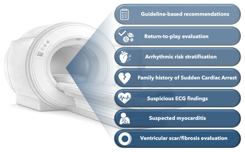
Figure 1. Indications for Cardiac Magnetic Resonance imaging in athletes.
Applsci 16 04330 g001
Figure 2. Graphical resemble of the basic and advanced Cardiac Magnetic Resonance imaging techniques used for diagnostic evaluation of Athletes. ARVC, Arrhythmogenic Right Ventricular Cardiomyopathy; CMR, Cardiac Magnetic Resonance; ECV, Extra-cellular Volume; LGE, Late Gadolinium Enhancement; PA, Pulmonary Artery; RV, Right Ventricle; SSFP, Steady State Free Precession; 4D, Four-Dimensional.
Applsci 16 04330 g002
Figure 3. Overview of the principal cardiac magnetic resonance sequences employed in the assessment of the athlete’s heart (cine Steady State Free Precession ((SSFP)) for functional evaluation, phase contrast for aortic and pulmonary flow evaluation, T1/T2 mapping for tissue characterization, and late gadolinium enhancement for fibrosis detection), with illustrative examples of the most frequently encountered findings in athlete’s physiological adaptations. (A,B) cine 4-Chambers and cine Mid-Short-axis (SAX)showing balanced mild enlargement of the cardiac chambers; (C) modified cine SAX for aortic valvular plane showing coronary artery ostia with emerging right coronary artery (RCA) and left main coronary artery (LMCA) (red arrows); (D) phase-contrast flow imaging at the aortic plane; (E) post-contrast T1 mapping Mid-SAX showing septal-junctional late gadolinium enhancement (LGE) (cyan arrow); (F) Native T1 mapping (4-chambers view) demonstrating T1 values approaching the lower limit of normal.
Applsci 16 04330 g003
Table 1. Overview of the main Cardiac Magnetic Resonance imaging techniques used for diagnostic evaluation of Athletes: comparison between physiological adaptation (athlete’s heart) and pathological findings.
CMR
Technique
Main indication in AthletesPhysiological Adaptation
(Athlete’s Heart)
Pathological Findings
Cine CMR (SSFP)Evaluation of ventricular volumes, mass, function and remodeling pattern, aortic valvular plane and coronary artery ostia evaluation
  • ↑ LV/RV volumes
  • ↑ LV mass
  • preserved or ↑ EF
  • Disproportionate LV/RV dilatation
  • ↓ LVEF and/or RVEF
  • Regional wall motion abnormalities (hypokinesia, akinesia, dyskinesia, aneurysm)
  • Asymmetric hypertrophy (e.g., septal in HCM)
  • Segmental wall thinning (e.g., ARVC)
  • Aortic annulus dilation
  • Coronary artery anomalous origin
RV-focused CMRSuspected ARVC/exercise-induced RV
remodeling/Atrial septal defect/Anomalous pulmonary venous return
  • RV volumes ↑
  • preserved function
  • Regional RV akinesia/dyskinesia
  • RV aneurysms
  • ↓ RVEF
  • RV enlargement out of proportion to training load
Late gadolinium enhancement (LGE)Detection of focal fibrosis or scar/Aortic and Pulmonary artery flow analysis with phase contrast and Qp/Qs evaluationJunctional LGE
  • Focal myocardial fibrosis/scar
  • Subendocardial/transmural LGE (e.g., ischemic pattern)
  • Mid-wall LGE (e.g., DCM)
  • Subepicardial LGE (e.g., NILVS/myocarditis)
  • RV free wall LGE (e.g., ARVC)
  • Alterated Qp/Qs
Native T1 mappingDetection of diffuse fibrosis, inflammation and tissue compositionLower limit of normal, mildly ↓ or ↔
  • ↑ native T1 (e.g., diffuse fibrosis, edema, inflammation, infiltration)
  • ↑↑ T1 (e.g., amyloidosis)
  • ↓ T1 (e.g., lipid/iron overload)
Post-contrast T1/ECVEvaluation and detection of interstitial
Expansion, diffuse fibrosis, infiltrative diseases and oedema
↔ or mildly ↓
  • ↑ ECV (e.g., diffuse interstitial fibrosis)
  • ↑↑ ECV (e.g., amyloidosis)
T2 mappingDetection and evaluation of myocardial
oedema/inflammation
  • ↑ T2 values (e.g., myocardial edema, active inflammation)
CMR strain (feature tracking)Evaluation of subclinical/mild systolic
dysfunction
Lower limit of normal, mildly ↓ or ↔ GLS
  • ↓ GLS and/or regional strain impairment
  • Disproportionate strain reduction relative to EF
  • Mechanical dispersion (arrhythmogenic substrate)
RV-focused CMRSuspected ARVC/exercise-induced RV
remodeling/Atrial septal defect/Anomalous pulmonary venous return
  • RV volumes ↑
  • preserved function
  • Regional RV akinesia/dyskinesia
  • RV aneurysms
  • ↓ RVEF
  • RV enlargement out of proportion to training load
4D-flow CMREvaluation of hemodynamics, flow
efficiency and RV–PA coupling
Optimized flow patterns
  • Abnormal flow vortices
  • ↓ flow efficiency
  • ↑ turbulent kinetic energy
  • Impaired RV–PA coupling
Parametric
mapping
(integrated)
Early disease detection and/or equivocal cases
  • Normal myocardial tissue characterization
  • Absence of pathological LGE
  • Abnormal multiparametric pattern (↑ T1, ↑ T2, ↑ ECV)
  • Discordant mapping values suggesting early cardiomyopathy
  • Persistent abnormalities despite normal cine findings
ARVC, Arrhythmogenic Right Ventricular Cardiomyopathy; CMR, Cardiac Magnetic Resonance; ECV, Extra-cellular Volume; DCM, Dilated Cardiomyopathy; EF, Ejection Fraction; GLS, Global Longitudinal Strain; HCM, Hypertrophic Cardiomyopathy; LGE, Late Gadolinium Enhancement; LV, Left Ventricle; LVEF, Left Ventricular Ejection Fraction; NILVS, Non-ischemic Left Ventricular Scar; PA, Pulmonary Artery; Qp/Qs, Pulmonary-to-Systemic flow ratio; RV, Right Ventricle; RVEF, Right Ventricular Ejection Fraction; SSFP, Steady State Free Precession; 4D, Four-Dimensional; ↑, augmented; ↓, reduced; ↔, unchanged/normal.
Disclaimer/Publisher’s Note: The statements, opinions and data contained in all publications are solely those of the individual author(s) and contributor(s) and not of MDPI and/or the editor(s). MDPI and/or the editor(s) disclaim responsibility for any injury to people or property resulting from any ideas, methods, instructions or products referred to in the content.