3D-Printed and Bioprinted Scaffolds in Regenerative Endodontics: A Systematic Review

 ,
,  , , ,
, , ,  , and
, and 
Featured Application
Abstract
1. Introduction
2. Materials and Methods
2.1. Protocol and Registration
2.2. Research Question and Eligibility Framework (PICOS)
2.3. Information Sources and Search Strategy
2.4. Eligibility Criteria
2.5. Study Selection
2.6. Data Extraction and Synthesis
2.7. Quantitative Synthesis (Intra-Study Analysis)
2.8. Risk of Bias Assessment
3. Results
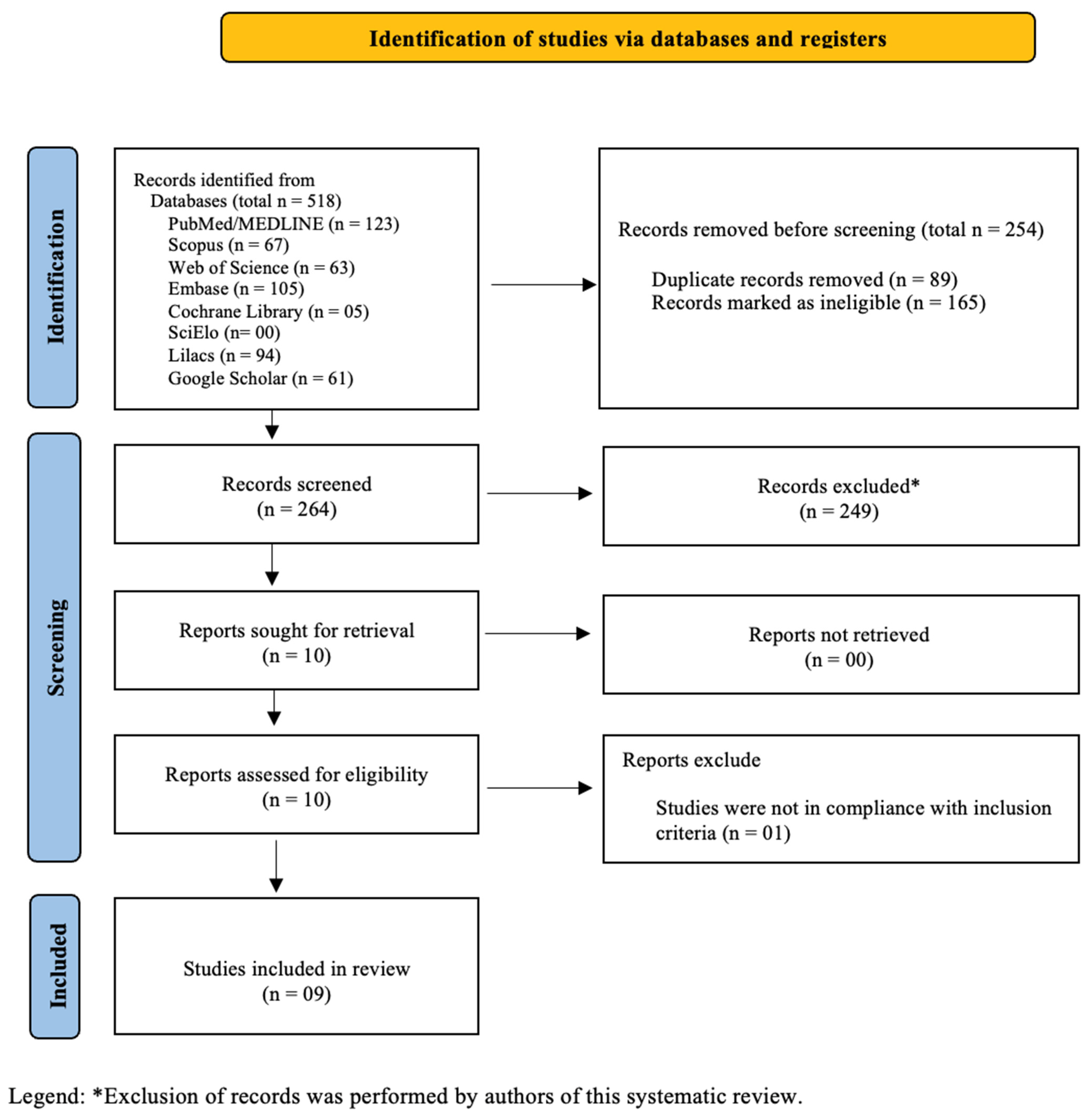
3.1. Study Selection
3.2. Characteristics of Included Studies

| Author (Year) | Main Objective | Experimental Model | Bioink/Scaffold Composition | Printing | Endodontic Focus | Conclusion |
|---|---|---|---|---|---|---|
| Cho et al. 2023 [2] | To evaluate whether a leptin-loaded, aminated PCL 3D-printed scaffold enhances adhesion, proliferation, and odontoblastic differentiation of human dental pulp cells. | hDPSCs | A 3D-printed PCL scaffold with 65% porosity and 6 mm disks was produced. Its surface was aminated with 1,6-hexanediamine and then functionalized with leptin using Traut’s reagent and Sulfo-SMCC. | Extrusion-based 3D printing (Dr. in vivo 4D6; 0.4 mm nozzle). | Regenerative endodontics and vital pulp therapy. | Leptin-loaded 3D-printed PCL increased wettability and enhanced dental pulp cell adhesion, proliferation, and odontoblastic differentiation, supporting its potential as a dental pulp transplant material pending further preclinical and clinical validation. |
| Ortega et al. 2025 [9] | This study describes the development of biocompatible 3D-printed hydrogel scaffolds intended for root canal disinfection. | hDPSCs | A 5% gelatin-based hydrogel was produced with added antimicrobial agents at concentrations between 3.9 and 1000 µg/mL, and corresponding freeze-dried versions were also prepared. | Extrusion-based 3D printing (BIOX6, Cellink®): | Regenerative endodontics. | The scaffolds showed good stability, controlled release, strong antimicrobial activity at 150 and 250 μg/mL, cell viability above 70%, and six-month stability after freeze-drying. |
| Karkehabadi et al. 2025 [11] | To design, fabricate, and characterize a novel nanocomposite scaffold based on alginate-xanthan containing copper-doped bioactive glass nanoparticles. | Bioactive glass nanoparticles with varying copper concentrations (0, 0.5, 2.5 and 5 wt%). | Alginate-xanthan polymer matrix + mesoporous bioactive glass nanoparticles doped with Cu at 0, 0.5, 2.5, and 5 wt%. | Extrusion-based 3D printing (0.4 mm extrusion width; 12 mm3/s speed; 3-layer scaffolds; 35–45% infill) followed by freeze-drying. | Regenerative endodontics. | Scaffolds containing 2.5 wt% copper-doped bioactive glass performed best, whereas 5 wt% led to nanoparticle aggregation, reduced antioxidant activity, and higher hemolysis, showing that copper is beneficial at low levels but toxic at higher doses. |
| Yu et al. 2019 [12] | To compare the effects of Alginate/Gelatin (Alg-Gel) hydrogel vs 3D-printed Alg-Gel scaffolds on adhesion, proliferation, and osteogenic/odontogenic differentiation of hDPSCs. | hDPSCs | Hydrogel composed of 20% gelatin + 4% alginate, crosslinked with CaCl2 and glutaraldehyde. | Extrusion-based 3D bioprinting using a Bioplotter (Envisiontec); nozzle 400 µm; 37 °C printing. | Regenerative endodontics evaluation of scaffolds for dentin–pulp complex regeneration. | These results indicate that 3D printed alginate-gelatin scaffolds better support stem cell growth and more effectively promote cell proliferation and differentiation than their non-printed counter parts. |
| Chiu et al. 2017 [23] | To assess whether an MTA-PCL hybrid 3D scaffold supports osteogenic differentiation of human dental pulp stem cells and how this composite affects the scaffold’s biological performance. | hDPSCs | MTA and PCL hybrid scaffold (60/40 wt%). | Extrusion-based 3D printing (BioScaffolder 3.1; nozzle 500 µm; layer-by-layer deposition). | Regenerative endodontics-pulp tissue regeneration and osteogenic differentiation. | The 3D-printed MTA/PCL scaffold showed excellent physicochemical properties and enhanced osteogenic differentiation, indicating its strong potential as a biomaterial for bone tissue engineering. |
| Athirasala et al. 2018 [33] | To develop and characterize a dentin-derived extracellular matrix hydrogel bioink for 3D bioprinting of cell-laden scaffolds and evaluate its printability, cytocompatibility, and odontogenic potential. | Mouse odontoblast-like cells and SCAPs | Alginate (3% w/v) blended with insoluble dentin matrix proteins at ratios 2:1, 1:1, and 1:2 (Alg:Dent). Soluble dentin molecules added at 1–100 μg/mL. | Extrusion-based bioprinting using a customized Hyrel3D coaxial nozzle system. | Regenerative endodontics-engineering scaffolds for regeneration of the pulp–dentin complex. | The dentin-derived hybrid bioinks showed good printability, supported cell viability, and enhanced odontogenic differentiation, indicating potential for pulp–dentin and craniofacial tissue regeneration. |
| Leveque et al. 2024 [35] | This study aims to design a simple and standardized model to identify parameters that influence the early apical release kinetic of molecules from endodontic hydrogels. | 3D-printed ERI model | Fibrin hydrogels were produced by combining fibrinogen with sodium chloride, calcium chloride, and a fluorescent tracer, then adding thrombin to yield final fibrin concentrations of 1.5–6 mg/mL. | 3D printing (Formlabs 3B printer; 405 nm photopolymer resin; 50 μm layer thickness). | Drug/molecule release in regenerative endodontics. | ERI use enables investigation of the parameters influencing release kinetics from endodontic hydrogels. Further investigations are necessary to evaluate the interaction of these parameters with each other in animal models and clinic. |
| Ho et al. 2018 [43] | To determine whether a 3D-printed Biodentine-PCL scaffold promotes human dental pulp cell proliferation and odontogenic differentiation, while also assessing its physicochemical characteristics. | hDPSCs | Biodentine + PCL (60 wt% Biodentine in PCL). | Extrusion-based 3D printing (BioScaffolder 3.1; nozzle 500 µm; layer-by-layer deposition). | Regenerative endodontics-odontogenesis/dentin–pulp complex repair. | The composite scaffolds showed Biodentine-like bioactivity and promoted stem cell differentiation, indicating strong potential for dental and bone regeneration. |
| Dawood & Mahdee, 2025 [39] | To fabricate and characterize 3D-printed PLA porous scaffolds coated with nHA, NAR, or their combination (nHA/NAR), evaluating mechanical properties, antibacterial activity, cytotoxicity, and bioactivity for pulp regeneration. | FDM-printed PLA structures | PLA scaffolds with 300 or 700 µm pores were coated via PVA assisted dip-coating with nHA, NAR, or a combination of both. | Fused deposition modeling 3D printing (Creality CR-10S; 210 °C extrusion; 0.1 mm layer thickness). | Regenerative endodontics/pulp regeneration. | The 3D-printed PLA scaffold coated with nHA and NAR showed improved surface features, mechanical strength, antibacterial effects, and biocompatibility, indicating strong potential for regenerative applications. |
| Author (Year) | Groups | Viability Assays | Differentiation/Markers | Mineralization/Bioactivity | Physicochemical Properties | Antimicrobial Tests |
|---|---|---|---|---|---|---|
| Cho et al. 2023 [2] | PCL Aminated PCL Leptin-loaded PCL | EZ-Cytox (%) 24 h Cell proliferation was significantly higher in the Leptin-loaded PCL group than in both the control and Aminated PCL groups (p < 0.001 and p < 0.0001). | ALP 7 d | Alizarin red S 14 d | The scaffold has 270–340 μm pores, increased hydrophilicity after amination, crystal formation after leptin conjugation, and a controlled leptin release reaching about 96 percent in 14 days. | n.a. |
| Ortega et al. 2025 [9] | Gel GL5 GL5-Q125 GL5-Q250 GL5-Q500 GL5-Q1000 Freeze-dried FGL5 FGL5-Q125 FGL5-Q250 FGL5-Q500 FGL5-Q1000 | MTT assay (%) 24 h, 72 h and 7 d | n.a. | n.a. | The bioinks showed initial elasticity, shear-thinning behavior, and thermal instability near 26 °C. BDMDAC enhanced structural strength and viscosity, forming denser networks. The analysis was descriptive without statistical testing. | CFU/mL MRSA 24 h GL5: ≅107 GL5-Q3.9: ≅107 GL5-Q7.8: ≅107 GL5-Q15: n.a. GL5-Q31: n.a. 24 h FGL5: ≅108 FGL5-Q3.9: ≅108 FGL5-Q7.8: ≅108 FGL5-Q15: n.a. FGL5-Q31: n.a. P. aeruginosa 24 h GL5: ≅107 GL5-Q125 ≅ 107 GL5-Q250: ≅106 *** GL5-Q500: ≅103 *** GL5-Q1000: n.a. 24 h FGL5: ≅109 FGL5-Q125 ≅ 109 FGL5-Q250: ≅108 *** FGL5-Q500: ≅108 *** FGL5-Q1000: n.a. P. gingivalis 24 h GL5: ≅103 GL5-Q15 ≅ 103 GL5-Q31: ≅103 GL5-Q62: n.a. GL5-Q125: n.a. 24 h FGL5: ≅103 FGL5-Q15: ≅103 FGL5-Q31: ≅103 FGL5-Q62: n.a. FGL5-Q125: n.a. E. faecalis 24 h GL5: ≅108 GL5-Q31: ≅197 GL5-Q62: ≅ 106 *** GL5-Q125: n.a. GL5-Q250: n.a. 24 h FGL5: ≅109 FGL5-Q31: ≅107 FGL5-Q62: ≅106 *** FGL5-Q125: n.a. FGL5-Q250: n.a. S. mutans 24 h GL5: ≅107 GL5-Q31: ≅197 GL5-Q62: ≅105 *** GL5-Q125: n.a. GL5-Q250: n.a. 24 h FGL5: ≅107 FGL5-Q31: ≅107 FGL5-Q62: ≅107 FGL5-Q125: n.a. FGL5-Q250: n.a. |
| Karkehabadi et al. 2025 [11] | B0 B0.5 B2.5 B5 | n.a. | n.a. | n.a. | FTIR Copper caused band shifts and reduced OH-band intensity, indicating its incorporation into the glass network. The composite scaffolds showed additional band shifts, suggesting strong polymer bioglass interactions. EDS EDS confirmed copper doping in the nanoparticles (B0.5, B2.5, B5). In the scaffolds, A-X contained C, Ca, O, and N; A-XB0 added Si; and A-XB0.5, A-XB2.5, and A-XB5 incorporated Cu, confirming successful integration. XRD The XRD diffractogram revealed a high degree of similarity among the four nanoparticle samples, indicating that all of them exhibit a predominantly amorphous character. | |
| Yu et al. 2019 [12] | NC Alg-Gel 3 D-printed-Alg-Gel | DSPP The 3D Alg-Gel group showed significantly higher levels than both the NC and Alg-Gel groups, and the conventional Alg-Gel group also exceeded the NC group (p < 0.001). CCK-8 1 d, 3 d and 5 d Cell proliferation was significantly higher in the Alg-Gel and 3D Alg-Gel groups than in NC, with the 3D scaffold showing the best results at both 3 and 5 days (p < 0.001). | Real-time PCR ALP OCN The 3D Alg-Gel group showed significantly higher levels than both the NC and Alg-Gel groups, and the conventional Alg-Gel group also exceeded the NC group (p < 0.001) across both periods and analyses. | Alizarin red S 7 d and 14 d | Elemental analysis Calcium Phosphorus | n.a. |
| Chiu et al. 2017 [23] | Control PCL MTA/PCL | PrestoBlue 3 h, 6 h, 12 h, 1 d, 3 d and 7 d | ALP 3 d and 7 d OC (pg/mL) 7 d and 14 d | Alizarin red S 0 d, 7 d and 14 d Ion release Si-PCL, Si-MTA/PCL, Ca-PCL and Ca-MTA/PCL 0 h, 1 d, 2 d, 3 d, 4 d, 5 d and 6 d SBF immersion test (bioactivity) Apatite deposition increased from approximately 15% at 0.5 days to about 80% at 7 days. | Pore structure & morphology Pore size ~500 µm; 70% porosity; fully interconnected structure. Mechanical strength (MPa) The scaffold showed a compressive strength of about 4.5 MPa, which increased during the first 30 days in SBF but declined with longer immersion. XRD MTA and PCL retain their original crystalline structure after processing. | n.a. |
| Athirasala et al. 2018 [33] | Alginate 3% Alg-Dent 2:1, 1:1 and 1:2 | Live dead Alginate 3% Alg-Dent 2:1, 1:1 and 1:2 1 d, 3 d and 5 d | ALP 7 d Alg-Dent 1:1 An increase in protein-level ALP activity was observed in the bioinks containing 100 μg/mL compared with the 1:1 Alg-Dent control group (p < 0.05). | n.a. | Pure alginate exhibited a compression modulus of approximately 6 kPa, whereas the addition of 2, 1, or ½ parts of dentin to each part of alginate significantly reduced this value (p < 0.05) to around 1–2 kPa. | n.a. |
| Leveque et al. 2024 [35] | Apical diameter 0.5 mm, 1.0 mm and 2.0 mm Apical solution PBS and Serum Hydrogel type Agarose 2.5% Fibrin 1.5, 3 and 6 mg/mL Molecule type Fluorescein and BDP-500 Nanoencapsulation: BDP-500 Nanoencapsulated BDP-500 | n.a. | n.a. | Apical solution 0 h, 8 h, 16 h and 24 h A statistically significant difference was observed between the solutions (p < 0.001). Hydrogel type 0 h, 8 h, 16 h and 24 h No significant difference was observed between hydrogel types, but reducing fibrin from 3 to 1.5 mg/mL resulted in a significant change (p < 0.001). Molecule type 0 h, 8 h, 16 h and 24 h A statistically significant difference was observed between the solutions (p < 0.05). | Apical diameter 0 h, 8 h, 16 h and 24 h All apical diameters exhibited statistically significant differences between each Other (p < 0.05). Nanoencapsulation 0 h, 8 h, 16 h and 24 h A statistically significant difference was observed between the solutions (p < 0.05). | n.a. |
| Ho et al. 2018 [43] | Control PCL BD/PCL | PrestoBlue 1 d, 3 d and 7 d | ALP 3 d and 7 d OC (pg/mL) 7 d and 14 d | Alizarin red S 0 d, 7 d and 14 d SBF immersion test (bioactivity) Analyses showed ~500 μm struts and ~550 μm pores, with SBF immersion producing early precipitates and a dense apatite layer by 7 days. | Mechanical strength (MPa) Scaffolds showed ~500 μm struts, 550 μm pores, compressive strength ~6.5 MPa, and rapid apatite formation in SBF. | n.a. |
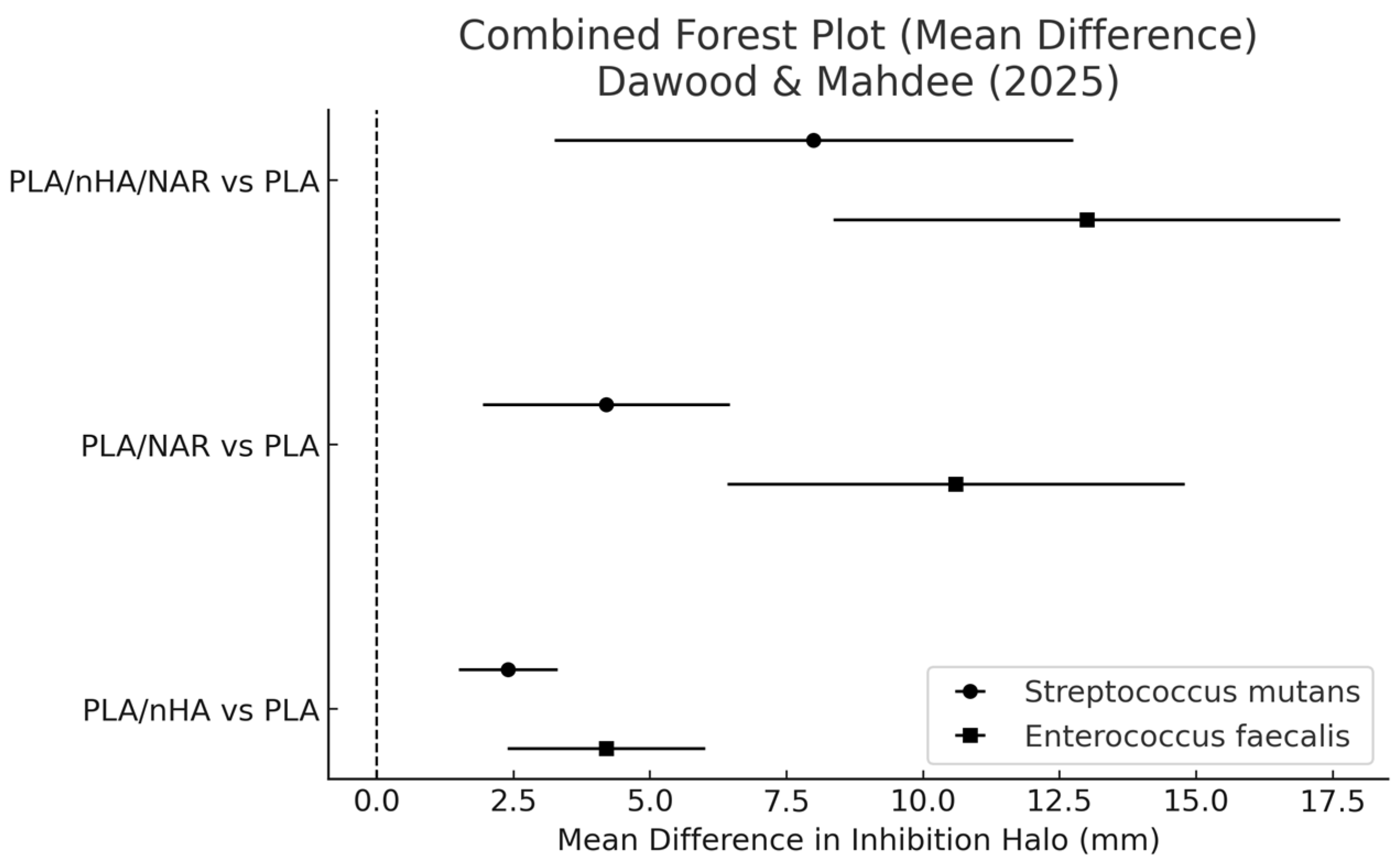
| Dawood & Mahdee, 2025 [39] | PLA PLA/nHA PLA/NAR PLA/nHA/NAR | MTT assay The PLA scaffold was biocompatible (p > 0.05), NAR was less cytotoxic than nHA, and the NAR + nHA combination remained biocompatible with 27.15% cytotoxicity and synergistic activity. | n.a. | It was observed that a calcium phosphate apatite layer formed on the surface of the PLA/nHA and PLA/nHA/NAR scaffolds after immersion in SBF. | EDX analysis Pure PLA showed only carbon and oxygen. nHA added calcium and phosphorus, and NAR increased carbon. After SBF immersion, calcium rose in all groups and phosphorus rose only with nHA. No statistical tests were performed. Compressive and flexural strength Compression and Flexural P300 and P700 Strength differed significantly across pore sizes (p < 0.000), but coating types showed no differences when pore size was constant (p < 0.000). | Halo/mm S. mutans E. Faecalis Different superscript letters within each bacterial test indicate statistically significant differences (p < 0.05). |
3.3. Viability Assays
3.4. Differentiation and Marker Expression
3.5. Mineralization and Bioactivity
3.6. Physicochemical Properties
3.7. Antimicrobial Tests
3.8. Intra-Study Quantitative Assessment
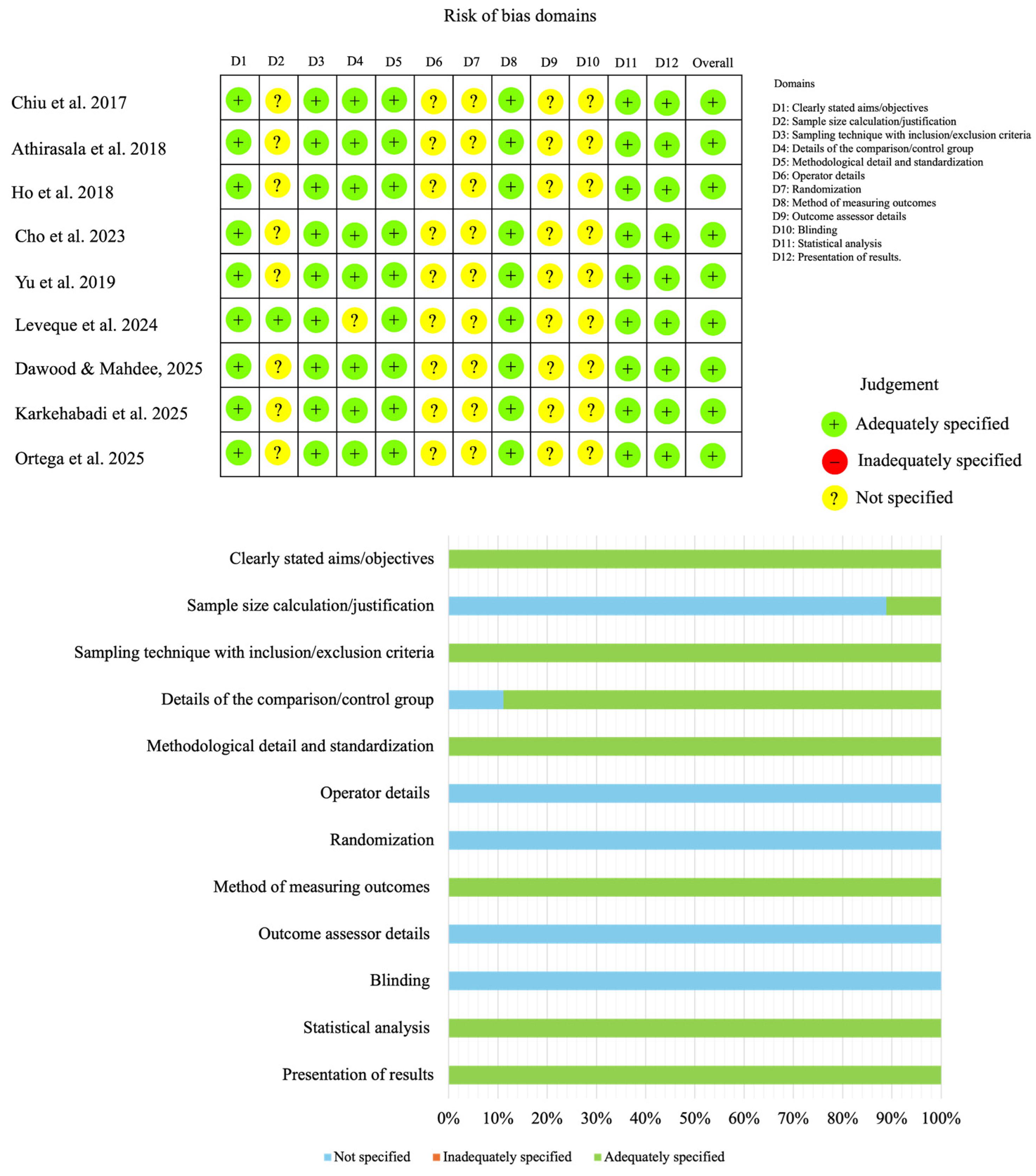
3.9. Risk of Bias Assessment of the Included Studies
4. Discussion
5. Conclusions
Supplementary Materials
Author Contributions
Funding
Institutional Review Board Statement
Informed Consent Statement
Data Availability Statement
Conflicts of Interest
Abbreviations
| 3D | Three-dimensional |
| Alg-Gel | Hybrid alginate–gelatin |
| ALP | Alkaline phosphatase |
| AI | Artificial Intelligence |
| BD | Biodentine |
| BDMDAC | Benzyldimethyldodecylammonium chloride |
| C | Comparison |
| DSPP | Dentin sialophosphoprotein |
| FDM | Fused deposition modeling |
| hDPSCs | Human dental pulp stem cells |
| I | Intervention |
| MD | Mean differences |
| MeSH | Medical subject headings |
| MTA | Mineral trioxide aggregate |
| MTT | 3-[4,5-dimethylthiazol-2-yl]-2,5-diphenyl tetrazolium bromide |
| NA | Not applicable |
| NAR | Naringin |
| nHA | Nano-hydroxyapatite |
| O | Outcome |
| OCN | Osteocalcin |
| OSF | Open science framework |
| P | Population |
| PCL | Poly-ε-caprolactone |
| PEG | Polyethylene glycol |
| PLA | Polylactic acid |
| PRISMA | Preferred Reporting Items for Systematic Reviews and Meta-Analysis |
| REPs | Regenerative endodontic procedures |
| S | Study design |
| SBF | Simulated body fluid |
| SCAPS | Stem cells from the apical papilla |
References
- Nakashima, M.; Akamine, A. The application of tissue engineering to regeneration of pulp and dentin in endodontics. J. Endod. 2005, 31, 711–718. [Google Scholar] [CrossRef] [Scilit] [PubMed]
- Cho, E.H.; Kim, Y.S.; Kim, Y.R.; Kang, J.H.; Park, S.W.; Lim, H.P.; Yun, K.D.; Jang, W.H.; Koh, J.T.; Park, C.; et al. A leptin-loaded poly-ϵ-caprolactone 3D printing scaffold for odontoblastic differentiation in human dental pulp cells. Biomed. Mater. 2023, 19, 011001. [Google Scholar] [CrossRef] [Scilit]
- Santos, J.M.; Diogo, P.; Dias, S.; Marques, J.A.; Palma, P.J.; Ramos, J.C. Long-term outcome of nonvital immature permanent teeth treated with apexification and corono-radicular adhesive restoration: A case series. J. Endod. 2022, 48, 1191–1199. [Google Scholar] [CrossRef] [Scilit]
- Widbiller, M.; Knüttel, H.; Meschi, N.; Durán-Sindreu Terol, F. Effectiveness of endodontic tissue engineering in treatment of apical periodontitis: A systematic review. Int. Endod. J. 2023, 56, 533–548. [Google Scholar] [CrossRef] [Scilit]
- Sabeti, M.; Ghobrial, D.; Zanjir, M.; da Costa, B.R.; Young, Y.; Azarpazhooh, A. Treatment outcomes of regenerative endodontic therapy in immature permanent teeth with pulpal necrosis: A systematic review and network meta-analysis. Int. Endod. J. 2024, 57, 238–255. [Google Scholar] [CrossRef] [Scilit]
- Duncan, H.F.; Kirkevang, L.L.; Peters, O.A.; El-Karim, I.; Krastl, G.; Del Fabbro, M.; Chong, B.S.; Galler, K.M.; Segura-Egea, J.J.; Kebschull, M. Treatment of pulpal and apical disease: The European Society of Endodontology (ESE) S3-level clinical practice guideline. Int. Endod. J. 2023, 56, 238–295. [Google Scholar] [CrossRef] [Scilit]
- Lin, L.M.; Kahler, B. A review of regenerative endodontics: Current protocols and future directions. J Istanb Univ Fac Dent. 2017, 2, S41–S51. [Google Scholar] [CrossRef] [Scilit]
- Arruda, M.E.F.; Neves, M.A.S.; Diogenes, A.; Mdala, I.; Guilherme, B.P.S.; Siqueira, J.F., Jr.; Rôças, I.N. Infection control in teeth with apical periodontitis using a triple antibiotic solution or calcium hydroxide with chlorhexidine: A randomized clinical trial. J. Endod. 2018, 44, 1474–1479. [Google Scholar] [CrossRef] [Scilit]
- Ortega, M.D.; Aveyard, J.; Abdelgawad, R.M.; El-Gendy, R.; Ciupa, A.; Whetnall, D.; Behnsen, J.; Poole, R.J.; D’Sa, R.A. Antimicrobial 3D printed gelatin scaffolds for root canal disinfection in regenerative endodontics procedures. Biomater. Sci. 2025, 13, 3795. [Google Scholar] [CrossRef] [Scilit] [PubMed]
- Nowicka, A.; Miller-Burchacka, M.; Lichota, D.; Metlerska, J.; Gońda-Domin, M. Tissue engineering application in regenerative endodontics. Pomeranian J. Life Sci. 2021, 67, 10–17. [Google Scholar] [CrossRef] [Scilit]
- Karkehabadi, H.; Sharifi, E.; Khoshbin, E. Alginate-xanthan nanocomposite scaffolds incorporating copper-doped bioactive glass for novel tissue engineering potential in regenerative endodontics. J. Clust. Sci. 2025, 36, 180. [Google Scholar] [CrossRef] [Scilit]
- Yu, H.; Zhang, X.; Song, W.; Pan, T.; Wang, H.; Ning, T.; Wei, Q.; Xu, H.H.K.; Wu, B.; Ma, D. Effects of 3-dimensional bioprinting alginate/gelatin hydrogel scaffold extract on proliferation and differentiation of human dental pulp stem cells. J. Endod. 2019, 45, 706–715. [Google Scholar] [CrossRef] [Scilit] [PubMed]
- Sequeira, D.B.; Oliveira, A.R.; Seabra, C.M.; Palma, P.J.; Ramos, C.; Figueiredo, M.H.; Santos, A.C.; Cardoso, A.L.; Peça, J.; Santos, J.M. Regeneration of pulp-dentin complex using human stem cells of the apical papilla: In vivo interaction with two bioactive materials. Clin. Oral. Investig. 2021, 25, 5317–5329. [Google Scholar] [CrossRef] [Scilit]
- Atalayin, C.; Tezel, H.; Dagci, T.; Karabay Yavasoglu, N.U.; Oktem, G.; Kose, T. In vivo performance of different scaffolds for dental pulp stem cells induced for odontogenic differentiation. Braz. Oral. Res. 2016, 30, e120. [Google Scholar] [CrossRef] [Scilit]
- Gathani, K.M.; Raghavendra, S.S. Scaffolds in regenerative endodontics: A review. Dent. Res. J. 2016, 13, 379–386. [Google Scholar] [CrossRef] [Scilit]
- Nemati, S.; Rezabakhsh, A.; Khoshfetrat, A.B.; Nourazarian, A.; Biray Avci, Ç.; Goker Bagca, B.; Alizadeh Sardroud, H.; Khaksar, M.; Ahmadi, M.; Delkhosh, A.; et al. Alginate-gelatin encapsulation of human endothelial cells promoted angiogenesis in in vivo and in vitro milieu. Biotechnol. Bioeng. 2017, 114, 2920–2930. [Google Scholar] [CrossRef] [Scilit]
- Sequeira, D.B.; Diogo, P.; Gomes, B.P.F.A.; Peça, J.; Santos, J.M. Scaffolds for dentin–pulp complex regeneration. Medicina 2024, 60, 7. [Google Scholar] [CrossRef] [Scilit]
- Echave, M.C.; Saenz del Burgo, L.; Pedraz, J.L.; Orive, G. Gelatin as biomaterial for tissue engineering. Curr. Pharm. Des. 2017, 23, 3567–3584. [Google Scholar] [CrossRef] [Scilit] [PubMed]
- Lewandowska-Łańcucka, J.; Mystek, K.; Mignon, A.; Van Vlierberghe, S.; Łatkiewicz, A.; Nowakowska, M. Alginate- and gelatin-based bioactive photocross-linkable hybrid materials for bone tissue engineering. Carbohydr. Polym. 2017, 157, 1714–1722. [Google Scholar] [CrossRef] [Scilit] [PubMed]
- Salehi, S.; Cooper, P.; Smith, A.; Ferracane, J. Dentin matrix components extracted with phosphoric acid enhance cell proliferation and mineralization. Dent. Mater. 2016, 32, 334–342. [Google Scholar] [CrossRef] [Scilit]
- Conde, M.C.M.; Chisini, L.A.; Sarkis-Onofre, R.; Schuch, H.S.; Nör, J.E.; Demarco, F.F. A scoping review of root canal revascularization: Relevant aspects for clinical success and tissue formation. Int. Endod. J. 2017, 50, 860–874. [Google Scholar] [CrossRef] [Scilit]
- Cheng, Y.L.; Chen, Y.W.; Wang, K.; Shie, M.Y. Enhanced adhesion and differentiation of human mesenchymal stem cell inside apatite-mineralized/poly(dopamine)-coated poly(ε-caprolactone) scaffolds by stereolithography. J. Mater. Chem. B 2016, 4, 6307–6315. [Google Scholar] [CrossRef] [Scilit] [PubMed]
- Chiu, Y.C.; Fang, H.Y.; Hsu, T.T.; Lin, C.Y.; Shie, M.Y. The characteristics of mineral trioxide aggregate/polycaprolactone 3-dimensional scaffold with osteogenesis properties for tissue regeneration. J. Endod. 2017, 43, 923–929. [Google Scholar] [CrossRef] [Scilit]
- Margunato, S.; Taşlı, P.N.; Aydın, S.; Karapınar Kazandağ, M.; Şahin, F. In vitro evaluation of ProRoot MTA, Biodentine, and MM-MTA on human alveolar bone marrow stem cells in terms of biocompatibility and mineralization. J. Endod. 2015, 41, 1646–1652. [Google Scholar] [CrossRef] [Scilit]
- Gomes-Cornélio, A.L.; Rodrigues, E.M.; Salles, L.P.; Mestieri, L.B.; Faria, G.; Guerreiro-Tanomaru, J.M.; Tanomaru-Filho, M. Bioactivity of MTA Plus, Biodentine and an experimental calcium silicate-based cement on human osteoblast-like cells. Int. Endod. J. 2017, 50, 39–47. [Google Scholar] [CrossRef] [Scilit]
- Silva, E.J.; Senna, P.M.; De-Deus, G.; Zaia, A.A. Cytocompatibility of Biodentine using a three-dimensional cell culture model. Int. Endod. J. 2016, 49, 574–580. [Google Scholar] [CrossRef] [Scilit]
- Prasopthum, A.; Deng, Z.; Khan, I.M.; Yin, Z.; Guo, B.; Yang, J. Three dimensional printed degradable and conductive polymer scaffolds promote chondrogenic differentiation of chondroprogenitor cells. Biomater. Sci. 2020, 8, 4287–4298. [Google Scholar] [CrossRef] [Scilit]
- Shie, M.Y.; Chang, W.C.; Wei, L.J.; Huang, Y.H.; Chen, C.H.; Shih, C.T.; Chen, Y.W.; Shen, Y.F. 3D printing of cytocompatible water-based light-cured polyurethane with hyaluronic acid for cartilage tissue engineering applications. Materials 2017, 10, 136. [Google Scholar] [CrossRef] [Scilit] [PubMed]
- Tyler, B.; Gullotti, D.; Mangraviti, A.; Utsuki, T.; Brem, H. Polylactic acid (PLA) controlled delivery carriers for biomedical applications. Adv. Drug. Deliv. Rev. 2016, 107, 163–175. [Google Scholar] [CrossRef] [Scilit] [PubMed]
- Wang, W.; Zhang, B.; Li, M.; Li, J.; Zhang, C.; Han, Y.; Wang, L.; Wang, K.; Zhou, C.; Liu, L.; et al. 3D printing of PLA/n-HA composite scaffolds with customized mechanical properties and biological functions for bone tissue engineering. Compos. Part. B Eng. 2021, 224, 109192. [Google Scholar] [CrossRef] [Scilit]
- Ribeiro, J.S.; Sanz, C.K.; Münchow, E.A.; Kalra, N.; Dubey, N.; Suárez, C.E.C.; Fenno, J.C.; Lund, R.G.; Bottino, M.C. Photocrosslinkable methacrylated gelatin hydrogel as a cell-friendly injectable delivery system for chlorhexidine in regenerative endodontics. Dent. Mater. 2022, 38, 1507–1517. [Google Scholar] [CrossRef] [Scilit]
- Kumar Tiwari, S.; Guo, X.; Huang, Y.; Zhou, X.; Xu, H.H.K.; Ren, B.; Peng, X.; Weir, M.D.; Li, M.; Cheng, L. The inhibitory effect of quaternary ammonium salt on bacteria in root canal. Sci. Rep. 2019, 9, 12463. [Google Scholar] [CrossRef] [Scilit]
- Athirasala, A.; Tahayeri, A.; Thrivikraman, G.; França, C.M.; Monteiro, N.; Tran, V.; Ferracane, J.; Bertassoni, L.E. A dentin-derived hydrogel bioink for 3D bioprinting of cell-laden scaffolds for regenerative dentistry. Biofabrication 2018, 10, 024101. [Google Scholar] [CrossRef] [Scilit] [PubMed]
- Kharchi, A.S.; Tagiyeva-Milne, N.; Kanagasingam, S. Regenerative endodontic procedures, disinfectants and outcomes: A systematic review. Prim. Dent. J. 2020, 9, 65–84. [Google Scholar] [CrossRef] [Scilit] [PubMed]
- Leveque, M.; Bekhouche, M.; Farges, J.C.; Richert, R.; Ducret, M. Investigation of the early apical release from endodontic hydrogels: A 3D printed model. Int. Endod. J. 2024, 57, 943–950. [Google Scholar] [CrossRef] [Scilit] [PubMed]
- Moher, D.; Liberati, A.; Tetzlaff, J.; Altman, D.G.; PRISMA Group. Preferred reporting items for systematic reviews and meta-analyses: The PRISMA statement. PLoS Med. 2009, 6, e1000097. [Google Scholar] [CrossRef] [Scilit]
- Chaves, H.G.D.S.; Mendes, A.C.S.; Titato, P.C.G.; Dias, V.C.M.; Figueiredo, B.; Duarte, M.A.H.; Cintra, L.T.A.; André, C.B.; Morgan, L.F.S.A.; Santos, J.M.M.d.; et al. A comparison of niobium with other radiopacifying agents in endodontic cements: A systematic review of physicochemical properties. Appl. Sci. 2026, 16, 1722. [Google Scholar] [CrossRef] [Scilit]
- Batista, L.A.S.; Dos Reis-Prado, A.H.; Chaves, H.G.D.S.; De Arantes, L.C.; Morgan, L.F.S.A.; André, C.B.; Suzuki, T.Y.; Benetti, F. Can different agents reduce the damage caused by bleaching gel to pulp tissue? A systematic review of basic research. Restor. Dent. Endod. 2023, 48, e39. [Google Scholar] [CrossRef] [Scilit]
- Dawood, R.M.; Mahdee, A.F. Fabrication and characterization of 3D-printed polymeric-based scaffold coated with bioceramic and naringin for a potential use in dental pulp regeneration (in vitro study). Int. Endod. J. 2025, 58, 627–642. [Google Scholar] [CrossRef] [Scilit]
- Sheth, V.H.; Shah, N.P.; Jain, R.; Bhanushali, N.; Bhatnagar, V. Development and validation of a risk-of-bias tool for assessing in vitro studies conducted in dentistry: The QUIN. J. Prosthet. Dent. 2024, 131, 1038–1042. [Google Scholar] [CrossRef] [Scilit]
- Cabrera-Fernández, A.; dos Santos Chaves, H.G.; Díaz-Cuenca, A.; Segura-Egea, J.J.; Martín-González, J.; Peça, J.; Sequeira, D.B.; dos Santos, J.M.M. Histological tissue response to calcium silicate-based cements assessed in human tooth culture models: A systematic review. J. Funct. Biomater. 2026, 17, 78. [Google Scholar] [CrossRef] [Scilit] [PubMed]
- Parihar, A.S.; Nandanan, N.; Patadiya, H.H.; Jain, M.; Arya, A.; Laddha, R.; Kumar, S. Efficacy of 3D-printed scaffolds in guided tissue regeneration for combined endo-perio lesions: A clinical and radiographic study. J. Pharm. Bioallied Sci. 2025, 17, S568–S570. [Google Scholar] [CrossRef] [Scilit] [PubMed]
- Ho, C.C.; Fang, H.Y.; Wang, B.; Huang, T.H.; Shie, M.Y. The effects of Biodentine/polycaprolactone three-dimensional scaffold with odontogenesis properties on human dental pulp cells. Int. Endod. J. 2018, 51, e291–e300. [Google Scholar] [CrossRef] [Scilit]
- Moussa, M.; Carrel, J.-P.; Scherrer, S.; Cattani-Lorente, M.; Wiskott, A.; Durual, S. Medium-term function of a 3D printed TCP/HA structure as a new osteoconductive scaffold for vertical bone augmentation: A simulation by BMP-2 activation. Materials 2015, 8, 2174–2190. [Google Scholar] [CrossRef] [Scilit]
- Mott, E.J.; Busso, M.; Luo, X.; Dolder, C.; Wang, M.O.; Fisher, J.P.; Dean, D. Digital micromirror device (DMD)-based 3D printing of poly(propylene fumarate) scaffolds. Mater. Sci. Eng. C Mater. Biol. Appl. 2016, 61, 301–311. [Google Scholar] [CrossRef] [Scilit]
- Mohanty, S.; Alm, M.; Hemmingsen, M.; Dolatshahi-Pirouz, A.; Trifol, J.; Thomsen, P.; Dufva, M.; Wolff, A.; Emnéus, J. 3D printed silicone-hydrogel scaffold with enhanced physicochemical properties. Biomacromolecules 2016, 17, 1321–1329. [Google Scholar] [CrossRef] [Scilit] [PubMed]
- Wang, X.; Xu, S.; Zhou, S.; Xu, W.; Leary, M.; Choong, P.; Qian, M.; Brandt, M.; Xie, Y.M. Topological design and additive manufacturing of porous metals for bone scaffolds and orthopaedic implants: A review. Biomaterials 2016, 83, 127–141. [Google Scholar] [CrossRef] [Scilit]
- Laurent, P.; Camps, J.; About, I. Biodentine(TM) induces TGF-β1 release from human pulp cells and early dental pulp mineralization. Int. Endod. J. 2012, 45, 439–448. [Google Scholar] [CrossRef] [Scilit]
- Zanini, M.; Sautier, J.M.; Berdal, A.; Simon, S. Biodentine induces immortalized murine pulp cell differentiation into odontoblast-like cells and stimulates biomineralization. J. Endod. 2012, 38, 1220–1226. [Google Scholar] [CrossRef] [Scilit]
- Luo, Z.; Kohli, M.R.; Yu, Q.; Kim, S.; Qu, T.; He, W.X. Biodentine induces human dental pulp stem cell differentiation through mitogen-activated protein kinase and calcium-/calmodulin-dependent protein kinase II pathways. J. Endod. 2014, 40, 937–942. [Google Scholar] [CrossRef] [Scilit]
- Chen, Y.W.; Yeh, C.H.; Shie, M.Y. Stimulatory effects of the fast setting and suitable degrading Ca–Si–Mg cement on both cementogenesis and angiogenesis differentiation of human periodontal ligament cells. J. Mater. Chem. B 2015, 3, 7099–7108. [Google Scholar] [CrossRef] [Scilit]
- Huang, M.H.; Shen, Y.F.; Hsu, T.T.; Huang, T.H.; Shie, M.Y. Physical characteristics, antimicrobial and odontogenesis potentials of calcium silicate cement containing hinokitiol. Mater. Sci. Eng. C Mater. Biol. Appl. 2016, 65, 1–8. [Google Scholar] [CrossRef] [Scilit]
- Li, J.; Zhai, D.; Lv, F.; Yu, Q.; Ma, H.; Yin, J.; Yi, Z.; Liu, M.; Chang, J.; Wu, C. Preparation of copper-containing bioactive glass/eggshell membrane nanocomposites for improving angiogenesis, antibacterial activity and wound healing. Acta Biomater. 2016, 36, 254–266. [Google Scholar] [CrossRef] [Scilit] [PubMed]
- Kao, C.T.; Shie, M.Y.; Huang, T.H.; Ding, S.J. Properties of an accelerated mineral trioxide aggregate-like root-end filling material. J. Endod. 2009, 35, 239–242. [Google Scholar] [CrossRef] [Scilit] [PubMed]
- Ji, L.; Wang, W.; Jin, D.; Zhou, S.; Song, X. In vitro bioactivity and mechanical properties of bioactive glass nanoparticles/polycaprolactone composites. Mater. Sci. Eng. C Mater. Biol. Appl. 2015, 46, 1–9. [Google Scholar] [CrossRef] [Scilit]
- No, Y.J.; Roohani-Esfahani, S.I.; Lu, Z.; Schaer, T.; Zreiqat, H. Injectable radiopaque and bioactive polycaprolactone-ceramic composites for orthopedic augmentation. J. Biomed. Mater. Res. B Appl. Biomater. 2015, 103, 1465–1477. [Google Scholar] [CrossRef] [Scilit] [PubMed]
- Bekhouche, M.; Bolon, M.; Charriaud, F.; Lamrayah, M.; Da Costa, D.; Primard, C.; Costantini, A.; Pasdeloup, M.; Gobert, S.; Mallein-Gerin, F.; et al. Development of an antibacterial nanocomposite hydrogel for human dental pulp engineering. J. Mater. Chem. B 2020, 8, 8422–8432. [Google Scholar] [CrossRef] [Scilit]
- Namazi, S.S.; Mahmoud, A.H.; Dal-Fabbro, R.; Han, Y.; Xu, J.; Sasaki, H.; Fenno, J.C.; Bottino, M.C. Multifunctional and biodegradable methacrylated gelatin/Aloe vera nanofibers for endodontic disinfection and immunomodulation. Biomater. Adv. 2023, 150, 213427. [Google Scholar] [CrossRef] [Scilit]
- Mustafa, S.; Meheissen, M.A.; Moussa, S.; ElBackly, R. Effect of ultrasonically activated irrigation protocols used for regenerative endodontics on removal of dual species biofilm in a three-dimensionally printed tooth model: In vitro study. BMC Oral. Health 2025, 25, 98. [Google Scholar] [CrossRef] [Scilit]
- Almutairi, W.; Yassen, G.H.; Aminoshariae, A.; Williams, K.A.; Mickel, A. Regenerative endodontics: A systematic analysis of the failed cases. J. Endod. 2019, 45, 567–577. [Google Scholar] [CrossRef] [Scilit]
- Albuquerque, M.T.P.; Nagata, J.; Bottino, M.C. Antimicrobial efficacy of triple antibiotic-eluting polymer nanofibers against multispecies biofilm. J. Endod. 2017, 43, S51–S56. [Google Scholar] [CrossRef] [Scilit] [PubMed]
- Tănase, M.; Veres, C.; Szabo, D.-A. Artificial intelligence in biomedical 3D printing: Mapping the evidence. J. Manuf. Mater. Process 2025, 9, 407. [Google Scholar] [CrossRef] [Scilit]
- Liang, Z.; Liao, X.; Zong, H.; Zeng, X.; Liu, H.; Wu, C.; Keremane, K.; Poudel, B.; Yin, J.; Wang, K.; et al. Pioneering the future of dentistry: AI-driven 3D bioprinting for next-generation clinical applications. Transl. Dent. Res. 2025, 1, 100005. [Google Scholar] [CrossRef] [Scilit]
- Daghrery, A.; Soares, I.P.M.; Reis-Prado, A.H.; Araújo, I.J.S.; Dal-Fabbro, R.; Bottino, M.C. Advances in 3D printed scaffolds for periodontal regeneration. Curr. Oral. Health Rep. 2026, 13, 1. [Google Scholar] [CrossRef] [Scilit]
- Page, M.J.; McKenzie, J.E.; Bossuyt, P.M.; Boutron, I.; Hoffmann, T.C.; Mulrow, C.D.; Shamseer, L.; Tetzlaff, J.M.; Akl, E.A.; Brennan, S.E.; et al. The PRISMA 2020 statement: An updated guideline for reporting systematic reviews. BMJ 2021, 372, n71. [Google Scholar] [CrossRef] [Scilit]


| Domain | Inclusion Criteria | Exclusion Criteria |
|---|---|---|
| Study type | Original experimental studies (in vitro, ex vivo, in vivo or clinical) | Narrative/systematic/scoping reviews; editorials; letters; conference abstracts; book chapters |
| Scope/Relevance | Studies addressing endodontic procedures and/or dental pulp regeneration | Applications exclusively in other dental fields without endodontic/pulp relevance |
| Technology of interest | 3D bioprinting involving bioprinted constructs and/or bioinks, including cell-laden or biologically functionalized inks intended for pulp/endodontic applications | Non-biological 3D printing applications |
| Experimental model | Cell-based and/or bioactive strategies relevant to pulp/endodontic tissue engineering | Studies not relevant to endodontics or without relevance to pulpal/periapical tissues |
| Outcomes reported | At least one relevant outcome: cell viability, proliferation, differentiation, ECM/mineralization, vascularization proxies, biocompatibility, printability linked to biological function | Outcomes unrelated to pulp/endodontic goals with no pulp/endodontic component |
| Language/Timeframe | No restrictions on language or publication date | Duplicates or multiple reports of the same dataset |
| Availability | Full text accessible | Full text unavailable after reasonable efforts |
Disclaimer/Publisher’s Note: The statements, opinions and data contained in all publications are solely those of the individual author(s) and contributor(s) and not of MDPI and/or the editor(s). MDPI and/or the editor(s) disclaim responsibility for any injury to people or property resulting from any ideas, methods, instructions or products referred to in the content. |
© 2026 by the authors. Licensee MDPI, Basel, Switzerland. This article is an open access article distributed under the terms and conditions of the Creative Commons Attribution (CC BY) license.
Share and Cite
Chaves, H.G.d.S.; Sequeira, D.B.; Dias, V.C.M.; Cabrera-Fernández, A.; Peça, J.; Benetti, F.; Santos, J.M.M.d. 3D-Printed and Bioprinted Scaffolds in Regenerative Endodontics: A Systematic Review. Appl. Sci. 2026, 16, 3940. https://doi.org/10.3390/app16083940
Chaves HGdS, Sequeira DB, Dias VCM, Cabrera-Fernández A, Peça J, Benetti F, Santos JMMd. 3D-Printed and Bioprinted Scaffolds in Regenerative Endodontics: A Systematic Review. Applied Sciences. 2026; 16(8):3940. https://doi.org/10.3390/app16083940
Chicago/Turabian StyleChaves, Hebertt Gonzaga dos Santos, Diana B. Sequeira, Vilton Cardozo Moreira Dias, Alberto Cabrera-Fernández, João Peça, Francine Benetti, and João Miguel Marques dos Santos. 2026. "3D-Printed and Bioprinted Scaffolds in Regenerative Endodontics: A Systematic Review" Applied Sciences 16, no. 8: 3940. https://doi.org/10.3390/app16083940
APA StyleChaves, H. G. d. S., Sequeira, D. B., Dias, V. C. M., Cabrera-Fernández, A., Peça, J., Benetti, F., & Santos, J. M. M. d. (2026). 3D-Printed and Bioprinted Scaffolds in Regenerative Endodontics: A Systematic Review. Applied Sciences, 16(8), 3940. https://doi.org/10.3390/app16083940