Article

First Molecular and Metagenomic Investigation of the Italian Honey Bee (Apis mellifera) Microbiome †

by
Fulvio Bordin
1,*,‡,
Arianna Peruzzo
2,‡,
Gianpiero Zamperin
3,‡,
Elisa Palumbo
3,
Adelaide Milani
3,
Massimiliano Orsini
2,§,
Alice Fusaro
3,
Michela Bertola
1,
Paola Mogliotti
4,
Monica Pierangela Cerioli
5,
Giovanni Formato
6,
Luciano Ricchiuti
7,
Anna Cerrone
8,
Pasquale Troiano
9,
Antonio Salvaggio
10,
Antonio Pintore
11,
Franco Mutinelli
1 and
Anna Granato
1
1
National Reference Laboratory for Honey Bee Health, Istituto Zooprofilattico Sperimentale delle Venezie, 35020 Legnaro, Italy
2
Laboratory of Microbial Ecology and Genomics, Istituto Zooprofilattico Sperimentale delle Venezie, 35020 Legnaro, Italy
3
Department of Virology, Research and Innovation Health, Istituto Zooprofilattico Sperimentale delle Venezie, 35020 Legnaro, Italy
4
Istituto Zooprofilattico Sperimentale del Piemonte, Liguria e Valle d’Aosta, 14100 Asti, Italy
5
Istituto Zooprofilattico Sperimentale della Lombardia e dell’Emilia Romagna “Bruno Ubertini”, 25124 Brescia, Italy
6
Istituto Zooprofilattico Sperimentale del Lazio e della Toscana, 00178 Roma, Italy
7
Istituto Zooprofilattico Sperimentale dell’Abruzzo e del Molise “G. Caporale”, 66034 Lanciano, Italy
8
Istituto Zooprofilattico Sperimentale del Mezzogiorno, 80055 Portici, Italy
9
Istituto Zooprofilattico Sperimentale della Puglia e della Basilicata, 71121 Foggia, Italy
10
Istituto Zooprofilattico Sperimentale della Sicilia, 95125 Catania, Italy
11
Istituto Zooprofilattico Sperimentale della Sardegna, 07100 Sassari, Italy
*
Author to whom correspondence should be addressed.
This paper is an extended version of our paper published in Bordin, F.; Zamperin, G.; Peruzzo, A.; et al. Identificazione e Caratterizzazione Di Patogeni Noti, Emergenti e Simbionti Di Apis mellifera Nel Territorio Italiano Mediante Protocolli Molecolari e Tecnologie NGS: Risultati Preliminari. In Proceedings of the XXII Congresso Nazionale S.I.Di.L.V. Società Italiana Diagnostica Di Laboratorio Veterinaria, Brescia, Italy, 11–13 October 2023.
These authors contributed equally to this work.
§
Deceased author.
Appl. Sci. 2026, 16(7), 3521; https://doi.org/10.3390/app16073521
Submission received: 17 February 2026 / Revised: 24 March 2026 / Accepted: 31 March 2026 / Published: 3 April 2026
(This article belongs to the Special Issue Advances in Honeybee and Their Biological and Environmental Threats)

Abstract

The western honey bee (Apis mellifera) represents a key pollinator for both crops and wild plants, and its global decline raises serious concerns for ecosystem stability and agricultural productivity. Several biotic and abiotic factors are responsible for colony losses, including alterations in the bee microbiota, which is essential for host metabolism, development, and immune responses. In this study, we employed both molecular protocols and metagenomic approaches based on Next-Generation Sequencing (NGS) to characterize the microbial composition and identify commensal, symbiotic, and pathogenic microorganisms, both known and emerging, associated with A. mellifera colonies from 20 apiaries across the Italian territory. Molecular screening revealed Vairimorpha ceranae, Lotmaria passim, Crithidia mellificae and several viruses, including Sacbrood virus (SBV), Black Queen Cell virus (BQCV), Deformed Wing virus (DWV), Chronic Bee Paralysis virus (CBPV) and Acute Bee Paralysis virus (ABPV). 16S rRNA gene sequencing highlighted a bacterial community mainly composed of the Lactobacillus, Gilliamella, and Snodgrassella genera. Virome analysis detected members belonging to the families Dicistroviridae and Iflaviridae, as well as previously unreported viruses in Italy, such as Apis rhabdovirus (ARV-1, ARV-2), Bee Macula-like virus (BeeMLV), and Lake Sinai virus (LSV). This research expands current knowledge of the A. mellifera metagenome, offering valuable insights for epidemiological surveillance and diagnostic assay development.

1. Introduction

Honey bees (Apis mellifera, Linnaeus, 1758) are among the most important insect pollinators, playing an essential role in maintaining biodiversity and agricultural productivity [1,2]. Beyond their essential function in pollination, they act as sensitive bioindicators of environmental quality. Due to their foraging behavior and close interaction with diverse ecosystems, they effectively reflect changes in habitat integrity [3,4]. The ecological importance of honey bees is closely linked to their physiological health and biological status, which are increasingly compromised by several stressors. Among these, pathogens (viruses, bacteria, fungi, and parasites) as well as exposure to pesticides and environmental pollutants have been identified as significant drivers of global honey bee colony declines [5,6]. These stressors not only threaten honey bee populations but may also disrupt their native microbiota, the diverse microbial communities living in and on the bees’ bodies, and, more broadly, their microbiome, which includes these communities together with their collective genomes, interactions, and surrounding microenvironment. The honey bee microbiome can be further divided into bacteriome and virome.
The bacteriome consists of a complex and diverse bacterial community, present mainly in the gut, that influences honey bee health, development, and reproduction; most importantly, it contributes significantly to metabolic processes and immune system modulation [7]. Major bacterial phyla found in the honey bee gut include Firmicutes, Proteobacteria, Bacteroidetes, and Actinobacteria. Genera such as Lactobacillus, Bifidobacterium, Gilliamella and Snodgrassella are involved in nutrient metabolism, detoxification, and immune system modulation, including host immune responses mediated through the production of antimicrobial peptides and pathogen competition [8]. Lactobacillus and Bifidobacterium species are well known for their ability to ferment carbohydrates and produce beneficial metabolites, such as short-chain fatty acids, which lower the intestinal pH, thus creating an inhospitable environment for the proliferation of pathogenic microorganisms. Snodgrassella and Gilliamella contribute to the degradation of complex carbohydrates from pollen, which are essential for honey bee nutrition and play a role in the detoxification of harmful substances [9,10]. Beyond their direct effects on digestion and detoxification, core gut microbiota bacteria contribute to honey bee resistance against fungal infections by modulating immune pathways and maintaining an intestinal environment unfavorable to the proliferation of pathogens such as Vairimorpha (recently phylogenetically reclassified from the genus Nosema to Vairimorpha [11,12,13]), Ascosphaera, and Aspergillus species [14,15], which are known to negatively affect honey bee health. It has also been demonstrated that the gut microbiota can provide viral tolerance: honey bees with dysbiosis exhibit significantly shorter lifespans following Deformed Wing virus (DWV) infection [16]. However, the role of the microbiota in viral infections remains poorly understood and is still under investigation.
Environmental factors such as floral diversity, seasonality, and agricultural intensification play a significant role in shaping the composition of the honey bee microbiome. This composition varies according to bee age, foraging activity, diet, and landscape diversity, all of which can modulate microbial communities either positively or negatively [17,18,19,20,21,22,23]. Several studies have shown that bees feeding on a diverse diet tend to harbor more diverse and resilient microbiomes compared to bees exposed to monocultures or environments with limited floral diversity [3]. In such less diverse environments, particularly when associated with intensive pesticide use, dysbiosis is more likely to occur, increasing susceptibility to infections and pathogens [24,25]. In line with these findings, colonies with poor diets and higher fungal loads have been reported to exhibit altered microbiota and to be more frequently infected by Vairimorpha ceranae and Neogregarines, whereas pollen-rich forage supports healthier bacterial communities and greater disease resilience [26].
While the bacteriome is fundamental for bee health, the virome has been studied more extensively in recent years [27,28,29,30,31]. The honey bee virome comprises both known and emerging viral species, many of which are transmitted by vectors such as Varroa destructor, the parasitic mite that has exacerbated viral infections in honey bee colonies [32,33,34]. These viruses contribute to colony collapse, by impairing foraging and reducing pollination efficiency [35]. Some of the most well-known and extensively studied honey bee viral species include DWV, Black Queen Cell virus (BQCV), Sacbrood virus (SBV), Acute Bee Paralysis virus (ABPV) and Chronic Bee Paralysis virus (CBPV) [36,37]. Infections caused by these viruses can be identified easily using available diagnostic tests; however, for many other viruses, the true variability of circulation remains largely unknown, as no diagnostic tests have been developed yet. Recent advancements in NGS technologies, particularly RNA sequencing (RNA-seq), have enhanced the ability to detect and characterize viral species in honey bees. RNA-seq provides a sensitive and comprehensive approach for identifying both known and novel viral species by sequencing viral RNA from bee samples. This method has led to the discovery of several new or emerging viruses, such as Bee Macula-like virus (BeeMLV), Apis rhabdovirus (ARV) and Lake Sinai virus (LSV), which had previously gone undetected. Moreover, it provided a more complete view of the viral landscape in honey bees, although their roles in colony collapse and interaction with other pathogens remain subjects of ongoing investigation [27,29,38,39,40,41,42,43].
In Italy, beekeeping is an integral part of the agricultural economy and cultural heritage and the presence in the national territory of four subspecies of A. mellifera (A. m. ligustica, A. m. siciliana, A. m. mellifera, and A. m. carnica) [44] makes Italy particularly relevant to investigate the relationship between these and local biodiversity and landscape. Thus, by employing high-throughput genomic techniques, this study aimed to provide for the first time a comprehensive and detailed assessment of the health status of Italian honey bee populations, investigating the bacteriome, the virome and pathogen occurrence across multiple colonies. Furthermore, this research will contribute to enhancing our understanding of the honey bee microbiota composition, characterizing the viral species circulating within Italy, and facilitating the identification of novel viruses through the development of molecular protocols.

2. Materials and Methods

2.1. Sample Collection

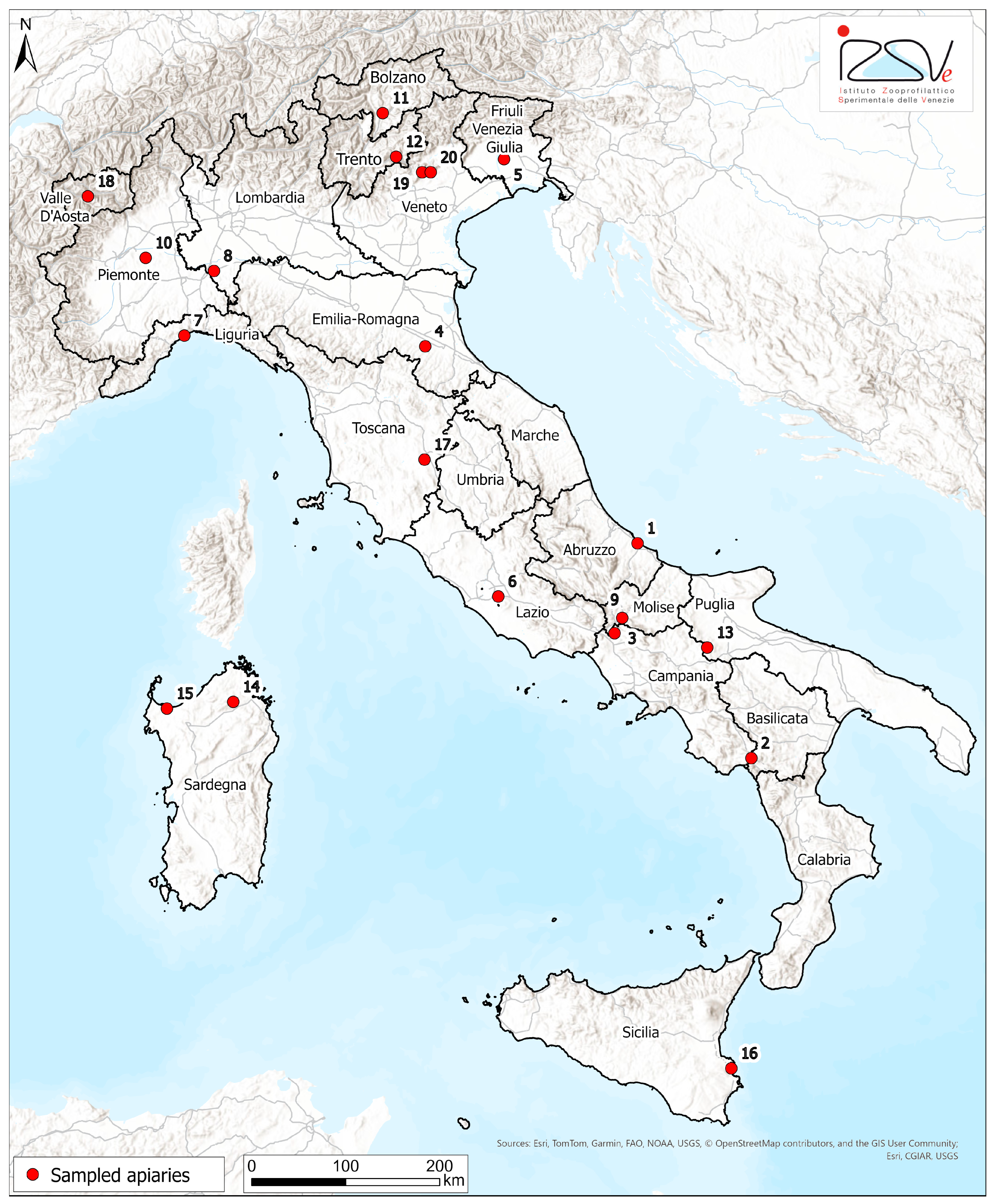
Twenty healthy honey bee apiaries (each with at least 5 hives per apiary) distributed across 17 regions of Italy, to represent different geographical areas, were selected based on the local beekeepers’ availability (Figure 1). An ad hoc questionnaire was developed to collect data from the period just before or during honey bee sampling, including information on the apiary (e.g., geo-referencing, territory type, agronomic and vegetation characteristics, health status, etc.), colony strength (assessed through both on-site inspection during sampling and anamnesis data collection), and the beekeeper’s management practices (e.g., control against Varroa mite infestation, other treatments).
Between July and August 2021, from each apiary, adult honey bees were sampled from the flight boards of 5 selected hives, yielding approximately 200 specimens per apiary, and then quickly stored at −80 °C until shipment on dry ice to the National Reference Laboratory for Honey Bee Health at Istituto Zooprofilattico Sperimentale delle Venezie (Legnaro, Italy) for analyses. Unfortunately, due to issues during shipment, the samples from Sicily (apiary 16) arrived in a condition unsuitable for analysis. Consequently, of the 20 selected apiaries, only 19 were subjected to analysis.

2.2. Nucleic Acid Extraction

Two different pools of 30 honey bees were prepared from the specimens collected in each apiary by randomly selecting 6 bees from each of the 5 sampled hives. One pool was used for DNA extraction, while the other pool was used for RNA extraction, in order to perform molecular investigations for the detection of known pathogens and to carry out metagenomic analyses.
For DNA extraction, each pool of 30 bees was homogenized in 30 mL of sterile phosphate-buffered saline (PBS) solution using a Stomacher® 400 Circulator lab blender (Seward Ltd, Worthing, United Kingdom) (2 cycles of 2 min each) and the resulting suspension was used for DNA extraction using the QIAamp DNA Mini Kit (Qiagen, Hilden, Germany), following the manufacturer’s instructions.
For RNA extraction, each pool of 30 bees was resuspended in 21 mL of RA1 buffer from the NucleoSpin RNA Kit (Macherey-Nagel™, Dueren, Germany) and homogenized using the TissueRuptor II (Qiagen, Hilden, Germany). Total RNA was extracted from the resulting homogenate using the NucleoSpin RNA Kit (Macherey-NagelTM, Dueren, Germany), following the manufacturer’s instructions. Qualitative (260/280 nm and 260/230 nm ratios) and quantitative (ng/µL) analyses of nucleic acids extracted from the bee pools were performed using a Nanodrop™ OneC spectrophotometer (Thermo Fisher Scientific, Waltham, MA, USA).
The DNA and RNA samples were then stored at −20 °C and −80 °C, respectively, until analysis. Negative process controls were included in each DNA and RNA extraction session.

2.3. Vairimorpha spp., Lotmaria passim, Crithidia mellificae PCR Detection

The presence of Vairimorpha spp. was evaluated initially by microscopic analysis of the homogenate to detect spores of this microsporidian. Subsequently, all samples underwent PCR analysis to confirm the presence or absence and to identify the species V. apis/V. ceranae. Amplification of a portion of the 16S rRNA gene was performed on a Veriti™ 96-Well Thermal Cycler (Applied Biosystems™, Waltham, MA, USA) using the protocol, primers and reagent concentrations reported by Bordin et al. [45]. To assess the presence of trypanosomatids Lotmaria passim and Crithidia mellificae by PCR, specific primers targeting a region of the RNA polymerase II large subunit (RPB1) gene of L. passim and the glyceraldehyde-3-phosphate dehydrogenase (GAPDH) gene of C. mellificae were used following the PCR conditions reported by Bordin et al. [45]. Negative and positive controls were included in each PCR session. Amplification products (218-219 bp V. ceranae; 321 bp V. apis; 254 bp L. passim; 177 bp C. mellificae) were analyzed by capillary electrophoresis on LabChip GX Touch HT® (Perkin Elmer, Waltham, MA, USA).
To confirm the detected positivity for L. passim and C. mellificae, the PCR products were sequenced using the Sanger method. Specifically, amplicons were purified using the ExoSAP-IT Express PCR Product Cleanup kit (Applied Biosystems™, Waltham, MA, USA), and the sequencing reaction was set up using a BrilliantDye™ Terminator Cycle Sequencing kit v3.1 (Nimagen, Nijmegen, The Netherlands). The sequencing reaction products, purified using CENTRI-SEP 96-Well Plates (Princeton Separations, Monmouth Junction, NJ, USA), were loaded onto an ABI PRISM® 3130xl Genetic Analyzer (Applied Biosystems™, Waltham, MA, USA). The obtained sequences were analyzed using SeqScape® v3 software (Applied Biosystems™, Waltham, MA, USA) and compared with sequences available in the GenBank database using BLAST (Basic Local Alignment Search Tool, NCBI) online tool (accessed on November 2023) [46].

2.4. RT-PCR Virus Analysis

Identification of honey bee viruses was performed by amplifying RNA extracted from each honey bee pool using both real-time one-step RT-PCR (rRT-PCR) (ABPV, CBPV, DWV variant A—DWV-A, DWV variant B—DWV-B, SBV and BQCV) and one-step end-point RT-PCR (Israeli Acute Paralysis virus—IAPV and Kashmir Bee virus—KBV). The rRT-PCR was carried out on a CFX96 Real-Time detection System (Bio-Rad Laboratories Inc., Hercules, CA, USA) using 250 ng of RNA and the QuantiTect Probe RT-PCR Kit (Qiagen, Hilden, Germany). For IAPV and KBV detection, the RT-PCR was performed on a Veriti™ 96-Well Thermal Cycler (Applied Biosystems™, Waltham, MA, USA) using 250 ng of RNA and the OneStep RT-PCR Kit (Qiagen, Hilden, Germany). Primers and probes sequences and concentrations, as well as the amplification conditions, are reported by Bordin et al. [45]. Negative and positive controls were included in each PCR. Amplicons for IAPV (767 bp) and KBV (659 bp) were analyzed by capillary electrophoresis on LabChip GX Touch HT® (Perkin Elmer, Waltham, MA, USA).

2.5. 16S rRNA Gene Sequencing and Bioinformatics Analyses

To determine the composition of the bacterial community of the 19 apiaries under analysis, total DNA extracted from each honey bee pool was subjected to NGS on an Illumina platform. The hypervariable regions V3-V4 of the 16S rRNA gene were amplified by PCR using degenerate primers (forward: 5′-CCTACGGGNGGCWGCAG-3′; reverse: 5′-GACTACHVGGGTATCTAATCC-3′) according to the instructions provided by Klindworth et al. [47] and following the Illumina 16S Metagenomic sequencing Library Preparation protocol. The PCR thermal profile was as follows: denaturation at 95 °C for 3 min; 30 cycles consisting of: 95 °C for 30 s, 55 °C for 30 s, 72 °C for 30 s; final extension at 72 °C for 5 min. PCR products (~550 bp) were subjected to electrophoresis on a 2% agarose gel and then visualized using Midori dye (Nippon Genetics, Dueren, Germany) as an intercalating agent. Libraries were prepared following instruction of the Nextera® XT DNA Library Prep kit (Illumina, San Diego, CA, USA), and their quality and quantity were verified using the dsDNA High Sensitivity Kit for the Qubit™ 2.0 fluorimeter (Thermo Fisher Scientific, Waltham, MA, USA) and the Agilent TapeStation 2200 System (Agilent Technologies, Santa Clara, CA, USA), respectively. Samples were pooled equimolarly, and sequencing (600 cycles, 2 × 300 bp, paired-end reads) was performed on an Illumina MiSeq™ instrument with Illumina MiSeq™ 600 V3 kit (Illumina, San Diego, CA, USA).
After sequencing, the raw data generated were further processed in RStudio using the DADA2 package [48]. Reads were first filtered and trimmed based on the quality profiles. The filtered sequences were then de-replicated to combine identical sequencing reads into unique sequences. Then, the forward and the reverse reads were merged to obtain the full denoised sequences and the chimeras were removed. SILVA ribosomal database (version 138.1) [49] was used to assign taxonomy to the identified Amplicon Sequence Variants (ASVs). For subsequent statistical analyses, only ASVs present in at least 10% of the samples were retained. The DNA raw data generated in this study have been deposited in the Sequence Read Archive (SRA) database (https://www.ncbi.nlm.nih.gov/sra (accessed on 15 January 2026)) under the accession number PRJNA1403243.

2.6. RNA Sequencing and Bioinformatics Analysis

Total RNA was treated with DNase I, and its integrity was assessed by determining the RNA Integrity Number (RIN) using the Agilent Bioanalyzer platform (Agilent Technologies, Santa Clara, CA, USA). From each apiary, 100 ng of total RNA was processed by applying a ribosomal RNA (rRNA) depletion protocol using the Illumina Stranded Total RNA Prep Ligation with Ribo-Zero Plus kit (Illumina, San Diego, CA, USA) optimized with a spike-in of 86 probes specific (Table S1) for A. mellifera rRNA designed on the reference genomes available in EnsemblMetazoa (Amel_HAv3.1; https://metazoa.ensembl.org/index.html). Sequencing libraries were prepared and sequenced on a high-throughput NovaSeq™ 6000 platform (Illumina, San Diego, CA, USA) in 2 × 150 paired-end mode, with sequencing output designed to target 100 million reads per sample.
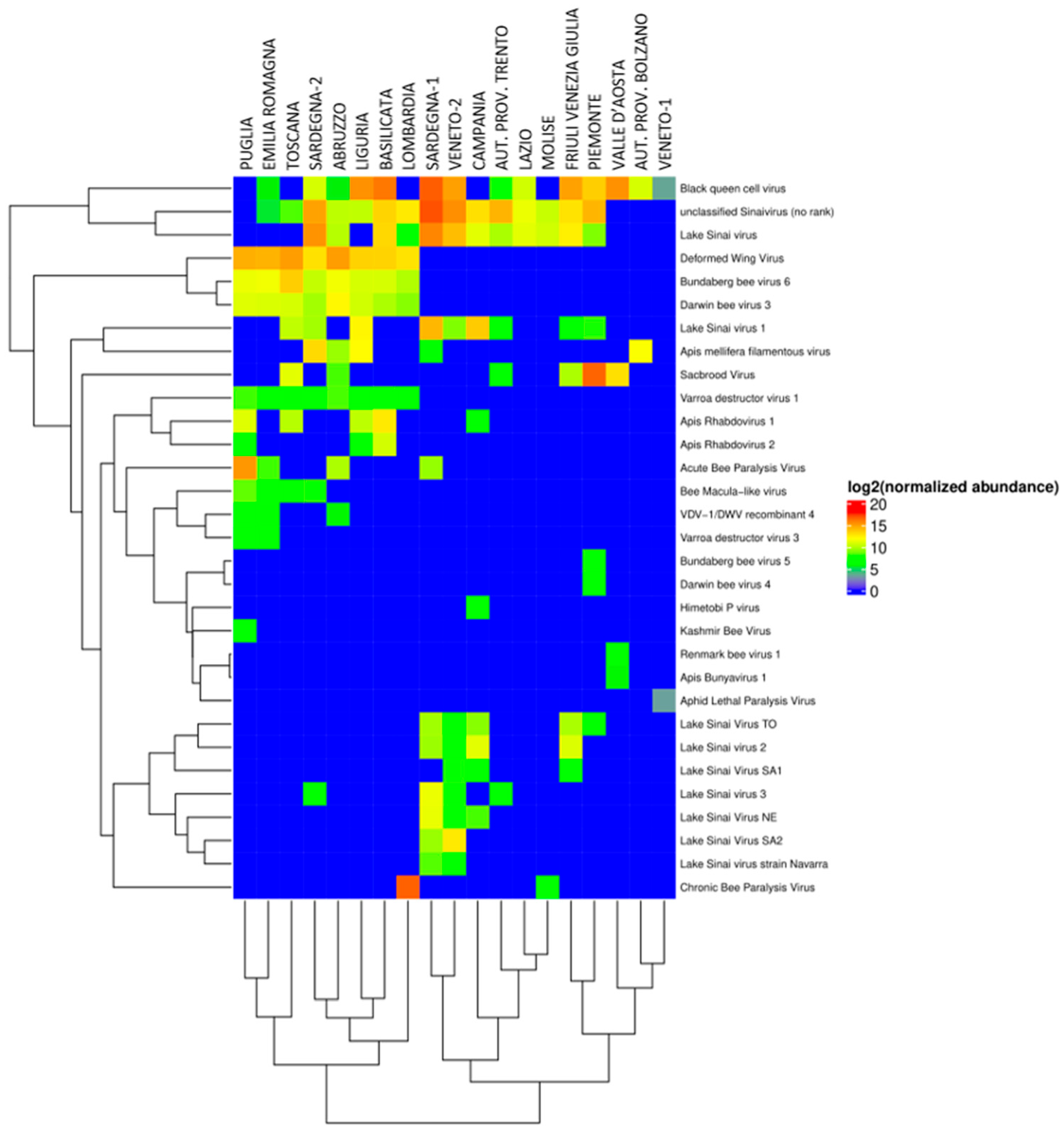
After sequencing, data quality was checked with FastQC v0.11.2 [50] and the raw data were appropriately filtered with Scythe v0.991 [51] and Sickle v1.33 [52]. The data were further filtered to remove host reads (A. mellifera) using STAR v2.7.9a [53] with standard parameters. To the high-quality reads thus obtained, a taxonomic classification was assigned through metagenomic analysis following the published “best practices” [54]: (i) aligning the high-quality, host-cleaned reads against the nr protein database (v 23/02/2020) using diamond v0.9.17 [55]; (ii) discarding alignments with e-value > 1·10−3; (iii) assigning the taxonomic level to each read with the LCA (Lowest Common Ancestor) algorithm implemented in MEGAN v.6.18.50 [56]. The heatmap of read abundance (log2 normalized) of the viral species found in the 19 apiaries was produced using the ComplexHeatmap library in the R environment v4.2 [57]. The RNA-seq data generated in this study have been deposited in the SRA database (https://www.ncbi.nlm.nih.gov/sra (accessed on 14 January 2026)) under the accession number PRJNA1402780.

2.7. Bacterial Community Confirmation: Identification of the Species Belonging to the Melissococcus Genus

To confirm the presence of Melissococcus plutonius, the causative agent of European foulbrood (EFB) in honey bees, all samples, tested both positive and negative for the Melissococcus genus following NGS analysis of the 16S rRNA gene, were further examined by PCR. DNA amplification was performed using the AmpliTaq™ Gold Kit (Applied Biosystems™, Waltham, MA, USA) with the following components (final reaction volume of 50 µL): MgCl2 [2.5 mM], dNTPs [0.2 mM], specific forward and reverse primers for the 16S rRNA subunit [0.5 µM] [58], 2 U of AmpliTaq Gold DNA polymerase. Amplification was carried out on a Veriti™ 96-Well Thermal Cycler (Applied Biosystems™, Waltham, MA, USA) with the following thermal cycling profile: Taq polymerase activation at 95 °C for 10 min; 35 amplification cycles consisting of denaturation at 94 °C for 30 s, annealing at 60 °C for 30 s, extension at 72 °C for 30 s; final extension at 72 °C for 7 min. The amplification products (~812 bp) were analyzed by 7% acrylamide gel electrophoresis and visualized by silver staining.

2.8. Viral Species Confirmation Assays

Some of the viral species identified through NGS analysis of the viral communities (Apis mellifera Filamentous virus—AmFV, BeeMLV, ARV-1, ARV-2, Aphid Lethal Paralysis virus—ALPV, Varroa destructor virus 3—VDV-3 and LSV variants) were confirmed using specific oligonucleotide sequences available in the literature (Table S2). The PCR and RT-PCR conditions (Table S2) were optimized using some of the samples that tested positive in the NGS analysis. The molecular protocols were then used to verify the presence of these viruses in all samples, including those that had tested negative by NGS analysis.
To detect the presence of AmFV, PCR reaction was performed on a Veriti™ 96-Well Thermal Cycler (Applied Biosystems™, Waltham, MA, USA) using the AmpliTaq™ Gold kit (Applied Biosystems™, Waltham, MA, USA) in a final volume of 50 µL with the following components: MgCl2 [1.5 mM], dNTPs [0.4 mM], forward and reverse primers [0.4 µM], 1.25 U of AmpliTaq Gold DNA polymerase, and 50 ng of DNA. The thermal profile is reported in Table S2. For ARV-1, ARV-2, ALPV, VDV-3 and LSV variants detection, reverse transcription of 1 µg of RNA was carried out on a Veriti™ 96-Well Thermal Cycler (Applied Biosystems™, Waltham, MA, USA) using the SuperScript™ IV Reverse Transcriptase kit (Invitrogen™, Waltham, MA, USA), following the manufacturer’s instructions. cDNA amplification reaction was performed using the AmpliTaq™ Gold kit (Applied Biosystems™, Waltham, MA, USA), mixing the following components in a final volume of 50 µL: 2 µL of cDNA, 1.25 U of Taq polymerase and: (i) ARV-1/ARV-2: MgCl2 [1.5 mM], dNTPs [0.4 mM], primer [0.2 µM]; (ii) ALPV: MgCl2 [1.5 mM], dNTPs [0.2 mM], primer [0.2 µM]; (iii) VDV-3: MgCl2 [2.5 mM], dNTPs [0.4 mM], primer [0.4 µM]; (iv) LSV variants: MgCl2 [1.5 mM], dNTPs [0.2 mM], primer [0.2 µM]. The amplification was performed on a Veriti™ 96-Well Thermal Cycler (Applied Biosystems™, Waltham, MA, USA) with the thermal profiles reported in Table S2 for each virus. For BeeMLV detection, the RT-PCR was performed on a Veriti™ 96-Well Thermal Cycler (Applied Biosystems™, Waltham, MA, USA) using the One-step RT-PCR Kit (Qiagen, Hilden, Germany), specific primer [0.4 µM] and 100 ng of RNA. The thermal profile is reported in Table S2.
Amplification products were analyzed by 7% acrylamide gel electrophoresis, visualized by silver staining and further sequenced using the Sanger method as previously described. The obtained sequences were analyzed in SeqScape® v3 software (Applied Biosystems™, Waltham, MA, USA) and compared with those available in the GenBank database, using the BLAST online software [46].

2.9. Statistical Analyses

The statistical analyses conducted on the ASVs obtained from 16S rRNA sequencing were performed using the phyloseq package (version 1.44.0) [59] of the RStudio software (version R 4.1.0—RStudio Team, 2020), while graphs and figures were created using the ggplot2 package (version 3.4.4) [60] of the RStudio software. Alpha diversity indices (richness, Pielou’s index and Shannon’s index) were calculated after normalizing the ASVs table using the Geometric Mean of Pairwise Ratios (GMPR) method. To compare the alpha diversity indices among the 19 apiaries, the non-parametric Kruskal–Wallis test was used, and if this test was statistically significant (p-value < 0.05), the Wilcoxon test was then applied to further evaluate whether the differences between the various samples were statistically significant. The p-value correction method used was the False Discovery Rate (FDR). Beta diversity was measured based on the Bray–Curtis dissimilarity index and visualized using Non-metric Multi-dimensional Scaling (NMDS) plots. To assess beta diversity differences, a permutational analysis of variance (PERMANOVA) was performed, considering it significant with a p-value < 0.05.
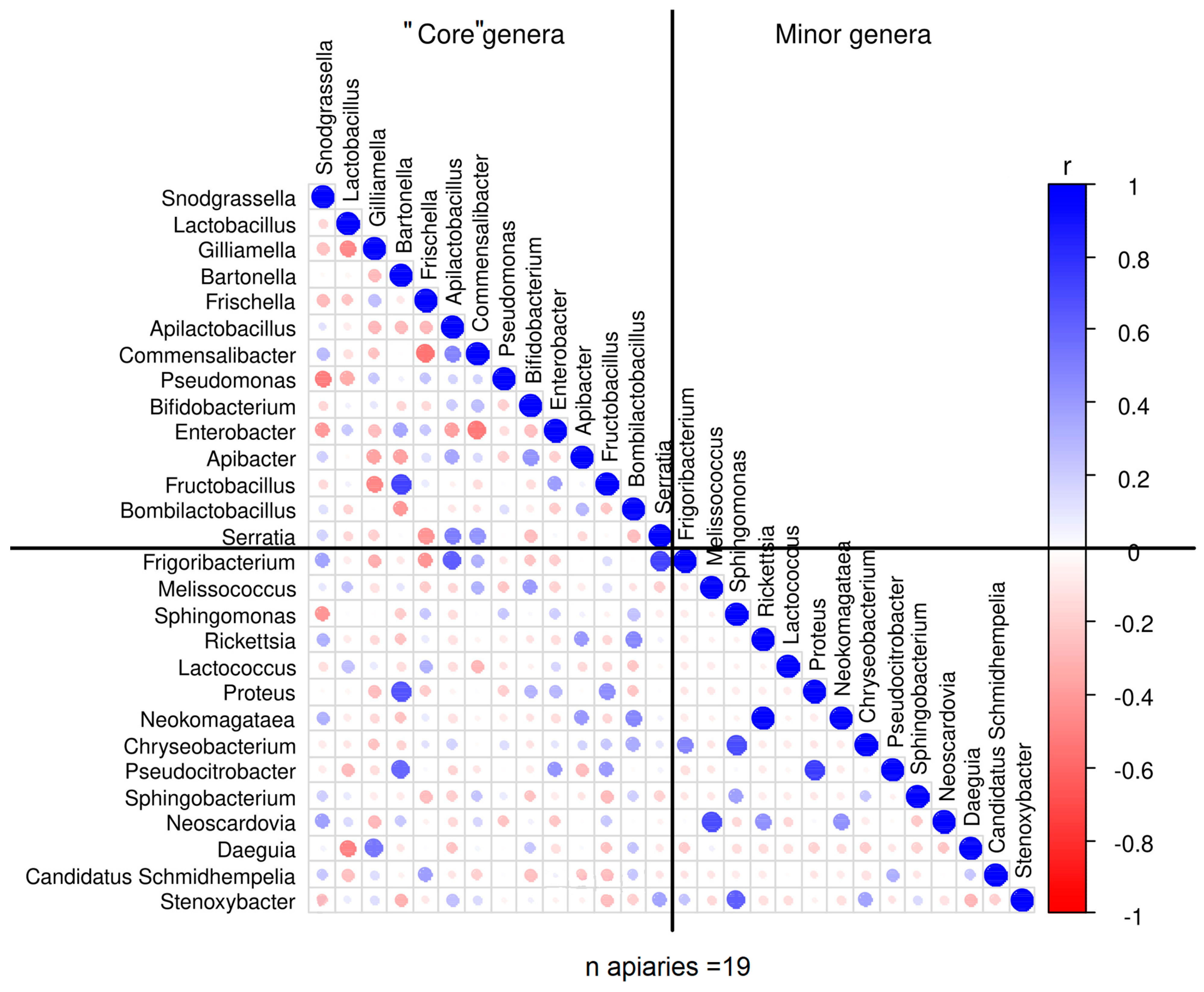
An analysis of variance (ANOVA) was performed to determine whether significant differences exist in the abundance of bacterial genera across the 19 apiaries, and to assess if the mean abundance of specific bacterial genera differs significantly between these groups. Bacteria were classified into “core genera” (those up to the 75th percentile) and “minor genera.” A correlation matrix was generated to examine and visualize the relationships between core and minor bacterial genera using Pearson’s r coefficient, after excluding variables with zero variance. The strongest correlations (r > |0.7|) were further explored using regression analysis (p-value < 0.05). Additionally, a correlation analysis was conducted on all the microorganisms (including bacteria, viruses, fungi, and protozoa) detected in the 19 apiaries by both NGS and PCR analyses, using presence/absence data at the colony level. For binary 0/1 data, Pearson’s r coefficient corresponds to the φ coefficient, commonly employed to assess co-occurrence patterns among microbial taxa, whereas for continuous data, it reflects standard linear correlations. ANOVA, correlation matrix, regression analyses, and graphical outputs were carried out in the R environment (R version 4.4.0).

3. Results

3.1. Sampling Observations (Questionnaires)

The data derived from sampling observations collected through questionnaires reported that, in 16 apiaries, no abnormal behavior or signs of disease were observed during sampling. Aggressive behavior was noted in the two apiaries in Sardegna, while shaking bees were seen in one hive in Piemonte. All hives in Liguria showed low flight activity. Information on hive strength was incomplete for some apiaries, due to missing evaluations, and thus could not be included in further analysis. No mortality events occurred in 12 apiaries; where reported, they were caused in the period preceding sampling, by European foulbrood (EFB) (Veneto-2), American Foulbrood (AFB) (Basilicata), Varroa mite infestation (Puglia, Sardegna-1 and Sardegna-2), malnutrition from adverse weather (Friuli Venezia Giulia) or unexplained (Liguria).

3.2. Detection of Vairimorpha spp., Lotmaria passim and Crithidia mellificae by PCR

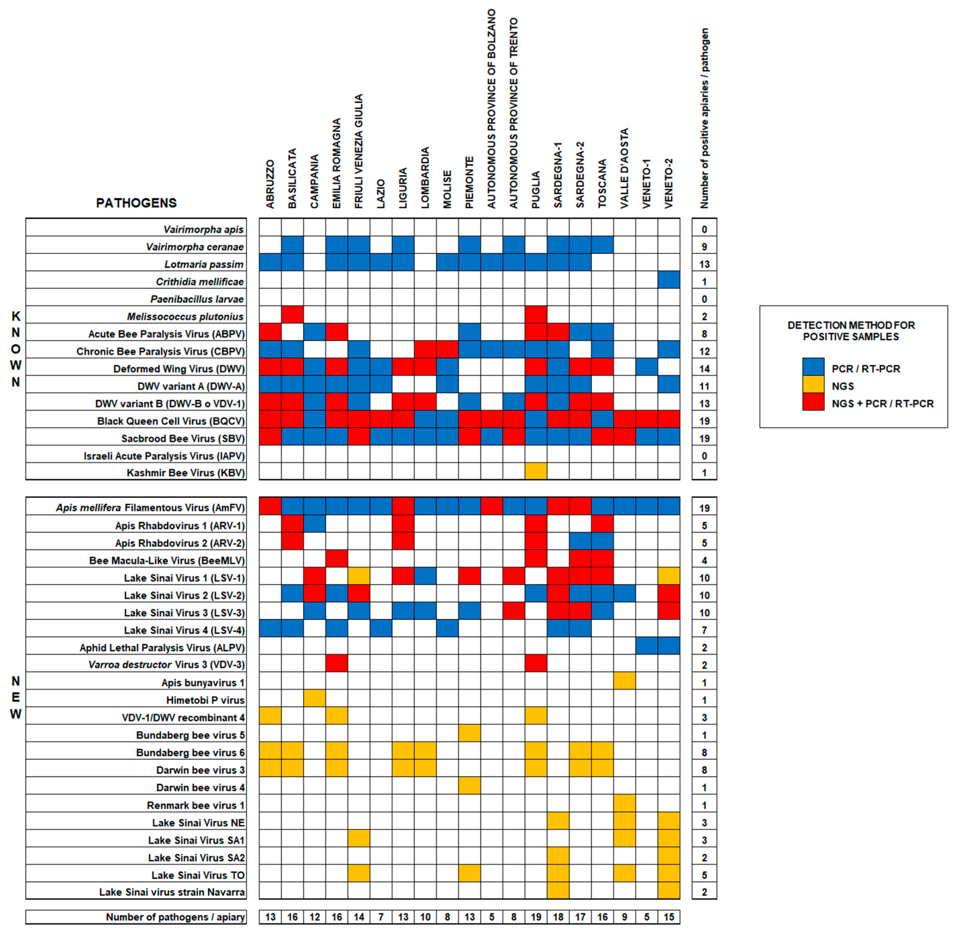
Vairimorpha spp. spores were detected in 9 out of the 19 apiaries analyzed (9/19 = 47%), further confirmed by PCR analysis of all 19 samples, revealing the presence of only V. ceranae species in all the positive samples. Trypanosomatids C. mellificae and L. passim were detected in 1/19 apiaries (Veneto-2) and in 13/19 apiaries (68%), respectively. The sequencing of amplification products confirmed 100% identity with the respective species. The results for each apiary are summarized in Figure 2.

3.3. Virus Detection by RT-PCR

KBV and IAPV viruses were not detected in any of the apiaries analyzed, while SBV and BQCV were detected in the 100% of the apiaries. Other viruses (ABPV, CBPV, DWV-A and DWV-B) were identified at different frequencies: ABPV, the least frequent virus, in 8/19 apiaries (42%); CBPV in 12/19 apiaries (63%); DWV-A variant in 11/19 apiaries (58%); DWV-B in 13/19 apiaries (68%). Both DWV variants were simultaneously present in 8 apiaries (42%). The results for each apiary are reported in Figure 2.

3.4. Honey Bee Bacteriome Analysis

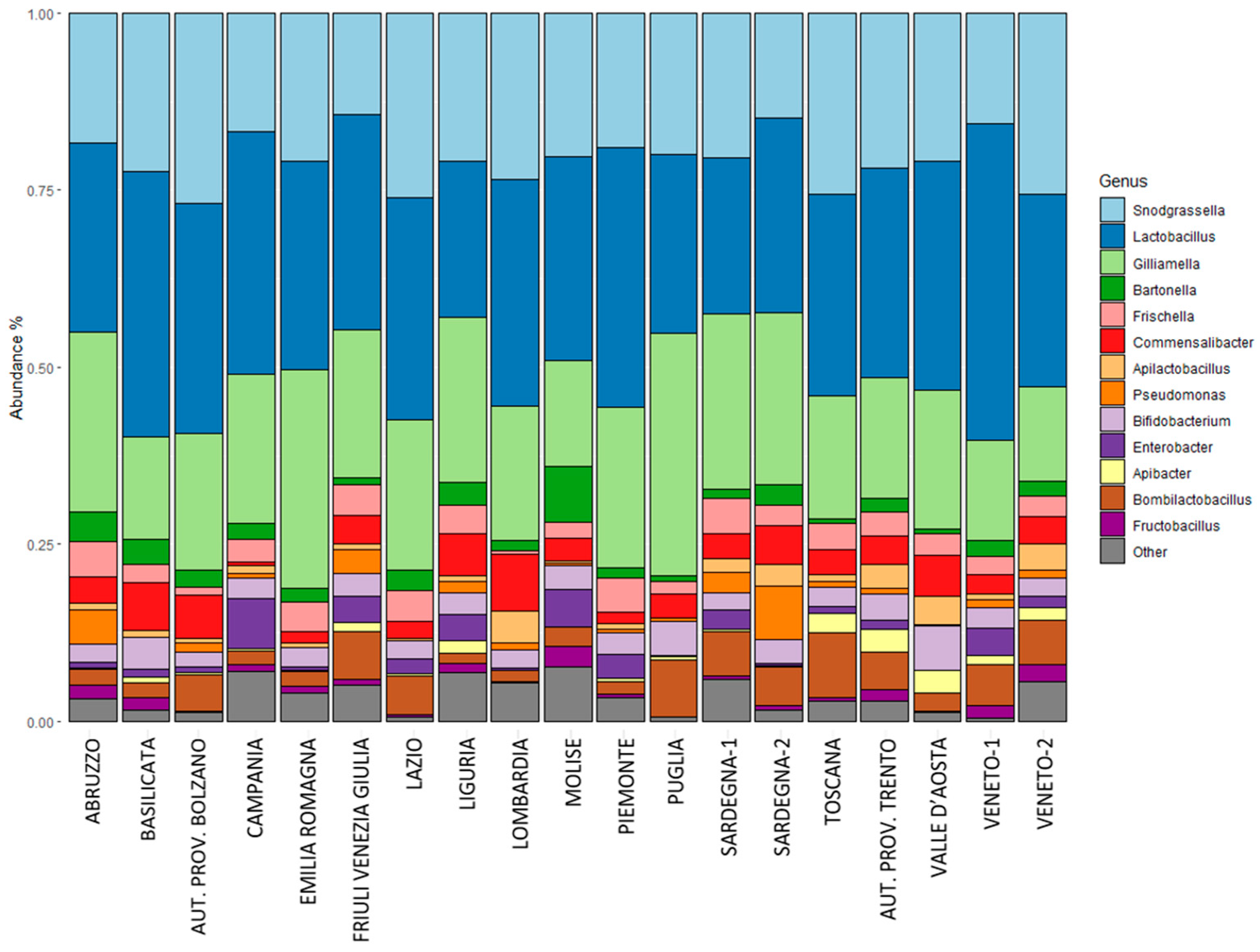
16S rRNA gene sequencing detected a total of 2763 ASVs. Metataxonomic analysis has shown that the bee microbiome does not present statistically significant differences between samples from different Italian regions, both in terms of biodiversity indices and taxonomic composition. From a taxonomic perspective, the microbial community of all samples is mostly characterized by bacterial genera such as Snodgrassella, Lactobacillus, Gilliamella, Bartonella, Frischella, Commensalibacter, Apilactobacillus, Bifidobacterium and Bombilactobacillus (Figure 3). These bacterial genera, together with others such as Pseudomonas and Fructolactobacillus, represent approximately 90% of the microbial composition of the entire dataset considered. The remaining genera included microorganisms often associated with other arthropods, plants or present in the environment where bees live and feed. Sequences belonging to certain opportunistic pathogenic bacteria, such as Serratia and Escherichia-Shigella, were detected in some samples. Additionally, the analysis identified sequences belonging to the genus Melissococcus in two apiaries (Basilicata and Puglia), whose species M. plutonius is the etiological agent of European foulbrood.
Relative abundance values (expressed as percentages) of bacterial genera among all 19 apiaries showed that the most abundant genera were Lactobacillus (22–44.6%), Gilliamella (13.3–25.4%), Snodgrassella (14.3–26.9%), Bombilactobacillus (1.4–9.2%), Commensalibacter (0.4–8.1%), Frischella (0.4–4.9%), Bifidobacterium (2.1–6.3%), Bartonella (0.6–8%), and Enterobacter (0.1–7.1%).
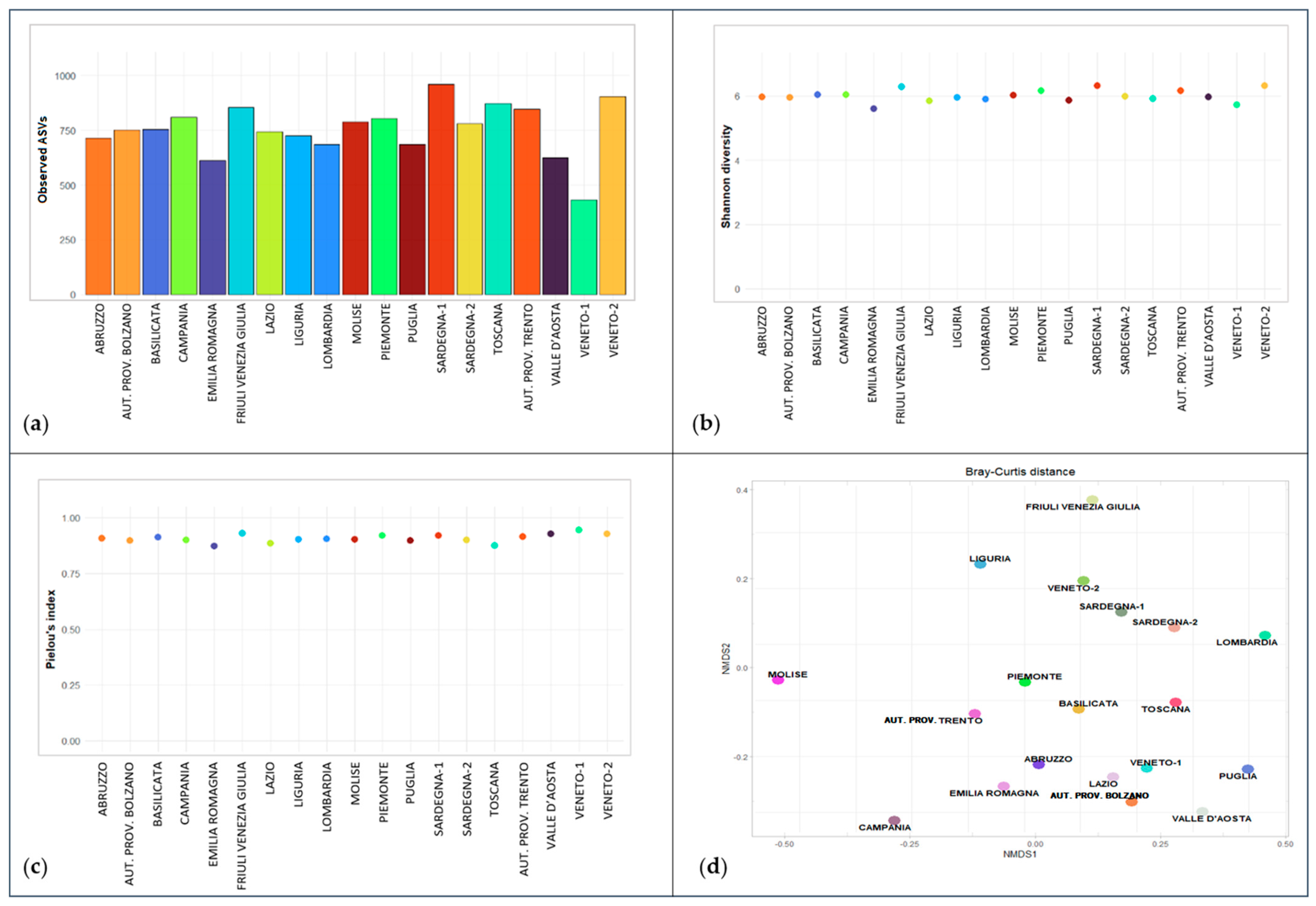
Regarding the diversity of the microbial community within each sample, defined as alpha diversity (Figure 4a–c), none of the three selected indices (richness, Shannon index and Pielou’s index) showed statistically significant differences between samples from different regions (richness: p-value > 0.05; Shannon index: p-value > 0.05; Pielou’s index: p-value > 0.05). Similarly, for beta diversity, which evaluated the bacterial communities between samples (Figure 4d), the result of the PERMANOVA test (p-value > 0.05) did not reveal statistically significant differences between the various apiaries.

3.5. Bacterial Community: Identification of the Species Belonging to the Genus Melissococcus

The sequencing of the hypervariable V3–V4 regions of the 16S rRNA gene revealed the presence of the genus Melissococcus in samples from two apiaries (Basilicata and Puglia). PCR analysis confirmed that these bacteria belonged to the species M. plutonius, the etiological agent of European foulbrood (EFB) in honey bees. In the remaining 17 apiaries, the absence of this pathogen was confirmed.

3.6. Honey Bee Viral Community

The RNA concentration and RNA Integrity Number (RIN) values of the samples ranged between 131 ng/µL and 901 ng/µL, and 5.9 and 9.7, respectively.
High-throughput sequencing produced a total of 834 gigabases (Gb) of data, with an average of 247 million paired-end reads per sample: 75.1% of the reads originated from the host (A. mellifera), and 1.9% corresponded to host ribosomal RNA (rRNA), indirectly confirming the effectiveness of the rRNA depletion protocol performed before library preparation (Table S3). After removing the host-related data, the number of reads used for metagenomic analysis averaged 39 million paired-end reads per sample, ranging from 3 to 110 million.
The metagenomic analysis provided an average of 17% of viral reads on the total number of reads, ranging from 1% (Molise) to 59% (Sardegna-1), while the remaining reads mainly belonged to bacteria, fungi, and other eukaryotic organisms (mainly trypanosomes and other unicellular flagellates). These proportions were maintained up to the genus classification level, but at the species level, about half of these reads could not be classified more precisely. Consequently, the fraction of viral reads with complete classification at the species level decreased to an average of 7.8%, ranging from 0.3% (Molise) to 34.3% (Sardegna-1). The total number of reads assigned to each taxonomic classification level for each analyzed apiary is shown in Table S4.
Both RNA genome viruses, which constitute 98.8% of the identified viruses, and DNA genome viruses were characterized. Approximately 45% of the total viral reads were assigned to the order Picornavirales, while the remaining reads were classified into other orders within the Riboviria domain (Mononegavirales, Bunyavirales, Articulavirales, and Tymovirales) or remained unclassified. Within the order Picornavirales, reads were distributed among the families Iflaviridae and Dicistroviridae, which include the majority of honey bee viruses. Reads assigned to other orders included families such as Rhabdoviridae, Peribunyaviridae, Sinhaliviridae, Tymoviridae, as well as other taxonomically identified viral families (Secoviridae, Bromoviridae, Leviviridae, Partitiviridae, Solemoviridae, and Virgaviridae). Furthermore, some reads corresponded to unclassified viruses, mainly having plants and other arthropods as natural hosts. Of the 81 viral species identified by NGS, 30 had already been identified and characterized in A. mellifera and in other apoideans or wasps and are shown in Table S2. Of the remaining 51 identified species, 27 belong to plant viruses, and 24 were found in other arthropods. The complete list of viral species detected in the 19 analyzed apiaries is illustrated in Table 1. In addition to well-known viruses, several emerging and previously uninvestigated viruses were identified in the 19 apiaries. All of them are single-stranded RNA viruses, except for AmFV, which is a double-stranded DNA virus.
BQCV and LSV (the latter present in numerous variants) were the most prevalent viruses detected in the analyzed samples, with 14/19 (73.7%) and 12/19 (63.1%) positive apiaries, respectively. In contrast, Himetobi P virus, KBV, Bundaberg bee virus, Darwin bee virus, Apis Bunyavirus, ALPV, and Renmark bee virus were each detected only in one apiary, located in different regions, with low transcript abundance. A high number of viral co-infections, including even phylogenetically diverse viruses, was observed. In particular, the apiaries of Sardegna-1 and Sardegna-2, Abruzzo, Puglia, Emilia Romagna, and Liguria exhibited the highest levels of viral co-infections, with 10 or more viral species detected simultaneously in each apiary. In contrast, apiaries in Veneto (Veneto-1), the Autonomous Province of Bolzano, Molise, and Lazio exhibited only two or three viral species (Figure 5) simultaneously in each apiary. The variability of the detected viral species does not seem to correlate with the number of viral reads per sample.

3.7. Verification and Confirmation Analysis of the Viral Community Identified by Metagenomic Analysis

Some selected viruses from the virome analysis were investigated using molecular protocols to confirm their species identity and assess their presence in samples that tested negative. Viruses were selected based on their spread and prevalence in other geographic areas/countries, their epidemiological relevance, and the availability of published molecular protocols with specific pairs of oligonucleotides capable of amplifying genome fragments suitable for sequencing. The investigated viral species included ARV-1 and ARV-2, BeeMLV, LSV-1, LSV-2, LSV-3, AmFV, ALPV, and VDV-3. Sanger sequencing confirmed the NGS data analysis of the viral community and additionally detected some of these viruses in samples lacking assigned NGS reads. The results are summarized in Figure 2.
Reads belonging to ARV-1 and ARV-2 were detected by metagenomic analysis in 5/19 (26.3%) and 3/19 (15.8%) apiaries, respectively. Confirmation RT-PCR identified two additional ARV-2 positive apiaries. Moreover, the simultaneous presence of both ARV-1 and ARV-2 variants was detected in four of the five apiaries. Regarding BeeMLV, NGS data were confirmed by RT-PCR, detecting the virus in only 4/19 apiaries (21%). RNA-seq detected the occurrence of VDV-3 in two apiaries (10.5%), which was confirmed by RT-PCR. The presence of ALPV was revealed by NGS in one apiary of Veneto (Veneto-1), but also honey bee specimens from Veneto-2 tested positive after RT-PCR.
Among the LSV variants identified by NGS analysis, LSV-1, LSV-2, and LSV-3 were selected to confirm their presence through RT-PCR and sequencing. Before focusing on individual analysis of the three viral LSV strains, all the RNA samples were amplified using degenerate primers targeting a common region spanning the ORF1/RdRp genes. LSV positivity was detected in 13/19 apiaries (68.4%), slightly increasing the NGS-positive apiaries (12/19). Specific RT-PCR for each of the LSV variants then revealed LSV-2 and LSV-3 as the most prevalent (10/19 apiaries; 52.6%). Finally, LSV-1 was detected in 8/19 apiaries (42.1%). These results also demonstrate that multiple variants of the same virus can be present within the same sample. For each variant, the sequence identity with those deposited in the GenBank database ranged from 94.73 to 98.77% for LSV-2, 97.32 to 98.94% for LSV-3 and 93.17 to 94.86% for LSV-1. Sequence analysis of RT-PCR amplification products using LSV-1 and LSV-3 specific primers also revealed the presence of LSV-4, another LSV strain, in some apiaries. To assess it, specific LSV-4 primers [61] were used and, after amplification and Sanger sequencing, positivity to this viral strain was confirmed in the previously mentioned apiaries and also in the two apiaries of Sardegna, for a total of 7/19 apiaries (36.8%).
For all other viruses analyzed, Sanger sequencing of RT-PCR amplification products showed the following identity ranges with GenBank-deposited sequences: ARV1: 97.45–99.79%; ARV-2: 99.19–99.54%; BeeMLV: 92.92–98.85%; VDV-3: 94.56–97.23%; and ALPV: 94.95–95.33%.
Finally, RNA-seq detected the presence of AmFV in 5/19 apiaries (26.3%). Amplification of the DNA extracted from each pool of the 19 apiaries, followed by Sanger sequencing of amplicons, confirmed AmFV presence in both the five NGS-positive apiaries and the remaining 14, suggesting ubiquitous distribution. Sanger-derived sequences showed 95.16 to 98.85% identity with those deposited in GenBank.

3.8. Statistical Analysis

The results from ANOVA showed significant variation in the mean percentages of abundance among the different bacterial genera (p-value < 0.001), indicating that the genus-level identity strongly influences the observed values. In contrast, no significant variation was attributable to the apiaries, suggesting that the distribution of bacterial abundance percentages did not differ meaningfully among apiaries.
The correlation analysis of the bacteriome in A. mellifera from the 19 analyzed apiaries showed no significant negative correlations, while identifying numerous strong positive associations among bacterial genera. Notably, Neokomagataea and Rickettsia showed a perfect positive correlation (r = 1, p-value < 0.001), even if detected only in the apiary from the Autonomous Province of Trento. Enterococcus displayed very high correlations with Erwinia (r = 0.996) and Spiroplasma (r = 0.97), while several other pairs, including gut-associated or opportunistic bacteria such as Raoultella, Citrobacter, Proteus, Leuconostoc, Weissella, Bartonella, Rosenbergiella, Fructobacillus, Frigoribacterium and Serratia also exhibited significant positive strong correlations (r > |0.7|, p-value < 0.001), suggesting co-occurrence and potential interactions within the gut ecosystem. The correlation matrix of the bacterial genera is represented in Figure 6.
A correlation analysis was performed on all detected microorganisms at the apiary level using presence/absence data, including bacteria, fungi, and viruses. Pearson correlation coefficients, corresponding to the φ coefficient for binary variables, were calculated to assess co-occurrence patterns among microbial taxa. Only strong correlations (r > |0.7|) that were statistically significant (p-value < 0.05) were considered, and no significant negative correlations were detected. The results revealed, again, a perfect positive correlation between Neokomagataea and Rickettsia (r = 1.000, p-value < 0.001), indicating a highly consistent co-occurrence. Additional strong positive correlations included those between viruses LSV-1 and LSV-3 (r = 0.809), ARV-1 and ARV-2 (r = 0.729), as well as bacterial genera such as Erwinia with Frigoribacterium (r = 0.792) and Enterococcus with Erwinia (r = 0.792). Positive correlations were also observed between Gluconobacter and Weissella, Lonsdalea and Zymobacter, and Arsenophonus and Erwinia, all with r-values above 0.7 and highly significant p-values (p-value < 0.001). These associations suggest frequent co-colonization or potential synergistic interactions among the microbial taxa in the honeybee gut microbiota. Conversely, no significant correlations were detected when microbiological data were integrated with questionnaire-derived variables, including apiary characteristics and localization, colony strength, and beekeeping management practices, likely due to the limited sample size (n = 19 apiaries), which may have reduced the statistical power of the analysis.

4. Discussion

The global health status of honey bees is critical, driven by a complex interplay of environmental and biological stressors. Consequently, there is a need to expand research focus on the intricate host–microbe interactions among honey bees and a wide range of microbial agents simultaneously, including bacteria, viruses, fungi, and parasites, in order to understand their impact on bee health and disease progression. In this context, this study, which employed both standard molecular methods and modern “-omics” approaches, such as NGS technologies, has allowed, for the first time, the characterization of communities of known, new, and emerging commensals, symbionts, and pathogens in Italian honey bees [62,63]. Notably, previously unreported microorganisms, particularly viruses, were detected in the national territory.
The compilation of an ad hoc questionnaire during the honey bee sampling phase, although sometimes partially incomplete, provided valuable insight into apiary management. The general absence of abnormal behaviors or disease signs in 16 out of 19 apiaries suggested a positive health baseline among most hives, notwithstanding some behavioral alterations, such as aggressiveness in Sardinian apiaries, trembling bees in Piemonte or reduced flight activity observed across Liguria hives. These symptoms may indicate potential environmental stressors, the presence of predators such as Vespa velutina nigrithorax (in Liguria) [64] or ongoing viral infections like CBPV or DWV, whose presence was later confirmed by diagnostic tests. However, the lack of data on hive strength limits a comprehensive assessment of colony health across all apiaries, highlighting the need for standardized evaluation criteria in future studies.
The study involved 19 out of the 20 selected apiaries from 16 Italian regions, as the sample from Sicily was excluded due to unsuitable conditions upon arrival.
The results of the metagenomic analysis of the whole honey bee body allowed us to characterize the microbiome of the insect without excluding microbial communities that may affect specific tissues. DNA and RNA extracted from different pools of honey bees revealed not only eukaryotic microorganisms but also numerous bacterial and viral communities.
Molecular assays using PCR and RT-PCR, targeting known hive pathogens, confirmed previous data on Italian epidemiological pathogen distributions [45,65,66,67,68,69,70]. Specifically, our investigation of the two microsporidian fungi species, recently phylogenetically reclassified, V. apis and V. ceranae [11,12,13], showed the absence of V. apis, although recently detected in Southwestern Italy [71], while nearly half of the apiaries tested positive for V. ceranae. These pathogens replicate in the bee midgut, causing diverse detrimental effects including altered behavior or brood management, compromised productivity and survival [72]. Less known are the effects of trypanosomatids L. passim and C. mellificae, obligate unicellular parasitic protozoa that colonize the digestive system and have been linked to colony losses, particularly when associated with V. ceranae [73]. PCR analysis confirmed the widespread presence of L. passim in A. mellifera, which has been reported globally [45,74,75,76,77,78,79,80,81], compared to C. mellificae, which was only detected in one of two apiaries in the Veneto region, supporting its sporadic occurrence in honey bees [82]. Several studies [73,83,84,85,86,87,88] showed that trypanosomatids could also exhibit positive correlations with microsporidium V. ceranae in honey bee colonies, enhancing immune suppression and contributing to winter colony declines. However, the statistical analyses conducted revealed no significant correlations among these microorganisms in any of the apiaries. Even if it remains unresolved as to whether trypanosomatids act as direct pathogens or opportunistically follow V. ceranae infection, the synergistic interaction likely weakens the co-infected hosts [89], making them more susceptible to other pathogens. In fact, the analysis of known viral pathogens in the sampled honey bees revealed the presence of SBV and BQCV in all apiaries, along with other viruses such as DWV-A and DWV-B variants, CBPV, and ABPV. While PCR-based methods are highly effective for detecting known and specific pathogens, the metagenomic analysis of the whole honey bee body enabled a comprehensive characterization of both known and novel bacterial and viral taxa, revealing some correlation between microorganisms in bee colonies.
To study the symbiotic and commensal bacterial communities, we employed 16S rRNA gene sequencing targeting the hypervariable V3–V4 regions, a well-established method for assessing bacterial diversity in biological samples. This approach provided valuable data on the presence and distribution of major bacterial taxa across various taxonomic levels.
As previously reported, the microbiota of the collected A. mellifera specimens was predominantly composed of approximately 8-10 core bacterial genera [8,90,91,92,93,94], which represented approximately 90% of the prokaryotic organisms present. These genera—Snodgrassella, Lactobacillus, Gilliamella, Frischella, Bartonella, Commensalibacter, and Bifidobacterium—are often associated with bee health [9]. In particular, Gilliamella and Snodgrassella are the main bacterial genera in the honey bee midgut and hindgut, playing crucial roles in food metabolism, detoxification of dietary toxins, and protection against intestinal parasites and pathogens [8,92,95]. These bacteria coexist in a mutualistic relationship that enhances food metabolism and maintains gut microbial stability [95]. Other bacteria, such as Lactobacillus and Bifidobacterium, are essential for digestion, including the processing of nectar during honey production and the digestion of complex carbohydrates. They also contribute to the production of short-chain fatty acids that are beneficial for gut health and may have a protective function by producing antimicrobial compounds that inhibit the growth of pathogens [8].
Bartonella is a genus of symbiotic bacteria found in various insects, involved in the degradation of secondary plant metabolites in pollen and nectar, as well as protein and nitrogen metabolism [93]. Commensalibacter bacteria are intestinal symbionts that can also colonize the bees’ environment, having been detected in bee bread and honeycombs, with their abundance correlating with colony health [96]. Frischella, a lesser-studied genus, plays a role in bee digestion, health, and potentially the modulation of their immune systems [92]. Its prevalence varies with environmental conditions and colony health, and it may become pathogenic in stressed bees, leading to dysbiosis [95].
In addition to the core members, minor bacterial genera were found: Bombella, often detected in the bee oral microbiome, especially in the late summer/autumn season when the colony is more active in foraging [90,95]; Apilactobacillus and Fructobacillus, associated with sugar fermentation and pathogen defense and commonly found in various environmental niches, including flowers and plants [97]; and Melissococcus, Serratia, Pseudomonas, and Escherichia-Shigella, known opportunistic pathogens in honey bees, insects and mammals. These bacteria usually cohabit with their host at low levels without causing disease, but they can overgrow and become pathogenic when the host’s immune system is impaired, contributing to the colony’s weakening [92]. Indeed, a strong correlation between V. ceranae infection and Serratia development has recently been highlighted under cage conditions [98], while environmental microorganisms, such as Apilactobacillus, may reduce it, underscoring the role of microbial interactions in controlling pathogen development. In this study, no significant correlation was found between these two pathogens, even though in 6/19 apiaries, they were simultaneously detected. Regarding the genus Melissococcus detected in two apiaries (Puglia and Basilicata), PCR-based species identification confirmed M. plutonius, the causative agent of European foulbrood. However, neither apiary showed any signs of infection or disease.
Alpha and beta diversity analyses, used to assess the abundance and diversity of microbiome within (alpha diversity: Shannon and Pielou’s indexes) and between (beta diversity: Bray–Curtis dissimilarity) the 19 sampled apiaries, showed no significant differences within or between the sampling sites. The ANOVA of the mean percentages of abundance across different bacterial genera (p-value < 0.001) also showed no significant variation across different apiaries. These results suggest that, despite the apiaries being situated in heterogeneous environments (mountain, hill, or plain; natural, agricultural, or urban settings), these factors do not greatly affect the bee microbiome. This stability is consistent with prior works suggesting that the A. mellifera microbiome is largely resilient [94], being more influenced by factors such as caste [99], seasonality [100], or shifts in bacterial taxa associated with flowers or nectar [101,102] rather than geographical location. Differences between sampling sites might become more evident if sampling had been conducted at different times of the year, as seasonal variation is known to influence honey bee microbiota and pathogen dynamics, or by focusing on microbiome composition at the species or strain level using more sensitive sequencing techniques. Moreover, the number of apiaries included (n = 19), although geographically distributed, and the single sampling point per site may limit the generalizability of the findings.
Further correlation analyses of the bacteriome demonstrated a network of strong positive associations (r > 0.7, p-value < 0.001) among bacterial genera (Figure 6). A striking, perfect positive correlation found between Neokomagataea and Rickettsia (r = 1.0, p-value < 0.001) was restricted to the apiary of the Autonomous Province of Trento, while other gut-associated or opportunistic bacteria exhibited significant positive correlations (r > 0.7), suggesting co-occurrence or sharing similar metabolic functions. However, this perfect correlation, while statistically significant, was restricted to 1 out of 19 apiaries and should be weighed and evaluated cautiously in light of the small sample size of the apiaries analyzed.
Viruses represent a significant component of the bee microbial community and can cause serious health issues. The RNA-seq and viral sequence assembly revealed the presence of 81 viral species. Half of these comprised plant and insect viruses, suggesting that honey bees may play a role in the transmission and spread of other non-species-specific viruses. Although honey bees are frequently affected by viral infections, the co-infection with multiple viral species, especially pathogenic ones, could contribute to weakening and colony losses. Furthermore, the presence of viral communities, often found in synergy with other pathogens such as Vairimorpha, may exacerbate the impact of co-infections, accelerating the spread of pathogens within colonies [103,104]. However, although the heatmap (Figure 5) suggests a clustering pattern among some apiaries in the distribution of certain viral species, it has not been possible to statistically demonstrate their significance with respect to potential influencing factors (geographic, trophic, genetic or others) that may have contributed to/influenced the presence of viruses in the 19 sampling sites.
The majority of the viruses detected in this study belong to the families Iflaviridae and Dicistroviridae, with BQCV, SBV, and DWV being the most prevalent viruses, as reported globally [37,105,106,107]. BQCV and SBV are primarily associated with brood mortality, although they can also affect adult bees. DWV, one of the most widespread viruses worldwide, was detected in nearly all analyzed apiaries in this study, both by metagenomic analysis and real-time RT-PCR. In addition to the DWV-A variant, already broadly reported in Europe [34,108], our results confirmed the presence of the DWV-B variant, also known as Varroa destructor virus 1 or VDV-1, which is spreading rapidly throughout Italian honey bee colonies and causing significant harm partly due to its high virulence and easy transmission via the V. destructor mite. This emerging variant has become dominant in many regions, including Europe and North America, often replacing the older DWV-A strain [109,110]. Our findings further highlight that DWV-B can also be detected in asymptomatic colonies, as well as in colonies with no apparent Varroa infestation [111], and frequently in co-infection with DWV-A. The viruses ABPV, KBV, and IAPV belong to the “AKI group”, which is closely related to the family Dicistroviridae [112]. In our study, IAPV and KBV were not detected by RT-PCR, although KBV transcripts were identified by NGS in a single apiary (Puglia). In contrast, ABPV was detected in fewer than half of the apiaries, and, similar to other countries worldwide, is considered a common infectious agent of honey bees, also in apparently healthy colonies [113]. In our samples, ABPV was occasionally detected in co-infection with CBPV [114], a positive-sense single-stranded RNA (+ss-RNA) virus that has not yet been assigned to a family or genus. The presence of these viruses in Italian apiaries has been previously reported [45,68,115], even in the absence of clinical symptoms. Interestingly, although not statistically significant, the greatest viral diversity was observed in samples collected from apiaries where beekeepers reported Varroa infestation or colony collapse/mortality in the months prior to sampling. This suggests that a general weakening of the colonies, due to the presence of parasites or pathogens, may have contributed to viral co-infections. Another noteworthy finding was the detection of AmFV, the only DNA virus identified across all apiaries. Belonging to the family Baculoviridae [116], AmFV was initially detected by NGS in five apiaries; however, the targeted PCR protocol followed by Sanger sequencing, developed in this study, confirmed its presence in all 19 apiaries, suggesting a widespread distribution across Italy. This observation is consistent with previous reports indicating that AmFV is globally distributed in honey bee colonies [117,118,119,120,121,122,123,124,125], although clinical symptoms are typically sporadic and may become more evident under conditions of stress or colony weakening. The discrepancy between NGS and PCR detection may reflect both methodological sensitivity and biological factors, such as viral replication dynamics. While the RNA-seq metagenomic approach primarily detects viral transcripts and therefore identifies viruses that are actively replicating, the targeted PCR assay performed on DNA extracts can detect the presence of the viral genome even when the virus is not in a replicative phase. Consistently, AmFV transcripts were detected only in a subset of apiaries, suggesting that active viral replication may occur only in some colonies.
One of the most intriguing results from the analysis of the A. mellifera virome was the detection of several viral species not previously investigated or described in Italy, although reported elsewhere. These include Bee Macula-like virus (BeeMLV), Apis rhabdovirus-1 (ARV-1), Apis rhabdovirus-2 (ARV-2), various Lake Sinai virus (LSV) variants, Darwin bee virus, Bundaberg bee virus, Renmark bee virus, Aphid Lethal Paralysis virus (ALPV), Apis Bunyavirus-1, and Varroa destructor virus 3 (VDV-3).
BeeMLV, a positive-sense single-stranded RNA (+ss-RNA) virus from the family Tymoviridae, has been detected in bee and Varroa samples from the USA, China, the Republic of Korea, Brazil, Europe, and, more recently, in other countries [30,126,127]. Currently, there is no strong evidence that BeeMLV alone causes significant disease in bee colonies; however, since it is frequently reported in Varroa, the mite could transmit it to the bees and contribute to colony stress when combined with other pathogens/stressors [126]. Its infection route and genetics remain under investigation.
ARV-1 and ARV-2 are negative single-strand RNA (-ss-RNA) viruses recently discovered in bee populations across North America, Europe, the Middle East, Africa, Asia, and the South Pacific [29,38,39]. Primarily transmitted by arthropod vectors, they can infect a wide range of species. In our study, ARV-1 and ARV-2 were significantly correlated (r = 0.729; p-value < 0.001), suggesting interdependency between the variants, a shared transmission route or another unknown mechanism [29,128] as their impact on honey bee health remains poorly defined.
Lake Sinai virus (LSV) is a group of +ss-RNA viruses classified (in part) in the family Sinhaliviridae [111,112,113]. LSV gained scientific interest in recent years due to its high worldwide prevalence and association with honey bee colony declines, especially during severe winter losses [29,107,129,130,131,132,133,134,135]. Although no clinical symptoms have yet been associated with LSV, this viral group exhibits significant genetic diversity and wide geographic distribution, highlighting its importance for epidemiological studies [135,136]. Moreover, previous research [137,138,139] suggested that LSV may be present in pollen or potentially transmitted via interactions with V. destructor mites. Phylogenetic analyses of LSV indicate that there are two main clusters, LSV-1 and LSV-2, with several additional lineages (LSV-3 to LSV-8) identified since then [27,39,84,130,140,141]. LSV variants were among the most frequently detected viruses in this study, highlighting their stable association with the host, despite the absence of overt symptoms of colony weakening or poor bee fitness. However, some surveys suggested an inverse relationship between colony health and LSV-2 abundance [107,130,142]. In any case, LSV’s role within honey bee colonies and its interactions with other pathogens have recently been a research focus [143]. In the samples from the Italian apiaries, LSV variants predominantly included LSV-1, LSV-2, and LSV-3. However, some sequences obtained via Sanger sequencing were assigned to the LSV-4 variant, which was not detected by NGS. The statistical analysis revealed a strong, positive, and significant correlation between the presence of LSV-1 and LSV-3 in the same apiaries (r = 0.809; p-value < 0.001), corroborating what was previously found by Faurot-Daniels et al. [107] in a longitudinal monitoring in the USA, providing insight into potential interactions between these two viral strains or recombination among different LSV groups.
Among the newly detected viruses in Italy, ALPV and VDV-3 were confirmed by RT-PCR and Sanger sequencing. ALPV belongs to the Dicistroviridae family, and it was originally discovered in aphids [144] and later detected in honey bees, with the first report in Spain [145] and subsequent identification worldwide [84,129,146,147]. There is no strong evidence that ALPV causes significant disease or obvious clinical signs in honey bees, and its low prevalence in our samples (detected only in two apiaries in the Veneto region), as in other studies, makes it difficult to assess its impact on honey bee health [40]. VDV-3, a recently identified picorna-like virus that infects both Varroa mites and honey bees [38,127,148,149], was detected in only two apiaries (Puglia and Emilia Romagna) in this study. Its prevalence was low, even in other surveys [148,149], and further research is needed to understand its role in honey bee colony health.
Due to limited or incomplete knowledge, including genetic data, some viruses (Apis Bunyavirus 1, Bundaberg bee virus 5, Bundaberg bee virus 6, Darwin bee virus 3, Darwin bee virus 4, Himetobi P virus, and Renmark bee virus 1) remain poorly studied. These viruses could represent key targets for future research aimed at developing molecular diagnostic methods for their detection and characterization. Collectively, these results highlight the importance of microbial community structure and co-occurrence patterns for the health and resilience of A. mellifera colonies.
Finally, this study confirmed that while real-time PCR is more sensitive for detecting low levels of viral transcripts, it is limited to known sequences; NGS can detect both known and novel viral sequences and variants, although it is less sensitive than PCR and requires more data processing. Despite being expensive and requiring specialized personnel for bioinformatics analysis, NGS remains a powerful tool for high-throughput viral detection.

5. Conclusions

In this study, integrative “-omics” approaches allowed, for the first time, a comprehensive characterization of the microbial communities associated with A. mellifera across Italian territory, revealing a stable core bacteriome dominated by key genera such as Lactobacillus, Snodgrassella, and Gilliamella. It confirmed the resilience of the microbial ecosystem despite the honey bee colonies being raised in different geographical and environmental contexts. Metagenomic workflows enabled the detection of both well-known viruses and emerging viral taxa previously unreported in Italy (ARV-1, ARV-2, BeeMLV, and LSV strains), highlighting viral co-infection in honey bee populations.
This combined high-throughput sequencing and molecular diagnostics framework expanded baseline microbiome knowledge and provided tools for enhanced pathogen surveillance and honey bee health monitoring.
Moreover, further studies based on larger sample sizes and longitudinal sampling are needed to better elucidate the interactions among pathogens, microbiota, and environmental factors affecting honey bee health.
Finally, our findings emphasize the need for continued research on environmental influences on microbial communities and collaborative efforts among researchers, beekeepers, and policymakers to improve beekeeping practices, ensuring the vital ecosystem services provided by honey bees for both environmental conservation and agricultural sustainability.

Supplementary Materials

The following supporting information can be downloaded at: https://www.mdpi.com/article/10.3390/app16073521/s1, Table S1: List of the 86 probes specific for A. mellifera rRNA depletion; Table S2: Oligonucleotide sequences and amplification conditions used for PCR/RT-PCR and sequencing of novel viral species found after NGS; Table S3: Summary of RNA-seq read statistics; Table S4: Total number of reads assigned to each taxonomic classification level for each analyzed apiary.

Author Contributions

Conceptualization, A.G.; methodology, A.G., F.B., A.P. (Arianna Peruzzo), G.Z., A.M., A.F. and M.O.; software, A.P. (Arianna Peruzzo), G.Z., M.O. and M.B.; validation, F.B., A.P. (Arianna Peruzzo), G.Z. and E.P.; formal analysis, F.B., A.P. (Arianna Peruzzo), G.Z. and M.B.; investigation, F.B., A.P. (Arianna Peruzzo), G.Z., E.P., A.M., P.M.; M.P.C., G.F., L.R., A.C., P.T., A.S., A.P. (Antonio Pintore) and A.G.; resources, F.B., A.P. (Arianna Peruzzo), G.Z., E.P., A.M., M.O., A.F., M.B., P.M., M.P.C., G.F., L.R., A.C., P.T., A.S., A.P. (Antonio Pintore), F.M. and A.G.; data curation, F.B., A.P. (Arianna Peruzzo), G.Z., E.P. and M.B.; writing—original draft preparation, F.B., A.P. (Arianna Peruzzo), G.Z., E.P. and M.B.; writing—review and editing, F.B., A.P. (Arianna Peruzzo), G.Z., E.P., A.M., M.O., A.F., M.B., P.M., M.P.C., G.F., L.R., A.C., P.T., A.S., A.P. (Antonio Pintore), F.M. and A.G.; visualization, F.B., A.P. (Arianna Peruzzo), G.Z. and M.B.; supervision, A.G., M.O. and A.F.; project administration, A.G.; funding acquisition, A.G. Author M.O. passed away prior to the publication of this manuscript. All authors have read and agreed to the published version of the manuscript.

Funding

This study was part of the project IZS VE 09/20 RC “Application of Next-Generation Sequencing (NGS) technologies for the identification and characterization of emerging pathogens and symbionts of Apis mellifera in Italy, and to enhance knowledge of the bee transcriptome” funded by the Italian Ministry of Health.

Institutional Review Board Statement

Not applicable.

Informed Consent Statement

Not applicable.

Data Availability Statement

The DNA raw data generated in this study have been deposited in the Sequence Read Archive (SRA) database (https://www.ncbi.nlm.nih.gov/sra (accessed on 15 January 2026)) under the accession number PRJNA1403243, while the RNA-seq data generated in this study have been deposited in the SRA database (https://www.ncbi.nlm.nih.gov/sra (accessed on 14 January 2026) under the accession number PRJNA1402780.

Acknowledgments

The authors would like to thank Claudia Casarotto from the GIS (Geographic Information System) Laboratory for producing the geographical maps of Italy. We wish to thank all the beekeepers who made their apiaries available for this study. During the preparation of this manuscript, the author used SciDraw (https://sci-draw.com, accessed on 30 March 2026) to assist in the generation of some of the graphical abstract images. The authors have reviewed and edited the output and take full responsibility for the content of this publication.

Conflicts of Interest

The authors declare no conflicts of interest. The funders had no role in the design of the study; in the collection, analyses, or interpretation of data; in the writing of the manuscript; or in the decision to publish the results.

Abbreviations

The following abbreviations are used in this manuscript:
NGSNext-Generation Sequencing
DWVDeformed Wing virus
BQCVBlack Queen Cell virus
SBVSacbrood virus
ABPVAcute Bee Paralysis virus
CBPVChronic Bee Paralysis virus
RNA-seqRNA sequencing
BeeMLVBee Macula-like virus
ARVApis rhabdovirus
LSVLake Sinai virus
IAPVIsraeli Acute Paralysis virus
KBVKashmir Bee virus
ASVAmplicon Sequence Variant
RINRNA Integrity Number
LCALowest Common Ancestor
EFBEuropean foulbrood
ALPVAphid Lethal Paralysis virus
VDVVarroa destructor virus
AmFVApis mellifera Filamentous virus
NMDSNon-metric Multi-dimensional Scaling
ANOVAAnalysis of variance
PERMANOVAPermutational analysis of variance
AFBAmerican Foulbrood
-ss-RNANegative single-strand RNA
+ss-RNAPositive single-strand RNA

References

  1. Potts, S.G.; Imperatriz-Fonseca, V.; Ngo, H.T.; Aizen, M.A.; Biesmeijer, J.C.; Breeze, T.D.; Dicks, L.V.; Garibaldi, L.A.; Hill, R.; Settele, J.; et al. Safeguarding Pollinators and Their Values to Human Well-Being. Nature 2016, 540, 220–229. [Google Scholar] [CrossRef]
  2. Hung, K.-L.J.; Kingston, J.M.; Albrecht, M.; Holway, D.A.; Kohn, J.R. The Worldwide Importance of Honey Bees as Pollinators in Natural Habitats. Proc. Biol. Sci. 2018, 285, 20172140. [Google Scholar] [CrossRef]
  3. Goulson, D.; Nicholls, E.; Botías, C.; Rotheray, E.L. Bee Declines Driven by Combined Stress from Parasites, Pesticides, and Lack of Flowers. Science 2015, 347, 1255957. [Google Scholar] [CrossRef] [PubMed]
  4. IPBES. Summary for Policymakers of the Assessment Report of the Intergovernmental Science-Policy Platform on Biodiversity and Ecosystem Services on Pollinators, Pollination and Food Production. Zenodo. 2016. Available online: https://zenodo.org/records/2616458 (accessed on 30 March 2026).
  5. Vanengelsdorp, D.; Evans, J.D.; Saegerman, C.; Mullin, C.; Haubruge, E.; Nguyen, B.K.; Frazier, M.; Frazier, J.; Cox-Foster, D.; Chen, Y.; et al. Colony Collapse Disorder: A Descriptive Study. PLoS ONE 2009, 4, e6481. [Google Scholar] [CrossRef] [PubMed]
  6. Hristov, P.; Shumkova, R.; Palova, N.; Neov, B. Factors Associated with Honey Bee Colony Losses: A Mini-Review. Vet. Sci. 2020, 7, 166. [Google Scholar] [CrossRef] [PubMed]
  7. Smutin, D.; Lebedev, E.; Selitskiy, M.; Panyushev, N.; Adonin, L. Micro”bee”ota: Honey Bee Normal Microbiota as a Part of Superorganism. Microorganisms 2022, 10, 2359. [Google Scholar] [CrossRef]
  8. Kwong, W.K.; Moran, N.A. Gut Microbial Communities of Social Bees. Nat. Rev. 2016, 14, 374–384. [Google Scholar] [CrossRef]
  9. Bonilla-Rosso, G.; Engel, P. Functional Roles and Metabolic Niches in the Honey Bee Gut Microbiota. Curr. Opin. Microbiol. 2018, 43, 69–76. [Google Scholar] [CrossRef]
  10. Motta, E.V.S.; Moran, N.A. The Honeybee Microbiota and Its Impact on Health and Disease. Nat. Rev. 2024, 22, 122–137. [Google Scholar] [CrossRef]
  11. Tokarev, Y.S.; Huang, W.-F.; Solter, L.F.; Malysh, J.M.; Becnel, J.J.; Vossbrinck, C.R. A Formal Redefinition of the Genera Nosema and Vairimorpha (Microsporidia: Nosematidae) and Reassignment of Species Based on Molecular Phylogenetics. J. Invertebr. Pathol. 2020, 169, 107279. [Google Scholar] [CrossRef]
  12. Bartolomé, C.; Higes, M.; Hernández, R.M.; Chen, Y.P.; Evans, J.D.; Huang, Q. The Recent Revision of the Genera Nosema and Vairimorpha (Microsporidia: Nosematidae) Was Flawed and Misleads the Bee Scientific Community. J. Invertebr. Pathol. 2024, 206, 108146. [Google Scholar] [CrossRef]
  13. Bojko, J.; Becnel, J.; Bessette, E.; Edwards, S.; Gao, J.; Huang, W.-F.; Katanić, N.; Khalaf, A.; Li, T.; Snow, J.W.; et al. Nosema or Vairimorpha: Genomic/Proteomic Support to a Complex Socio-Economic Issue Rooted in Taxonomic Change. J. Invertebr. Pathol. 2025, 212, 108376. [Google Scholar] [CrossRef]
  14. Iorizzo, M.; Lombardi, S.J.; Ganassi, S.; Testa, B.; Ianiro, M.; Letizia, F.; Succi, M.; Tremonte, P.; Vergalito, F.; Cozzolino, A.; et al. Antagonistic Activity against Ascosphaera apis and Functional Properties of Lactobacillus kunkeei Strains. Antibiotics 2020, 9, 262. [Google Scholar] [CrossRef] [PubMed]
  15. Nowak, A.; Szczuka, D.; Gorczynska, A.; Motyl, I.; Kregiel, D. Characterization of Apis mellifera Gastrointestinal Microbiota and Lactic Acid Bacteria for Honeybee Protection—A Review. Cells 2021, 10, 701. [Google Scholar] [CrossRef] [PubMed]
  16. Dosch, C.; Manigk, A.; Streicher, T.; Tehel, A.; Paxton, R.J.; Tragust, S. The Gut Microbiota Can Provide Viral Tolerance in the Honey Bee. Microorganisms 2021, 9, 871. [Google Scholar] [CrossRef]
  17. Donkersley, P.; Rhodes, G.; Pickup, R.W.; Jones, K.C.; Wilson, K. Bacterial Communities Associated with Honeybee Food Stores Are Correlated with Land Use. Ecol. Evol. 2018, 8, 4743–4756. [Google Scholar] [CrossRef] [PubMed]
  18. Jones, B.; Shipley, E.; Arnold, K.E. Social Immunity in Honeybees-Density Dependence, Diet, and Body Mass Trade-Offs. Ecol. Evol. 2018, 8, 4852–4859. [Google Scholar] [CrossRef]
  19. Kesnerova, L.; Emery, O.; Troilo, M.; Liberti, J.; Erkosar, B.; Engel, P. Gut Microbiota Structure Differs between Honeybees in Winter and Summer. ISME J. 2020, 14, 801–814. [Google Scholar] [CrossRef]
  20. Papp, M.; Bekesi, L.; Farkas, R.; Makrai, L.; Judge, M.F.; Maroti, G.; Tozser, D.; Solymosi, N. Natural Diversity of the Honey Bee (Apis mellifera) Gut Bacteriome in Various Climatic and Seasonal States. PLoS ONE 2022, 17, e0273844. [Google Scholar] [CrossRef]
  21. Santorelli, L.A.; Wilkinson, T.; Abdulmalik, R.; Rai, Y.; Creevey, C.J.; Huws, S.; Gutierrez-Merino, J. Beehives Possess Their Own Distinct Microbiomes. Environ. Microbiome 2023, 18, 1. [Google Scholar] [CrossRef]
  22. Vernier, C.L.; Nguyen, L.A.; Gernat, T.; Ahmed, A.C.; Chen, Z.; Robinson, G.E. Gut Microbiota Contribute to Variations in Honey Bee Foraging Intensity. ISME J. 2024, 18, wrae030. [Google Scholar] [CrossRef] [PubMed]
  23. Luo, S.; Zhang, X.; Zhou, X. Temporospatial Dynamics and Host Specificity of Honeybee Gut Bacteria. Cell Rep. 2024, 43, 114408. [Google Scholar] [CrossRef] [PubMed]
  24. Cuesta-Mate, A.; Renelies-Hamilton, J.; Kryger, P.; Jensen, A.B.; Sinotte, V.M.; Poulsen, M. Resistance and Vulnerability of Honeybee (Apis mellifera) Gut Bacteria to Commonly Used Pesticides. Front. Microbiol. 2021, 12, 717990. [Google Scholar] [CrossRef] [PubMed]
  25. Hotchkiss, M.Z.; Poulain, A.J.; Forrest, J.R.K. Pesticide-Induced Disturbances of Bee Gut Microbiotas. FEMS Microbiol. Rev. 2022, 46, fuab056. [Google Scholar] [CrossRef]
  26. Ptaszyńska, A.A.; Latoch, P.; Hurd, P.J.; Polaszek, A.; Michalska-Madej, J.; Grochowalski, Ł.; Strapagiel, D.; Gnat, S.; Załuski, D.; Gancarz, M.; et al. Amplicon Sequencing of Variable 16S rRNA from Bacteria and ITS2 Regions from Fungi and Plants, Reveals Honeybee Susceptibility to Diseases Results from Their Forage Availability under Anthropogenic Landscapes. Pathogens 2021, 10, 381. [Google Scholar] [CrossRef]
  27. Thaduri, S.; Locke, B.; Granberg, F.; De Miranda, J.R. Temporal Changes in the Viromes of Swedish Varroa-Resistant and Varroa-Susceptible Honeybee Populations. PLoS ONE 2018, 13, e0206938. [Google Scholar] [CrossRef]
  28. Regan, T.; Barnett, M.W.; Laetsch, D.R.; Bush, S.J.; Wragg, D.; Budge, G.E.; Highet, F.; Dainat, B.; De Miranda, J.R.; Watson, M.; et al. Characterisation of the British Honey Bee Metagenome. Nat. Commun. 2018, 9, 4995. [Google Scholar] [CrossRef]
  29. Kadlečková, D.; Tachezy, R.; Erban, T.; Deboutte, W.; Nunvář, J.; Saláková, M.; Matthijnssens, J. The Virome of Healthy Honey Bee Colonies: Ubiquitous Occurrence of Known and New Viruses in Bee Populations. mSystems 2022, 7, e00072-22. [Google Scholar] [CrossRef]
  30. Kwon, M.; Jung, C.; Kil, E.-J. Metagenomic Analysis of Viromes in Honey Bee Colonies (Apis mellifera; Hymenoptera: Apidae) after Mass Disappearance in Korea. Front. Cell. Infect. Microbiol. 2023, 13, 1124596. [Google Scholar] [CrossRef]
  31. Kwon, M.; Kwon, S.-H.; Jang, H.; Oh, H.; Sun, S.; Jung, C.; Kil, E.-J. Landscape-Scale Virome Analysis Uncovers Endemic and Emerging Honey Bee Viruses in the Silk-Road Hub of Uzbekistan. J. Invertebr. Pathol. 2026, 214, 108436. [Google Scholar] [CrossRef]
  32. Martin, S.J.; Highfield, A.C.; Brettell, L.; Villalobos, E.M.; Budge, G.E.; Powell, M.; Nikaido, S.; Schroeder, D.C. Global Honey Bee Viral Landscape Altered by a Parasitic Mite. Science 2012, 336, 1304–1306. [Google Scholar] [CrossRef]
  33. Francis, R.M.; Nielsen, S.L.; Kryger, P. Varroa-Virus Interaction in Collapsing Honey Bee Colonies. PLoS ONE 2013, 8, e57540. [Google Scholar] [CrossRef] [PubMed]
  34. Wilfert, L.; Long, G.; Leggett, H.C.; Schmid-Hempel, P.; Butlin, R.; Martin, S.J.M.; Boots, M. Deformed Wing Virus Is a Recent Global Epidemic in Honeybees Driven by Varroa Mites. Science 2016, 351, 594–597. [Google Scholar] [CrossRef] [PubMed]
  35. McMenamin, A.J.; Flenniken, M.L. Recently Identified Bee Viruses and Their Impact on Bee Pollinators. Curr. Opin. Insect Sci. 2018, 26, 120–129. [Google Scholar] [CrossRef] [PubMed]
  36. McMenamin, A.J.; Genersch, E. Honey Bee Colony Losses and Associated Viruses. Curr. Opin. Insect Sci. 2015, 8, 121–129. [Google Scholar] [CrossRef]
  37. Beaurepaire, A.; Piot, N.; Doublet, V.; Antunez, K.; Campbell, E.; Chantawannakul, P.; Chejanovsky, N.; Gajda, A.; Heerman, M.; Panziera, D.; et al. Diversity and Global Distribution of Viruses of the Western Honey Bee, Apis mellifera. Insects 2020, 11, 239. [Google Scholar] [CrossRef]
  38. Levin, S.; Sela, N.; Chejanovsky, N. Two Novel Viruses Associated with the Apis mellifera Pathogenic Mite Varroa destructor. Sci. Rep. 2016, 6, 37710. [Google Scholar] [CrossRef]
  39. Remnant, E.J.; Shi, M.; Buchmann, G.; Blacquière, T.; Holmes, E.C.; Beekman, M.; Ashe, A. A Diverse Range of Novel RNA Viruses in Geographically Distinct Honey Bee Populations. J. Virol. 2017, 91, e00158-17. [Google Scholar] [CrossRef]
  40. Roberts, J.M.K.; Anderson, D.L.; Durr, P.A. Metagenomic Analysis of Varroa-Free Australian Honey Bees (Apis mellifera) Shows a Diverse Picornavirales Virome. J. Gen. Virol. 2018, 99, 818–826. [Google Scholar] [CrossRef]
  41. Ryabov, E.V.; Childers, A.K.; Lopez, D.; Grubbs, K.; Posada-Florez, F.; Weaver, D.; Girten, W.; vanEngelsdorp, D.; Chen, Y.; Evans, J.D. Dynamic Evolution in the Key Honey Bee Pathogen Deformed Wing Virus: Novel Insights into Virulence and Competition Using Reverse Genetics. PLoS Biol. 2019, 17, e3000502. [Google Scholar] [CrossRef]
  42. Lester, P.J.; Felden, A.; Baty, J.W.; Bulgarella, M.; Haywood, J.; Mortensen, A.N.; Remnant, E.J.; Smeele, Z.E. Viral Communities in the Parasite Varroa destructor and in Colonies of Their Honey Bee Host (Apis mellifera) in New Zealand. Sci. Rep. 2022, 12, 8809. [Google Scholar] [CrossRef]
  43. Kadlečková, D.; Saláková, M.; Erban, T.; Tachezy, R. Discovery and Characterization of Novel DNA Viruses in Apis mellifera: Expanding the Honey Bee Virome through Metagenomic Analysis. mSystems 2024, 9, e00088-24. [Google Scholar] [CrossRef]
  44. Fontana, P.; Costa, C.; Di Prisco, G.; Ruzzier, E.; Annoscia, D.; Battisti, A.; Caoduro, G.; Carpana, E.; Contessi, A.; Dal Lago, A.; et al. Appeal for Biodiversity Protection of Native Honey Bee Subspecies of Apis mellifera in Italy (San Michele All’Adige Declaration). Bull. Insectology 2018, 71, 257–271. [Google Scholar]
  45. Bordin, F.; Zulian, L.; Granato, A.; Caldon, M.; Colamonico, R.; Toson, M.; Trevisan, L.; Biasion, L.; Mutinelli, F. Presence of Known and Emerging Honey Bee Pathogens in Apiaries of Veneto Region (Northeast of Italy) during Spring 2020 and 2021. Appl. Sci. 2022, 12, 2134. [Google Scholar] [CrossRef]
  46. BLAST. Available online: https://blast.ncbi.nlm.nih.gov/Blast.cgi (accessed on 1 November 2023).
  47. Klindworth, A.; Pruesse, E.; Schweer, T.; Peplies, J.; Quast, C.; Horn, M.; Glöckner, F.O. Evaluation of General 16S Ribosomal RNA Gene PCR Primers for Classical and Next-Generation Sequencing-Based Diversity Studies. Nucleic Acids Res. 2013, 41, e1. [Google Scholar] [CrossRef] [PubMed]
  48. Callahan, B.J.; McMurdie, P.J.; Rosen, M.J.; Han, A.W.; Johnson, A.J.A.; Holmes, S.P. DADA2: High-Resolution Sample Inference from Illumina Amplicon Data. Nat. Methods 2016, 13, 581–583. [Google Scholar] [CrossRef]
  49. Quast, C.; Pruesse, E.; Yilmaz, P.; Gerken, J.; Schweer, T.; Yarza, P.; Peplies, J.; Glöckner, F.O. The SILVA Ribosomal RNA Gene Database Project: Improved Data Processing and Web-Based Tools. Nucleic Acids Res. 2012, 41, D590–D596. [Google Scholar] [CrossRef]
  50. FastQC. Available online: https://www.bioinformatics.babraham.ac.uk/projects/fastqc/ (accessed on 1 August 2023).
  51. Scythe. Available online: https://github.com/vsbuffalo/Scythe (accessed on 1 April 2023).
  52. Sickle. Available online: https://github.com/najoshi/Sickle (accessed on 1 April 2023).
  53. Dobin, A.; Davis, C.A.; Schlesinger, F.; Drenkow, J.; Zaleski, C.; Jha, S.; Batut, P.; Chaisson, M.; Gingeras, T.R. STAR: Ultrafast Universal RNA-Seq Aligner. Bioinformatics 2013, 29, 15–21. [Google Scholar] [CrossRef]
  54. Bağcı, C.; Patz, S.; Huson, D.H. DIAMOND+MEGAN: Fast and Easy Taxonomic and Functional Analysis of Short and Long Microbiome Sequences. Curr. Protoc. 2021, 1, e59. [Google Scholar] [CrossRef]
  55. Buchfink, B.; Xie, C.; Huson, D.H. Fast and Sensitive Protein Alignment Using DIAMOND. Nat. Methods 2015, 12, 59–60. [Google Scholar] [CrossRef]
  56. Huson, D.H.; Beier, S.; Flade, I.; Górska, A.; El-Hadidi, M.; Mitra, S.; Ruscheweyh, H.-J.; Tappu, R. MEGAN Community Edition—Interactive Exploration and Analysis of Large-Scale Microbiome Sequencing Data. PLoS Comput. Biol. 2016, 12, e1004957. [Google Scholar] [CrossRef] [PubMed]
  57. ComplexHeatmap. Available online: https://bioconductor.org/packages/release/bioc/html/ComplexHeatmap.html (accessed on 1 April 2023).
  58. Govan, V.A.; Brözel, V.; Allsopp, M.H.; Davison, S. A PCR Detection Method for Rapid Identification of Melissococcus Pluton in Honeybee Larvae. Appl. Environ. Microbiol. 1998, 64, 1983–1985. [Google Scholar] [CrossRef] [PubMed]
  59. McMurdie, P.J.; Holmes, S. Phyloseq: An R Package for Reproducible Interactive Analysis and Graphics of Microbiome Census Data. PLoS ONE 2013, 8, e61217. [Google Scholar] [CrossRef]
  60. Wickham, H. Data Analysis. In ggplot2; Use R! Springer International Publishing: Cham, Switzerland, 2016; pp. 189–201. ISBN 978-3-319-24275-0. [Google Scholar]
  61. Cavigli, I.; Daughenbaugh, K.F.; Martin, M.; Lerch, M.; Banner, K.; Garcia, E.; Brutscher, L.M.; Flenniken, M.L. Pathogen Prevalence and Abundance in Honey Bee Colonies Involved in Almond Pollination. Apidologie 2016, 47, 251–266. [Google Scholar] [CrossRef] [PubMed]
  62. Bordin, F.; Zamperin, G.; Peruzzo, A.; Palumbo, E.; Milani, A.; Orsini, M.; Fusaro, A.; Mogliotti, P.; Cerioli, M.P.; Formato, G.; et al. Identificazione e Caratterizzazione Di Patogeni Noti, Emergenti e Simbionti Di Apis mellifera Nel Territorio Italiano Mediante Protocolli Molecolari e Tecnologie NGS: Risultati Preliminari. In Proceedings of the XXII Congresso Nazionale S.I.Di.L.V. Società Italiana Diagnostica Di Laboratorio Veterinaria, Brescia, Italy, 11–13 October 2023. [Google Scholar]
  63. Bordin, F.; Peruzzo, A.; Zamperin, G.; Bertola, M.; Palumbo, E.; Milani, A.; Orsini, M.; Fusaro, A.; Mogliotti, P.; Cerioli, M.P.; et al. EAVLD 2024. In Proceedings of the 7th Congress of the European Association of Veterinary Laboratory Diagnosticians (EAVLD), Padova, Italy, 21–23 October 2024. [Google Scholar]
  64. Stop Velutina. Available online: https://www.stopvelutina.it/ (accessed on 1 December 2025).
  65. Ferroglio, E.; Zanet, S.; Peraldo, N.; Tachis, E.; Trisciuoglio, A.; Laurino, D.; Porporato, M. Nosema ceranae Has Been Infecting Honey Bees Apis mellifera in Italy since at Least 1993. J. Apic. Res. 2013, 52, 60–61. [Google Scholar] [CrossRef]
  66. Maiolino, P.; Iafigliola, L.; Rinaldi, L.; De Leva, G.; Restucci, B.; Martano, M. Histopathological Findings of the Midgut in European Honey Bee (Apis mellifera L.) Naturally Infected by Nosema spp. Vet. Med. Anim. Sci. 2014, 2, 4. [Google Scholar] [CrossRef]
  67. Porrini, C.; Mutinelli, F.; Bortolotti, L.; Granato, A.; Laurenson, L.; Roberts, K.; Gallina, A.; Silvester, N.; Medrzycki, P.; Renzi, T.; et al. The Status of Honey Bee Health in Italy: Results from the Nationwide Bee Monitoring Network. PLoS ONE 2016, 11, e0155411. [Google Scholar] [CrossRef]
  68. Cilia, G.; Tafi, E.; Zavatta, L.; Caringi, V.; Nanetti, A. The Epidemiological Situation of the Managed Honey Bee (Apis mellifera) Colonies in the Italian Region Emilia-Romagna. Vet. Sci. 2022, 9, 437. [Google Scholar] [CrossRef]
  69. Cilia, G.; Flaminio, S.; Zavatta, L.; Ranalli, R.; Quaranta, M.; Bortolotti, L.; Nanetti, A. Occurrence of Honey Bee (Apis mellifera L.) Pathogens in Wild Pollinators in Northern Italy. Front. Cell. Infect. Microbiol. 2022, 12, 907489. [Google Scholar] [CrossRef]
  70. Zavatta, L.; Bortolotti, L.; Catelan, D.; Granato, A.; Guerra, I.; Medrzycki, P.; Mutinelli, F.; Nanetti, A.; Porrini, C.; Sgolastra, F.; et al. Spatiotemporal Evolution of the Distribution of Chronic Bee Paralysis Virus (CBPV) in Honey Bee Colonies. Virology 2024, 598, 110191. [Google Scholar] [CrossRef]
  71. Sgroi, G.; D’Auria, L.J.; Lucibelli, M.G.; Mancusi, A.; Proroga, Y.T.R.; Esposito, M.; Rea, S.; Signorelli, D.; Gargano, F.; D’Alessio, N.; et al. Bees on the Run: Nosema spp. (Microsporidia) in Apis mellifera and Related Products, Italy. Front. Vet. Sci. 2025, 11, 1530169. [Google Scholar] [CrossRef]
  72. Biganski, S.; Kurze, C.; Müller, M.Y.; Moritz, R.F.A. Social Response of Healthy Honeybees towards Nosema ceranae-Infected Workers: Care or Kill? Apidologie 2018, 49, 325–334. [Google Scholar] [CrossRef]
  73. Tritschler, M.; Retschnig, G.; Yañez, O.; Williams, G.R.; Neumann, P. Host Sharing by the Honey Bee Parasites Lotmaria passim and Nosema ceranae. Ecol. Evol. 2017, 7, 1850–1857. [Google Scholar] [CrossRef] [PubMed]
  74. Schwarz, R.S.; Bauchan, G.R.; Murphy, C.A.; Ravoet, J.; De Graaf, D.C.; Evans, J.D. Characterization of Two Species of Trypanosomatidae from the Honey Bee Apis mellifera: Crithidia mellificae Langridge and McGhee, and Lotmaria passim n. Gen., n. sp. J. Eukaryot. Microbiol. 2015, 62, 567–583. [Google Scholar] [CrossRef] [PubMed]
  75. Stevanovic, J.; Schwarz, R.S.; Vejnovic, B.; Evans, J.D.; Irwin, R.E.; Glavinic, U.; Stanimirovic, Z. Species-Specific Diagnostics of Apis mellifera Trypanosomatids: A Nine-Year Survey (2007–2015) for Trypanosomatids and Microsporidians in Serbian Honey Bees. J. Invertebr. Pathol. 2016, 139, 6–11. [Google Scholar] [CrossRef] [PubMed]
  76. Castelli, L.; Branchiccela, B.; Invernizzi, C.; Tomasco, I.; Basualdo, M.; Rodriguez, M.; Zunino, P.; Antúnez, K. Detection of Lotmaria passim in Africanized and European Honey Bees from Uruguay, Argentina and Chile. J. Invertebr. Pathol. 2019, 160, 95–97. [Google Scholar] [CrossRef]
  77. Hall, R.J.; Pragert, H.; Phiri, B.J.; Fan, Q.-H.; Li, X.; Parnell, A.; Stanislawek, W.L.; McDonald, C.M.; Ha, H.J.; McDonald, W.; et al. Apicultural Practice and Disease Prevalence in Apis mellifera, New Zealand: A Longitudinal Study. J. Apic. Res. 2021, 60, 644–658. [Google Scholar] [CrossRef]
  78. Mráz, P.; Hýbl, M.; Kopecký, M.; Bohatá, A.; Hoštičková, I.; Šipoš, J.; Vočadlová, K.; Čurn, V. Screening of Honey Bee Pathogens in the Czech Republic and Their Prevalence in Various Habitats. Insects 2021, 12, 1051. [Google Scholar] [CrossRef]
  79. Buendía-Abad, M.; Martín-Hernández, R.; Higes, M. Trypanosomatids in Honey Bee Colonies in Spain: A New Specific qPCR Method for Specific Quantification of Lotmaria passim, Crithidia mellificae and Crithidia bombi. J. Invertebr. Pathol. 2023, 201, 108004. [Google Scholar] [CrossRef]
  80. Rudelli, C.; Isani, G.; Andreani, G.; Tedesco, P.; Galuppi, R. Detection of Lotmaria passim in Honeybees from Emilia Romagna (Italy) Based on a Culture Method. J. Invertebr. Pathol. 2023, 201, 108007. [Google Scholar] [CrossRef]
  81. Williams, M.-K.; Cleary, D.; Szalanski, A. Occurrence of Lotmaria passim in Africanized and European Honey Bee, Apis mellifera, Lineages from the United States. J. Apic. Sci. 2024, 68, 57–63. [Google Scholar] [CrossRef]
  82. Tiritelli, R.; Cilia, G.; Gómez-Moracho, T. The Trypanosomatid (Kinetoplastida: Trypanosomatidae) Parasites in Bees: A Review on Their Environmental Circulation, Impacts and Implications. Curr. Res. Insect Sci. 2025, 7, 100106. [Google Scholar] [CrossRef] [PubMed]
  83. Schwarz, R.S.; Evans, J.D. Single and Mixed-Species Trypanosome and Microsporidia Infections Elicit Distinct, Ephemeral Cellular and Humoral Immune Responses in Honey Bees. Dev. Comp. Immunol. 2013, 40, 300–310. [Google Scholar] [CrossRef] [PubMed]
  84. Ravoet, J.; Maharramov, J.; Meeus, I.; Smet, L.D.; Wenseleers, T.; Smagghe, G.; de Graaf, D.C. Comprehensive Bee Pathogen Screening in Belgium Reveals Crithidia mellificae as a New Contributory Factor to Winter Mortality. PLoS ONE 2013, 8, e72443. [Google Scholar] [CrossRef]
  85. Runckel, C.; DeRisi, J.; Flenniken, M.L. A Draft Genome of the Honey Bee Trypanosomatid Parasite Crithidia mellificae. PLoS ONE 2014, 9, e95057. [Google Scholar] [CrossRef]
  86. Higes, M.; Rodríguez-García, C.; Gómez-Moracho, T.; Meana, A.; Bartolomé, C.; Maside, X.; Barrios, L.; Martín-Hernández, R. Short Communication: Survival of Honey Bees (Apis mellifera) Infected with Crithidia mellificae (Langridge and McGhee: ATCC® 30254™) in the Presence of Nosema ceranae. Span. J. Agric. Res. 2016, 14, e05SC02. [Google Scholar] [CrossRef]
  87. Vejnovic, B.; Stevanovic, J.; Schwarz, R.S.; Aleksic, N.; Mirilovic, M.; Jovanovic, N.M.; Stanimirovic, Z. Quantitative PCR Assessment of Lotmaria passim in Apis mellifera Colonies Co-Infected Naturally with Nosema ceranae. J. Invertebr. Pathol. 2018, 151, 76–81. [Google Scholar] [CrossRef]
  88. Arismendi, N.; Caro, S.; Castro, M.P.; Vargas, M.; Riveros, G.; Venegas, T. Impact of Mixed Infections of Gut Parasites Lotmaria passim and Nosema ceranae on the Lifespan and Immune-Related Biomarkers in Apis mellifera. Insects 2020, 11, 420. [Google Scholar] [CrossRef]
  89. Antúnez, K.; Martín-Hernández, R.; Prieto, L.; Meana, A.; Zunino, P.; Higes, M. Immune Suppression in the Honey Bee (Apis mellifera) Following Infection by Nosema ceranae (Microsporidia). Environ. Microbiol. 2009, 11, 2284–2290. [Google Scholar] [CrossRef]
  90. Martinson, V.G.; Danforth, B.N.; Minckley, R.L.; Rueppell, O.; Tingek, S.; Moran, N.A. A Simple and Distinctive Microbiota Associated with Honey Bees and Bumble Bees. Mol. Ecol. 2011, 20, 619–628. [Google Scholar] [CrossRef]
  91. Moran, N.A.; Hansen, A.K.; Powell, J.E.; Sabree, Z.L. Distinctive Gut Microbiota of Honey Bees Assessed Using Deep Sampling from Individual Worker Bees. PLoS ONE 2012, 7, e36393. [Google Scholar] [CrossRef] [PubMed]
  92. Raymann, K.; Moran, N.A. The Role of the Gut Microbiome in Health and Disease of Adult Honey Bee Workers. Curr. Opin. Insect Sci. 2018, 26, 97–104. [Google Scholar] [CrossRef] [PubMed]
  93. Subotic, S.; Boddicker, A.M.; Nguyen, V.M.; Rivers, J.; Briles, C.E.; Mosier, A.C. Honey Bee Microbiome Associated with Different Hive and Sample Types over a Honey Production Season. PLoS ONE 2019, 14, e0223834. [Google Scholar] [CrossRef] [PubMed]
  94. Almeida, E.L.; Ribiere, C.; Frei, W.; Kenny, D.; Coffey, M.F.; O’Toole, P.W. Geographical and Seasonal Analysis of the Honeybee Microbiome. Microb. Ecol. 2023, 85, 765–778. [Google Scholar] [CrossRef]
  95. Engel, P.; Moran, N.A. Functional and Evolutionary Insights into the Simple yet Specific Gut Microbiota of the Honey Bee from Metagenomic Analysis. Gut Microbes 2013, 4, 60–65. [Google Scholar] [CrossRef]
  96. Botero, J.; Sombolestani, A.S.; Cnockaert, M.; Peeters, C.; Borremans, W.; De Vuyst, L.; Vereecken, N.J.; Michez, D.; Smagghe, G.; Bonilla-Rosso, G.; et al. A Phylogenomic and Comparative Genomic Analysis of Commensalibacter, a Versatile Insect Symbiont. Anim. Microbiome 2023, 5, 25. [Google Scholar] [CrossRef]
  97. Simsek, D.; Kiymaci, M.E.; Tok, K.C.; Gumustas, M.; Altanlar, N. Investigation of the Probiotic and Metabolic Potential of Fructobacillus tropaeoli and Apilactobacillus kunkeei from Apiaries. Arch. Microbiol. 2022, 204, 432. [Google Scholar] [CrossRef]
  98. Braglia, C.; Alberoni, D.; Garrido, P.M.; Porrini, M.P.; Baffoni, L.; Scott, D.; Eguaras, M.J.; Di Gioia, D.; Mifsud, D. Vairimorpha (Nosema) ceranae Can Promote Serratia Development in Honeybee Gut: An Underrated Threat for Bees? Front. Cell. Infect. Microbiol. 2024, 14, 1323157. [Google Scholar] [CrossRef]
  99. Kapheim, K.M.; Rao, V.D.; Yeoman, C.J.; Wilson, B.A.; White, B.A.; Goldenfeld, N.; Robinson, G.E. Caste-Specific Differences in Hindgut Microbial Communities of Honey Bees (Apis mellifera). PLoS ONE 2015, 10, e0123911. [Google Scholar] [CrossRef]
  100. Ludvigsen, J.; Rangberg, A.; Avershina, E.; Sekelja, M.; Kreibich, C.; Amdam, G.; Rudi, K. Shifts in the Midgut/Pyloric Microbiota Composition within a Honey Bee Apiary throughout a Season. Microbes Environ. 2015, 30, 235–244. [Google Scholar] [CrossRef]
  101. Castelli, L.; Branchiccela, B.; Garrido, M.; Invernizzi, C.; Porrini, M.; Romero, H.; Santos, E.; Zunino, P.; Antúnez, K. Impact of Nutritional Stress on Honeybee Gut Microbiota, Immunity, and Nosema ceranae Infection. Microb. Ecol. 2020, 80, 908–919. [Google Scholar] [CrossRef] [PubMed]
  102. Meehan, D.E.; O’Toole, P.W. A Review of Diet and Foraged Pollen Interactions with the Honeybee Gut Microbiome. Microb. Ecol. 2025, 88, 54. [Google Scholar] [CrossRef] [PubMed]
  103. Graystock, P.; Goulson, D.; Hughes, W.O.H. Parasites in Bloom: Flowers Aid Dispersal and Transmission of Pollinator Parasites within and between Bee Species. Proc. R. Soc. B Biol. Sci. 2015, 282, 20151371. [Google Scholar] [CrossRef] [PubMed]
  104. Tiritelli, R.; Flaminio, S.; Zavatta, L.; Ranalli, R.; Giovanetti, M.; Grasso, D.A.; Leonardi, S.; Bonforte, M.; Boni, C.B.; Cargnus, E.; et al. Ecological and Social Factors Influence Interspecific Pathogens Occurrence among Bees. Sci. Rep. 2024, 14, 5136. [Google Scholar] [CrossRef]
  105. Chen, Y.P.; Siede, R. Honey Bee Viruses. Adv. Virus Res. 2007, 70, 33–80. [Google Scholar] [CrossRef]
  106. Zhang, X.; He, S.Y.; Evans, J.D.; Pettis, J.S.; Yin, G.F.; Chen, Y.P. New Evidence That Deformed Wing Virus and Black Queen Cell Virus Are Multi-Host Pathogens. J. Invertebr. Pathol. 2012, 109, 156–159. [Google Scholar] [CrossRef]
  107. Faurot-Daniels, C.; Glenny, W.; Daughenbaugh, K.F.; McMenamin, A.J.; Burkle, L.A.; Flenniken, M.L. Longitudinal Monitoring of Honey Bee Colonies Reveals Dynamic Nature of Virus Abundance and Indicates a Negative Impact of Lake Sinai Virus 2 on Colony Health. PLoS ONE 2020, 15, e0237544. [Google Scholar] [CrossRef]
  108. Ongus, J.R.; Peters, D.; Bonmatin, J.-M.; Bengsch, E.; Vlak, J.M.; Van Oers, M.M. Complete Sequence of a Picorna-like Virus of the Genus Iflavirus Replicating in the Mite Varroa destructor. J. Gen. Virol. 2004, 85, 3747–3755. [Google Scholar] [CrossRef]
  109. McMahon, D.P.; Natsopoulou, M.E.; Doublet, V.; Fürst, M.; Weging, S.; Brown, M.J.F.; Gogol-Döring, A.; Paxton, R.J. Elevated Virulence of an Emerging Viral Genotype as a Driver of Honeybee Loss. Proc. R. Soc. B Biol. Sci. 2016, 283, 20160811. [Google Scholar] [CrossRef]
  110. Paxton, R.J.; Schäfer, M.O.; Nazzi, F.; Zanni, V.; Annoscia, D.; Marroni, F.; Bigot, D.; Laws-Quinn, E.R.; Panziera, D.; Jenkins, C.; et al. Epidemiology of a Major Honey Bee Pathogen, Deformed Wing Virus: Potential Worldwide Replacement of Genotype A by Genotype B. Int. J. Parasitol. Parasites Wildl. 2022, 18, 157–171. [Google Scholar] [CrossRef]
  111. Norton, A.M.; Remnant, E.J.; Tom, J.; Buchmann, G.; Blacquiere, T.; Beekman, M. Adaptation to Vector-based Transmission in a Honeybee Virus. J. Anim. Ecol. 2021, 90, 2254–2267. [Google Scholar] [CrossRef] [PubMed]
  112. De Miranda, J.R.; Cordoni, G.; Budge, G. The Acute Bee Paralysis Virus–Kashmir Bee Virus–Israeli Acute Paralysis Virus Complex. J. Invertebr. Pathol. 2010, 103, S30–S47. [Google Scholar] [CrossRef] [PubMed]
  113. Bakonyi, T.; Grabensteiner, E.; Kolodziejek, J.; Rusvai, M.; Topolska, G.; Ritter, W.; Nowotny, N. Phylogenetic Analysis of Acute Bee Paralysis Virus Strains. Appl. Environ. Microbiol. 2002, 68, 6446–6450. [Google Scholar] [CrossRef] [PubMed]
  114. Ribière, M.; Olivier, V.; Blanchard, P. Chronic Bee Paralysis: A Disease and a Virus like No Other? J. Invertebr. Pathol. 2010, 103, S120–S131. [Google Scholar] [CrossRef]
  115. Bellucci, V.; Lucci, S.; Bianco, P.; Ubaldi, A.; Felicioli, A.; Porrini, C.; Mutinelli, F.; Battisti, S.; Spallucci, V.; Cersini, A.; et al. Monitoring Honey Bee Healthin Five Natural Protected Areas in Italy. Vet. Ital. 2019, 55, 15–25. [Google Scholar] [CrossRef]
  116. Gauthier, L.; Cornman, S.; Hartmann, U.; Cousserans, F.; Evans, J.; De Miranda, J.; Neumann, P. The Apis mellifera Filamentous Virus Genome. Viruses 2015, 7, 3798–3815. [Google Scholar] [CrossRef]
  117. Hartmann, U.; Forsgren, E.; Charrière, J.-D.; Neumann, P.; Gauthier, L. Dynamics of Apis mellifera Filamentous Virus (AmFV) Infections in Honey Bees and Relationships with Other Parasites. Viruses 2015, 7, 2654–2667. [Google Scholar] [CrossRef]
  118. Zana, B.; Geiger, L.; Kepner, A.; Földes, F.; Urbán, P.; Herczeg, R.; Kemenesi, G.; Jakab, F. First Molecular Detection of Apis mellifera Filamentous Virus in Honey Bees (Apis mellifera) in Hungary. Acta Vet. Hung. 2019, 67, 151–157. [Google Scholar] [CrossRef]
  119. Gebremedhn, H.; Deboutte, W.; Schoonvaere, K.; Demaeght, P.; De Smet, L.; Amssalu, B.; Matthijnssens, J.; De Graaf, D.C. Metagenomic Approach with the NetoVIR Enrichment Protocol Reveals Virus Diversity within Ethiopian Honey Bees (Apis mellifera simensis). Viruses 2020, 12, 1218. [Google Scholar] [CrossRef]
  120. Yang, D.; Wang, J.; Wang, X.; Deng, F.; Diao, Q.; Wang, M.; Hu, Z.; Hou, C. Genomics and Proteomics of Apis mellifera Filamentous Virus Isolated from Honeybees in China. Virol. Sin. 2022, 37, 483–490. [Google Scholar] [CrossRef]
  121. Arzumanyan, H.; Avagyan, H.; Voskanyan, H.; Simonyan, L.; Simonyan, J.; Semirjyan, Z.; Karalyan, Z. First Molecular Detection of the Presence of Honey Bee Viruses in Insects, Varroa destructor Mites, and Pollinated Plants in an Isolated Region of Armenia. Vet. World 2023, 16, 1029–1034. [Google Scholar] [CrossRef]
  122. Papp, M.; Tóth, A.G.; Békési, L.; Farkas, R.; Makrai, L.; Maróti, G.; Solymosi, N. Apis mellifera Filamentous Virus from a Honey Bee Gut Microbiome Survey in Hungary. Sci. Rep. 2024, 14, 5803. [Google Scholar] [CrossRef]
  123. Deboutte, W.; De Smet, L.; Brunain, M.; Basler, N.; De Rycke, R.; Smets, L.; De Graaf, D.C.; Matthijnssens, J. Known and Novel Viruses in Belgian Honey Bees: Yearly Differences, Spatial Clustering, and Associations with Overwintering Loss. Microbiol. Spectr. 2024, 12, e03581-23. [Google Scholar] [CrossRef] [PubMed]
  124. Nguyen, T.-T.; Yoo, M.-S.; Lee, H.-S.; Truong, A.-T.; Youn, S.-Y.; Lee, S.-J.; Kim, J.; Cho, Y.S. First Detection and Prevalence of Apis mellifera Filamentous Virus in Apis mellifera and Varroa destructor in the Republic of Korea. Sci. Rep. 2024, 14, 14105. [Google Scholar] [CrossRef] [PubMed]
  125. Arredondo, D.; Grecco, S.; Panzera, Y.; Zunino, P.; Antúnez, K. Honey Bee Viromes From Varroa destructor-Resistant and Susceptible Colonies. Environ. Microbiol. Rep. 2025, 17, e70097. [Google Scholar] [CrossRef] [PubMed]
  126. De Miranda, J.; Cornman, R.; Evans, J.; Semberg, E.; Haddad, N.; Neumann, P.; Gauthier, L. Genome Characterization, Prevalence and Distribution of a Macula-Like Virus from Apis mellifera and Varroa destructor. Viruses 2015, 7, 3586–3602. [Google Scholar] [CrossRef]
  127. Da Silva, L.A.; De Camargo, B.R.; Rodrigues, B.M.P.; Berlitz, D.L.; Fiuza, L.M.; Ardisson-Araújo, D.M.P.; Ribeiro, B.M. Exploring Viral Infections in Honey Bee Colonies: Insights from a Metagenomic Study in Southern Brazil. Braz. J. Microbiol. 2023, 54, 1447–1458. [Google Scholar] [CrossRef]
  128. Eliash, N.; Suenaga, M.; Mikheyev, A.S. Vector-Virus Interaction Affects Viral Loads and Co-Occurrence. BMC Biol. 2022, 20, 284. [Google Scholar] [CrossRef]
  129. Granberg, F.; Vicente-Rubiano, M.; Rubio-Guerri, C.; Karlsson, O.E.; Kukielka, D.; Belák, S.; Sánchez-Vizcaíno, J.M. Metagenomic Detection of Viral Pathogens in Spanish Honeybees: Co-Infection by Aphid Lethal Paralysis, Israel Acute Paralysis and Lake Sinai Viruses. PLoS ONE 2013, 8, e57459. [Google Scholar] [CrossRef]
  130. Daughenbaugh, K.; Martin, M.; Brutscher, L.; Cavigli, I.; Garcia, E.; Lavin, M.; Flenniken, M. Honey Bee Infecting Lake Sinai Viruses. Viruses 2015, 7, 3285–3309. [Google Scholar] [CrossRef]
  131. Šimenc, L.; Knific, T.; Toplak, I. The Comparison of Honeybee Viral Loads for Six Honeybee Viruses (ABPV, BQCV, CBPV, DWV, LSV3 and SBV) in Healthy and Clinically Affected Honeybees with TaqMan Quantitative Real-Time RT-PCR Assays. Viruses 2021, 13, 1340. [Google Scholar] [CrossRef]
  132. Shojaei, A.; Nourian, A.; Khanjani, M.; Mahmoodi, P. The First Molecular Characterization of Lake Sinai Virus in Honey Bees (Apis mellifera) and Varroa destructor Mites in Iran. J. Apic. Res. 2023, 62, 1176–1182. [Google Scholar] [CrossRef]
  133. Čukanová, E.; Moutelíková, R.; Prodělalová, J. First Detection of Lake Sinai Virus in the Czech Republic: A Potential Member of a New Species. Arch. Virol. 2022, 167, 2213–2222. [Google Scholar] [CrossRef]
  134. Kitamura, Y.; Asai, T. First Detection of Lake Sinai Virus in Honeybees (Apis mellifera) and Wild Arthropods in Japan. J. Vet. Med. Sci. 2022, 84, 346–349. [Google Scholar] [CrossRef] [PubMed]
  135. Nguyen, T.-T.; Yoo, M.-S.; Truong, A.-T.; Youn, S.Y.; Kim, D.-H.; Lee, S.-J.; Yoon, S.-S.; Cho, Y.S. Prevalence and Genome Features of Lake Sinai Virus Isolated from Apis mellifera in the Republic of Korea. PLoS ONE 2024, 19, e0299558. [Google Scholar] [CrossRef] [PubMed]
  136. McAfee, A.; Alavi-Shoushtari, N.; Labuschagne, R.; Tran, L.; Common, J.; Higo, H.; Pernal, S.F.; Giovenazzo, P.; Hoover, S.E.; Guzman-Novoa, E.; et al. Regional Patterns and Climatic Predictors of Viruses in Honey Bee (Apis mellifera) Colonies over Time. Sci. Rep. 2025, 15, 286. [Google Scholar] [CrossRef]
  137. Mazzei, M.; Carrozza, M.L.; Luisi, E.; Forzan, M.; Giusti, M.; Sagona, S.; Tolari, F.; Felicioli, A. Infectivity of DWV Associated to Flower Pollen: Experimental Evidence of a Horizontal Transmission Route. PLoS ONE 2014, 9, e113448. [Google Scholar] [CrossRef]
  138. Fürst, M.A.; McMahon, D.P.; Osborne, J.L.; Paxton, R.J.; Brown, M.J.F. Disease Associations between Honeybees and Bumblebees as a Threat to Wild Pollinators. Nature 2014, 506, 364–366. [Google Scholar] [CrossRef]
  139. Ravoet, J.; De Smet, L.; Wenseleers, T.; De Graaf, D.C. Genome Sequence Heterogeneity of Lake Sinai Virus Found in Honey Bees and Orf1/RdRP-Based Polymorphisms in a Single Host. Virus Res. 2015, 201, 67–72. [Google Scholar] [CrossRef]
  140. Cornman, R.S.; Tarpy, D.R.; Chen, Y.; Jeffreys, L.; Lopez, D.; Pettis, J.S.; vanEngelsdorp, D.; Evans, J.D. Pathogen Webs in Collapsing Honey Bee Colonies. PLoS ONE 2012, 7, e43562. [Google Scholar] [CrossRef]
  141. Hou, C.; Liang, H.; Chen, C.; Zhao, H.; Zhao, P.; Deng, S.; Li, B.; Yang, D.; Yang, S.; Wilfert, L. Lake Sinai Virus Is a Diverse, Globally Distributed but Not Emerging Multi-strain Honeybee Virus. Mol. Ecol. 2023, 32, 3859–3871. [Google Scholar] [CrossRef] [PubMed]
  142. Glenny, W.; Cavigli, I.; Daughenbaugh, K.F.; Radford, R.; Kegley, S.E.; Flenniken, M.L. Honey Bee (Apis mellifera) Colony Health and Pathogen Composition in Migratory Beekeeping Operations Involved in California Almond Pollination. PLoS ONE 2017, 12, e0182814. [Google Scholar] [CrossRef] [PubMed]
  143. Hesketh-Best, P.J.; Fowler, P.D.; Odogwu, N.M.; Milbrath, M.O.; Schroeder, D.C. Sacbrood Viruses and Select Lake Sinai Virus Variants Dominated Apis mellifera Colonies Symptomatic for European Foulbrood. Microbiol. Spectr. 2024, 12, e00656-24. [Google Scholar] [CrossRef] [PubMed]
  144. Wamonje, F.O.; Michuki, G.N.; Braidwood, L.A.; Njuguna, J.N.; Musembi Mutuku, J.; Djikeng, A.; Harvey, J.J.W.; Carr, J.P. Viral Metagenomics of Aphids Present in Bean and Maize Plots on Mixed-Use Farms in Kenya Reveals the Presence of Three Dicistroviruses Including a Novel Big Sioux River Virus-like Dicistrovirus. Virol. J. 2017, 14, 188. [Google Scholar] [CrossRef]
  145. Chen, Y.; Zhao, Y.; Hammond, J.; Hsu, H.; Evans, J.; Feldlaufer, M. Multiple Virus Infections in the Honey Bee and Genome Divergence of Honey Bee Viruses. J. Invertebr. Pathol. 2004, 87, 84–93. [Google Scholar] [CrossRef]
  146. Runckel, C.; Flenniken, M.L.; Engel, J.C.; Ruby, J.G.; Ganem, D.; Andino, R.; DeRisi, J.L. Temporal Analysis of the Honey Bee Microbiome Reveals Four Novel Viruses and Seasonal Prevalence of Known Viruses, Nosema, and Crithidia. PLoS ONE 2011, 6, e20656. [Google Scholar] [CrossRef]
  147. Cepero, A.; Ravoet, J.; Gómez-Moracho, T.; Bernal, J.L.; Del Nozal, M.J.; Bartolomé, C.; Maside, X.; Meana, A.; González-Porto, A.V.; De Graaf, D.C.; et al. Holistic Screening of Collapsing Honey Bee Colonies in Spain: A Case Study. BMC Res. Notes 2014, 7, 649. [Google Scholar] [CrossRef]
  148. Herrero, S.; Millán-Leiva, A.; Coll, S.; González-Martínez, R.M.; Parenti, S.; González-Cabrera, J. Identification of New Viral Variants Specific to the Honey Bee Mite Varroa destructor. Exp. Appl. Acarol. 2019, 79, 157–168. [Google Scholar] [CrossRef]
  149. Li, N.; Li, C.; Hu, T.; Li, J.; Zhou, H.; Ji, J.; Wu, J.; Kang, W.; Holmes, E.C.; Shi, W.; et al. Nationwide Genomic Surveillance Reveals the Prevalence and Evolution of Honeybee Viruses in China. Microbiome 2023, 11, 6. [Google Scholar] [CrossRef]
Figure 1. Map of Italy indicating honey bee (A. mellifera) sampling locations. Red dots and progressive numbers represent the sampled apiaries distributed across Italian regions: (1) Abruzzo; (2) Basilicata; (3) Campania; (4) Emilia Romagna; (5) Friuli Venezia Giulia; (6) Lazio; (7) Liguria; (8) Lombardia; (9) Molise; (10) Piemonte; (11) Autonomous Province of Bolzano; (12) Autonomous Province of Trento; (13) Puglia; (14) Sardegna-1; (15) Sardegna-2; (16) Sicilia; (17) Toscana; (18) Valle d’Aosta; (19) Veneto-1; (20) Veneto-2.
Applsci 16 03521 g001
Figure 2. Known and new pathogens of A. mellifera detected in the sampled apiaries using PCR/RT-PCR (blue) and NGS (yellow). Some of them were first identified by NGS and then confirmed by PCR/RT-PCR and Sanger sequencing of amplification products (red). The number of positive apiaries for each tested pathogen and the number of pathogens detected per apiary are shown. The white squares indicate an absence of pathogens.
Applsci 16 03521 g002
Figure 3. Taxonomic composition of A. mellifera’s microbial community from the sampled apiaries. The bar plots represent the relative abundance (expressed as percentages) of the bacterial genera detected in at least the 5% of apiaries and whose total count was >20.
Applsci 16 03521 g003
Figure 4. Alpha and beta diversity of bacterial community. Alpha diversity, calculated in terms of (a) richness (observed ASVs), (b) Shannon index and (c) Pielou’s index. (d) Beta diversity, based on the Bray–Curtis dissimilarity index, is visualized using a Non-metric Multi-dimensional Scaling (NMDS) plot.
Applsci 16 03521 g004
Figure 5. Heatmap of normalized read abundances (log2 scale) of viral species detected in sampled apiaries. Viral species assigned after metagenomics analysis are listed on the right. The bioinformatics analysis also allowed for the visualization of the clustering of apiaries (bottom) and the viruses (left) using a distance matrix-based algorithm.
Applsci 16 03521 g005
Figure 6. Bacterial genera correlation matrix to visualize the relationships between “core” (≥75th percentile) and minor bacterial genera using Pearson’s r coefficient, after excluding variables with zero variance. Negative correlations are shown as shaded red circles, while positive correlations are shaded blue. Larger and darker circles indicate stronger correlations. The strongest correlations (r > |0.7|) were further explored using regression analysis (p-value < 0.05). The sample size used in the correlation analyses is n = 19 apiaries.
Applsci 16 03521 g006
Table 1. List of viral species detected by NGS in the A. mellifera specimens from the sampled apiaries.
Honey Bee Viruses Viruses of Other ArthropodsPlant Viruses
Acute Bee Paralysis virus (ABPV)Heliconius erato iflavirusBroad bean wilt virus 1
Aphid Lethal Paralysis virus (ALPV)Helicoverpa armigera iflavirusBlackcurrant reversion virus
Apis Bunyavirus 1Lymantria dispar iflavirus 1Blueberry leaf mottle virus
Apis mellifera Filamentous virus (AmFV)Euscelidius variegatus virus 1Caraway yellows virus
Apis rhabdovirus-1 (ARV-1)Graminella nigrifrons virus 1Grapevine Bulgarian latent virus
Apis rhabdovirus-2 (ARV-2)King virusHobart nepovirus 2
Bee Macula-like virus (BeeMLV)La Jolla virusTomato ringspot virus
Black Queen Cell virus (BQCV)Lampyris noctiluca iflavirus 2Unclassified nepovirus (no rank)
Bundaberg bee virus 5Laodelphax striatellus iflavirus 1Brevicoryne brassicae virus
Bundaberg bee virus 6Nesidiocoris tenuis iflavirus 1Alfalfa mosaic virus
Chronic Bee Paralysis virus (CBPV)Sogatella furcifera honeydew virusMelandrium yellow fleck virus
Darwin bee virus 3Changjiang picorna-like virus 1Peanut virus C
Darwin bee virus 4Hubei arthropod virus 1Tobacco streak virus
Deformed Wing virus (DWV)Hubei coleoptera virus 1Unclassified ilarvirus (no rank)
Himetobi P virusHubei odonate virus 4Pear alphapartitivirus
Kashmir Bee virus (KBV)Hubei partiti-like virus 34Unclassified alphapartitivirus (no rank)
Lake Sinai virus (LSV)Hubei picorna-like virus 26Crimson clover cryptic virus 2
Lake Sinai virus 1 (LSV-1)Hubei picorna-like virus 27White clover cryptic virus 2
Lake Sinai virus 2 (LSV-2)Hubei picorna-like virus 29Pelargonium flower break virus
Lake Sinai virus 3 (LSV-3)Hubei picorna-like virus 34Raspberry bushy dwarf virus
Lake Sinai virus NEHubei picorna-like virus 35Sowbane mosaic virus
Lake Sinai virus SA1Shahe heteroptera virus 2Turnip rosette virus
Lake Sinai virus SA2Wenzhou picorna-like virus 47Gentian ovary ringspot virus
Lake Sinai virus strain NavarraWuhan insect virus 21Actinidia virus X
Lake Sinai virus TO Alstroemeria virus X
Renmark bee virus 1 Asparagus virus 3
Sacbrood virus (SBV) Lettuce virus X
Unclassified sinaivirus (no rank)
Varroa destructor virus 1 (VDV-1)
Varroa destructor virus 3 (VDV-3)
VDV-1/DWV recombinant 4
Disclaimer/Publisher’s Note: The statements, opinions and data contained in all publications are solely those of the individual author(s) and contributor(s) and not of MDPI and/or the editor(s). MDPI and/or the editor(s) disclaim responsibility for any injury to people or property resulting from any ideas, methods, instructions or products referred to in the content.