Review

Effects of Physical Stimulation and Bioactive Agents in Bone Regenerative Medicine: Recent Advances and Synergistic Approaches


by
Elisa Borsani
1,2,3,*,
Federica Re
2,3,4,5,
Nathalie Steimberg
2,6 and
Francesca Bonomini
1,2
1
Division of Anatomy and Physiopathology, Department of Clinical and Experimental Sciences, University of Brescia, Viale Europa n. 11, 25123 Brescia, Italy
2
Interdepartmental University Center of Research “Adaptation and Regeneration of Tissues and Organs” (ARTO), University of Brescia, Viale Europa n. 11, 25123 Brescia, Italy
3
University Center of Research “STem Cells, bioENgineering and Regenerative MEDicine” (STENMED), University of Brescia, Viale Europa n. 11, 25123 Brescia, Italy
4
Unit of Blood Diseases and Cell Therapies, Department of Clinical and Experimental Sciences, University of Brescia, ASST-Spedali Civili Hospital, 25123 Brescia, Italy
5
“Angelo Nocivelli” Institute for Molecular Medicine, ASST Spedali Civili, 25123 Brescia, Italy
6
Laboratory of Tissue Engineering, Department of Clinical and Experimental Sciences, University of Brescia, Viale Europa n. 11, 25123 Brescia, Italy
*
Author to whom correspondence should be addressed.
Appl. Sci. 2026, 16(7), 3403; https://doi.org/10.3390/app16073403
Submission received: 1 March 2026 / Revised: 26 March 2026 / Accepted: 28 March 2026 / Published: 31 March 2026
(This article belongs to the Special Issue Advances in Bone Metabolism, Remodeling and Regeneration)

Featured Application

The use of combined strategies for bone regeneration represents a new interesting frontier that could be considered for future research directions.

Abstract

Bone tissue regeneration represents a complex biological process regulated by mechanical, biochemical, and cellular interactions. It remains a major challenge in regenerative medicine due to the limited self-healing capacity of large or complex bone defects. Recent advances have highlighted the pivotal role of biophysical stimuli, such as mechanical loading, electromagnetic fields, ultrasound, and photobiomodulation, in promoting osteogenic differentiation and tissue remodeling. At the same time, bioactive substances, including growth factors, peptides delivered primarily via extracellular vesicles, and biomaterial-based delivery systems, have shown a potential role in enhancing cellular responses and modulating the microenvironment to promote regeneration. This review aims to provide a comprehensive overview of the latest progress in understanding how biophysical and bioactive stimuli converge to regulate bone regeneration and to consider their synergistic approaches that integrate physical stimulation with the controlled release of bioactive molecules. These combined strategies are promising to improve tissue integration and reduce healing times and complications, representing a future research direction.

1. Introduction

The management of bone defects is a key objective for regenerative medicine. In particular, critical-sized bone defects that exceed the natural healing capacity represent a major clinical challenge. They commonly are the consequence of a high-energy trauma, an infection-related bone loss, an oncologic resection, or complex deformities. The patients are subjected to prolonged disability, chronic pain, and reduced quality of life [1,2]. In the presence of a lack of great bone volume, the healing process is delayed or can also result in a non-union because spontaneous bone healing is not adequate. Consequently, the clinical and socioeconomic commitment is very demanding, often requiring multiple surgical interventions and long-term rehabilitation, with uncertain functional outcomes [3,4].
The inherent inability of large bone defects to heal spontaneously is due to a multifactorial element leading to a hostile microenvironment such as persistent inflammation, compromised vascularity, and impaired cellular recruitment [5,6]. In particular, the reduction in endothelial and progenitor cells and the dysregulation of angiogenic signaling pathways, including diminished expression of Secreted Phosphoprotein 1 or osteopontin (OPN) and Cxcl12 C-X-C Motif Chemokine Ligand 12 (Cxcl12), limit neovascularization and nutrient delivery, compromising the integration of graft vitality. Furthermore, the increase in fibrotic and pro-inflammatory cell types leads to a persistent inflammatory signaling and excessive pro-inflammatory cytokines, such as Tumor Necrosis Factor alpha (TNF-α) and Interleukin-1 beta (IL-1β), inhibiting osteogenesis and promoting fibrotic tissue deposition, leading to a non-union [7,8].
Another point to consider is the elevated levels of reactive oxygen species (ROS) and oxidative stress, which compromise cell viability and differentiation. Recent preclinical studies have demonstrated that scaffolds that can scavenge ROS while promoting angiogenesis can overcome some of these hostile conditions and enhance bone formation in models of refractory nonunion [9]. Additionally, mechanical instability increases the risk of fixation failure, implant loosening, and non-union [5,10].
Taken together, these findings underline how these multifactorial barriers can only be addressed by therapeutic strategies that combine mechanical stabilization with the promotion of angiogenesis and immune modulation, as well as the restoration of a pro-regenerative niche, to achieve reliable and effective bone regeneration.
The therapeutic methods examined in this review are organized into two interconnected domains: (i) physical stimulation techniques and (ii) biological/molecular modulation (such as growth factors, cytokines, and gene or vesicle-based therapies) and their carriers. Therefore, understanding the mechanistic overlap among these strategies is essential for the development of effective combinational therapies.

1.1. Limitation of Current Therapies

Significant limitations constrain the conventional clinical approaches.
The current gold standard, autologous bone grafting, is limited by donor-site morbidity, inadequate graft volume for large defects, and variable osteogenic and osteoconductive properties [10,11]. Moreover, they are frequently inadequate for segmental defects exceeding 4–5 cm [6,12].
Despite offering greater availability and eliminating the need for a donor-site harvest, allogenic bone grafts also present considerable challenges. They demonstrate poor biological incorporation, characterized by slow or incomplete remodeling, potential immune rejection, and a residual risk of pathogen transmission [10,13]. Synthetic scaffolds, while customizable, frequently lack the mechanical strength and biological cues necessary for robust vascularized bone formation [10,14].
Another point to consider is that non-union and critical-sized defects often necessitate complex surgical intervention such as vascularized bone transfers, distraction osteogenesis, or the induced membrane (Masquelet) technique, which can carry a lot of complications, prolonged treatment timelines (often exceeding 12 months), and elevated reoperation rates [5,14].
Recently, 3D-printed scaffold systems designed to deliver tailored mechanical support and spatial guidance for vascularized tissue ingrowth have been proposed. The preclinical and early clinical findings are promising. Still, the widespread clinical use is hindered by stringent regulatory requirements, variability in long-term outcomes, and a lack of standardized clinical settings for scaffold design, fabrication, and surgical deployment [15,16]. In particular, the persistent challenge of balancing load-bearing mechanical demands with the necessity of a proficient microenvironment remains a limitation for the use of advanced scaffold systems. It is widely recognized that overcoming this barrier is essential for enabling next-generation scaffolds to transition from experimental use to routine clinical practice [17].

1.2. Rationale for Combining Physical and Biochemical Stimuli

Looking ahead, overcoming these challenges will require a multidisciplinary, integrative approach. Currently, no single therapy can reliably restore the structure and function of large bone defects with a low risk of complications.
The combination of physical and biochemical stimuli in the promotion of bone regeneration aims to promote the natural interplay between the mechanical forces and molecular signals that regulate bone healing and remodeling. Physical stimuli, such as mechanical loading, magnetic or photothermal cues, can directly influence cell behaviour modulating signaling pathways, to promote osteogenic differentiation and vascularisation at the defect site [18,19,20,21]. Biochemical stimuli, including growth factors, cytokines, and bioactive molecules, are essential for creating a positive microenvironment that promotes regeneration [22,23]. The synergistic integration of these stimuli more closely mimics the physiological bone microenvironment with respect to the stimulus alone, overcoming the limitations of single-modality therapies by promoting cellular activity and tissue-level responses, such as angiogenesis and mineralisation [20,24,25]. For example, biomaterials that respond to external physical triggers, such as pulsed electromagnetic fields, can be combined with the controlled release of bioactive molecules or ions to enhance osteogenic-angiogenic coupling, which is critical for repairing large defects [20]. This dual approach also enables spatiotemporal control over the regenerative process, improving outcomes in both small and large bone defects by simultaneously addressing mechanical stability and biochemical signaling [19,26].
Future strategies are likely to combine next-generation scaffold architectures with the precise delivery of bioactive molecules and physical stimuli for personalised medicine. Equally important will be the development of standardised manufacturing pipelines, validated preclinical models for better replicating human bone biology, and defined clinical protocols. The convergence of materials science, cell biology, and regenerative medicine could ultimately provide reliable and reproducible solutions tailored to individual patients for the treatment of critical-sized bone defects [1,27,28].

2. Biology of Bone Regeneration

Bone is a dynamic, metabolically active organ that fulfills structural, endocrine, and homeostatic functions. Osteogenesis is a complex, well-orchestrated process, controlled at cellular, molecular, and physical levels, that forms mineralized bone. This process occurs throughout life, from embryogenesis to postnatal growth, mechanical adaptation, injury repair, and continuous remodeling, and is regulated at cellular, molecular, and biochemical levels. Bone forms through two principal mechanisms: intramembranous ossification, in which mesenchymal cells directly differentiate into osteoblasts to produce mineralized matrix (typical of flat bones), and endochondral ossification, in which a transient hyaline cartilage is progressively replaced by bone (typical of long bones). These processes are regulated by transcription factors such as Runt-related transcription factor 2 (RUNX2) and osterix, as well as signaling molecules, including bone morphogenetic proteins (BMPs). A subset of osteoblasts becomes embedded within the mineralized matrix and differentiates into osteocytes. Endochondral Ossification is a multistep process characterized by the differentiation of mesenchymal cells into chondrocytes, which proliferate, hypertrophy, undergo matrix mineralization, and secrete alkaline phosphatase and vascular endothelial growth factor (VEGF), thereby favoring vascular invasion. Due to limited nutrient diffusion within this calcified matrix, chondrocytes undergo apoptosis, leaving free cavities that are invaded by blood vessels and osteoprogenitor cells, leading to the formation of primary and secondary ossification centers, enabling longitudinal growth at the epiphyseal plate until skeletal maturity. Bone homeostasis, defined as the balance between bone formation and bone resorption, results from intricate interactions within the bone microenvironment, mediated by bone cells, immune cells, blood vessels, nerves, and numerous soluble biomolecules. Bone homeostasis relies on the tight coupling of bone formation and resorption within basic multicellular units (BMUs), which comprise osteoblasts, osteocytes, osteoclasts, capillary networks, lining cells, and macrophages that coordinate the bone reconstruction [29]. Within these transient BMUs, coordinated cellular and molecular interactions optimize skeletal maintenance by coupling osteoblast and osteoclast activity, thereby adjusting the bony micro- and macro-architecture and repairing microdamage [30,31,32].
Each BMU must be activated to initiate osteoclastic resorption, creating small cavities that are subsequently refilled by osteoblast-derived osteoid, which further mineralizes to restore bone integrity. Continuous bone remodeling is crucial for accommodating changing physiological and pathological demands, including fracture healing and mitigating microdamage within the bone matrix. Furthermore, as the skeleton serves as the primary reservoir for calcium, bone remodeling is essential for mineral homeostasis [33] and the regulation of hematopoietic stem cell niche and hematopoiesis [34,35]. This tightly coupled process depends on extensive cellular crosstalk, growth factors, osteokines, and other signaling molecules that guide the formation of new bone tissue.

2.1. Cell Component of the Bone Microenvironment

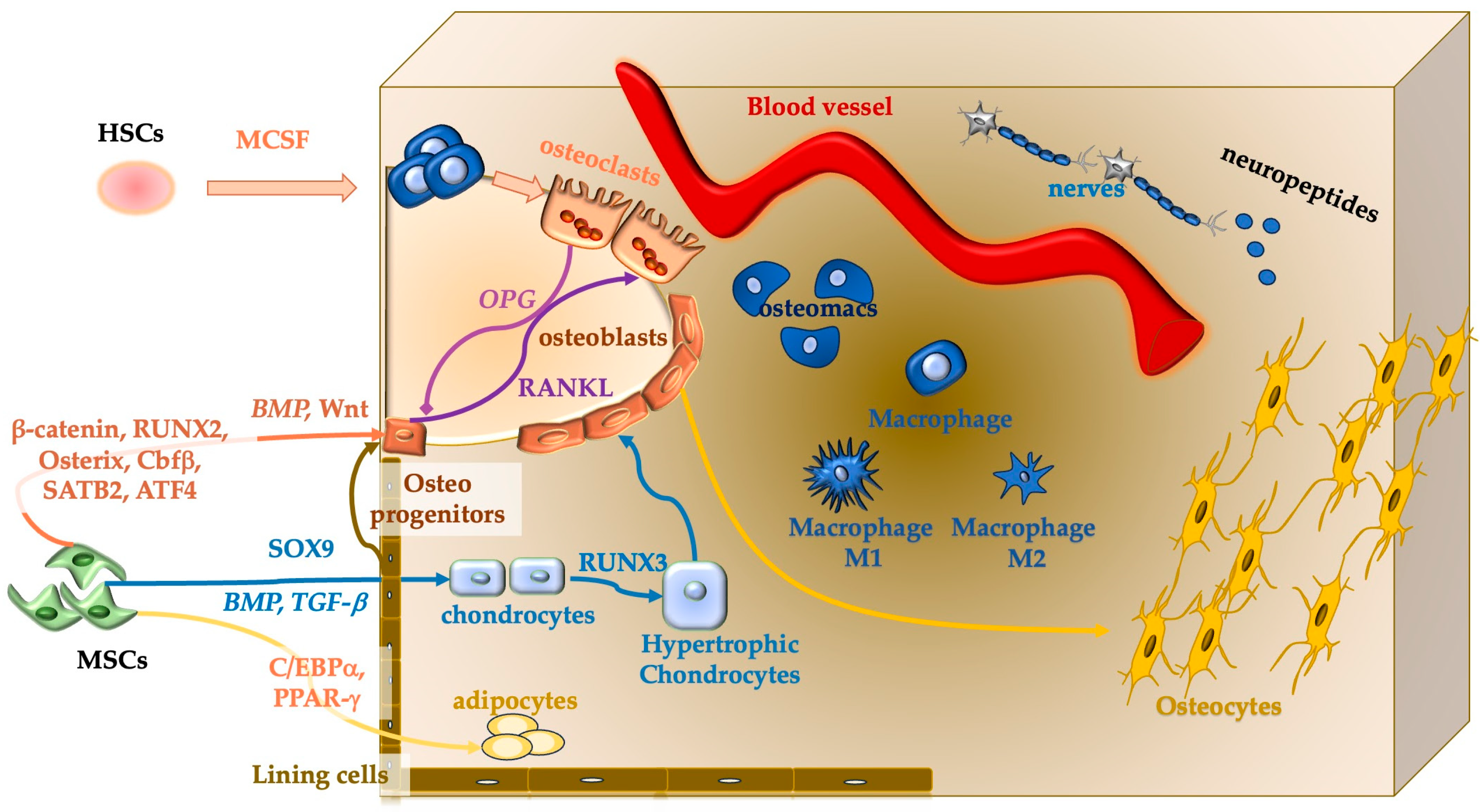
Multiple cell populations derived from mesenchymal precursors and hematopoietic stem cells coordinate osteogenesis and continuous bone remodeling (Figure 1).
Osteoblasts, the primary bone-forming cells, arise from mesenchymal stem cells (MSCs) through osteoprogenitor and preosteoblast [36].
They form osteoid, including type I collagen and non-collagenous proteins (e.g., osteocalcin (OCN), bone sialoprotein (BSP), matrix Gla protein, osteonectin (OSN), OPN, fibronectin, thrombospondin, proteoglycans, and alkaline phosphatase), regulate matrix mineralization, and control hydroxyapatite deposition. Osteoblast differentiation and activity are tightly governed by signaling pathways, including but not limited to the Wingless integration glycoproteins (Wnt), BMPs, transforming growth factor-β (TGF-β), parathyroid hormone (PTH), fibroblast growth factors (FGFs), ephrin and ephrin ligands, Hedgehog, Notch, Hippo, and mechanosensitive Piezo1/2 channels. Osteoblasts deposit new bone matrix, promote its mineralization, and regulate hydroxyapatite deposition, contributing to osteon formation. They also secrete paracrine factors (e.g., TGF-β, BMPs, insulin-like growth factors I and II (IGF-I and IGF-II), platelet-derived growth factor (PDGF), FGFs, receptor activator of nuclear factor κB (NF-κB) ligand (RANKL), and macrophage colony-stimulating factor (M-CSF) that regulate bone cell proliferation and osteoclastogenesis via the RANKL/osteoprotegerin (OPG) axis. Furthermore, osteoblasts may differentiate into bone-lining cells or, upon becoming fully embedded within the mineralized matrix, terminally differentiate into osteocytes.
Osteocytes, the most abundant bone cells, act as master regulators of bone homeostasis.
Embedded within the hard and calcified bone matrix they have synthesized, osteocytes communicate through an extensive network of long dendritic processes that extend through canaliculi, forming an important lacunocanalicular network that integrates signals from other bone cells and the vasculature present in this matrix. They function as mechanosensors that detect mechanical loading, fluid shear stress, and microdamage, thereby modulating bone mass, elasticity, and strength. Osteocytes regulate mineral metabolism and systemic phosphate balance through factors such as FGF23 and sclerostin and orchestrate remodeling by secreting signals (e.g., RANKL and sclerostin) that influence osteoblast- and osteoclast-mediated processes.
Osteoclasts, derived from the hematopoietic lineage, specialize in bone resorption, secreting acids and proteolytic enzymes that degrade mineralized matrix. Their differentiation is primarily controlled by M-CSF and RANKL [37], linking immune activation to bone loss.
In addition to classical osteoclast properties, other bone-resident macrophages (osteomacs) support osteoblast differentiation, intramembranous mineralization and fracture healing [38,39].
Osteomacs and sensory nerve fibers further regulate bone repair and regeneration. Osteomacs modulate immune response, promote efferocytosis, angiogenesis, and MSCs recruitment, and balance osteoblast-osteoclast activity; their depletion leads to osteopenia. PTH promotes macrophage polarization from the proinflammatory M1 to the reparative M2 phenotype [31,40,41,42,43,44,45,46]. Nociceptor-mediated neuropeptide and growth factors release following injury promotes angiogenesis and osteogenesis and may act indirectly via immune modulation [46].
Bone homeostasis also depends on systemic crosstalk between bone and other organs, including bone marrow, muscles [47], adipose tissue, the brain [48], the gut and its microbiota [49], endocrine glands, the immune system [50], the kidney, pancreas [51], and liver [52]. The osteocyte lacunocanalicular network facilitates molecular exchange between bone and the bloodstream, supporting whole-body mineral and metabolic regulation.

2.2. Molecular Pathways Regulating Bone Homeostasis

Bone homeostasis depends on coordinated cellular activity and molecular crosstalk that balance bone formation and resorption. This network comprises growth and transcription factors, hormones, cytokines, extracellular matrix (ECM) components, and signaling molecules vesicles (Figure 2). Both systemic and local factors regulate osteogenesis and bone repair. Key pathways Wnt, BMP, FGF, and TGF-β activate specific intracellular cascades (Wnt/β-catenin, MAPK/ERK, and Smad), which in turn induce osteogenic transcription factors, including RUNX2 and Activating Transcription Factor 4 (ATF4), driving osteoblast commitment, maturation, and differentiation and promoting ECM synthesis.
The main regulatory pathways include Wnt, Hedgehog (Hh), Notch, and RANKL/RANK/OPG.
Wnt signaling (canonical and non-canonical) governs osteogenesis from MScs while suppressing adipogenic and chondrogenic fates [53].
Wnt3a and Wnt5a enhance osteoblastogenesis and modulate osteoclast activity; Wnt5a signals via ROR2/RhoA/JNK (ROR2, Receptor Tyrosine Kinase-Like Orphan Receptor 2; RhoA: Ras homolog family member A; JNK, c-Jun N-terminal kinase) and can indirectly inhibit osteoclastogenesis by increasing OPG expression. R-spondin 2 (RSPO2) amplifies canonical Wnt signaling by preventing the receptor degradation, whereas sclerostin and Dickkopf-1 (DKK1) reduce bone formation [54,55].
The Hh signaling, mediated by Indian Hedgehog (Ihh) and Sonic Hedgehog (Shh), regulates skeletal development, endochondral ossification, and fracture repair through Runx2 and osterix activation. Although Hh activity declines after birth, it can be reactivated during fracture healing to support regeneration.
Notch Signaling pathway is a key factor of skeletal development, regeneration, and the coupling of angiogenesis with osteogenesis [56,57]. It works through cell–cell interactions and pathway crosstalk [58,59].
The RANKL/RANK/OPG axis is the main regulator of osteoclastogenesis and bone resorption. RANKL binding to RANK on osteoclast precursors promotes differentiation and activation, whereas OPG acts as a decoy receptor that inhibits this interaction [60,61].
The RANKL/OPG ratio determines osteoclast formation and is critical in pathological bone loss, such as osteoporosis. RANKL secreted by osteoblasts, osteocytes, and immune cells also mediates osteocyte responses to mechanical stimuli [62,63]. Conversely, elevated RANKL expression in cancer cells enhances bone resorption, thereby preparing the bone microenvironment for homing and establishment of bone metastases [64].

2.2.1. Growth Factors

Bone Morphogenetic Proteins—Transforming Growth Factor-β
BMPs and TGF-β are potent osteoinductive cytokines regulating bone formation and repair via canonical (Smad-dependent) and non-canonical (MAPK) pathways.
The BMPs promote osteoblast differentiation by activating Runx2 and osterix, and guide MSCs’ commitment, especially during fracture healing, where they contribute to callus formation and are regulated by vascular endothelial growth factor (VEGF) [65,66].
TGF-β, released from bone matrix during resorption, couples bone resorption to formation, promotes MSCs proliferation and differentiation, stimulates ECM production, and modulates inflammation, improving repair outcomes [67].
Insulin-like Growth Factors
Insulin-like Growth Factors (IGFs), IGF-I and IGF-II, released from bone matrix during remodeling, promote osteoblasts and chondroprogenitor proliferation and differentiation, couple resorption to formation, and mediate anabolic effects of growth hormone (GH) and PTH through IGF receptor/insulin receptor substrate (IGR-R/IRS) signaling [68,69]. IGF signaling supports, longitudinal bone growth, mineralization, fracture repair, and metabolic regulation, potentially interacting with the gut microbiota [49].
Fibroblast Growth Factors
FGFs regulate skeletal development, osteogenesis, angiogenesis, and repair [70]. Canonical, endocrine, and intracellular FGFs act in a spatiotemporal manner, with FGF2 and FGF23 specifically associated with the osteoblast lineage [71]; FGF23 also exerts endocrine control over renal phosphate handling [72,73].
Platelet-Derived Growth Factor
Platelet-derived growth factor (PDGF), and its cognate platelet-derived growth factor receptor beta (PDGFR-β) promote MSC proliferation, chemotaxis, angiogenesis, pericyte recruitment, and osteoblast survival while enhancing OPG production to limit bone resorption [74,75,76]. PDGF-BB accelerates fracture healing and is clinically approved by the Food and Drug Administration (FDA), although it may antagonize BMP-2-induced mineralization under certain conditions [77,78].
Angiogenic Factors
Angiogenesis is essential for bone development, repair, and remodeling [66,79]. VEGF, is indispensable for coupling angiogenesis with osteogenesis, enabling vascularization and recruitment of progenitor cells [80]. The Hypoxia-Inducible Factor 1-alpha (HIF-1α)/VEGF axis and PDGF signaling coordinate early inflammatory responses and efficient oxygen and nutrient delivery [81].

2.2.2. Transcription Factors

Transcription factors, including osterix, SRY-related HMG-box 9 (SOX9), ATF4, and β-catenin, orchestrate osteoblast commitment, maturation, and matrix production. RUNX2 acts as the master regulator of osteoblast differentiation and integrates signals from Wnt, FGFs, Hh and PTH pathway, while ATF4 regulates collagen synthesis, mineralization, and RANKL expression [65,82,83,84]. For osteoclast commitment, the primary transcription factor is NFATc1.

2.2.3. Hormones Regulating Bone Remodeling

Hormonal regulation is central to bone remodeling. PTH and Parathyroid hormone-related protein (PTHrP) regulate osteoblast differentiation and osteoclastogenesis via Parathyroid Hormone Receptor 1 (PTHR1) signaling and RANKL modulation, with intermittent PTH exerting anabolic effects [85,86,87]. Estrogens preserve bone mass by suppressing osteoclast activity and interacting with Wnt/β-catenin signaling; deficiency leads to postmenopausal osteoporosis [88,89,90,91,92]. Oxytocin enhances bone formation, and in osteoblasts, its expression increases in response to estrogen exposure [93,94,95]. The combined application of photobiomodulation therapy and oxytocin has been shown to improve MSCs viability and mineralization by modulating the expression of RUNX2, OCN, and OPG [96]. Vitamin D maintains calcium-phosphate homeostasis and mineralization [97], while insulin promotes osteoblast proliferation and systemic metabolic coupling through OCN activation [98,99,100]. Treatment of patients with osteoporosis may provide greater benefits when combined with vitamin D and calcium supplementation [101].

2.2.4. Extracellular Vesicles

Extracellular vesicles (EVs) mediate intercellular communication by transporting a wide range of molecules, such as proteins, lipids, metabolites, nucleic acids, mRNA, and microRNA (miRNA), that regulate osteoblast and osteoclast activity, including RANKL, OPG, sclerostin, and miRNA (e.g., EVs rich in miR-214-3p; miR-141-3p, and miR-940) that modulate bone formation [102,103,104,105].

2.2.5. Immune Cytokines & Inflammatory Mediators

Bone healing integrates immune and skeletal systems, with cytokines such as TNF-α, Interleukin (IL)-1 and IL-6 regulating MSC recruitment and osteoclast activity [50,106]. Controlled inflammation and macrophage polarization are essential for fracture healing [107]. M1-macrophages govern early inflammation, while M2-like macrophages support resolution angiogenesis, efferocytosis, and tissue remodeling [107].
Bone regeneration proceeds through four principal stages, coordinated by growth factors, cytokines, hormones, and mechanotransductive signals. Inflammation stage: begins immediately after fracture with hematoma formation [108]. TGF-β, PDGF, other growth factors, cytokines, and chemokines initiate the regenerative healing cascade and can trigger tissue regeneration following injury [109]. Angiogenesis is also required at this stage and is initiated by growth factors such as VEGF. TNF-α, alone or in combination with other inflammatory factors, promotes MSC activation. The following stage involved the formation of the soft callus, which lasts approximately 1–3 weeks. During this phase, recruited MSCs differentiate into chondrocytes and fibroblasts, forming a fibrocartilaginous tissue that bridges the gap between the bone fragments and stabilizes the fracture site. Chondrogenesis is activated by HIF-1α and sustained by SOX9 and TGF-β expression. The callus progressively hardens over weeks to months as osteoblasts differentiate under the control of RUNX2, osterix, and BMPs. The cartilage framework is replaced by bone tissue, which is ultimately remodeled to restore healthy, functional bone through coupled osteoblast-osteoclast activity, with additional neural regulation of somatosensory neurons via FGF9-FGFR signaling [43]. This late phase can take months to years.

2.2.6. Support and Matrix Molecules

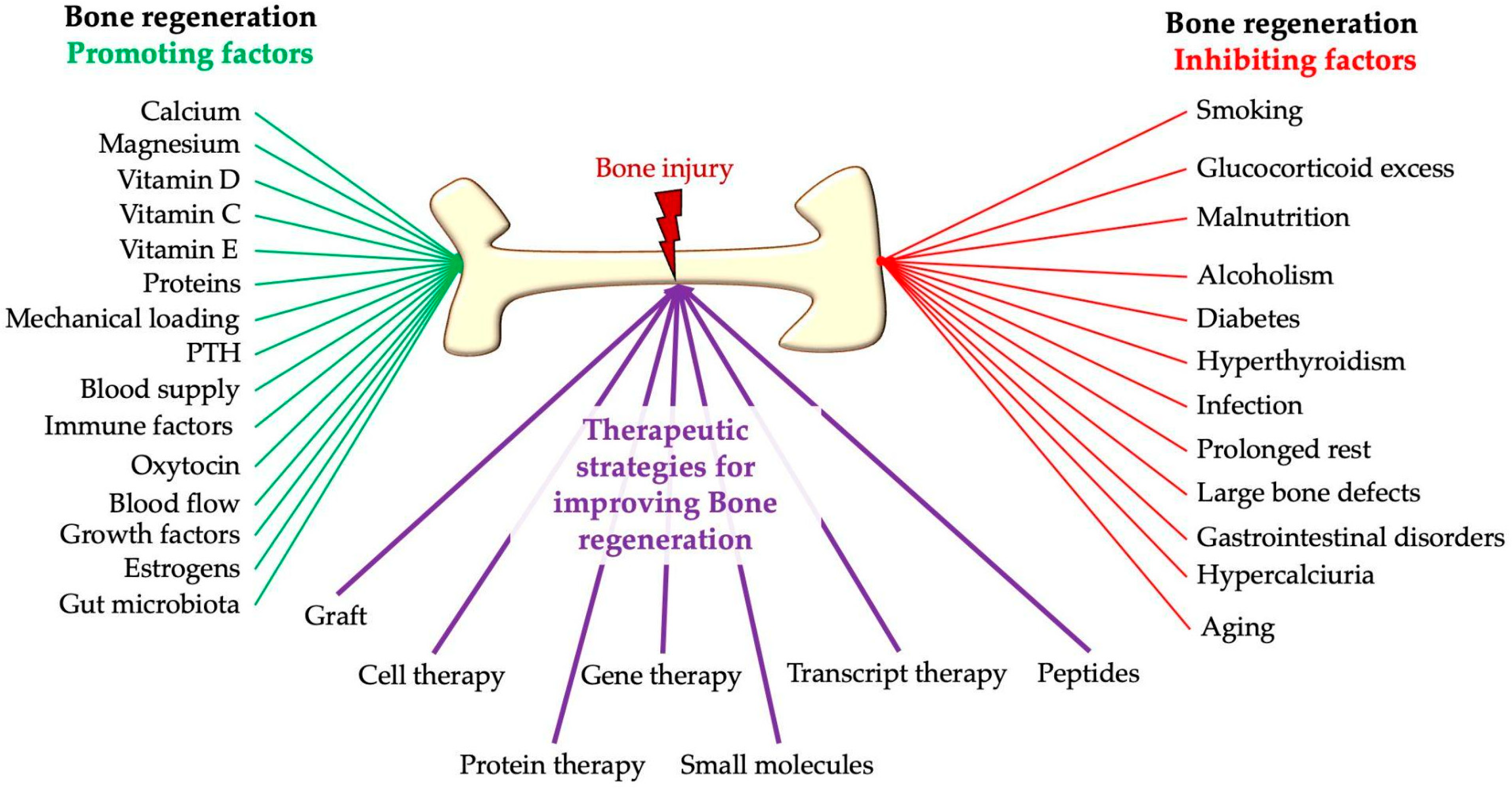
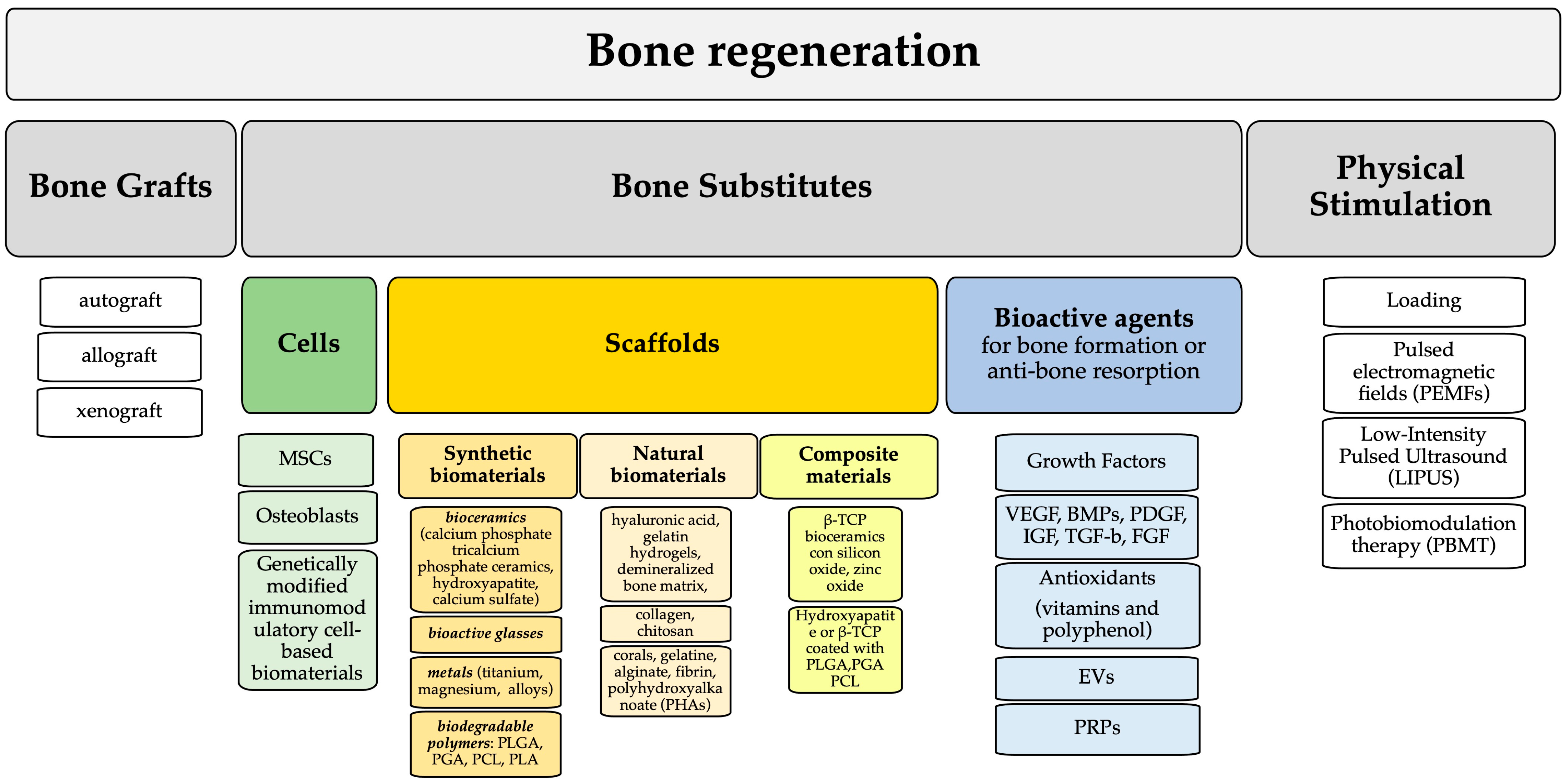
Bone healing after a fracture is not always straightforward and can be complicated by severe issues, especially in cases of malunion or non-union. Bone repair can be supported through proper nutrition—such as adequate intake of calcium, proteins, vitamins D, E, and K, magnesium, and omega-3 fatty acids—and lifestyle changes like quitting smoking and reducing alcohol intake. However, in cases of extensive bone loss or critical-sized defects, advanced clinical treatments and therapies are still needed. Tissue engineering shows great potential for treating large bone defects and can help improve bone healing (Figure 3).
Tissue engineering for bone regeneration leverages molecular, biophysical, angiogenic, and neuroregulatory insights to select optimal cell sources, scaffolds, and bioactive molecules. The purpose is to promote vascularization, osteogenesis, and functional repair by enabling recruitment, engraftment, proliferation, and differentiation of osteogenic cells.
Ideal biomaterials must be biocompatible, biodegradable, and highly porous mimicking the native bone structure, mechanical properties and microenvironment while supporting cell osteoconduction, osteoinduction, and osteogenesis. Most investigations are still performed in vitro and ex vivo, demonstrating that cell sources such as MSCs or osteoblasts represent the first choice; however, coculture of MSCs or osteoblasts with other cell types, including endothelial cells or immune cells, has been shown to enhance cell proliferation [110,111].
Regarding scaffold characteristics, synthetic biomaterials (e.g., calcium phosphate ceramics, biodegradable polymers: poly(lactic-co-glycolic acid) (PLGA), polycaprolactone (PCL), and polylactic acid (PLA), natural biomaterials (e.g., collagen, chitosan, hyaluronan, gelatin hydrogels), and composite scaffolds have been investigated.
Nevertheless, these materials still present limitations, such as weak mechanical properties and low osteoconductivity, that restrict clinical use in larger defects [112]. Consequently, translation from bench to bedside is still hampered. To improve outcomes, bioactive molecules (growth factors, recombinant proteins, small molecules, and blood-derived products) have been employed to promote MSCS differentiation, osteogenesis, and angiogenesis. These molecules generally favor bone healing; several were FDA-approved, including recombinant human Bone Morphogenetic Protein-2 (rhBMP-2), peptide P-15, and PDGF-BB.
In some cases, adverse effects, uncontrolled release, short half-life, batch-to-batch variability, and heterotypic ossification were observed [113,114]. Emerging evidence underscores the pivotal role of immune modulation in bone homeostasis and repair, setting the development of immunoregulatory biomaterials as a critical and ongoing challenge [115].

2.3. Overview

Collectively, these molecular and cellular mechanisms highlight that successful bone regeneration depends on the coordinated regulation of three fundamental processes: osteogenesis, angiogenesis, and immune modulation. Disruption of any of these axes, as observed in critical-sized defects, leads to impaired healing. Therefore, most emerging therapeutic strategies aim to simultaneously target these interconnected pathways rather than focusing on a single biological process.

3. Physical Stimulation Techniques

Physical stimulation techniques for bone regeneration are external-stimulus-responsive therapies that enhance bone healing by using mechanical, electrical, electromagnetic, or light-based signals to bypass traditional biochemical interventions and directly activate the body’s cellular repair mechanisms.

3.1. Mechanical Loading

Bone is particularly notable for its remarkable ability to respond and adapt to physical forces. Four key cell types in bone tissue, including osteoblasts, osteoclasts, bone lining cells, and osteocytes, play critical roles in responding to mechanical stimuli. In addition, other cell lineages, including myocytes, platelets, fibroblasts, endothelial cells, and chondrocytes, also exhibit mechanosensitivity.
Among these, osteocytes serve as the primary mechanosensors and play a central role in detecting mechanical changes within the bone matrix.
In particular, mechanotransduction converts physical load into biochemical signals that alter cell function and morphology, ECM synthesis, and gene expression. Mechanical forces are sensed by specialized structures such as dendrites or cilia, which function as mechanoreceptors [116,117].
Mechanical loading-mediated bone remodeling is not an independent process involving a single factor. Instead, it is strongly influenced by pathophysiological factors such as age, sex, estrogen deficiency, disuse, and different disease states, all of which influence mechanotransduction and bone remodeling outcomes.
Upon mechanical loading, osteocytes detect interstitial fluid flow within the lacuno–canalicular network and subsequently generate and transmit signaling molecules that regulate the osteogenic/osteoclastic functions of osteoclasts and osteoblasts, respectively, thereby affecting bone remodeling [118].
By comparing variations in fluid transport with variations in wall shear stress, Bakker and collaborators demonstrated that the flow-derived stimulus (Pulsatile Fluid Flow, PFF, 0.7 ± 0.02 Pa, 5 Hz, 0.4 Pa/s) that activates the bone cells is shear stress rather than streaming potentials or chemotransport [119].
Extracellular matrix transmembrane receptors like integrins and CD44 receptors, which can be identified on the osteocyte membrane and attached to both the extracellular matrix and the cytoskeleton, are considered the most promising candidates as mechanotransducers for the conversion of mechanical stimuli into intracellular signals by osteocytes [120].
Signal molecules including prostaglandins, in particular Prostaglandin E2 (PGE2), and nitric oxide (NO) are then produced and secreted. Specifically, one-hour PFF treatment stimulated the release of PGE2 by 3.5-fold and Prostacyclin (PGI2) by 2.2-fold in elderly women [121]. Prostaglandin production continued for several hours after stress was stopped. For this reason, a limited number of stress cycles per day is sufficient to induce an adaptive response in animal studies [122]. The upregulation of prostaglandin release in response to mechanical stress was associated with an induction of cyclooxygenase-2 (COX-2) [123]. In a study of Joldersma and colleagues, PFF also increased the expression of COX-2 mRNA by 2.9-fold but did not change cyclooxygenase-1 (COX-1) mRNA [121]. While NO likely plays a role as a local inhibitor of osteoclastic attack during bone remodeling [121].
Numerous studies in bone mechanobiology have aimed to recreate biomimetic in vitro conditions that replicate the mechanical environment of bone cells to investigate the relations between cellular behaviour at the microscopic scale and the surrounding physical milieu.
Among the mechanical stimuli applied in both in vitro and in vivo models are: (a) dynamic bioreactor, including rotating wall vessel reactor, pinner flask, and flow perfusion, which provide physical signals that promote stem cell differentiation and ECM production [124]; (b) ultrasound-based mechanical stimulation, which exploits acoustic wave propagation through the placement of a piezoelectric transducer in culture media. Low-intensity ultrasound (0.5–50 mW/cm2) is commonly used for diagnostic purposes, whereas higher intensities (0.2–100 W/cm2) are applied therapeutically to enhance bone healing and improve drug efficacy; (c) magnetic, optical, electric, aerodynamic, and acoustic levitation-based approaches, which enable contact-free mechanical stimulation and precise control of physical forces [125].
Osteocytes are considered the primary mechanosensors in bone tissue. They can detect mechanical forces via fluid shear stress within the lacuno–canalicular network [126] leading to the activation of intracellular pathways that control bone remodeling. Fluid shear stress stimulates the release of signaling molecules such as nitric oxide (NO), prostaglandins (e.g., PGE2), and adenosine triphosphate (ATP), which act in autocrine and paracrine manners to regulate osteoblast and osteoclast activity [127,128]. At the molecular level, mechanical stimuli, including fluid shear stress and cyclic strain, activate the Wnt/β-catenin pathway by inhibiting sclerostin expression in osteocytes, consequently promoting osteoblast differentiation and bone formation [126,127]. Concurrently, mechanical loading induces intracellular calcium (Ca2+) oscillations through mechanosensitive ion channels such as Piezo1, which regulate downstream signaling cascades, including MAPK/ERK [129,130].
These mechanosensitive pathways establish a key mechanism for the effectiveness of physical stimulation in bone repair and regeneration [129,131].

3.2. Electrical Stimulation (Direct Current and Capacitive Coupling)

Electrical stimulation, including direct current (DC) and capacitive coupling (CC), represents one of the earliest clinically applied physical approaches for enhancing bone regeneration [132]. These techniques have been shown to promote osteogenesis by modulating transmembrane potentials, enhancing calcium ion influx primarily through voltage-gated calcium channels (VGCCs), and activating intracellular signaling pathways such as MAPK and Wnt/β-catenin [132].
DC stimulation consists of its continuous application of low intensity to the bone surface through implantable electrodes [133]. This technique is considered invasive with the risk of infection and the high costs limiting the widespread clinical use [133] even if the results were promising. Recently, Pettersen and collaborators reviewed the literature and found that these effects have not been conclusively demonstrated or optimized in humans due to inconsistent and incomplete stimulation parameters [134].
On the other hand, CC generates an alternating electric field that stimulates cell differentiation and mineralization, acting via application of electrodes on the skin surface, so it has been considered a non-invasive technique [135]. In an in vivo study on dogs, animals received tibial corticotomy and distraction, with one group undergoing 28 days of CC. Statistically significant changes were reported: 37% lower maximum torque capacity, 40% decrease in strain energy to failure, lower modulus of rigidity (37% decrease) and a smaller percentage of active osteoid perimeter (20% decrease) in the stimulated group compared with the nonstimulated group [136].
Clinically, these approaches have demonstrated improved union rates in fracture healing and non-union cases. Nevertheless, despite their clinical relevance, variability in stimulation parameters and limited mechanistic standardization have restricted their broader application [137] compared to newer modalities such as pulsed electromagnetic fields (PEMFs).

3.3. Electromagnetic Fields

PEMFs represent a safe, noninvasive physical modality for modulating the osseous microenvironment. Therefore, this therapeutic approach was approved by the FDA in 1979 for the treatment of non-unions of bone fractures and related complications. Nowadays, the use of PEMFs has been considered an important tool in orthopedics for bone defect repair and the induction of osteogenesis [138,139]. Numerous clinical and scientific studies have shown the effects of PEMFs on angiogenesis, osteogenesis, and bone remodeling in various diseases, although several mechanisms remain under investigation and are not fully understood [140].
Among the biological bases and key pathways involved, it has been shown that physical PEMF stimulation acts on different cells such as osteoblasts, osteoclasts, endothelial cells, MSCs through membrane receptors, ion channels, and key intracellular signaling pathways. All these pathways have been considered key aspects in promoting bone repair and tissue regeneration [138,140,141,142].
There are multiple mechanisms of action underlying PEMFs’ beneficial effects on bone healing. Among these, Varani and collaborators [141] recently demonstrated that induction of osteoblast membrane adenosine receptor activation leads to activation of the Wnt/β-catenin signaling pathway, which is considered critical for normal bone formation and development. This pathway is also involved in mechanosensation and transduction in bone, which are essential for cell viability, anti-apoptotic pathways, and communication [143,144].
Moreover, in different animal experimental studies on osteoporosis, it has been demonstrated that the beneficial effects of PEMFs are achieved by upregulating the canonical Wnt (Wnt/β-catenin) signaling pathway, which is considered crucial for bone regeneration [144,145,146,147].
In 2018, a pilot study by Catalano and collaborators investigated the effects of PEMF treatment in osteoporotic postmenopausal women, demonstrating treatment-related changes in bone metabolism and the involvement of the Wnt/β-Catenin and RANKL/OPG signaling pathways, suggesting an anabolic and antiresorptive function [148]. Furthermore, in 2025, it was demonstrated that PEMF treatment reduces pro-inflammatory cytokine expression and induces bone formation biomarkers in a lipopolysaccharide (LPS)-induced osteoporosis mouse model, highlighting its potential as a therapeutic strategy for osteoporosis [149].
Experimental in vitro studies have demonstrated that PEMF exposure can increase the area of mineralized matrix in osteoblasts. For example, during the proliferation stage, this increase is 51% compared to the control group. PEMF also significantly increases osteoblast proliferation compared to untreated controls. DNA content in the PEMF-stimulated group increased by 38.5% and 7.1% over the control group on the 2nd and 5th days of culture, respectively [150].

3.4. Low-Intensity Pulsed Ultrasound

Decades of research have shown that Low-intensity Pulsed Ultrasound (LIPUS) is a non-invasive physical intervention that uses low-intensity, pulsed ultrasound to specifically target tissue and has a therapeutic effect on diseases involving bone remodeling [151].
This physical stimulation at 1.5 MHz, maintaining a constant intensity of 30 mW/cm2 and 1 kHz, for 20 min/day, has been demonstrated to be effective in treating bone fractures [152,153].
Among the mechanisms of action of LIPUS in bone healing, some data in the literature highlight its effect in changing the mechanical stimulus in biochemical response. In particular, LIPUS activates integrins and different pathways involved in osteogenic differentiation, including an increase in alkaline phosphatase (ALP) activity, OCN, VEGF secretion, calcium nodule formation, and osteoblastic differentiation [152,154,155,156]. Moreover, a review by Puts and collaborators reported a LIPUS inflammatory-modulation resulting in anti-inflammatory and pro-regenerative effects [157].
Taken together, these data showed the effect of LIPUS on osteogenesis induction and on reduction in osteoclast-mediated bone resorption.
In animal models of osseous skull defects, LIPUS has been shown to induce almost complete closure of the defect [155]. Furthermore, in rabbit tibias, LIPUS stimuli have been considered a promising complementary treatment approach for non-irradiated bone regeneration procedures, enhancing bone healing [158].
In clinical conditions, there is controversy: some reviews reported a reduction in defect consolidation time, while a more recent, rigorous review paper reported no effects of this treatment on fresh bone fractures [159,160,161].
On the other hand, Leighton and collaborators [162] suggested LIPUS treatment as an alternative to surgery for established non-unions in patients for whom surgery is high risk, including elderly patients.
So, the LIPUS approach is probably more valuable in impaired healing conditions (osteoporosis or pseudoarthrosis) than in a fresh fracture.

3.5. Photobiomodulation Therapy

Phototherapy is a technique based on the controlled induction of inflammatory reactions through the use of specific light rays emitted by dedicated lamps, ranging from the UV spectrum (200–400 nm) to visible light (400–700 nm) and near-infrared (700–1300 nm), and capable of inducing photobiostimulation [163]. Important parameters to choose the light in clinical applications are related to its tissue penetration depth, which is wavelength-dependent [163].
Photobiomodulation therapy (PBMT) has increasingly gained recognition as a promising therapeutic approach to enhance bone regeneration in both maxillofacial and orthopedic surgery. PBMT often requires grafting materials or regenerative adjuvants to achieve predictable healing. In this context, PBMT has attracted growing interest due to its non-invasive profile and its ability to modulate key cellular and molecular pathways involved in tissue repair [164,165]. In an experimental model of calvarial critical-size bone defect, the use of collagen membrane and/or autologous bone with PBMT greatly enhanced the regenerative effect [164].
At the mechanistic level, PBMT exerts its regenerative effects through several intracellular signaling pathways. Light energy absorbed by mitochondrial chromophores, in particular cytochrome c oxidase, increases ATP production and modulates ROS levels. These mitochondrial responses trigger downstream cascades regulating gene expression associated with cell proliferation, migration, and differentiation. Systematic reviews report that PBMT promotes osteoblast differentiation by upregulating key osteogenic markers, including ALP, OCN, and RUNX2 [166,167]. PBMT also enhances angiogenesis by promoting endothelial cell proliferation and stimulating the release of pro-angiogenic growth factors, which are essential for oxygen and nutrient supply during tissue repair [167]. Additionally, in a carrageenan-induced inflammation model on the temporomandibular joint of rats, PBMT modulated inflammatory processes by reducing pro-inflammatory cytokines, inhibiting leukocyte chemotaxis in the TMJ, and increasing the protein level of IL-10, an anti-inflammatory cytokine [168].
However, despite its promising biological foundations, the clinical translation of PBMT remains limited by considerable heterogeneity in treatment parameters. Optimal Low-Level Laser Therapy (LLLT) parameters such as wavelength (600–980 nm), energy density (around 4–6 J/cm2), and treatment frequency are critical for maximizing bone-healing outcomes, particularly in early-stage osteogenesis. Variability in wavelength, energy density, irradiation duration, treatment frequency, and delivery techniques has led to inconsistent outcomes among studies. This lack of standardization complicates comparative analyses and hinders the development of evidence-based clinical protocols. Moreover, the biphasic dose–response effect, where both insufficient and excessive energy reduce therapeutic efficacy, highlights the need to optimize PBMT parameters [166,169].
Although preclinical research strongly supports the regenerative potential of PBMT, clinical results remain to be more deeply investigated. Some clinical studies report improved bone density, faster healing, and fewer postoperative complications following PBMT, while others show no significant differences compared with controls. For example, recently, a clinical trial using an 810 nm diode laser significantly improved implant stability, post-surgical healing, and reduced crestal bone loss around dental implants compared to controls [170]. On the contrary, another recent experimental clinical trial on implant stability did not report a significant effect using an 830 nm diode laser [168]. These discrepancies may be due to differences in study design, patient characteristics, defect type, and laser parameters. Many clinical trials also suffer from small sample sizes, methodological variability, and limited follow-up, which weaken the overall evidence base [164,171].
Beyond its role in bone repair, PBMT offers additional clinical benefits, including pain reduction, modulation of inflammation, and enhanced soft tissue healing. These effects may further improve patient comfort and reduce recovery times [164,172].
Future research should develop standardized PBMT protocols through rigorous randomized controlled trials with adequate sample sizes and long-term follow-up. Additional studies are needed to refine irradiation parameters, better understand dose–response relationships, and explore PBMT in combination with emerging regenerative approaches, including bioengineered scaffolds, gene-based therapies, and advanced stem cell delivery systems [165,173].
In summary, PBMT is a safe, non-invasive technique with significant potential to enhance bone regeneration and improve the management of large bone defects. While preclinical data robustly support its efficacy, further high-quality clinical research is essential to establish standardized protocols and confirm long-term clinical benefits.

3.6. Other Emerging Biophysical Stimuli

Microgravity provides a compelling model for elucidating the role of mechanical forces and loading in regulating bone homeostasis. Exposure to microgravity accelerates bone loss and reduces bone mineral density, while also impairing bone regeneration through the uncoupling of osteoblasts and osteoclast activity [174]. At the cellular level, microgravity shifts MSCs’ lineage commitment from osteogenic toward adipogenic pathways and diminishes the MSCs’ proliferative capacity and self-renewal. Wnt/β-catenin signaling is disrupted, characterized by reduced nuclear translocation of β-catenin and increased expression of sclerotin and DKK1. Under these conditions, osteoblast differentiation and survival are compromised, and osteoblasts exhibit reduced ECM synthesis and mineralization capacity. Osteocytes display altered morphology, impairing their mechanosensory function and consequent anabolic activity. In murine models, a 30-day spaceflight not only decreases trabecular bone volume but also induces osteocyte apoptosis [175]. Furthermore, osteoclastogenesis is enhanced through an increased RANKL/OPG ratio. In astronauts who perform resistance exercise using an advanced resistive exercise device and maintain adequate energy intake and vitamin D status, bone mass loss is attenuated [176].
The use of various physical cues (such as optical, electrical, and magnetic) has been studied since the early 1970s to develop a totally non-invasive method of tracking cell proliferation and differentiation in three-dimensional environments. In this context, electrochemical sensors, which connect electrical parameter variations with certain cell cycle events, dominated as the most appealing approach among all the suggested techniques [177].
The literature highlights two potential methods to monitor cell activities: (i) making the scaffold conductive or (ii) monitoring the cell and scaffold dielectric characteristics by putting external conductive materials in contact with the scaffold itself. Nevertheless, despite encouraging outcomes, integrating them with biocompatibility and biodegradability is still difficult, and other approaches, like the use of external sensing, remain widespread with a minimally invasive sensing strategy [178]. Thus, to monitor the attachment and growth of MSCs seeded into gelatin-chitosan hybrid hydrogel scaffolds, Tonello and collaborators developed sensors with a biocompatible carbon-based ink on foldable polyimide substrates using an innovative aerosol jet printing approach [178].
In recent years, electrically active biomaterials like piezoelectric scaffolds and electro-conductive hydrogels have attracted attention for their ability to modulate cellular behavior. The degree of piezoelectric activity affects the development of MSCs, with higher voltage outputs promoting osteogenic differentiation and lower voltage outputs favoring chondrogenic differentiation [179]. Barium titanate (BaTiO3), poly l-lactic acid (PLLA), poly (vinylidene fluoride-trifluoro ethylene) (PVDF-TrFE), and diphenylalanine (FF) are examples of piezoelectric scaffolds [180].
Promising approaches to improve piezoelectricity while preserving biocompatibility include the use of composite techniques, polymer blends, and piezoelectric and non-piezoelectric fillers.
Several decades ago, Ikada and collaborators reported that stabilizing a tibia fracture with an intermedullary piezoelectric PLLA rod increased bone callus development and fracture healing in a cat model. They also demonstrated that piezoelectric PLLA scaffolds significantly promoted osteoblast proliferation in vitro [181].
Liu and collaborators develop a piezoelectric-conductive scaffold based on cartilage decellularized ECM and modified gelatin, where piezoelectricity is induced by FF and conductivity by poly(3,4-ethylenedioxythiophene). In vitro, MSCs show biphasic differentiation, while in vivo, the scaffold significantly improves osteochondral repair, guiding cell migration and chondrogenic differentiation in the upper layer and osteogenic differentiation in the lower layer [180].
In a study by Zheng and collaborators, conductivity and piezoelectricity were combined in an organic–inorganic composite cryogel based on BaTiO3, and Ca2+ and Mn4+ were added to enhance BaTiO3’s ability to regenerate bone. By mimicking the electrophysiological features of normal bone tissue, this combination successfully facilitated charge migration and produced a potential electroactive milieu for bone regeneration [182].
However, several challenges remain in fully unlocking the potential of piezoelectric materials for translational medicine, such as the limitations in the mechanical properties of bone regenerative constructs [183].

3.7. Overview

The physical stimulation techniques have an overall positive biological effect with an improvement in quantitative outcomes (Table 1), even if clinical studies should be increased.
Interestingly, despite differences in the type of stimulus, many physical therapies converge on common intracellular pathways, including Wnt/β-catenin signaling, MAPK cascades, and modulation of ROS levels. These shared mechanisms may explain the potential for synergistic effects when combining physical stimulation with biochemical or biomaterial-based approaches.
Overall, physical stimulation strategies offer significant advantages as non-invasive or adjunctive therapies, with demonstrated potential to enhance bone repair. Nonetheless, variability in quantitative outcomes and a lack of standardized protocols continue to limit their widespread and consistent clinical translation. Future efforts should focus on optimizing stimulation parameters and combining these approaches with biological and biomaterial-based therapies to achieve synergistic regenerative effects.

4. Bioactive Agents and Their Carriers

Bioactive agents are able to stimulate a response in living tissue, inducing a biological response. They could be of natural [184] or synthetic [185] origin, able to promote tissue regeneration. Here, interesting categories for bone regeneration have been included, such as growth factors and antioxidants [186], nucleic acids [187], but also the active involvement of EVs and exosomes, which represent natural carriers for a bioactive cargo [188], and bioactive scaffold which could also act as modulators of the microenvironment [189].

4.1. Growth Factors

Numerous cytokines and growth factors, such as BMP-2, BMP-7, TGF-β, FGF-2, VEGF, PDGF, and IGF-1, are involved in bone tissue regeneration. These signaling molecules act together through interconnected pathways and extensive molecular crosstalk that regulate bone formation and remodeling in vivo.
The FDA has approved rhBMP-2 and rhBMP-7, which are currently used in clinical settings as osteoinductive agents to treat orthopedic disorders such as spinal fusion, acute open fractures, and non-union [190]. However, the extensive therapeutic use of BMPs has been hindered by adverse effects like inflammatory edema, ectopic ossification, seroma, and radiculitis linked to high doses of BMP application [113].
TGF-β plays a dual role in bone regeneration by promoting the proliferation and early differentiation of osteoprogenitor cells while inhibiting osteoblast maturation and matrix mineralization. In addition, TGF-β stimulates MSCs and supports chondrogenesis [191]. Consequently, combinational delivery strategies using TGF-β with BMPs have been shown to significantly enhance new bone formation [192].
One of the most crucial growth factors for angiogenesis and vascular development is VEGF. Previous reports have demonstrated the effectiveness of combining BMPs and VEGF by facilitating the replacement of cartilaginous tissue with bone tissue during endochondral ossification, as well as subsequent bone remodeling maintenance [193].
PDGF, secreted from platelet α-granules, exists as five dimeric isoforms (AA, AB, BB, CC, and DD). Among these, PDGF-BB has the strongest osteoregenerative ability and was approved by the FDA for ankle and hindfoot fusion surgeries in 2015 [194]. PDGF has a several-fold stronger chemotactic effect on MSCs recruitment, which can synergistically enhance the osteogenic effect of BMPs [195].
IGF-1 is particularly involved in bone formation, promoting osteoblast differentiation [196].
Platelet concentrates, such as platelet-rich plasma (PRP) or human platelet lysate (HPL), contain many of these growth factors and have been widely demonstrated to enhance the healing of cartilage lesions, reduce joint inflammation, and alleviate various musculoskeletal pathological conditions. To optimize the therapeutic efficacy of PRP in orthopedic applications, careful standardization and characterization of the product are required. To date, some studies have addressed the biochemical characterization of PRP and HPL formulations for clinical translation [197].

4.2. Antioxidants

Among bioactive compounds, antioxidants, such as vitamins, polyphenolic compounds, and various nanostructures that scavenge ROS, have been studied in the context of bone repair. Increase in ROS in osseous milieu induces apoptosis of osteoblasts and osteocytes, inducing osteoclastogenesis, so lead to osseous tissue degradation, osteoporosis, and an increase in bone fracture risk [17,198].
Oxidative stress is considered a key factor for regenerative and anti-osteoporotic strategies.
For this reason, among the strategies, the use of biological compounds, such as antioxidants, is included. These compounds could be classified as natural substances, commonly found in medicinal plants and dietary sources, or as endogenous substances such as glutathione, superoxide dismutase, and catalase [199].
Many studies evaluated the potential protective effects of antioxidants in preventing bone resorption and pathological conditions. In particular, some authors evaluated the use of these substances in osteoporosis or inflammatory bone diseases, demonstrating that antioxidant intake induces bone remineralization and reduces osteoclastic activity, including also the ligand of receptor activator of NF-kB (RANKL)/OPG pathway modulation [200,201]. Moreover, Liu and collaborators [202], in glucocorticoid-treated osteoporotic rats, showed that an antioxidant compound, gastrodin, protect osteoblast through the actor Nuclear factor erythroid 2-related factor (Nrf2) regulated mitochondrial and ER stress-related signaling pathways, and enhances remineralization.
Another research study investigated the protective effect of pyrroloquinoline quinone (PQQ) on a femur fracture model in ovariectomized (OVX) mice. The results confirmed that PQQ played a preventive and protective role in OVX-induced delay of bone fracture healing, acting as an antioxidant; moreover, it promotes osteoblastic bone formation and inhibits osteoclastic action [23].
Overall, these findings support a beneficial effect of antioxidants in bone defects through osteogenesis and angiogenesis induction. The limitation is that these studies are almost all animal, or in vitro studies, and that clinical studies are poor and heterogeneous. So, further research is required to validate this claim.

4.3. Extracellular Vesicles and Exosomes

MSCs have been widely explored as a new therapeutic method for bone diseases. In regenerative medicine, MSC-derived EVs (MSCs-EVs) are suggested as a potential cell-free substitute for MSC cell treatment. According to recent research, MSCs-EVs are primarily responsible for their therapeutic benefits and are present in most biological fluids.
EVs are categorized as exosomes, microvesicles (MVs), and apoptotic bodies (ABs) based on different biogenesis. Exosomes (typically <150 nm in diameter) originate from the fusion of multivesicular bodies with the plasma membrane, leading to the release of intraluminal vesicles into the extracellular space. MVs (generally <1 µm) are generated by direct outward budding of the plasma membrane and transport locally derived cytosolic biomolecules. In contrast, ABs, which range in size from approximately 1 to 5 µm, are released during the apoptotic process and contain cellular debris, intact organelles, and nuclear fragments resulting from karyorrhexis [203].
MSCs-EVs are applied in different bone disorders, such as osteoarthritis, rheumatoid arthritis, osteoporosis, and bone fractures. Different methods based on distinct principles have been used to isolate EVs.
Traditional EV isolation techniques, such as ultracentrifugation, ultrafiltration, chromatography, and precipitation, are primarily based on EV characteristics (such as size and density) [204]. MSCs-EVs incorporated into scaffolds have been shown to enhance bone regeneration under pathological conditions. These effects are mediated by the promotion of angiogenesis, the upregulation of osteogenic gene expression—including RUNX2, OCN, OPN, Tuftelin 1 (TUFT1), Tuftelin Interacting Protein 11 (TFIP11), and Collagen Type I Alpha 1 Chain (COL1A1)—and the activation of key signaling pathways involved in bone regeneration, such as the Phosphoinositide 3-kinase (PI3K)/Protein Kinase B (AKT) and MAPK pathways. Additionally, both in vitro and in vivo investigations have shown that MSCs-EVs enable improved osteogenesis and tissue healing by decreasing pro-inflammatory cytokine levels [205].
Additionally, exosomes immunomodulate the inflammatory cytokines, influencing the ratio of M2/M1-type macrophages and the differentiation of M1- and M2-type macrophages. Macrophages are essential for bone remodeling as they regulate the resorption and formation of bone tissue. Pro-inflammatory cytokines, including TNF-α and IL-1, are released by M1 macrophages, which increase bone resorption and stimulate the production of the ligand for RANKL. M2 macrophages, on the other hand, release anti-inflammatory cytokines (IL-4, IL-10, and IL-13) that promote the formation of OPG, which suppresses RANKL activity and promotes bone mineralization and regeneration [206,207].
In these studies, the primary scaffolds used for MSCs-EVs delivery include hydrogels, collagen membranes, PLA combined with polyethylenimine, PLA, PCL, tricalcium phosphate–based scaffolds, titanium alloys, and demineralized bone matrix [205].

4.4. Peptide-Based Osteogenic Agents

Peptide-based osteogenic agents have emerged as advantageous alternatives due to their lower cost, improved stability, and reduced risk of adverse effects [208]. Examples include P-15, a synthetic peptide that mimics the cell-binding domain of collagen, and BMP-derived peptides that retain osteoinductive properties while minimizing the side effects associated with full-length proteins [208,209].
These peptides promote cell adhesion, osteoblast differentiation [209], and matrix mineralization, and can be incorporated into scaffolds or delivery systems for controlled release [208]. Their tunability and safety profile make them attractive candidates for advanced bone regenerative therapies.

4.5. Biomaterial-Based Controlled Release System

In recent years, many growth factors have been investigated in clinical trials for bone regeneration. However, the expected benefit to patients has sometimes not been proven by the outcomes [210,211,212].
To promote repair without these negative effects, there is a clear clinical need to develop alternative techniques for delivering one or more growth factors to the site of injury at sustainable, physiologically relevant dosages. Particularly, to improve treatment efficacy, the simultaneous or sequential administration of several growth factors has been used in addition to localized delivery of a single component or factor [213]. In fact, spatiotemporal control over the location and bioactivity of factors following administration into the body is essential [213]. An intriguing new generation of therapeutic agents for the treatment of a wide range of disorders is represented by sophisticated material systems that control the biological presentation of growth factors.
Numerous methods for conjugating growth factors to synthetic or natural compounds and biomaterials have been developed. Growth factors can be presented on extracellular matrices using two primary methods: covalent immobilization directly to the matrix and non-covalent physical adsorption via weak contacts. In both situations, adding cell-adhesive elements to the scaffold promotes intimate contact between growth agents and cells [213]. In tissue engineering, physical encapsulation is an easy and effective way of delivering growth factors locally with controlled release, providing an alternative to chemical conjugation. In order to support the creation of new tissue, methods like gas foaming and particle leaching produce porous scaffolds that enable the continuous release of active growth agents in vivo [213].
Among the studies, Freeman and collaborators describe a novel strategy using nanoparticle-functionalized bioinks for spatiotemporal regulation of growth factor delivery [214]. The method allows regulated tissue regeneration without supraphysiological doses by 3D-printing structures with specified growth factor gradients. When combined with targeted BMP-2 release, spatially patterned VEGF boosted angiogenesis in vivo and minimized heterotopic bone production while promoting massive bone defect healing [214].
Selecting the appropriate biomaterial is essential for creating effective growth factor encapsulation systems. Both natural and synthetic materials have been utilized extensively. While liposome-based carriers offer a biocompatible substitute by avoiding toxic organic solvents [215], synthetic polymers such as PLA, poly(glycoside) (PGA), PLGA, poly(anhydrides), and poly(amino acids) offer a range of options [216,217]. Encapsulation in synthetic porous materials can affect cell recruitment, proliferation, and differentiation in addition to delivering growth factors.
Collagen, gelatin, fibrin, chitosan, hyaluronic acid, alginate, and silk are examples of natural polymers that are widely used as carriers [213,218].
Growth factor release from degradable gels can be regulated by varying the rates of diffusion and degradation: slower degradation prolongs delivery, whereas faster degradation results in rapid release. However, it can be difficult to manage the characteristics of natural polymers. Recent techniques, such as partial oxidation of polymer chains and molecular weight modification, have increased control over breakdown and release behavior [213].

4.6. Gene Delivery Approaches

Recently, the use of Tetrahedral Framework Nucleic Acids (tFNAs), has exhibited numerous advantageous properties, including rapid internalization by cells via caveolin-mediated endocytosis, escape from lysosomal degradation [219] and potential as vectors for drug and gene delivery [220,221]. The use of tFNAs has been applied in bone regeneration with interesting results as a carrier for miRNA [222,223]. In particular, the use of miR-21-5p promotes a synergistic regulation of osteogenesis and angiogenesis [224]. tFNAs may also exert their effects by activating the AKT and ERK signaling pathways [225].
Moreover, IL-4 gene delivery via adeno-associated virus (AAV) vectors has been explored with positive results, with particular focus on modulating macrophage polarization and promoting osteogenic differentiation, creating a microenvironment useful to promote bone regeneration [226]. Using the AAV, the mediated gene delivery of α-calcitonin gene-related peptide also promotes osseointegration in an osteoporosis rat model [227].

4.7. Overview

Considering the overall proposed strategies (Table 2), the beneficial effects should be weighed against the therapy’s intrinsic limitations.
Despite advances in bone tissue engineering, clinical translation of bioactive strategies remains limited. This is due to the intrinsic complexity of bone regeneration and the balance between efficacy, safety, and delivery control. Growth factors such as BMPs remain the benchmark for osteoinduction because they potently activate SMAD signaling pathways. However, their use has well-documented adverse effects. These include ectopic bone formation and the need for high doses, raising safety and cost concerns. PDGF and IGF help early-stage repair by recruiting mesenchymal stem cells and promoting osteoblast proliferation. Yet, their short half-lives and narrow therapeutic windows make clinical use challenging.
Angiogenesis is crucial, and VEGF plays a central role in linking vascularization to bone formation. VEGF breaks down quickly and can cause abnormal vessel growth. This shows the need for regulated delivery systems. Biomaterial-based controlled-release platforms are important tools but achieving precise spatial and temporal control remains challenging.
Peptide-based approaches, such as P-15, offer greater stability and lower immunogenicity than full-length proteins. However, their lower bioactivity limits them as standalone therapies.
Extracellular vesicles (EVs) are gaining attention as a cell-free alternative. They deliver complex signals, including miRNAs. Preclinical data are promising, but a lack of standardized protocols remains a limitation for clinical use.
Adjunctive therapies, including antioxidant delivery systems, aim to modulate the inflammatory and oxidative microenvironment; however, their role in bone regeneration remains insufficiently validated in clinical settings. Gene therapy offers a theoretically powerful approach to sustained pathway modulation, yet concerns about vector safety, off-target effects, and regulatory approval continue to impede its progress.
These limitations show that single-factor approaches are insufficient to match the complex mechanism of bone healing. Research is shifting to combinational strategies.

5. Synergistic Approaches

The rationale for combinational strategies in bone regeneration lies in the intrinsic complexity of the healing process, which involves tightly coordinated biological, mechanical, and biochemical events. Single-modality approaches often fail to fully reproduce this complexity. In contrast, synergistic strategies aim to simultaneously target multiple regenerative axes, including cellular metabolism, signaling pathways, ECM remodeling, and vascularization.
Despite the significant advances in both physical stimulation techniques and bioactive agent delivery, each approach presents intrinsic limitations when used as a standalone therapy. Physical stimulation alone may not provide sufficient biological cues for sustained osteogenesis, while bioactive agents often suffer from rapid degradation, limited spatial control, and suboptimal delivery efficiency.
These limitations highlight the need for combinational strategies that can simultaneously address multiple aspects of the bone healing process. By integrating physical and biochemical stimuli, synergistic approaches aim to enhance cellular responsiveness, improve microenvironmental conditions, and achieve more robust and predictable regenerative outcomes.
From a mechanistic perspective, synergistic effects arise when different therapies act on complementary pathways. For example, physical stimuli such as PBMT or PEMFs enhance cellular metabolism and signaling sensitivity, thereby increasing the responsiveness of cells to growth factors. Similarly, biomaterial scaffolds provide a structural and biochemical niche that amplifies the effects of delivered bioactive molecules and supports cell recruitment and differentiation.

Combinational Strategies

Mechanical loading has been demonstrated to induce an antioxidant response modulating NRF2 activity and antioxidant function in osteocytes [228]. These results suggest that combining pharmacological approaches with mechanical loading could offer more effective treatments to maintain oxidative homeostasis [228]. These recent data are also supported by previous studies showing that mechanical stretch induces antioxidant responses in human bone marrow-derived-MSCs via activation of silent information regulator type 1 (SIRT1) and AMP-activated protein kinase (AMPK) [229]. Moreover, intracellular ROS were decreased while antioxidant enzymes, especially superoxide dismutase 1 (SOD1), were upregulated and osteogenesis was improved [229]. Also, the application of PEMFs modulates antioxidative defense enzymes [230]; new studies could address this strategy. Very recently, a system integrating a structurally engineered bimetallic FeCu-metal–organic framework within a PLA/hydroxyapatite scaffold has been tested both in vitro and in vivo [20]. It is able to program and sustain the co-release of Fe3+ and Cu2+ ions, providing tailored chemical signals and the synergistic stimulation with PEMF further enhance the scaffold’s bioactivity and successfully achieve synergistic angiogenic-osteogenic coupling, offering a promising approach for complex defect scenarios [20]. For instance, there is no experimental evidence or suggestions on LIPUS and the use of exogenous antioxidants; nevertheless, this technique could upregulate endogenous antioxidant defenses [231,232]. The same considerations can be applied to PMBT, where the antioxidant effect alone has been demonstrated experimentally, but no classical antioxidant has been associated with it.
Nevertheless, positive synergistic effects have been observed combining PMBT with platelet concentrates, in particular with leukocyte-PRF (L-PRF) in rat calvarial defects [171] and with PRF in an in vitro model [233] improving calcium deposition leading to enhanced cellular proliferation, increased vascularization, and accelerated ECM deposition [171]. Comparable benefits have also been documented when PBMT is combined with adipose-derived mesenchymal stem cells, yielding improved osteogenic differentiation and greater bone formation than stem cell therapy alone [234].
Moreover, 3D mechanical-chemical model of bone regeneration has been tested [235]. It combines multiple factors, including mechanical stimulation, scaffold degradation, and VEGF release. The study underlines the importance of coupling mechanical and chemical factors in bone regeneration and suggests that the findings can guide future scaffold designs and therapeutic strategies [235].
In addition, the integration of PBMT with biomaterials represents another promising frontier. Ceramic scaffolds, particularly those based on hydroxyapatite, are widely used due to their biocompatibility, osteoconductive properties, and structural similarity to natural bone mineral. Preclinical models have demonstrated that PBMT combined with hydroxyapatite scaffolds promotes greater bone formation, improves scaffold integration, and accelerates mineralization [236]. These findings support the use of PBMT as an adjunct in guided bone regeneration and implant dentistry.
Moreover, the use of scaffold itself has demonstrated good results enhanced by growth factors incorporated into a hydrogel scaffold combined with mechanotransduction (static magnetics), a mouse in vivo study [237]. In addition, recent papers demonstrated the beneficial effects of local antioxidants systems release in bone healing. In particular, the authors demonstrated the positive effects on bone regeneration from the incorporation of antioxidants into scaffolds, and the use of chitosan/gelatin films as local delivery systems, showing not only oxidative stress but also inflammation modulation [21,238,239].
Recent advances in biofabrication strategies further support the development of multifunctional platforms for bone and osteochondral regeneration. For instance, Bini and collaborators [240] highlighted the potential of integrated biofabrication approaches to resurface skeletal defects by combining biomaterials, cells, and bioactive signals within spatially controlled architectures. These approaches support the synergistic and multifunctional strategies to effectively reproduce the complexity of native tissue.
However, the simultaneous use of PEMFs with growth factors has not been explored, but experimental data support a positive synergistic concept [241]. Finally, studies combining photobiomodulation therapy with growth factors show promising but variable effects depending on the parameters used. In a recent in vitro study, the combining use of PMBT and PRF has been applied to MC3T3-E1 preosteoblasts with a clear synergistic effect in defined conditions [233]. The combined use has also been tested preclinically, in a rabbit critical-sized calvarial defect model, using concentrated growth factor (CGF), but the synergistic effect had not been observed [242]. On the contrary, in a rat model of a critical bone defect, the synergy of PBMT, BMPs, and a bovine biological membrane has been reported [243].
However, despite promising preclinical evidence, the translation of combinational strategies into clinical practice remains limited. Major challenges include the lack of standardized protocols, difficulty in optimizing dosing and timing of combined therapies, and variability in patient-specific responses. Future research should focus on defining precise spatiotemporal treatment regimens and identifying predictive biomarkers to guide personalized regenerative approaches.

6. Conclusions

Taken together, the data collected in this paper showed the effects of different approaches to bone regeneration. Moreover, the overall evidence shows an incomplete research panel on the synergistic use of different methods in bone healing, even though preclinical studies suggest a possible positive outcome compared with a single approach. Future research could address these new topics.

Funding

This research received no external funding.

Data Availability Statement

Not applicable.

Acknowledgments

The authors have reviewed and edited the output and take full responsibility for the content of this publication.

Conflicts of Interest

The authors declare no conflicts of interest.

Abbreviations

The following abbreviations are used in this manuscript:
AAVAdeno-associated virus
AbsApoptotic bodies
AKTProtein kinase B
ALPAlkaline phosphatase
AMPKAMP-activated protein kinase
ATF4Activating transcription factor 4
BaTiO3Barium titanate
BMUsBasic multicellular units
BMPsBone morphogenetic proteins
BSPBone sialoprotein
CbfβCore-binding factor beta subunit
C/EPBαCCAAT/enhancer-binding protein alpha
CCcapacitive coupling
c-FosCellular Fos proto-oncogene
CGFConcentrated growth factor
COL1A1Collagen type I alpha 1 chain
COX-1Cyclooxygenase-1
COX-2Cyclooxygenase-2
CXCL12C-X-C motif chemokine ligand 12
DCdirect current
DKK1Dickkopf-1
ECMExtracellular matrix
EGFEpidermal growth factor
EVsExtracellular vesicles
FDAFood and Drug Administration
FFDiphenylalanine
FGFsFibroblast growth factors
GHGrowth hormone
HhHedgehog
HIF-1αHypoxia-inducible factor 1-alpha
HPLHuman platelet lysate
HSCsHematopoietic Stem Cells
IGF-I/IGF-IIInsulin-like growth factors I and II
IGR-R/IRSIGF receptor/insulin receptor substrate
IhhIndian Hedgehog
ILInterleukin
IL-1βInterleukin-1 beta
JNKc-Jun N-terminal kinase
L-PRFLeukocyte platelet-rich fibrin
LLLTLow-level laser therapy
LPSLipopolysaccharide
LRP5/6Low-density lipoprotein receptor-related protein 5/6
M-CSFMacrophage colony-stimulating factor
miRNAmicroRNA
MSCsMesenchymal stem/stromal cells
MSCs-EVsMesenchymal stem cell-derived extracellular vesicles
MVsMicrovesicles
NFATc1Nuclear factor of activated T-cells, cytoplasmic 1
NF-κBNuclear factor kappa B
NOnitric oxide
Nrf2Nuclear factor erythroid 2-related factor
OCNOsteocalcin
OPGOsteoprotegerin
OPNOsteopontin
OSNOsteonectin
OVXOvariectomized
PBMTPhotobiomodulation therapy
PCLPolycaprolactone
PDGFPlatelet-derived growth factor
PDGFR-βPlatelet-derived growth factor receptor beta
PFFPulsatile Fluid Flow
PGAPolyglycolic acid
PGE2Prostaglandin E2
PGI2Prostacyclin
PI3KPhosphoinositide 3-kinase
PLAPolylactic acid
PLLAPoly l-lactic acid
PLGAPoly(lactic-co-glycolic acid)
PPAR-γPeroxisome Proliferator-Activated Receptor gamma
PRPPlatelet-rich plasma
PTCHPatched receptors
PQQPyrroloquinoline quinone
PTHParathyroid hormone
PTHrPParathyroid hormone-related protein
PTHR1Parathyroid hormone receptor 1
PVDF-TrFEPoly (vinylidene fluoride-trifluoro ethylene
RANKLReceptor activator of nuclear factor κB ligand
RhoARas homolog family member A
ROR2Receptor tyrosine kinase-like orphan receptor 2
ROSReactive oxygen species
RSPO2R-spondin 2
RUNX2/3Runt-related transcription factor 2/3
SATB2Special AT-rich sequence-binding protein 2
ShhSonic Hedgehog
SOX9SRY-related HMG-box transcription factor 9
SOD1Superoxide dismutase 1
TFIP11Tuftelin interacting protein 11
TGF-βTransforming growth factor beta
tFNAsTetrahedral framework nucleic acids
TNF-αTumor necrosis factor alpha
TUFT1Tuftelin 1
VEGFVascular endothelial growth factor
VGCCvoltage-gated calcium channel
WntWingless integration glycoproteins

References

  1. De Pace, R.; Molinari, S.; Mazzoni, E.; Perale, G. Bone Regeneration: A Review of Current Treatment Strategies. J. Clin. Med. 2025, 14, 1838. [Google Scholar] [CrossRef] [Scilit]
  2. Tsang, S.J.; Epstein, G.Z.; Ferreira, N. Critical Bone Defect Affecting the Outcome of Management in Anatomical Type IV Chronic Osteomyelitis. Strateg. Trauma. Limb Reconstr. 2024, 19, 26–31. [Google Scholar] [CrossRef] [Scilit]
  3. Benulic, C.; Canton, G.; Gril, I.; Murena, L.; Kristan, A. Management of acute bone loss following high grade open tibia fractures. Acta Biomed. 2020, 91, e2020012. [Google Scholar] [CrossRef] [Scilit]
  4. Bowers, K.M.; Anderson, D.E. Delayed Union and Nonunion: Current Concepts, Prevention, and Correction: A Review. Bioengineering 2024, 11, 525. [Google Scholar] [CrossRef] [Scilit]
  5. Vidal, L.; Kampleitner, C.; Brennan, M.; Hoornaert, A.; Layrolle, P. Reconstruction of Large Skeletal Defects: Current Clinical Therapeutic Strategies and Future Directions Using 3D Printing. Front. Bioeng. Biotechnol. 2020, 8, 61. [Google Scholar] [CrossRef] [Scilit]
  6. Stahl, A.; Yang, Y.P. Regenerative Approaches for the Treatment of Large Bone Defects. Tissue Eng. Part B Rev. 2021, 27, 539–547. [Google Scholar] [CrossRef] [Scilit]
  7. Newman, H.; Shih, Y.V.; Varghese, S. Resolution of inflammation in bone regeneration: From understandings to therapeutic applications. Biomaterials 2021, 277, 121114. [Google Scholar] [CrossRef] [Scilit]
  8. Wang, C.; Ying, J.; Nie, X.; Zhou, T.; Xiao, D.; Swarnkar, G.; Abu-Amer, Y.; Guan, J.; Shen, J. Targeting angiogenesis for fracture nonunion treatment in inflammatory disease. Bone Res. 2021, 9, 29. [Google Scholar] [CrossRef] [Scilit]
  9. Liang, Z.; Xue, F.; Zhang, J.; Pan, Q.; Hu, X.; Hu, Y.; Fan, Z.; Wang, W.; Guo, X.; Qian, Z.; et al. The Treatment of Large Bone Defects with Atrophic Nonunion by Bioactive Scaffolds Remodeling the Regeneration Microenvironment. Adv. Health Mater. 2025, 14, e2500120. [Google Scholar] [CrossRef] [Scilit]
  10. Yu, D.; Shen, W.; Dai, J.; Zhu, H. Treatment of large bone defects in load-bearing bone: Traditional and novel bone grafts. J. Zhejiang Univ. Sci. B 2025, 26, 421–447. [Google Scholar] [CrossRef] [Scilit]
  11. Fernandez de Grado, G.; Keller, L.; Idoux-Gillet, Y.; Wagner, Q.; Musset, A.M.; Benkirane-Jessel, N.; Bornert, F.; Offner, D. Bone substitutes: A review of their characteristics, clinical use, and perspectives for large bone defects management. J. Tissue Eng. 2018, 9, 2041731418776819. [Google Scholar] [CrossRef] [Scilit]
  12. Giannoudis, P.V.; Faour, O.; Goff, T.; Kanakaris, N.; Dimitriou, R. Masquelet technique for the treatment of bone defects: Tips-tricks and future directions. Injury 2011, 42, 591–598. [Google Scholar] [CrossRef] [Scilit]
  13. Migliorini, F.; La Padula, G.; Torsiello, E.; Spiezia, F.; Oliva, F.; Maffulli, N. Strategies for large bone defect reconstruction after trauma, infections or tumour excision: A comprehensive review of the literature. Eur. J. Med. Res. 2021, 26, 118. [Google Scholar] [CrossRef] [Scilit]
  14. Schulze, F.; Lang, A.; Schoon, J.; Wassilew, G.I.; Reichert, J. Scaffold Guided Bone Regeneration for the Treatment of Large Segmental Defects in Long Bones. Biomedicines 2023, 11, 325. [Google Scholar] [CrossRef] [Scilit]
  15. Laubach, M.; Suresh, S.; Herath, B.; Wille, M.L.; Delbrück, H.; Alabdulrahman, H.; Hutmacher, D.W.; Hildebrand, F. Clinical translation of a patient-specific scaffold-guided bone regeneration concept in four cases with large long bone defects. J. Orthop. Transl. 2022, 34, 73–84. [Google Scholar] [CrossRef] [Scilit]
  16. Laubach, M.; Hildebrand, F.; Suresh, S.; Wagels, M.; Kobbe, P.; Gilbert, F.; Kneser, U.; Holzapfel, B.M.; Hutmacher, D.W. The Concept of Scaffold-Guided Bone Regeneration for the Treatment of Long Bone Defects: Current Clinical Application and Future Perspective. J. Funct. Biomater. 2023, 14, 341. [Google Scholar] [CrossRef] [Scilit]
  17. Luo, J.; Li, L.; Shi, W.; Xu, K.; Shen, Y.; Dai, B. Oxidative stress and inflammation: Roles in osteoporosis. Front. Immunol. 2025, 16, 1611932. [Google Scholar] [CrossRef] [Scilit]
  18. Fernandez-Yague, M.A.; Abbah, S.A.; McNamara, L.; Zeugolis, D.I.; Pandit, A.; Biggs, M.J. Biomimetic approaches in bone tissue engineering: Integrating biological and physicomechanical strategies. Adv. Drug Deliv. Rev. 2015, 84, 1–29. [Google Scholar] [CrossRef] [Scilit]
  19. Wei, H.; Cui, J.; Lin, K.; Xie, J.; Wang, X. Recent advances in smart stimuli-responsive biomaterials for bone therapeutics and regeneration. Bone Res. 2022, 10, 17. [Google Scholar] [CrossRef] [Scilit]
  20. Guo, D.; Wang, W.; Zhao, D.; Chen, T.; Ma, X.; Li, Y.; Zhang, X. Synergistic dual chemophysical FeCu-MOF scaffold with PEMF stimulation drives angiogenic-osteogenic coupling for bone regeneration. Mater. Today Bio 2025, 35, 102324. [Google Scholar] [CrossRef] [Scilit]
  21. Liu, X.; Zhou, C.; Xie, Q.; Xia, L.; Liu, L.; Bao, W.; Lin, H.; Xiong, X.; Zhang, H.; Zheng, Z.; et al. Recent advances in layer-by-layer assembly scaffolds for co-delivery of bioactive molecules for bone regeneration: An updated review. J. Transl. Med. 2024, 22, 1001. [Google Scholar] [CrossRef] [Scilit] [PubMed]
  22. Vermeulen, S.; Tahmasebi Birgani, Z.; Habibovic, P. Biomaterial-induced pathway modulation for bone regeneration. Biomaterials 2022, 283, 121431. [Google Scholar] [CrossRef] [Scilit] [PubMed]
  23. Wu, M.; Liu, H.; Li, D.; Zhu, Y.; Wu, P.; Chen, Z.; Chen, F.; Chen, Y.; Deng, Z.; Cai, L. Smart-Responsive Multifunctional Therapeutic System for Improved Regenerative Microenvironment and Accelerated Bone Regeneration via Mild Photothermal Therapy. Adv. Sci. 2024, 11, e2304641. [Google Scholar] [CrossRef] [Scilit] [PubMed]
  24. Percival, K.M.; Paul, V.; Husseini, G.A. Recent Advancements in Bone Tissue Engineering: Integrating Smart Scaffold Technologies and Bio-Responsive Systems for Enhanced Regeneration. Int. J. Mol. Sci. 2024, 25, 6012. [Google Scholar] [CrossRef] [Scilit]
  25. Chen, S.; Qiao, M.; Liu, Y.; He, Z.; Huang, S.; Xu, Z.; Xie, W.; Wang, J.; Zhu, Z.; Wan, Q. Adhesive hydrogel barriers synergistically promote bone regeneration by self-constructing microstress and mineralization microenvironment. J. Mater. Chem. B 2025, 13, 7129–7145. [Google Scholar] [CrossRef] [Scilit]
  26. Tianyi, L.; Bowen, T.; Jinfeng, L.; Kun, S.; Liangju, N. A review on external physical stimuli with biomaterials for bone repair. Chem. Eng. J. 2024, 496, 153749. [Google Scholar] [CrossRef] [Scilit]
  27. Ho-Shui-Ling, A.; Bolander, J.; Rustom, L.E.; Johnson, A.W.; Luyten, F.P.; Picart, C. Bone regeneration strategies: Engineered scaffolds, bioactive molecules and stem cells current stage and future perspectives. Biomaterials 2018, 180, 143–162. [Google Scholar] [CrossRef] [Scilit]
  28. Su, N.; Villicana, C.; Yang, F. Immunomodulatory strategies for bone regeneration: A review from the perspective of disease types. Biomaterials 2022, 286, 121604. [Google Scholar] [CrossRef] [Scilit]
  29. Bolamperti, S.; Villa, I.; Rubinacci, A. Bone remodeling: An operational process ensuring survival and bone mechanical competence. Bone Res. 2022, 10, 48. [Google Scholar] [CrossRef] [Scilit]
  30. Kular, J.; Tickner, J.; Chim, S.M.; Xu, J. An overview of the regulation of bone remodelling at the cellular level. Clin. Biochem. 2012, 45, 863–873. [Google Scholar] [CrossRef] [Scilit]
  31. Batoon, L.; Millard, S.M.; Raggatt, L.J.; Pettit, A.R. Osteomacs and Bone Regeneration. Curr. Osteoporos. Rep. 2017, 15, 385–395. [Google Scholar] [CrossRef] [Scilit] [PubMed]
  32. Tresguerres, F.G.F.; Torres, J.; López-Quiles, J.; Hernández, G.; Vega, J.A.; Tresguerres, I.F. The osteocyte: A multifunctional cell within the bone. Ann. Anat. 2020, 227, 151422. [Google Scholar] [CrossRef] [Scilit] [PubMed]
  33. Peacock, M. Calcium metabolism in health and disease. Clin. J. Am. Soc. Nephrol. 2010, 5 (Suppl. S1), S23–S30. [Google Scholar] [CrossRef] [Scilit] [PubMed]
  34. Calvi, L.M.; Adams, G.B.; Weibrecht, K.W.; Weber, J.M.; Olson, D.P.; Knight, M.C.; Martin, R.P.; Schipani, E.; Divieti, P.; Bringhurst, F.R.; et al. Osteoblastic cells regulate the haematopoietic stem cell niche. Nature 2003, 425, 841–846. [Google Scholar] [CrossRef] [Scilit]
  35. Visnjic, D.; Kalajzic, Z.; Rowe, D.W.; Katavic, V.; Lorenzo, J.; Aguila, H.L. Hematopoiesis is severely altered in mice with an induced osteoblast deficiency. Blood 2004, 103, 3258–3264. [Google Scholar] [CrossRef] [Scilit]
  36. Wolff, L.I.; Hartmann, C. A Second Career for Chondrocytes-Transformation into Osteoblasts. Curr. Osteoporos. Rep. 2019, 17, 129–137. [Google Scholar] [CrossRef] [Scilit]
  37. Ross, F.P.; Teitelbaum, S.L. alphavbeta3 and macrophage colony-stimulating factor: Partners in osteoclast biology. Immunol. Rev. 2005, 208, 88–105. [Google Scholar] [CrossRef] [Scilit]
  38. Chang, M.K.; Raggatt, L.J.; Alexander, K.A.; Kuliwaba, J.S.; Fazzalari, N.L.; Schroder, K.; Maylin, E.R.; Ripoll, V.M.; Hume, D.A.; Pettit, A.R. Osteal tissue macrophages are intercalated throughout human and mouse bone lining tissues and regulate osteoblast function in vitro and in vivo. J. Immunol. 2008, 181, 1232–1244. [Google Scholar] [CrossRef] [Scilit]
  39. Alexander, K.A.; Chang, M.K.; Maylin, E.R.; Kohler, T.; Müller, R.; Wu, A.C.; Van Rooijen, N.; Sweet, M.J.; Hume, D.A.; Raggatt, L.J.; et al. Osteal macrophages promote in vivo intramembranous bone healing in a mouse tibial injury model. J. Bone Min. Res. 2011, 26, 1517–1532. [Google Scholar] [CrossRef] [Scilit]
  40. Halper, J.; Dolfi, B.; Ivanov, S.; Madel, M.B.; Blin-Wakkach, C. Macrophages and osteoclasts: Similarity and divergence between bone phagocytes. Front. Immunol. 2025, 16, 1683872. [Google Scholar] [CrossRef] [Scilit]
  41. Parker, R.S.; Nazzal, M.K.; Morris, A.J.; Fehrenbacher, J.C.; White, F.A.; Kacena, M.A.; Natoli, R.M. Role of the Neurologic System in Fracture Healing: An Extensive Review. Curr. Osteoporos. Rep. 2024, 22, 205–216. [Google Scholar] [CrossRef] [Scilit] [PubMed]
  42. Tao, R.; Mi, B.; Hu, Y.; Lin, S.; Xiong, Y.; Lu, X.; Panayi, A.C.; Li, G.; Liu, G. Hallmarks of peripheral nerve function in bone regeneration. Bone Res. 2023, 11, 6. [Google Scholar] [CrossRef] [Scilit] [PubMed]
  43. Xu, M.; Li, Z.; Thottappillil, N.; Cherief, M.; Zhu, M.; Xing, X.; Gomez-Salazar, M.; Rao, C.; Ramesh, S.; Mwirigi, J.M.; et al. Mapping somatosensory afferent circuitry to bone identifies neurotrophic signals required for fracture healing. Science 2026, 391, eadr9608. [Google Scholar] [CrossRef] [Scilit] [PubMed]
  44. Cho, S.W.; Soki, F.N.; Koh, A.J.; Eber, M.R.; Entezami, P.; Park, S.I.; van Rooijen, N.; McCauley, L.K. Osteal macrophages support physiologic skeletal remodeling and anabolic actions of parathyroid hormone in bone. Proc. Natl. Acad. Sci. USA 2014, 111, 1545–1550. [Google Scholar] [CrossRef] [Scilit] [PubMed]
  45. Frade, B.B.; Dias, R.B.; Gemini Piperni, S.; Bonfim, D.C. The role of macrophages in fracture healing: A narrative review of the recent updates and therapeutic perspectives. Stem Cell Investig. 2023, 10, 4. [Google Scholar] [CrossRef] [Scilit]
  46. Zhao, Y.; Xu, K.; Wu, K.; Guo, Z.; Li, H.; Lin, N.; Ye, Z.; Huang, X.; Xu, J.; Huang, D. The roles of the nerve-immune axis in modulating bone regeneration. Bone Res. 2026, 14, 2. [Google Scholar] [CrossRef] [Scilit]
  47. Bonewald, L. Use it or lose it to age: A review of bone and muscle communication. Bone 2019, 120, 212–218. [Google Scholar] [CrossRef] [Scilit]
  48. Idelevich, A.; Baron, R. Brain to bone: What is the contribution of the brain to skeletal homeostasis? Bone 2018, 115, 31–42. [Google Scholar] [CrossRef] [Scilit]
  49. Yan, J.; Herzog, J.W.; Tsang, K.; Brennan, C.A.; Bower, M.A.; Garrett, W.S.; Sartor, B.R.; Aliprantis, A.O.; Charles, J.F. Gut microbiota induce IGF-1 and promote bone formation and growth. Proc. Natl. Acad. Sci. USA 2016, 113, E7554–E7563. [Google Scholar] [CrossRef] [Scilit]
  50. Mori, G.; D’Amelio, P.; Faccio, R.; Brunetti, G. The Interplay between the bone and the immune system. Clin. Dev. Immunol. 2013, 2013, 720504. [Google Scholar] [CrossRef] [Scilit]
  51. Russo, V.; Chen, R.; Armamento-Villareal, R. Hypogonadism, Type-2 Diabetes Mellitus, and Bone Health: A Narrative Review. Front. Endocrinol. 2020, 11, 607240. [Google Scholar] [CrossRef] [Scilit] [PubMed]
  52. Nakchbandi, I.A. Osteoporosis and fractures in liver disease: Relevance, pathogenesis and therapeutic implications. World J. Gastroenterol. 2014, 20, 9427–9438. [Google Scholar] [CrossRef] [Scilit] [PubMed]
  53. Okamoto, M.; Udagawa, N.; Uehara, S.; Maeda, K.; Yamashita, T.; Nakamichi, Y.; Kato, H.; Saito, N.; Minami, Y.; Takahashi, N.; et al. Noncanonical Wnt5a enhances Wnt/β-catenin signaling during osteoblastogenesis. Sci. Rep. 2014, 4, 4493. [Google Scholar] [CrossRef] [Scilit] [PubMed]
  54. Delgado-Calle, J.; Sato, A.Y.; Bellido, T. Role and mechanism of action of sclerostin in bone. Bone 2017, 96, 29–37. [Google Scholar] [CrossRef] [Scilit]
  55. Pinzone, J.J.; Hall, B.M.; Thudi, N.K.; Vonau, M.; Qiang, Y.W.; Rosol, T.J.; Shaughnessy, J.D. The role of Dickkopf-1 in bone development, homeostasis, and disease. Blood 2009, 113, 517–525. [Google Scholar] [CrossRef] [Scilit]
  56. Majewski, J.; Schwartzentruber, J.A.; Caqueret, A.; Patry, L.; Marcadier, J.; Fryns, J.P.; Boycott, K.M.; Ste-Marie, L.G.; McKiernan, F.E.; Marik, I.; et al. Mutations in NOTCH2 in families with Hajdu-Cheney syndrome. Hum. Mutat. 2011, 32, 1114–1117. [Google Scholar] [CrossRef] [Scilit]
  57. Ramasamy, S.K.; Kusumbe, A.P.; Wang, L.; Adams, R.H. Endothelial Notch activity promotes angiogenesis and osteogenesis in bone. Nature 2014, 507, 376–380. [Google Scholar] [CrossRef] [Scilit]
  58. Wang, M.M. Notch signaling and Notch signaling modifiers. Int. J. Biochem. Cell Biol. 2011, 43, 1550–1562. [Google Scholar] [CrossRef] [Scilit]
  59. Andersson, E.R.; Sandberg, R.; Lendahl, U. Notch signaling: Simplicity in design, versatility in function. Development 2011, 138, 3593–3612. [Google Scholar] [CrossRef] [Scilit]
  60. Hofbauer, L.C.; Khosla, S.; Dunstan, C.R.; Lacey, D.L.; Boyle, W.J.; Riggs, B.L. The roles of osteoprotegerin and osteoprotegerin ligand in the paracrine regulation of bone resorption. J. Bone Min. Res. 2000, 15, 2–12. [Google Scholar] [CrossRef] [Scilit]
  61. Udagawa, N.; Koide, M.; Nakamura, M.; Nakamichi, Y.; Yamashita, T.; Uehara, S.; Kobayashi, Y.; Furuya, Y.; Yasuda, H.; Fukuda, C.; et al. Osteoclast differentiation by RANKL and OPG signaling pathways. J. Bone Min. Metab. 2021, 39, 19–26. [Google Scholar] [CrossRef] [Scilit] [PubMed]
  62. Nakashima, T.; Hayashi, M.; Fukunaga, T.; Kurata, K.; Oh-Hora, M.; Feng, J.Q.; Bonewald, L.F.; Kodama, T.; Wutz, A.; Wagner, E.F.; et al. Evidence for osteocyte regulation of bone homeostasis through RANKL expression. Nat. Med. 2011, 17, 1231–1234. [Google Scholar] [CrossRef] [Scilit] [PubMed]
  63. Takegahara, N.; Kim, H.; Choi, Y. RANKL biology. Bone 2022, 159, 116353. [Google Scholar] [CrossRef] [Scilit] [PubMed]
  64. Renema, N.; Navet, B.; Heymann, M.F.; Lezot, F.; Heymann, D. RANK-RANKL signalling in cancer. Biosci. Rep. 2016, 36, e00366. [Google Scholar] [CrossRef] [Scilit]
  65. Ducy, P.; Zhang, R.; Geoffroy, V.; Ridall, A.L.; Karsenty, G. Osf2/Cbfa1: A transcriptional activator of osteoblast differentiation. Cell 1997, 89, 747–754. [Google Scholar] [CrossRef] [Scilit]
  66. Maes, C.; Goossens, S.; Bartunkova, S.; Drogat, B.; Coenegrachts, L.; Stockmans, I.; Moermans, K.; Nyabi, O.; Haigh, K.; Naessens, M.; et al. Increased skeletal VEGF enhances beta-catenin activity and results in excessively ossified bones. EMBO J. 2010, 29, 424–441. [Google Scholar] [CrossRef] [Scilit]
  67. Sarahrudi, K.; Thomas, A.; Mousavi, M.; Kaiser, G.; Köttstorfer, J.; Kecht, M.; Hajdu, S.; Aharinejad, S. Elevated transforming growth factor-beta 1 (TGF-β1) levels in human fracture healing. Injury 2011, 42, 833–837. [Google Scholar] [CrossRef] [Scilit]
  68. Yakar, S.; Liu, J.L.; Stannard, B.; Butler, A.; Accili, D.; Sauer, B.; LeRoith, D. Normal growth and development in the absence of hepatic insulin-like growth factor I. Proc. Natl. Acad. Sci. USA 1999, 96, 7324–7329. [Google Scholar] [CrossRef] [Scilit]
  69. Ogata, N.; Chikazu, D.; Kubota, N.; Terauchi, Y.; Tobe, K.; Azuma, Y.; Ohta, T.; Kadowaki, T.; Nakamura, K.; Kawaguchi, H. Insulin receptor substrate-1 in osteoblast is indispensable for maintaining bone turnover. J Clin Investig. 2000, 105, 935–943. [Google Scholar] [CrossRef] [Scilit]
  70. Liu, Z.; Xu, J.; Colvin, J.S.; Ornitz, D.M. Coordination of chondrogenesis and osteogenesis by fibroblast growth factor 18. Genes. Dev. 2002, 16, 859–869. [Google Scholar] [CrossRef] [Scilit]
  71. Schmid, G.J.; Kobayashi, C.; Sandell, L.J.; Ornitz, D.M. Fibroblast growth factor expression during skeletal fracture healing in mice. Dev. Dyn. 2009, 238, 766–774. [Google Scholar] [CrossRef] [Scilit] [PubMed]
  72. Erben, R.G.; Andrukhova, O. FGF23-Klotho signaling axis in the kidney. Bone 2017, 100, 62–68. [Google Scholar] [CrossRef] [Scilit] [PubMed]
  73. Küng, C.J.; Haykir, B.; Schnitzbauer, U.; Egli-Spichtig, D.; Hernando, N.; Wagner, C.A. Fibroblast growth factor 23 leads to endolysosomal routing of the renal phosphate cotransporters NaPi-IIa and NaPi-IIc in vivo. Am. J. Physiol. Ren. Physiol. 2021, 321, F785–F798. [Google Scholar] [CrossRef] [Scilit] [PubMed]
  74. Graham, S.; Leonidou, A.; Lester, M.; Heliotis, M.; Mantalaris, A.; Tsiridis, E. Investigating the role of PDGF as a potential drug therapy in bone formation and fracture healing. Expert. Opin. Investig. Drugs 2009, 18, 1633–1654. [Google Scholar] [CrossRef] [Scilit]
  75. Abramsson, A.; Lindblom, P.; Betsholtz, C. Endothelial and nonendothelial sources of PDGF-B regulate pericyte recruitment and influence vascular pattern formation in tumors. J Clin Investig. 2003, 112, 1142–1151. [Google Scholar] [CrossRef] [Scilit]
  76. McCarthy, H.S.; Williams, J.H.; Davie, M.W.; Marshall, M.J. Platelet-derived growth factor stimulates osteoprotegerin production in osteoblastic cells. J. Cell Physiol. 2009, 218, 350–354. [Google Scholar] [CrossRef] [Scilit]
  77. Novak, S.; Madunic, J.; Shum, L.; Vucetic, M.; Wang, X.; Tanigawa, H.; Ghosh, M.; Sanjay, A.; Kalajzic, I. PDGF inhibits BMP2-induced bone healing. NPJ Regen. Med. 2023, 8, 3. [Google Scholar] [CrossRef] [Scilit]
  78. Hollinger, J.O.; Onikepe, A.O.; MacKrell, J.; Einhorn, T.; Bradica, G.; Lynch, S.; Hart, C.E. Accelerated fracture healing in the geriatric, osteoporotic rat with recombinant human platelet-derived growth factor-BB and an injectable beta-tricalcium phosphate/collagen matrix. J. Orthop. Res. 2008, 26, 83–90. [Google Scholar] [CrossRef] [Scilit]
  79. Hu, K.; Olsen, B.R. Osteoblast-derived VEGF regulates osteoblast differentiation and bone formation during bone repair. J Clin Investig. 2016, 126, 509–526. [Google Scholar] [CrossRef] [Scilit]
  80. Liu, Y.; Olsen, B.R. Distinct VEGF functions during bone development and homeostasis. Arch. Immunol. Ther. Exp. 2014, 62, 363–368. [Google Scholar] [CrossRef] [Scilit]
  81. Wang, Y.; Wan, C.; Deng, L.; Liu, X.; Cao, X.; Gilbert, S.R.; Bouxsein, M.L.; Faugere, M.C.; Guldberg, R.E.; Gerstenfeld, L.C.; et al. The hypoxia-inducible factor alpha pathway couples angiogenesis to osteogenesis during skeletal development. J. Clin. Investig. 2007, 117, 1616–1626. [Google Scholar] [CrossRef] [Scilit] [PubMed]
  82. Soltanoff, C.S.; Yang, S.; Chen, W.; Li, Y.P. Signaling networks that control the lineage commitment and differentiation of bone cells. Crit. Rev. Eukaryot. Gene Expr. 2009, 19, 1–46. [Google Scholar] [CrossRef] [Scilit] [PubMed]
  83. Yang, X.; Matsuda, K.; Bialek, P.; Jacquot, S.; Masuoka, H.C.; Schinke, T.; Li, L.; Brancorsini, S.; Sassone-Corsi, P.; Townes, T.M.; et al. ATF4 is a substrate of RSK2 and an essential regulator of osteoblast biology; implication for Coffin-Lowry Syndrome. Cell 2004, 117, 387–398. [Google Scholar] [CrossRef] [Scilit] [PubMed]
  84. Hallett, S.A.; Dixon, A.; Marrale, I.; Batoon, L.; Brenes, J.; Zhou, A.; Arbiv, A.; Kaartinen, V.; Allen, B.; Ono, W.; et al. RUNX2 is essential for maintaining synchondrosis chondrocytes and cranial base growth. Bone Res. 2025, 13, 57. [Google Scholar] [CrossRef] [Scilit]
  85. McSheehy, P.M.; Chambers, T.J. Osteoblast-like cells in the presence of parathyroid hormone release soluble factor that stimulates osteoclastic bone resorption. Endocrinology 1986, 119, 1654–1659. [Google Scholar] [CrossRef] [Scilit]
  86. Huang, J.C.; Sakata, T.; Pfleger, L.L.; Bencsik, M.; Halloran, B.P.; Bikle, D.D.; Nissenson, R.A. PTH differentially regulates expression of RANKL and OPG. J. Bone Min. Res. 2004, 19, 235–244. [Google Scholar] [CrossRef] [Scilit]
  87. Ponnapakkam, T.; Katikaneni, R.; Sakon, J.; Stratford, R.; Gensure, R.C. Treating osteoporosis by targeting parathyroid hormone to bone. Drug Discov. Today 2014, 19, 204–208. [Google Scholar] [CrossRef] [Scilit]
  88. Armstrong, V.J.; Muzylak, M.; Sunters, A.; Zaman, G.; Saxon, L.K.; Price, J.S.; Lanyon, L.E. Wnt/beta-catenin signaling is a component of osteoblastic bone cell early responses to load-bearing and requires estrogen receptor alpha. J. Biol. Chem. 2007, 282, 20715–20727. [Google Scholar] [CrossRef] [Scilit]
  89. Liedert, A.; Wagner, L.; Seefried, L.; Ebert, R.; Jakob, F.; Ignatius, A. Estrogen receptor and Wnt signaling interact to regulate early gene expression in response to mechanical strain in osteoblastic cells. Biochem. Biophys. Res. Commun. 2010, 394, 755–759. [Google Scholar] [CrossRef] [Scilit]
  90. Liedert, A.; Nemitz, C.; Haffner-Luntzer, M.; Schick, F.; Jakob, F.; Ignatius, A. Effects of Estrogen Receptor and Wnt Signaling Activation on Mechanically Induced Bone Formation in a Mouse Model of Postmenopausal Bone Loss. Int. J. Mol. Sci. 2020, 21, 8301. [Google Scholar] [CrossRef] [Scilit]
  91. Suthon, S.; Lin, J.; Perkins, R.S.; Crockarell, J.R.; Miranda-Carboni, G.A.; Krum, S.A. Estrogen receptor alpha and NFATc1 bind to a bone mineral density-associated SNP to repress WNT5B in osteoblasts. Am. J. Hum. Genet. 2022, 109, 97–115. [Google Scholar] [CrossRef] [Scilit]
  92. Mirza, F.S.; Padhi, I.D.; Raisz, L.G.; Lorenzo, J.A. Serum sclerostin levels negatively correlate with parathyroid hormone levels and free estrogen index in postmenopausal women. J. Clin. Endocrinol. Metab. 2010, 95, 1991–1997. [Google Scholar] [CrossRef] [Scilit] [PubMed]
  93. Elabd, C.; Basillais, A.; Beaupied, H.; Breuil, V.; Wagner, N.; Scheideler, M.; Zaragosi, L.E.; Massiéra, F.; Lemichez, E.; Trajanoski, Z.; et al. Oxytocin controls differentiation of human mesenchymal stem cells and reverses osteoporosis. Stem Cells 2008, 26, 2399–2407. [Google Scholar] [CrossRef] [Scilit] [PubMed]
  94. Fallahnezhad, S.; Piryaei, A.; Darbandi, H.; Amini, A.; Ghoreishi, S.K.; Jalalifirouzkouhi, R.; Bayat, M. Effect of low-level laser therapy and oxytocin on osteoporotic bone marrow-derived mesenchymal stem cells. J. Cell Biochem. 2018, 119, 983–997. [Google Scholar] [CrossRef] [Scilit] [PubMed]
  95. Tamma, R.; Colaianni, G.; Zhu, L.L.; DiBenedetto, A.; Greco, G.; Montemurro, G.; Patano, N.; Strippoli, M.; Vergari, R.; Mancini, L.; et al. Oxytocin is an anabolic bone hormone. Proc. Natl. Acad. Sci. USA 2009, 106, 7149–7154. [Google Scholar] [CrossRef] [Scilit]
  96. Fallahnezhad, S.; Jajarmi, V.; Shahnavaz, S.; Amini, A.; Ghoreishi, S.K.; Kazemi, M.; Chien, S.; Bayat, M. Improvement in viability and mineralization of osteoporotic bone marrow mesenchymal stem cell through combined application of photobiomodulation therapy and oxytocin. Lasers Med. Sci. 2020, 35, 557–566. [Google Scholar] [CrossRef] [Scilit]
  97. Laird, E.; Ward, M.; McSorley, E.; Strain, J.J.; Wallace, J. Vitamin D and bone health: Potential mechanisms. Nutrients 2010, 2, 693–724. [Google Scholar] [CrossRef] [Scilit]
  98. Ravindran, S.; Wong, S.K.; Mohamad, N.V.; Chin, K.Y. A Review of the Relationship Between Insulin and Bone Health. Biomedicines 2025, 13, 1504. [Google Scholar] [CrossRef] [Scilit]
  99. Wei, J.; Ferron, M.; Clarke, C.J.; Hannun, Y.A.; Jiang, H.; Blaner, W.S.; Karsenty, G. Bone-specific insulin resistance disrupts whole-body glucose homeostasis via decreased osteocalcin activation. J. Clin. Investig. 2014, 124, 1–13. [Google Scholar] [CrossRef] [Scilit]
  100. Komori, T. Functions of Osteocalcin in Bone, Pancreas, Testis, and Muscle. Int. J. Mol. Sci. 2020, 21, 7513. [Google Scholar] [CrossRef] [Scilit]
  101. Holick, M.F. Optimal vitamin D status for the prevention and treatment of osteoporosis. Drugs Aging 2007, 24, 1017–1029. [Google Scholar] [CrossRef] [Scilit]
  102. Li, D.; Liu, J.; Guo, B.; Liang, C.; Dang, L.; Lu, C.; He, X.; Cheung, H.Y.; Xu, L.; He, B.; et al. Osteoclast-derived exosomal miR-214-3p inhibits osteoblastic bone formation. Nat. Commun. 2016, 7, 10872. [Google Scholar] [CrossRef] [Scilit]
  103. Hashimoto, K.; Ochi, H.; Sunamura, S.; Kosaka, N.; Mabuchi, Y.; Fukuda, T.; Yao, K.; Kanda, H.; Ae, K.; Okawa, A.; et al. Cancer-secreted hsa-miR-940 induces an osteoblastic phenotype in the bone metastatic microenvironment via targeting ARHGAP1 and FAM134A. Proc. Natl. Acad. Sci. USA 2018, 115, 2204–2209. [Google Scholar] [CrossRef] [Scilit]
  104. Ye, Y.; Li, S.L.; Ma, Y.Y.; Diao, Y.J.; Yang, L.; Su, M.Q.; Li, Z.; Ji, Y.; Wang, J.; Lei, L.; et al. Exosomal miR-141-3p regulates osteoblast activity to promote the osteoblastic metastasis of prostate cancer. Oncotarget 2017, 8, 94834–94849. [Google Scholar] [CrossRef] [Scilit] [PubMed]
  105. Ikebuchi, Y.; Aoki, S.; Honma, M.; Hayashi, M.; Sugamori, Y.; Khan, M.; Kariya, Y.; Kato, G.; Tabata, Y.; Penninger, J.M.; et al. Coupling of bone resorption and formation by RANKL reverse signalling. Nature 2018, 561, 195–200. [Google Scholar] [CrossRef] [Scilit] [PubMed]
  106. Takayanagi, H. Osteoimmunology: Shared mechanisms and crosstalk between the immune and bone systems. Nat. Rev. Immunol. 2007, 7, 292–304. [Google Scholar] [CrossRef] [Scilit] [PubMed]
  107. Schlundt, C.; El Khassawna, T.; Serra, A.; Dienelt, A.; Wendler, S.; Schell, H.; van Rooijen, N.; Radbruch, A.; Lucius, R.; Hartmann, S.; et al. Macrophages in bone fracture healing: Their essential role in endochondral ossification. Bone 2018, 106, 78–89. [Google Scholar] [CrossRef] [Scilit]
  108. Opal, S.M. Phylogenetic and functional relationships between coagulation and the innate immune response. Crit. Care Med. 2000, 28, S77–S80. [Google Scholar] [CrossRef] [Scilit]
  109. Mountziaris, P.M.; Mikos, A.G. Modulation of the inflammatory response for enhanced bone tissue regeneration. Tissue Eng. Part B Rev. 2008, 14, 179–186. [Google Scholar] [CrossRef] [Scilit]
  110. Buschmann, J.; Welti, M.; Hemmi, S.; Neuenschwander, P.; Baltes, C.; Giovanoli, P.; Rudin, M.; Calcagni, M. Three-dimensional co-cultures of osteoblasts and endothelial cells in DegraPol foam: Histological and high-field magnetic resonance imaging analyses of pre-engineered capillary networks in bone grafts. Tissue Eng. Part A 2011, 17, 291–299. [Google Scholar] [CrossRef] [Scilit]
  111. Pirraco, R.P.; Reis, R.L.; Marques, A.P. Effect of monocytes/macrophages on the early osteogenic differentiation of hBMSCs. J. Tissue Eng. Regen. Med. 2013, 7, 392–400. [Google Scholar] [CrossRef] [Scilit] [PubMed]
  112. Donnaloja, F.; Jacchetti, E.; Soncini, M.; Raimondi, M.T. Natural and Synthetic Polymers for Bone Scaffolds Optimization. Polymers 2020, 12, 905. [Google Scholar] [CrossRef] [Scilit] [PubMed]
  113. James, A.W.; LaChaud, G.; Shen, J.; Asatrian, G.; Nguyen, V.; Zhang, X.; Ting, K.; Soo, C. A Review of the Clinical Side Effects of Bone Morphogenetic Protein-2. Tissue Eng. Part B Rev. 2016, 22, 284–297. [Google Scholar] [CrossRef] [Scilit] [PubMed]
  114. Safari, B.; Aghanejad, A.; Roshangar, L.; Davaran, S. Osteogenic effects of the bioactive small molecules and minerals in the scaffold-based bone tissue engineering. Colloids Surf. B Biointerfaces 2021, 198, 111462. [Google Scholar] [CrossRef] [Scilit]
  115. Lee, J.; Byun, H.; Madhurakkat Perikamana, S.K.; Lee, S.; Shin, H. Current Advances in Immunomodulatory Biomaterials for Bone Regeneration. Adv. Health Mater. 2019, 8, e1801106. [Google Scholar] [CrossRef] [Scilit]
  116. Huang, H.; Kamm, R.D.; Lee, R.T. Cell mechanics and mechanotransduction: Pathways, probes, and physiology. Am. J. Physiol. Cell Physiol. 2004, 287, C1–C11. [Google Scholar] [CrossRef] [Scilit]
  117. Wiedemann-Fodé, E.; Schiavi-Tritz, J.; Kerdjoudj, H.; Laurent, C. Effects of mechanical stimuli on bone cells for regenerative medicine: A review of recent experimental and computational methods. Med. Eng. Phys. 2025, 142, 104369. [Google Scholar] [CrossRef] [Scilit]
  118. Santos, A.; Bakker, A.D.; Zandieh-Doulabi, B.; Semeins, C.M.; Klein-Nulend, J. Pulsating fluid flow modulates gene expression of proteins involved in Wnt signaling pathways in osteocytes. J. Orthop. Res. 2009, 27, 1280–1287. [Google Scholar] [CrossRef] [Scilit]
  119. Bakker, A.D.; Soejima, K.; Klein-Nulend, J.; Burger, E.H. The production of nitric oxide and prostaglandin E(2) by primary bone cells is shear stress dependent. J. Biomech. 2001, 34, 671–677. [Google Scholar] [CrossRef] [Scilit]
  120. Ajubi, N.E.; Klein-Nulend, J.; Nijweide, P.J.; Vrijheid-Lammers, T.; Alblas, M.J.; Burger, E.H. Pulsating fluid flow increases prostaglandin production by cultured chicken osteocytes—A cytoskeleton-dependent process. Biochem. Biophys. Res. Commun. 1996, 225, 62–68. [Google Scholar] [CrossRef] [Scilit]
  121. Joldersma, M.; Burger, E.H.; Semeins, C.M.; Klein-Nulend, J. Mechanical stress induces COX-2 mRNA expression in bone cells from elderly women. J. Biomech. 2000, 33, 53–61. [Google Scholar] [CrossRef] [Scilit]
  122. Klein-Nulend, J.; van der Plas, A.; Semeins, C.M.; Ajubi, N.E.; Frangos, J.A.; Nijweide, P.J.; Burger, E.H. Sensitivity of osteocytes to biomechanical stress in vitro. FASEB J. 1995, 9, 441–445. [Google Scholar] [CrossRef] [Scilit]
  123. Klein-Nulend, J.; Helfrich, M.H.; Sterck, J.G.; MacPherson, H.; Joldersma, M.; Ralston, S.H.; Semeins, C.M.; Burger, E.H. Nitric oxide response to shear stress by human bone cell cultures is endothelial nitric oxide synthase dependent. Biochem. Biophys. Res. Commun. 1998, 250, 108–114. [Google Scholar] [CrossRef] [Scilit] [PubMed]
  124. Plunkett, N.; O’Brien, F.J. Bioreactors in tissue engineering. Technol. Health Care 2011, 19, 55–69. [Google Scholar] [CrossRef] [Scilit] [PubMed]
  125. Lee, S.; Jo, W.; Cho, Y.C.; Lee, H.H.; Lee, G.W. Solution electrostatic levitator for measuring surface properties and bulk structures of an extremely supersaturated solution drop above metastable zone width limit. Rev. Sci. Instrum. 2017, 88, 055101. [Google Scholar] [CrossRef] [Scilit] [PubMed]
  126. Ganesh, T.; Laughrey, L.E.; Niroobakhsh, M.; Lara-Castillo, N. Multiscale finite element modeling of mechanical strains and fluid flow in osteocyte lacunocanalicular system. Bone 2020, 137, 115328. [Google Scholar] [CrossRef] [Scilit]
  127. Kamel, M.A.; Picconi, J.L.; Lara-Castillo, N.; Johnson, M.L. Activation of β-catenin signaling in MLO-Y4 osteocytic cells versus 2T3 osteoblastic cells by fluid flow shear stress and PGE2: Implications for the study of mechanosensation in bone. Bone 2010, 47, 872–881. [Google Scholar] [CrossRef] [Scilit]
  128. Middleton, K.; Al-Dujaili, S.; Mei, X.; Günther, A.; You, L. Microfluidic co-culture platform for investigating osteocyte-osteoclast signalling during fluid shear stress mechanostimulation. J. Biomech. 2017, 59, 35–42. [Google Scholar] [CrossRef] [Scilit]
  129. Morrell, A.E.; Brown, G.N.; Robinson, S.T.; Sattler, R.L.; Baik, A.D.; Zhen, G.; Cao, X.; Bonewald, L.F.; Jin, W.; Kam, L.C.; et al. Mechanically induced Ca. Bone Res. 2018, 6, 6. [Google Scholar] [CrossRef] [Scilit]
  130. Yang, Z.; Duanwang, Y.; Zhang, A.; Gu, S.; Xie, Y.; Zhao, L.; Xue, Y.; Wang, Y.; Wu, X.; Zhang, M.; et al. Computational modeling of biomechanical response of osteocyte integrin and cytoskeleton based on the piezoelectricity of bone matrix. J. Biomech. 2025, 188, 112778. [Google Scholar] [CrossRef] [Scilit]
  131. Li, M.C.M.; Chow, S.K.H.; Wong, R.M.Y.; Qin, L.; Cheung, W.H. The role of osteocytes-specific molecular mechanism in regulation of mechanotransduction—A systematic review. J. Orthop. Transl. 2021, 29, 1–9. [Google Scholar] [CrossRef] [Scilit] [PubMed]
  132. Farjaminejad, S.; Dingle, A.M. The role of electrical stimulation in bone regeneration: Mechanistic insights and therapeutic advances. Bioelectron. Med. 2025, 11, 18. [Google Scholar] [CrossRef] [Scilit] [PubMed]
  133. Verma, N.; Le, T.; Mudge, J.; Nicksic, P.J.; Xistris, L.; Kasole, M.; Shoffstall, A.J.; Poore, S.O.; Ludwig, K.A.; Dingle, A.M. Efficacy of bone stimulators in large-animal models and humans may be limited by weak electric fields reaching fracture. Sci. Rep. 2022, 12, 21798. [Google Scholar] [CrossRef] [Scilit] [PubMed]
  134. Pettersen, E.; Anderson, J.; Ortiz-Catalan, M. Electrical stimulation to promote osseointegration of bone anchoring implants: A topical review. J. Neuroeng. Rehabil. 2022, 19, 31. [Google Scholar] [CrossRef] [Scilit]
  135. Nicksic, P.J.; Donnelly, D.T.; Verma, N.; Setiz, A.J.; Shoffstall, A.J.; Ludwig, K.A.; Dingle, A.M.; Poore, S.O. Electrical Stimulation of Acute Fractures: A Narrative Review of Stimulation Protocols and Device Specifications. Front. Bioeng. Biotechnol. 2022, 10, 879187. [Google Scholar] [CrossRef] [Scilit]
  136. Pepper, J.R.; Herbert, M.A.; Anderson, J.R.; Bobechko, W.P. Effect of capacitive coupled electrical stimulation on regenerate bone. J. Orthop. Res. 1996, 14, 296–302. [Google Scholar] [CrossRef] [Scilit]
  137. Bhavsar, M.B.; Han, Z.; DeCoster, T.; Leppik, L.; Costa Oliveira, K.M.; Barker, J.H. Electrical stimulation-based bone fracture treatment, if it works so well why do not more surgeons use it? Eur. J. Trauma. Emerg. Surg. 2020, 46, 245–264. [Google Scholar] [CrossRef] [Scilit]
  138. Caliogna, L.; Medetti, M.; Bina, V.; Brancato, A.M.; Castelli, A.; Jannelli, E.; Ivone, A.; Gastaldi, G.; Annunziata, S.; Mosconi, M.; et al. Pulsed Electromagnetic Fields in Bone Healing: Molecular Pathways and Clinical Applications. Int. J. Mol. Sci. 2021, 22, 7403. [Google Scholar] [CrossRef] [Scilit]
  139. Massari, L.; Benazzo, F.; Falez, F.; Perugia, D.; Pietrogrande, L.; Setti, S.; Osti, R.; Vaienti, E.; Ruosi, C.; Cadossi, R. Biophysical stimulation of bone and cartilage: State of the art and future perspectives. Int. Orthop. 2019, 43, 539–551. [Google Scholar] [CrossRef] [Scilit]
  140. Wang, A.; Ma, X.; Bian, J.; Jiao, Z.; Zhu, Q.; Wang, P.; Zhao, Y. Signalling pathways underlying pulsed electromagnetic fields in bone repair. Front. Bioeng. Biotechnol. 2024, 12, 1333566. [Google Scholar] [CrossRef] [Scilit]
  141. Varani, K.; Vincenzi, F.; Pasquini, S.; Blo, I.; Salati, S.; Cadossi, M.; De Mattei, M. Pulsed Electromagnetic Field Stimulation in Osteogenesis and Chondrogenesis: Signaling Pathways and Therapeutic Implications. Int. J. Mol. Sci. 2021, 22, 809. [Google Scholar] [CrossRef] [Scilit]
  142. Yuan, J.; Xin, F.; Jiang, W. Underlying Signaling Pathways and Therapeutic Applications of Pulsed Electromagnetic Fields in Bone Repair. Cell Physiol. Biochem. 2018, 46, 1581–1594. [Google Scholar] [CrossRef] [Scilit]
  143. Duan, P.; Bonewald, L.F. The role of the wnt/β-catenin signaling pathway in formation and maintenance of bone and teeth. Int. J. Biochem. Cell Biol. 2016, 77, 23–29. [Google Scholar] [CrossRef] [Scilit] [PubMed]
  144. Shao, X.; Yang, Y.; Tan, Z.; Ding, Y.; Luo, E.; Jing, D.; Cai, J. Amelioration of bone fragility by pulsed electromagnetic fields in type 2 diabetic KK-Ay mice involving Wnt/β-catenin signaling. Am. J. Physiol. Endocrinol. Metab. 2021, 320, E951–E966. [Google Scholar] [CrossRef] [Scilit] [PubMed]
  145. Cai, J.; Shao, X.; Yang, Q.; Yang, Y.; Yan, Z.; Luo, E.; Feng, X.; Jing, D. Pulsed electromagnetic fields modify the adverse effects of glucocorticoids on bone architecture, bone strength and porous implant osseointegration by rescuing bone-anabolic actions. Bone 2020, 133, 115266. [Google Scholar] [CrossRef] [Scilit] [PubMed]
  146. Li, J.; Zeng, Z.; Zhao, Y.; Jing, D.; Tang, C.; Ding, Y.; Feng, X. Effects of low-intensity pulsed electromagnetic fields on bone microarchitecture, mechanical strength and bone turnover in type 2 diabetic db/db mice. Sci. Rep. 2017, 7, 10834. [Google Scholar] [CrossRef] [Scilit]
  147. Li, J.; Cai, J.; Liu, L.; Wu, Y.; Chen, Y. Pulsed electromagnetic fields inhibit mandibular bone deterioration depending on the Wnt3a/β-catenin signaling activation in type 2 diabetic db/db mice. Sci. Rep. 2022, 12, 7217. [Google Scholar] [CrossRef] [Scilit]
  148. Catalano, A.; Loddo, S.; Bellone, F.; Pecora, C.; Lasco, A.; Morabito, N. Pulsed electromagnetic fields modulate bone metabolism via RANKL/OPG and Wnt/β-catenin pathways in women with postmenopausal osteoporosis: A pilot study. Bone 2018, 116, 42–46. [Google Scholar] [CrossRef] [Scilit]
  149. Nam, M.H.; Park, H.J.; Kim, T.W.; Lee, I.H.; Yun, H.D.; Chen, Z.; Seo, Y.K. Decreasing Bone Resorption by Inducing Anti-Osteoclastogenic IFN-γ and IL-10 Expression in the Spleen Through an Electromagnetic Field on LPS-Induced Osteoporosis Mice. Bioengineering 2025, 12, 923. [Google Scholar] [CrossRef] [Scilit]
  150. Diniz, P.; Shomura, K.; Soejima, K.; Ito, G. Effects of pulsed electromagnetic field (PEMF) stimulation on bone tissue like formation are dependent on the maturation stages of the osteoblasts. Bioelectromagnetics 2002, 23, 398–405. [Google Scholar] [CrossRef] [Scilit]
  151. Poolman, R.W.; Agoritsas, T.; Siemieniuk, R.A.; Harris, I.A.; Schipper, I.B.; Mollon, B.; Smith, M.; Albin, A.; Nador, S.; Sasges, W.; et al. Low intensity pulsed ultrasound (LIPUS) for bone healing: A clinical practice guideline. BMJ 2017, 356, j576. [Google Scholar] [CrossRef] [Scilit] [PubMed]
  152. McCarthy, C.; Camci-Unal, G. Low Intensity Pulsed Ultrasound for Bone Tissue Engineering. Micromachines 2021, 12, 1488. [Google Scholar] [CrossRef] [Scilit] [PubMed]
  153. Palanisamy, P.; Li, S.; Shea, Q.T.; Li, M.M.; Cheung, W.H.; Qin, L.; Chow, S.K.; Zheng, Y.P. Characterization of LIPUS Parameters Suitable for Hip Bone Fracture. Ultrasound Med. Biol. 2025, 51, 695–706. [Google Scholar] [CrossRef] [Scilit] [PubMed]
  154. Liang, C.; Liu, X.; Yan, Y.; Sun, R.; Li, J.; Geng, W. Effectiveness and Mechanisms of Low-Intensity Pulsed Ultrasound on Osseointegration of Dental Implants and Biological Functions of Bone Marrow Mesenchymal Stem Cells. Stem Cells Int. 2022, 2022, 7397335. [Google Scholar] [CrossRef] [Scilit]
  155. Volarić, D.; Žauhar, G.; Chen, J.; Jerbić Radetić, A.T.; Omrčen, H.; Raič, A.; Pirović, R.; Cvijanović Peloza, O. The Effect of Low-Intensity Pulsed Ultrasound on Bone Regeneration and the Expression of Osterix and Cyclooxygenase-2 during Critical-Size Bone Defect Repair. Int. J. Mol. Sci. 2024, 25, 3882. [Google Scholar] [CrossRef] [Scilit]
  156. Zong, B.; Sun, W.; Cai, C.; Shang, P. The Effects and Mechanisms of Low-Intensity Pulsed Ultrasound on Bone Remodeling: From Laboratory to Clinic. Biomolecules 2025, 15, 1351. [Google Scholar] [CrossRef] [Scilit]
  157. Puts, R.; Vico, R.; Beilfuß, N.; Shaka, M.; Padilla, F.; Raum, K. Pulsed ultrasound for bone regeneration—Outcomes and hurdles in the clinical application: A systematic review. Eur. Cell Mater. 2021, 42, 281–311. [Google Scholar] [CrossRef] [Scilit]
  158. Biewer, B.; Kleine-Borgmann, F.; Hammer, G.P.; Rompen, E.H.; Mittelbronn, M.; Quatresooz, P. Effects of Low Intensity Pulsed Ultrasound Stimulation on the Temporal Dynamics of Irradiated Bone Tissue Healing: A Histomorphometric Study in Rabbits. Int. J. Mol. Sci. 2022, 23, 12426. [Google Scholar] [CrossRef] [Scilit]
  159. Rutten, S.; van den Bekerom, M.P.J.; Sierevelt, I.N.; Nolte, P.A. Enhancement of Bone-Healing by Low-Intensity Pulsed Ultrasound: A Systematic Review. JBJS Rev. 2016, 4, e6. [Google Scholar] [CrossRef] [Scilit]
  160. Bashardoust Tajali, S.; Houghton, P.; MacDermid, J.C.; Grewal, R. Effects of low-intensity pulsed ultrasound therapy on fracture healing: A systematic review and meta-analysis. Am. J. Phys. Med. Rehabil. 2012, 91, 349–367. [Google Scholar] [CrossRef] [Scilit]
  161. Schandelmaier, S.; Kaushal, A.; Lytvyn, L.; Heels-Ansdell, D.; Siemieniuk, R.A.; Agoritsas, T.; Guyatt, G.H.; Vandvik, P.O.; Couban, R.; Mollon, B.; et al. Low intensity pulsed ultrasound for bone healing: Systematic review of randomized controlled trials. BMJ 2017, 356, j656. [Google Scholar] [CrossRef] [Scilit]
  162. Leighton, R.; Watson, J.T.; Giannoudis, P.; Papakostidis, C.; Harrison, A.; Steen, R.G. Healing of fracture nonunions treated with low-intensity pulsed ultrasound (LIPUS): A systematic review and meta-analysis. Injury 2017, 48, 1339–1347. [Google Scholar] [CrossRef] [Scilit] [PubMed]
  163. Wan, Z.; Zhang, P.; Lv, L.; Zhou, Y. NIR light-assisted phototherapies for bone-related diseases and bone tissue regeneration: A systematic review. Theranostics 2020, 10, 11837–11861. [Google Scholar] [CrossRef] [Scilit] [PubMed]
  164. Freitas, N.R.; Guerrini, L.B.; Esper, L.A.; Sbrana, M.C.; Dalben, G.D.S.; Soares, S.; Almeida, A.L.P.F. Evaluation of photobiomodulation therapy associated with guided bone regeneration in critical size defects. In vivo study. J. Appl. Oral. Sci. 2018, 26, e20170244. [Google Scholar] [CrossRef] [PubMed]
  165. Lu, P.; Peng, J.; Liu, J.; Chen, L. The role of photobiomodulation in accelerating bone repair. Prog. Biophys. Mol. Biol. 2024, 188, 55–67. [Google Scholar] [CrossRef] [Scilit]
  166. Saki, M.; Tahamtan, S.; Shavakhi, M.; Grzech-Leśniak, K.; Fekrazad, R. The effectiveness of photobiomodulation therapy on bone regeneration of oral and craniofacial defects. A systematic review of animal, and in-vitro studies. Lasers Med. Sci. 2025, 40, 384. [Google Scholar] [CrossRef] [Scilit]
  167. Escudero, J.S.B.; Perez, M.G.B.; de Oliveira Rosso, M.P.; Buchaim, D.V.; Pomini, K.T.; Campos, L.M.G.; Audi, M.; Buchaim, R.L. Photobiomodulation therapy (PBMT) in bone repair: A systematic review. Injury 2019, 50, 1853–1867. [Google Scholar] [CrossRef] [Scilit]
  168. Mazuqueli Pereira, E.S.B.; Basting, R.T.; Abdalla, H.B.; Garcez, A.S.; Napimoga, M.H.; Clemente-Napimoga, J.T. Photobiomodulation inhibits inflammation in the temporomandibular joint of rats. J. Photochem. Photobiol. B 2021, 222, 112281. [Google Scholar] [CrossRef] [Scilit]
  169. Huang, Y.Y.; Sharma, S.K.; Carroll, J.; Hamblin, M.R. Biphasic dose response in low level light therapy—An update. Dose Response 2011, 9, 602–618. [Google Scholar] [CrossRef] [Scilit]
  170. Sama, M.V.; Kumar, P.M.; Penmetsa, G.S.; Mantena, S.R.; Ksv, R.; Kumar, K.A. Effect Of Laser Photobiomodulation on Post-Surgical Healing, Implant Stability and Crestal Bone Levels around Implants: A Randomized Controlled Clinical Trial. Int. J. Oral. Maxillofac. Implant. 2025, 1–22. [Google Scholar] [CrossRef] [Scilit]
  171. Sadeghian, A.; Rohani, B.; Hasannia, S.; Mashhadiabbas, F.; Fekrazad, R. Synergistic effect of photobiomodulation and leukocyte-platelet rich fibrin on bone regeneration in rat critical defects. Photodiagn. Photodyn. Ther. 2025, 54, 104687. [Google Scholar] [CrossRef] [Scilit] [PubMed]
  172. Chow, R.T.; Johnson, M.I.; Lopes-Martins, R.A.; Bjordal, J.M. Efficacy of low-level laser therapy in the management of neck pain: A systematic review and meta-analysis of randomised placebo or active-treatment controlled trials. Lancet 2009, 374, 1897–1908. [Google Scholar] [CrossRef] [Scilit] [PubMed]
  173. Langella, L.G.; Casalechi, H.L.; Tomazoni, S.S.; Johnson, D.S.; Albertini, R.; Pallotta, R.C.; Marcos, R.L.; de Carvalho, P.T.C.; Leal-Junior, E.C.P. Photobiomodulation therapy (PBMT) on acute pain and inflammation in patients who underwent total hip arthroplasty-a randomized, triple-blind, placebo-controlled clinical trial. Lasers Med. Sci. 2018, 33, 1933–1940. [Google Scholar] [CrossRef] [Scilit] [PubMed]
  174. Collet, P.; Uebelhart, D.; Vico, L.; Moro, L.; Hartmann, D.; Roth, M.; Alexandre, C. Effects of 1- and 6-month spaceflight on bone mass and biochemistry in two humans. Bone 1997, 20, 547–551. [Google Scholar] [CrossRef] [Scilit]
  175. Gerbaix, M.; Gnyubkin, V.; Farlay, D.; Olivier, C.; Ammann, P.; Courbon, G.; Laroche, N.; Genthial, R.; Follet, H.; Peyrin, F.; et al. One-month spaceflight compromises the bone microstructure, tissue-level mechanical properties, osteocyte survival and lacunae volume in mature mice skeletons. Sci. Rep. 2017, 7, 2659. [Google Scholar] [CrossRef] [Scilit]
  176. Smith, S.M.; Heer, M.A.; Shackelford, L.C.; Sibonga, J.D.; Ploutz-Snyder, L.; Zwart, S.R. Benefits for bone from resistance exercise and nutrition in long-duration spaceflight: Evidence from biochemistry and densitometry. J. Bone Min. Res. 2012, 27, 1896–1906. [Google Scholar] [CrossRef] [Scilit]
  177. Kieninger, J.; Weltin, A.; Flamm, H.; Urban, G.A. Microsensor systems for cell metabolism—From 2D culture to organ-on-chip. Lab. Chip 2018, 18, 1274–1291. [Google Scholar] [CrossRef] [Scilit]
  178. Tonello, S.; Bianchetti, A.; Braga, S.; Almici, C.; Marini, M.; Piovani, G.; Guindani, M.; Dey, K.; Sartore, L.; Re, F.; et al. Impedance-Based Monitoring of Mesenchymal Stromal Cell Three-Dimensional Proliferation Using Aerosol Jet Printed Sensors: A Tissue Engineering Application. Materials 2020, 13, 2231. [Google Scholar] [CrossRef] [Scilit]
  179. Liu, Y.; Dzidotor, G.; Le, T.T.; Vinikoor, T.; Morgan, K.; Curry, E.J.; Das, R.; McClinton, A.; Eisenberg, E.; Apuzzo, L.N.; et al. Exercise-induced piezoelectric stimulation for cartilage regeneration in rabbits. Sci. Transl. Med. 2022, 14, eabi7282. [Google Scholar] [CrossRef] [Scilit]
  180. Liu, D.; Wang, X.; Gao, C.; Zhang, Z.; Wang, Q.; Pei, Y.; Wang, H.; Tang, Y.; Li, K.; Yu, Y.; et al. Biodegradable Piezoelectric-Conductive Integrated Hydrogel Scaffold for Repair of Osteochondral Defects. Adv. Mater. 2024, 36, e2409400. [Google Scholar] [CrossRef] [Scilit]
  181. Ikada, Y.; Shikinami, Y.; Hara, Y.; Tagawa, M.; Fukada, E. Enhancement of bone formation by drawn poly(L-lactide). J. Biomed. Mater. Res. 1996, 30, 553–558. [Google Scholar] [CrossRef]
  182. Zheng, T.; Pang, Y.; Zhang, D.; Wang, Y.; Zhang, X.; Leng, H.; Yu, Y.; Yang, X.; Cai, Q. Integrated Piezoelectric/Conductive Composite Cryogel Creates Electroactive Microenvironment for Enhanced Bone Regeneration. Adv. Health Mater. 2023, 12, e2300927. [Google Scholar] [CrossRef] [Scilit] [PubMed]
  183. Schönlein, R.; Bhattarai, P.; Raef, M.; Larrañaga, X.; Poudel, A.; Biggs, M.; Aguirresarobe, R.; Ugartemendia, J.M. Piezoelectric polylactic acid-based biomaterials: Fundamentals, challenges and opportunities in medical device design. Biomaterials 2026, 324, 123522. [Google Scholar] [CrossRef] [Scilit] [PubMed]
  184. Narayanankutty, A.; Famurewa, A.C.; Oprea, E. Natural Bioactive Compounds and Human Health. Molecules 2024, 29, 3372. [Google Scholar] [CrossRef] [Scilit]
  185. Jung, K.; Corrigan, N.; Wong, E.H.H.; Boyer, C. Bioactive Synthetic Polymers. Adv. Mater. 2022, 34, e2105063. [Google Scholar] [CrossRef] [Scilit]
  186. Viaña-Mendieta, P.; Sánchez, M.L.; Benavides, J. Rational selection of bioactive principles for wound healing applications: Growth factors and antioxidants. Int. Wound J. 2022, 19, 100–113. [Google Scholar] [CrossRef] [Scilit]
  187. Mendes, B.B.; Conniot, J.; Avital, A.; Yao, D.; Jiang, X.; Zhou, X.; Sharf-Pauker, N.; Xiao, Y.; Adir, O.; Liang, H.; et al. Nanodelivery of nucleic acids. Nat. Rev. Methods Primers 2022, 2, 24. [Google Scholar] [CrossRef] [Scilit]
  188. Tajafrooz, F.; Ghofrani, S.; Sadeghghomi, F.; El Hadi Chamas, A.; Rahimi, N.; Mirakhor, A.; Hooshiar, M.H.; Raee, A. Exosome-based therapeutics in bone regeneration: From fundamental biology to clinical translation. Stem Cell Res. Ther. 2025, 16, 555. [Google Scholar] [CrossRef] [Scilit]
  189. Pasini, C.; Pandini, S.; Re, F.; Ferroni, M.; Borsani, E.; Russo, D.; Sartore, L. New Poly(lactic acid)-Hydrogel Core-Shell Scaffolds Highly Support MSCs’ Viability, Proliferation and Osteogenic Differentiation. Polymers 2023, 15, 4631. [Google Scholar] [CrossRef] [Scilit]
  190. Cecchi, S.; Bennet, S.J.; Arora, M. Bone morphogenetic protein-7: Review of signalling and efficacy in fracture healing. J. Orthop. Transl. 2016, 4, 28–34. [Google Scholar] [CrossRef] [Scilit]
  191. Tang, Y.; Wu, X.; Lei, W.; Pang, L.; Wan, C.; Shi, Z.; Zhao, L.; Nagy, T.R.; Peng, X.; Hu, J.; et al. TGF-beta1-induced migration of bone mesenchymal stem cells couples bone resorption with formation. Nat. Med. 2009, 15, 757–765. [Google Scholar] [CrossRef] [Scilit] [PubMed]
  192. Bhattacharjee, P.; Naskar, D.; Maiti, T.K.; Bhattacharya, D.; Kundu, S.C. Investigating the potential of combined growth factors delivery, from non-mulberry silk fibroin grafted poly(ε-caprolactone)/hydroxyapatite nanofibrous scaffold, in bone tissue engineering. Appl. Mater. Today 2016, 5, 52–67. [Google Scholar] [CrossRef] [Scilit]
  193. Hu, K.; Olsen, B.R. The roles of vascular endothelial growth factor in bone repair and regeneration. Bone 2016, 91, 30–38. [Google Scholar] [CrossRef] [Scilit] [PubMed]
  194. Sun, H.; Lu, P.P.; Zhou, P.H.; Sun, S.W.; Zhang, H.T.; Liu, Y.J.; Yang, X.; Shen, X.F.; Yang, H.L. Recombinant human platelet-derived growth factor-BB versus autologous bone graft in foot and ankle fusion: A systematic review and meta-analysis. Foot Ankle Surg. 2017, 23, 32–39. [Google Scholar] [CrossRef] [Scilit]
  195. Habisch, H.J.; Fiedler, J.; Ludolph, A.C.; Storch, A.; Brenner, R.E. Altered migration and adhesion potential of pro-neurally converted human bone marrow stromal cells. Cytotherapy 2008, 10, 824–833. [Google Scholar] [CrossRef] [Scilit]
  196. Joshi, A.S.; Hatch, N.E.; Hayami, T.; Jheon, A.; Kapila, S. IGF-1 TMJ injections enhance mandibular growth and bone quality in juvenile rats. Orthod. Craniofac Res. 2022, 25, 183–191. [Google Scholar] [CrossRef] [Scilit]
  197. Re, F.; Sartore, L.; Moulisova, V.; Cantini, M.; Almici, C.; Bianchetti, A.; Chinello, C.; Dey, K.; Agnelli, S.; Manferdini, C.; et al. 3D gelatin-chitosan hybrid hydrogels combined with human platelet lysate highly support human mesenchymal stem cell proliferation and osteogenic differentiation. J. Tissue Eng. 2019, 10, 2041731419845852. [Google Scholar] [CrossRef] [Scilit]
  198. Bădilă, A.E.; Rădulescu, D.M.; Ilie, A.; Niculescu, A.G.; Grumezescu, A.M.; Rădulescu, A.R. Bone Regeneration and Oxidative Stress: An Updated Overview. Antioxidants 2022, 11, 318. [Google Scholar] [CrossRef] [Scilit]
  199. Wang, J.; Zhang, Y.; Tang, Q.; Yin, Y.; Chen, L. Application of Antioxidant Compounds in Bone Defect Repair. Antioxidants 2024, 13, 789. [Google Scholar] [CrossRef] [Scilit]
  200. Fontani, F.; Marcucci, G.; Iantomasi, T.; Brandi, M.L.; Vincenzini, M.T. Glutathione, N-acetylcysteine and lipoic acid down-regulate starvation-induced apoptosis, RANKL/OPG ratio and sclerostin in osteocytes: Involvement of JNK and ERK1/2 signalling. Calcif. Tissue Int. 2015, 96, 335–346. [Google Scholar] [CrossRef] [Scilit]
  201. Marcucci, G.; Domazetovic, V.; Nediani, C.; Ruzzolini, J.; Favre, C.; Brandi, M.L. Oxidative Stress and Natural Antioxidants in Osteoporosis: Novel Preventive and Therapeutic Approaches. Antioxidants 2023, 12, 373. [Google Scholar] [CrossRef] [Scilit]
  202. Liu, S.; Zhou, L.; Yang, L.; Mu, S.; Fang, T.; Fu, Q. Gastrodin alleviates glucocorticoid induced osteoporosis in rats via activating the Nrf2 signaling pathways. Oncotarget 2018, 9, 11528–11540. [Google Scholar] [CrossRef] [Scilit]
  203. Yan, H.C.; Yu, T.T.; Li, J.; Qiao, Y.Q.; Wang, L.C.; Zhang, T.; Li, Q.; Zhou, Y.H.; Liu, D.W. The Delivery of Extracellular Vesicles Loaded in Biomaterial Scaffolds for Bone Regeneration. Front. Bioeng. Biotechnol. 2020, 8, 1015. [Google Scholar] [CrossRef] [Scilit]
  204. Liu, S.; Xu, X.; Liang, S.; Chen, Z.; Zhang, Y.; Qian, A.; Hu, L. The Application of MSCs-Derived Extracellular Vesicles in Bone Disorders: Novel Cell-Free Therapeutic Strategy. Front. Cell Dev. Biol. 2020, 8, 619. [Google Scholar] [CrossRef] [Scilit]
  205. Re, F.; Gabusi, E.; Manferdini, C.; Russo, D.; Lisignoli, G. Bone Regeneration Improves with Mesenchymal Stem Cell Derived Extracellular Vesicles (EVs) Combined with Scaffolds: A Systematic Review. Biology 2021, 10, 579. [Google Scholar] [CrossRef] [Scilit] [PubMed]
  206. de Moraes, V.A.F.; Moreira, F.M.; Dos Santos Santinoni, C.; de Souza Batista, V.E.; Mori, G.G. Would Exosome Therapy be Effective for Bone Regeneration? Systematic Review and Meta-Analysis. Calcif. Tissue Int. 2025, 116, 127. [Google Scholar] [CrossRef] [Scilit] [PubMed]
  207. Wang, Z.; McCauley, L.K. Osteoclasts and odontoclasts: Signaling pathways to development and disease. Oral. Dis. 2011, 17, 129–142. [Google Scholar] [CrossRef] [Scilit]
  208. Cheng, C.T.; Vyas, P.S.; McClain, E.J.; Hoelen, T.A.; Arts, J.J.C.; McLaughlin, C.; Altman, D.T.; Yu, A.K.; Cheng, B.C. The Osteogenic Peptide P-15 for Bone Regeneration: A Narrative Review of the Evidence for a Mechanism of Action. Bioengineering 2024, 11, 599. [Google Scholar] [CrossRef] [Scilit] [PubMed]
  209. Lee, J.B.; Hong, J.Y.; Shim, H.; Kim, S.; Lee, D.W.; Kwon, B.; Yun, J.H. Bone morphogenetic protein-2-derived osteogenic peptide promotes bone regeneration via osteoblastogenesis. Regen. Ther. 2025, 30, 911–919. [Google Scholar] [CrossRef] [Scilit]
  210. Henry, T.D.; Annex, B.H.; McKendall, G.R.; Azrin, M.A.; Lopez, J.J.; Giordano, F.J.; Shah, P.K.; Willerson, J.T.; Benza, R.L.; Berman, D.S.; et al. The VIVA trial: Vascular endothelial growth factor in Ischemia for Vascular Angiogenesis. Circulation 2003, 107, 1359–1365. [Google Scholar] [CrossRef] [Scilit]
  211. Epstein, N.E. Complications due to the use of BMP/INFUSE in spine surgery: The evidence continues to mount. Surg. Neurol. Int. 2013, 4, S343–S352. [Google Scholar] [CrossRef] [Scilit] [PubMed]
  212. Shields, L.B.; Raque, G.H.; Glassman, S.D.; Campbell, M.; Vitaz, T.; Harpring, J.; Shields, C.B. Adverse effects associated with high-dose recombinant human bone morphogenetic protein-2 use in anterior cervical spine fusion. Spine 2006, 31, 542–547. [Google Scholar] [CrossRef] [Scilit] [PubMed]
  213. Lee, K.; Silva, E.A.; Mooney, D.J. Growth factor delivery-based tissue engineering: General approaches and a review of recent developments. J. R. Soc. Interface 2011, 8, 153–170. [Google Scholar] [CrossRef] [Scilit] [PubMed]
  214. Freeman, F.E.; Pitacco, P.; van Dommelen, L.H.A.; Nulty, J.; Browe, D.C.; Shin, J.Y.; Alsberg, E.; Kelly, D.J. 3D bioprinting spatiotemporally defined patterns of growth factors to tightly control tissue regeneration. Sci. Adv. 2020, 6, eabb5093. [Google Scholar] [CrossRef] [Scilit]
  215. Johnson, M.R.; Lee, H.J.; Bellamkonda, R.V.; Guldberg, R.E. Sustained release of BMP-2 in a lipid-based microtube vehicle. Acta Biomater. 2009, 5, 23–28. [Google Scholar] [CrossRef] [Scilit]
  216. Chen, R.R.; Mooney, D.J. Polymeric growth factor delivery strategies for tissue engineering. Pharm. Res. 2003, 20, 1103–1112. [Google Scholar] [CrossRef] [Scilit]
  217. Yilgor, P.; Tuzlakoglu, K.; Reis, R.L.; Hasirci, N.; Hasirci, V. Incorporation of a sequential BMP-2/BMP-7 delivery system into chitosan-based scaffolds for bone tissue engineering. Biomaterials 2009, 30, 3551–3559. [Google Scholar] [CrossRef] [Scilit]
  218. Santo, V.E.; Frias, A.M.; Carida, M.; Cancedda, R.; Gomes, M.E.; Mano, J.F.; Reis, R.L. Carrageenan-based hydrogels for the controlled delivery of PDGF-BB in bone tissue engineering applications. Biomacromolecules 2009, 10, 1392–1401. [Google Scholar] [CrossRef] [Scilit]
  219. Shao, X.; Ma, W.; Xie, X.; Li, Q.; Lin, S.; Zhang, T.; Lin, Y. Neuroprotective Effect of Tetrahedral DNA Nanostructures in a Cell Model of Alzheimer’s Disease. ACS Appl. Mater. Interfaces 2018, 10, 23682–23692. [Google Scholar] [CrossRef] [Scilit]
  220. Liang, L.; Li, J.; Li, Q.; Huang, Q.; Shi, J.; Yan, H.; Fan, C. Single-particle tracking and modulation of cell entry pathways of a tetrahedral DNA nanostructure in live cells. Angew. Chem. Int. Ed. Engl. 2014, 53, 7745–7750. [Google Scholar] [CrossRef] [Scilit]
  221. Tian, T.; Zhang, C.; Li, J.; Liu, Y.; Wang, Y.; Ke, X.; Fan, C.; Lei, H.; Hao, P.; Li, Q. Proteomic Exploration of Endocytosis of Framework Nucleic Acids. Small 2021, 17, e2100837. [Google Scholar] [CrossRef] [Scilit]
  222. Li, S.; Liu, Y.; Tian, T.; Zhang, T.; Lin, S.; Zhou, M.; Zhang, X.; Lin, Y.; Cai, X. Bioswitchable Delivery of microRNA by Framework Nucleic Acids: Application to Bone Regeneration. Small 2021, 17, e2104359. [Google Scholar] [CrossRef] [Scilit]
  223. Qi, L.; Hong, S.; Zhao, T.; Yan, J.; Ge, W.; Wang, J.; Fang, X.; Jiang, W.; Shen, S.G.; Zhang, L. DNA Tetrahedron Delivering miR-21-5p Promotes Senescent Bone Defects Repair through Synergistic Regulation of Osteogenesis and Angiogenesis. Adv. Health Mater. 2024, 13, e2401275. [Google Scholar] [CrossRef] [Scilit] [PubMed]
  224. Zhang, X.; Xing, H.; Qi, F.; Liu, H.; Gao, L.; Wang, X. Local delivery of insulin/IGF-1 for bone regeneration: Carriers, strategies, and effects. Nanotheranostics 2020, 4, 242–255. [Google Scholar] [CrossRef] [Scilit] [PubMed]
  225. Zhou, M.; Liu, N.; Zhang, Q.; Tian, T.; Ma, Q.; Zhang, T.; Cai, X. Effect of tetrahedral DNA nanostructures on proliferation and osteogenic differentiation of human periodontal ligament stem cells. Cell Prolif. 2019, 52, e12566. [Google Scholar] [CrossRef] [Scilit] [PubMed]
  226. Kim, H.; Kim, K.H.; Kang, L.; Seol, Y.J.; Chung, S.H.; Park, S.Y. Overexpression of interleukin-4 using adeno-associated virus is a potential strategy to enhance bone regeneration. JBMR Plus 2025, 9, ziaf060. [Google Scholar] [CrossRef] [Scilit]
  227. Guozhu, Y.; Yihua, W.; Zhu, Y.; Li, D.; Tianqi, Z.; Jia, L.; Shizhou, Z. Adenovirus mediated gene delivery of α-calcitonin gene-related peptide facilitates osseointegration of implant in ovariectomized rat. Regen. Ther. 2025, 29, 140–147. [Google Scholar] [CrossRef] [Scilit]
  228. Guo, Y.; Zhang, J.; Gong, L.; Liu, N.; Liu, Q.; Liu, Z.; Guo, B.; Yang, J. Mechanical Loading Induces NRF2 Nuclear Translocation to Epigenetically Remodel Oxidative Stress Defense in Osteocytes. Antioxidants 2025, 14, 346. [Google Scholar] [CrossRef] [Scilit]
  229. Chen, X.; Yan, J.; He, F.; Zhong, D.; Yang, H.; Pei, M.; Luo, Z.P. Mechanical stretch induces antioxidant responses and osteogenic differentiation in human mesenchymal stem cells through activation of the AMPK-SIRT1 signaling pathway. Free Radic. Biol. Med. 2018, 126, 187–201. [Google Scholar] [CrossRef] [Scilit]
  230. Ehnert, S.; Fentz, A.K.; Schreiner, A.; Birk, J.; Wilbrand, B.; Ziegler, P.; Reumann, M.K.; Wang, H.; Falldorf, K.; Nussler, A.K. Extremely low frequency pulsed electromagnetic fields cause antioxidative defense mechanisms in human osteoblasts via induction of •O2 and H2O2. Sci. Rep. 2017, 7, 14544. [Google Scholar] [CrossRef] [Scilit]
  231. Ying, S.; Tan, M.; Feng, G.; Kuang, Y.; Chen, D.; Li, J.; Song, J. Low-intensity Pulsed Ultrasound regulates alveolar bone homeostasis in experimental Periodontitis by diminishing Oxidative Stress. Theranostics 2020, 10, 9789–9807. [Google Scholar] [CrossRef] [Scilit]
  232. Tang, L.; Duan, Y.; Yang, K.; Wang, D.; Yao, H. LIPUS Protects PDLSCs From Oxidative Damage and Promotes Alveolar Bone Regeneration via FOXO1. Oral. Dis. 2025, 31, 3420–3436. [Google Scholar] [CrossRef] [Scilit]
  233. Chou, L.H.; Yang, T.S.; Wong, P.C.; Chen, Y.C. Synergistic effects of platelet-rich fibrin and photobiomodulation on bone regeneration in MC3T3-E1 Preosteoblasts. Photodiagn. Photodyn. Ther. 2025, 51, 104436. [Google Scholar] [CrossRef] [Scilit] [PubMed]
  234. Fekrazad, S.; Farzad-Mohajeri, S.; Mashhadiabbas, F.; Daghighi, H.; Arany, P.R.; Fekrazad, R. Bone Regeneration of Rat Critical-Sized Calvarial Defects by the Combination of Photobiomodulation and Adipose-Derived Mesenchymal Stem Cells. J. Lasers Med. Sci. 2024, 15, e31. [Google Scholar] [CrossRef] [Scilit] [PubMed]
  235. Wang, L.; Shi, Q.; Cai, Y.; Chen, Q.; Guo, X.; Li, Z. Mechanical-chemical coupled modeling of bone regeneration within a biodegradable polymer scaffold loaded with VEGF. Biomech. Model. Mechanobiol. 2020, 19, 2285–2306. [Google Scholar] [CrossRef] [Scilit] [PubMed]
  236. Magri, A.M.P.; Parisi, J.R.; de Andrade, A.L.M.; Rennó, A.C.M. Bone substitutes and photobiomodulation in bone regeneration: A systematic review in animal experimental studies. J. Biomed. Mater. Res. A 2021, 109, 1765–1775. [Google Scholar] [CrossRef] [Scilit]
  237. Gothard, D.; Rotherham, M.; Smith, E.L.; Kanczler, J.M.; Henstock, J.; Wells, J.A.; Roberts, C.A.; Qutachi, O.; Peto, H.; Rashidi, H.; et al. analysis of hybrid hydrogels containing dual growth factor combinations, and skeletal stem cells under mechanical stimulation for bone repair. Mechanobiol. Med. 2024, 2, 100096. [Google Scholar] [CrossRef] [Scilit]
  238. Sun, H.; Xu, J.; Wang, Y.; Shen, S.; Xu, X.; Zhang, L.; Jiang, Q. Bone microenvironment regulative hydrogels with ROS scavenging and prolonged oxygen-generating for enhancing bone repair. Bioact. Mater. 2023, 24, 477–496. [Google Scholar] [CrossRef] [Scilit]
  239. Xu, G.; Xi, L.; Huang, X.; Xie, Q.; Zhao, J.; Jiang, X.; Lu, Z.; Zheng, L. Anti-aging chitosan/gelatin film crosslinked by α-arbutin for bone regeneration by free radical scavenging to prevent osteoblast senescence. Biomed. Mater. 2025, 20, 025019. [Google Scholar] [CrossRef] [Scilit]
  240. Bini, F.; D’Alessandro, S.; Pica, A.; Marinozzi, F.; Cidonio, G. Harnessing Biofabrication Strategies to Re-Surface Osteochondral Defects: Repair, Enhance, and Regenerate. Biomimetics 2023, 8, 260. [Google Scholar] [CrossRef] [Scilit]
  241. Aaron, R.K.; Boyan, B.D.; Ciombor, D.M.; Schwartz, Z.; Simon, B.J. Stimulation of growth factor synthesis by electric and electromagnetic fields. Clin. Orthop. Relat. Res. 2004, 419, 30–37. [Google Scholar] [CrossRef] [Scilit]
  242. Xue, G.; Wang, S.; Liu, Q.; Zhang, K.; Xin, P. Analysis of the effects of concentrated growth factor and low-level laser therapy on the bone healing. Heliyon 2023, 9, e12800. [Google Scholar] [CrossRef] [Scilit]
  243. Gerbi, M.E.M.M.; Miranda, J.M.; Arruda, J.A.A.; Moreno, L.M.M.; Carneiro, V.S.M.; Brasilino, N.C.; Menezes, R.F.; Brugnera Junior, A.; Pinheiro, A.L.B. Photobiomodulation Therapy in Bone Repair Associated with Bone Morphogenetic Proteins and Guided Bone Regeneration: A Histomorphometric Study. Photomed. Laser Surg. 2018, 36, 581–588. [Google Scholar] [CrossRef] [Scilit]
Figure 1. Bone homeostasis is maintained through coordinated multicellular interactions and complex molecular crosstalk. BMP (Bone morphogenetic proteins), Cbfβ (core-binding factor beta subunit), C/EPBα (CCAAT/enhancer-binding protein alpha), HSCs (Hematopoietic Stem Cells), MCSF (Macrophage Colony-Stimulating Factor), MSCs (Mesenchymal stem/stromal cells), OPG (Osteoprotegerin), PPAR-γ (Peroxisome Proliferator-Activated Receptor gamma), RANKL (Receptor activator of nuclear factor κB ligand), RUNX3 (Runt-related transcription factor 3), SATB2 (Special AT-rich sequence-binding protein 2), SOX9 (SRY-related HMG-box transcription factor 9), TGF-β (Transforming growth factor beta), Wnt (Wingless integration glycoproteins).
Applsci 16 03403 g001
Figure 2. Bone fracture healing is influenced by a wide range of intrinsic and extrinsic factors including patient condition and therapeutic interventions, with grafting remaining as the standard approach and emerging alternatives under development or undergoing clinical evaluation.
Applsci 16 03403 g002
Figure 3. Bone regenerative strategies.
Applsci 16 03403 g003
Table 1. Overview of physical stimulation techniques in bone regeneration.
TechniqueMechanismBiological
Effect
Clinical
Status
Mechanical loadingFluid shear stress,
mechanotransduction
↑ osteogenesis, ECMPreclinical/
clinical
Electrical stimulationIon flux, membrane potential↑ proliferation, mineralizationClinical
PEMFsWnt/β-catenin
activation
↑ proliferation,
angiogenesis
Clinical
LIPUSIntegrin activation,
Ca2+ signaling
↑ differentiation,
↓ inflammation
Clinical
(controversial)
PBMTMitochondrial
activation, ATP ↑
↑ osteogenesis,
angiogenesis
Preclinical/
clinical
Piezoelectric scaffoldsElectromechanical coupling↑ differentiationPreclinical
↑: increase; ↓: decrease.
Table 2. Bioactive agents and delivery systems.
AgentMechanismDelivery
System
AdvantagesLimitations
BMPsSMAD
signaling
Scaffold,
injection
Strong
osteoinduction
Ectopic bone
VEGFAngiogenesisControlled releaseVascularizationInstability
PDGFMSC
recruitment
PRP, scaffoldHealing
acceleration
Dose control
IGFOsteoblast
proliferation
Matrix
release
Anabolic effectShort
half-life
Peptides
(P-15)
Cell adhesionScaffoldStability, safetyLower
potency
EVsmiRNA
delivery
Hydrogel/
scaffold
Cell-free
therapy
Standardization
AntioxidantsROS
scavenging
NanocarriersAnti-inflammatoryLimited clinical data
Gene therapyPathway
modulation
Viral/
non-viral
Long-term effectSafety concerns
Disclaimer/Publisher’s Note: The statements, opinions and data contained in all publications are solely those of the individual author(s) and contributor(s) and not of MDPI and/or the editor(s). MDPI and/or the editor(s) disclaim responsibility for any injury to people or property resulting from any ideas, methods, instructions or products referred to in the content.