Influence of Menstrual Cycle Phases on Muscle Activation in Women: A Systematic Review

 , ,
, ,  ,
,  and
and
Abstract
1. Introduction
2. Materials and Methods
2.1. Search Strategies and Sources Consulted
2.2. Inclusion and Exclusion Criteria
2.3. Quality Assessment of Included Studies
2.3.1. Physiological Verification Assessment
- (Q1) Were MC phases verified using blood samples?
- (Q2) Were urinary ovulation tests used to verify ovulation?
2.3.2. Level of Evidence
2.3.3. Critical Appraisal Assessment. Quality Assurance Process
3. Results
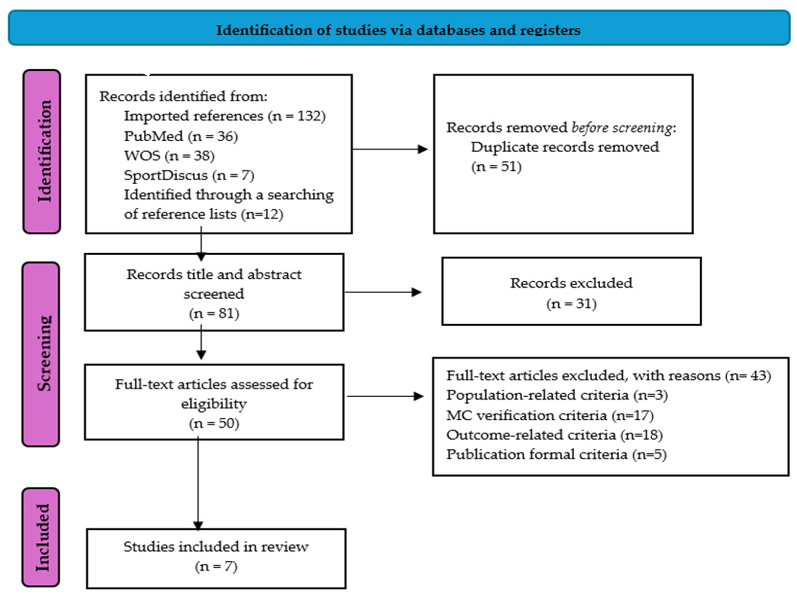
3.1. Selection of Articles
3.2. Study Characteristics
3.2.1. Population
3.2.2. Menstrual Cycle Phases
3.2.3. EMG Tasks, Variables Analyzed, and Normalized EMG Amplitudes
3.3. Methodological Quality Assessment
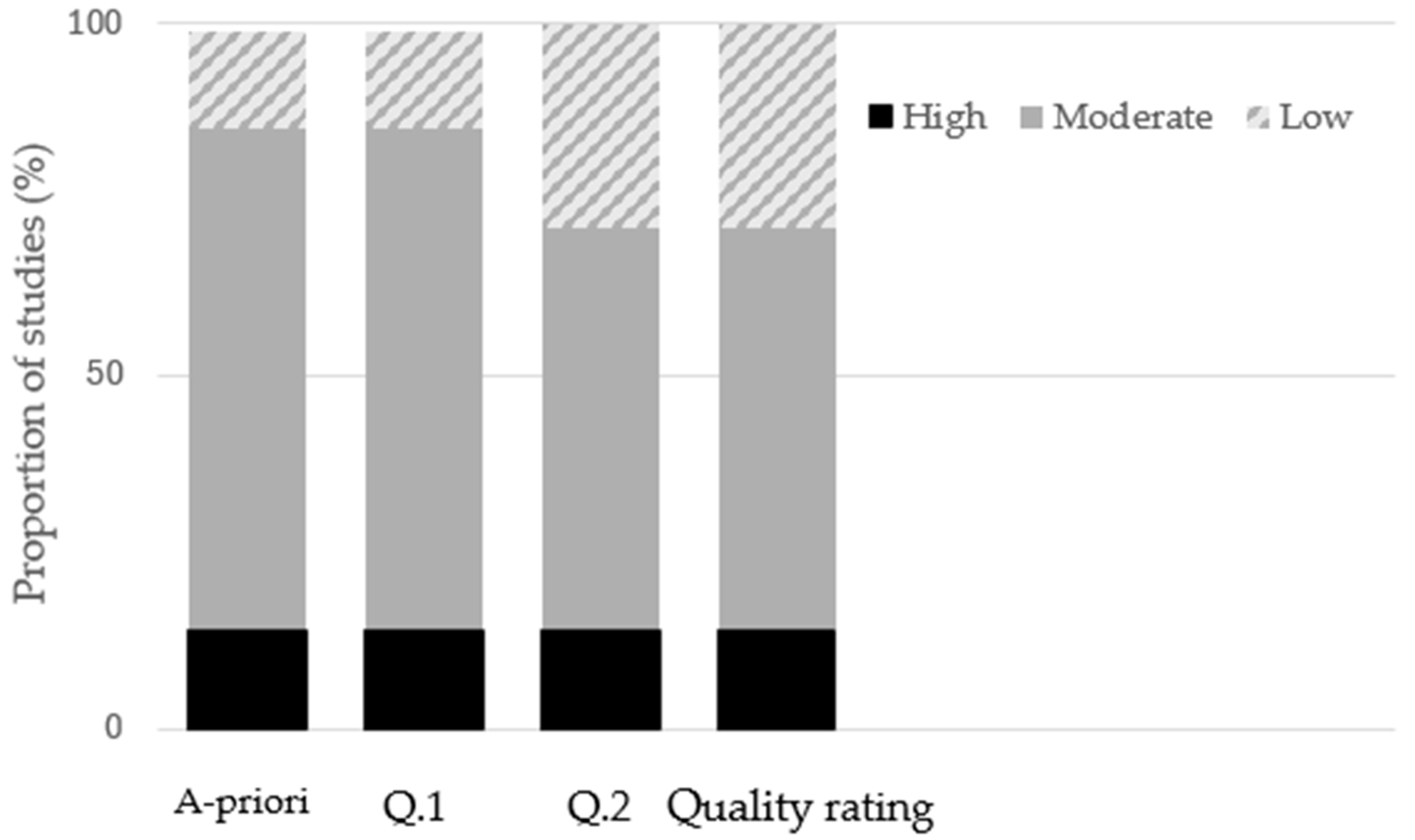
3.3.1. Physiological Verification of MC Phases
3.3.2. Critical Appraisal Assessment
3.4. Outcomes
Analysis EMG Outcomes Between Menstrual Cycle Phases
4. Discussion
4.1. Practical Application
4.2. Study Limitations and Strengths
5. Conclusions
Supplementary Materials
Author Contributions
Funding
Institutional Review Board Statement
Data Availability Statement
Conflicts of Interest
Abbreviations
| MC | Menstrual Cycle |
| M | Menstruation |
| FP | Follicular Phase |
| OV | Ovulation |
| LP | Luteal Phase |
| EFP | Early Follicular Phase |
| LFP | Late Follicular Phase |
| MP | Menstruation Phase |
| MVC | Maximum Voluntary Contraction |
| EMG | Electromyography |
| RMS | Root Mean Square |
| OC | Oral Contraceptive |
| RSE | Repeated Sprint Exercise |
| MUP | Motor Unit Potentials |
| PP | Premenstrual Phase |
| MLP | Mid-Luteal Phase |
| MDF | Median Frequency |
| NME | Neuromuscular Efficiency |
| AL | Voluntary Activation |
| ACL | Anterior Cruciate Ligament |
References
- European Commission. Gender Equality in Sport: Proposal for Strategic Actions 2014–2020. Available online: https://ec.europa.eu/assets/eac/sport/events/2013/documents/20131203-gender/final-proposal-1802_en.pdf (accessed on 28 May 2019).
- Fink, J.S. Female athletes, women’s sport, and the sport media commercial complex: Have we really “come a long way, baby”? Sport Manag. Rev. 2014, 18, 331–342. [Google Scholar] [CrossRef] [Scilit]
- Landen, S.; Hiam, D.; Voisin, S.; Jacques, M.; Lamon, S.; Eynon, N. Physiological and molecular sex differences in human skeletal muscle in response to exercise training. J. Physiol. 2023, 601, 419–434. [Google Scholar] [CrossRef] [Scilit] [PubMed]
- Thompson, B.M.; Drover, K.B.; Stellmaker, R.J.; Sculley, D.V.; Janse de Jonge, X.A.K. The effect of the menstrual cycle and oral contraceptive cycle on muscle performance and perceptual measures. Int. J. Environ. Res. Public Health 2021, 18, 10565. [Google Scholar] [CrossRef] [Scilit] [PubMed]
- Dawson, E.; Reilly, T. Menstrual cycle, exercise and health. Biol. Rhythm. Res. 2009, 40, 99–119. [Google Scholar] [CrossRef] [Scilit]
- de Jonge, X.J.; Thompson, B.; Ahreum, H.A.N. Methodological Recommendations for Menstrual Cycle Research in Sports and Exercise. Med. Sci. Sports Exerc. 2019, 51, 2610–2617. [Google Scholar] [CrossRef] [Scilit]
- McNulty, K.L.; Elliott-Sale, K.J.; Dolan, E.; Swinton, P.A.; Ansdell, P.; Goodall, S.; Thomas, K.; Hicks, K.M. The Effects of Menstrual Cycle Phase on Exercise Performance in Eumenorrheic Women: A Systematic Review and Meta-Analysis. Sports Med. 2020, 50, 1813–1827. [Google Scholar] [CrossRef] [Scilit]
- Ansdell, P.; Brownstein, C.G.; Skarabot, J.; Hicks, K.M.; Simoes, D.C.M.; Thomas, K.; Howatson, G.; Hunter, S.K.; Goodall, S. Menstrual cycle-associated modulations in neuromuscular function and fatigability of the knee extensors in eumenorrheic women. J. Appl. Physiol. 2019, 126, 1701–1712. [Google Scholar] [CrossRef] [Scilit]
- Blagrove, R.C.; Bruinvels, G.; Pedlar, C.R. Variations in strength-related measures during the menstrual cycle in eumenorrheic women: A systematic review and meta-analysis. J. Sci. Med. Sport 2020, 23, 1220–1227. [Google Scholar] [CrossRef] [Scilit]
- Florini, J.R. Hormonal control of muscle growth. Muscle Nerve 1987, 10, 577–598. [Google Scholar] [CrossRef] [Scilit]
- Deters, J.R.; Fietsam, A.C.; Workman, C.D.; Rudroff, T. High Estrogen Levels Cause Greater Leg Muscle Fatigability in Eumenorrheic Young Women after 4 mA Transcranial Direct Current Stimulation. Brain Sci. 2022, 12, 506. [Google Scholar] [CrossRef] [Scilit]
- Bencke, J.; Aagaard, P.; Zebis, M.K. Muscle activation during ACL injury risk movements in young female athletes: A narrative review. Front. Physiol. 2018, 9, 445. [Google Scholar] [CrossRef] [Scilit]
- Oosthuyse, T.; Bosch, A.N. The Effect of the Menstrual Cycle on Exercise Metabolism. Sports Med. 2010, 40, 207–227. [Google Scholar] [CrossRef] [Scilit] [PubMed]
- Constantini, N.W.; Dubnov, G.; Lebrun, C.M. The menstrual cycle and sport performance. Clin. Sports Med. 2005, 24, 51–82. [Google Scholar] [CrossRef] [Scilit]
- Hewett, T.E. Neuromuscular and hormonal factors associated with knee injuries in female athletes. Strategies for intervention. Sports Med. (Auckland N.Z.) 2000, 29, 313–327. [Google Scholar] [CrossRef] [Scilit]
- Hansen, M. Female hormones: Do they influence muscle and tendon protein metabolism? Proc. Nutr. Soc. 2018, 77, 32–41. [Google Scholar] [CrossRef] [Scilit]
- Bennal, A.S.; Chavan, V. Nerve Conduction Velocity across the phases of menstrual cycle. Indian J. Clin. Anat. Physiol. 2017, 4, 1–3. [Google Scholar]
- McClung, J.M.; Davis, J.M.; Wilson, M.A.; Goldsmith, E.C.; Carson, J.A. Estrogen status and skeletal muscle recovery from disuse atrophy. J. Appl. Physiol. 2006, 100, 2012–2023. [Google Scholar] [CrossRef] [Scilit]
- Wentorf, F.A.; Sudoh, K.; Moses, C.; Arendt, E.A.; Carlson, C.S. The effects of estrogen on material and mechanical properties of the intra- and extra-articular knee structures. Am. J. Sports Med. 2006, 34, 1948–1952. [Google Scholar] [CrossRef] [Scilit]
- Bailey, S.P.; Zacher, C.M.; Mittleman, K.D. Effect of menstrual cycle phase on carbohydrate supplementation during prolonged exercise to fatigue. J. Appl. Physiol. (Bethesda Md. 1985) 2000, 88, 690–697. [Google Scholar] [CrossRef] [Scilit] [PubMed]
- Iwamoto, Y.; Kubo, J.; Ito, M.; Takemiya, T.; Asami, T. Variation in maximal voluntary contraction during the menstrual cycle. Jpn. J. Phys. Fit. Sports Med. 2002, 51, 193–201. [Google Scholar] [CrossRef] [Scilit]
- Dedrick, G.S.; Sizer, P.S.; Merkle, J.N.; Hounshell, T.R.; Robert-McComb, J.J.; Sawyer, S.F.; Brismée, J.M.; Roger James, C. Effect of sex hormones on neuromuscular control patterns during landing. J. Electromyogr. Kinesiol. 2008, 18, 68–78. [Google Scholar] [CrossRef] [Scilit]
- Prado-Álvarez, R.; Estrada-barranco, C.; González-de-la-flor, Á.; De, M.; San, P.; Almazán-Polo, J.; Guerineau, F.; Giménez, M. Endurance and electromyographic assessment of abdominopelvic motor control in women with primary dysmenorrhea: A cross- sectional study. Sci. Rep. 2024, 14, 28836. [Google Scholar] [CrossRef] [Scilit]
- Romero-Moraleda, B.; Del Coso, J.; Gutiérrez-Hellín, J.; Ruiz-Moreno, C.; Grgic, J.; Lara, B. The influence of the menstrual cycle on muscle strength and power performance. J. Hum. Kinet. 2019, 68, 123–133. [Google Scholar] [CrossRef] [Scilit] [PubMed]
- Romero-Moraleda, B.; Morencos-Martínez, E.; Varón, P.; Lara, B.; Jiménez-Ormeño, E.; Peinado, A.B.; González-García, J. The influence of menstrual cycle phase on neuromuscular performance and subjective perception of effort in elite football players. Biol. Sport 2025, 42, 89–98. [Google Scholar] [CrossRef] [Scilit]
- Costa Pde, L.; Santos, F.P.; Rocha, C.C.D.; Da Silva, S.F. Study on the electromyographic activation of lower limbs during the menstrual cycle phases. J. Exerc. Physiol. Online 2015, 18, 52–57. [Google Scholar]
- Janse De Jonge, X.A.K.; Boot, C.R.L.; Thom, J.M.; Ruell, P.A.; Thompson, M.W. The influence of menstrual cycle phase on skeletal muscle contractile characteristics in humans. J. Physiol. 2001, 530, 161–166. [Google Scholar] [CrossRef] [Scilit]
- Kubo, K.; Miyamoto, M.; Tanaka, S.; Maki, A.; Tsunoda, N.; Kanehisa, H. Muscle and tendon properties during menstrual cycle. Int. J. Sports Med. 2009, 30, 139–143. [Google Scholar] [CrossRef] [Scilit]
- Samani, M.; Kordi Yoosefinejad, A.; Campos, M.H.; de Lira, C.A.B.; Motealleh, A. Changes in Knee Vastii Muscle Activity in Women with Patellofemoral Pain Syndrome During the Menstrual Cycle. PM&R 2020, 12, 382–390. [Google Scholar] [CrossRef] [Scilit]
- Pereira, H.M.; Larson, R.D.; Bemben, D.A. Menstrual Cycle Effects on Exercise-Induced Fatigability. Front. Physiol. 2020, 11, 517. [Google Scholar] [CrossRef] [Scilit] [PubMed]
- Kavanagh, J.J.; McFarland, A.J.; Taylor, J.L. Enhanced availability of serotonin increases activation of unfatigued muscle but exacerbates central fatigue during prolonged sustained contractions. J. Physiol. 2019, 597, 319–332. [Google Scholar] [CrossRef] [Scilit]
- Cordeiro, L.M.S.; Rabelo, P.C.R.; Moraes, M.M.; Teixeira-Coelho, F.; Coimbra, C.C.; Wanner, S.P.; Soares, D.D. Physical exercise-induced fatigue: The role of serotonergic and dopaminergic systems. Braz. J. Med. Biol. Res. Rev. Bras. De Pesqui. Medicas E Biol. 2017, 50, e6432. [Google Scholar] [CrossRef] [Scilit]
- Balachandar, V.; Marciniak, J.L.; Wall, O.; Balachandar, C. Effects of the menstrual cycle on lower-limb biomechanics, neuromuscular control, and anterior cruciate ligament injury risk: A systematic review. Muscles Ligaments Tendons J. 2017, 7, 136–146. [Google Scholar] [CrossRef] [Scilit] [PubMed]
- Shultz, S.J.; Schmitz, R.J.; Kong, Y.; Dudley, W.N.; Beynnon, B.D.; Nguyen, A.D.; Kim, H.; Montgomery, M.M. Cyclic variations in multiplanar knee laxity influence landing biomechanics. Med. Sci. Sports Exerc. 2012, 44, 900–909. [Google Scholar] [CrossRef] [Scilit] [PubMed]
- Belanger, M.J.; Moore, D.C.; Crisco, J.J., 3rd; Fadale, P.D.; Hulstyn, M.J.; Ehrlich, M.G. Knee laxity does not vary with the menstrual cycle, before or after exercise. Am. J. Sports Med. 2004, 32, 1150–1157. [Google Scholar] [CrossRef] [Scilit]
- Stijak, L.; Kadija, M.; Djulejic, V.; Aksic, M.; Petronijevic, N.; Markovic, B.; Radonjic, V.; Bumbasirevic, M.; Filipovic, B. The influence of sex hormones on anterior cruciate ligament rupture: Female study. Knee Surg. Sports Traumatol. Arthrosc. 2015, 23, 2742–2749. [Google Scholar] [CrossRef] [Scilit]
- Bencke, J.; Zebis, M.K. The influence of gender on neuromuscular pre-activity during side-cutting. J. Electromyogr. Kinesiol. 2011, 21, 371–375. [Google Scholar] [CrossRef] [Scilit] [PubMed]
- Chappell, J.D.; Creighton, R.A.; Giuliani, C.; Yu, B.; Garrett, W.E. Kinematics and electromyography of landing preparation in vertical stop-jump: Risks for noncontact anterior cruciate ligament injury. Am. J. Sports Med. 2007, 35, 235–241. [Google Scholar] [CrossRef] [Scilit]
- Nagano, Y.; Ida, H.; Akai, M.; Fukubayashi, T. Gender differences in knee kinematics and muscle activity during single limb drop landing. Knee 2007, 14, 218–223. [Google Scholar] [CrossRef] [Scilit]
- Pollard, C.D.; Sigward, S.M.; Powers, C.M. Limited hip and knee flexion during landing is associated with increased frontal plane knee motion and moments. Clin. Biomech. 2010, 25, 142–146. [Google Scholar] [CrossRef] [Scilit]
- Schultz, K.N.; Von Esenwein, S.A.; Hu, M.; Bennett, A.L.; Kennedy, R.T.; Musatov, S.; Toran-Allerand, C.D.; Kaplitt, M.G.; Young, L.J.; Becker, J.B. Viral vector-mediated overexpression of estrogen receptor- α in striatum enhances the estradiol-induced motor activity in female rats and estradiol-modulated GABA release. J. Neurosci. 2009, 29, 1897–1903. [Google Scholar] [CrossRef] [Scilit]
- Zazulak, B.T.; Ponce, P.L.; Straub, S.J.; Medvecky, M.J.; Avedisian, L.; Hewett, T.E. Gender comparison of hip muscle activity during single-leg landing. J. Orthop. Sports Phys. Ther. 2005, 35, 292–299. [Google Scholar] [CrossRef] [Scilit]
- Sharma, M.; Singh, A. Gender differences in muscle activation patterns during squat and countermovement jumps in soccer players. J. Phys. Educ. Sport 2024, 24, 2017–2024. [Google Scholar] [CrossRef]
- Page, M.J.; McKenzie, J.E.; Bossuyt, P.M.; Boutron, I.; Hoffmann, T.C.; Mulrow, C.D.; Shamseer, L.; Tetzlaff, J.M.; Akl, E.A.; Brennan, S.E.; et al. The PRISMA 2020 statement: An updated guideline for reporting systematic reviews. BMJ (Clin. Res. Ed.) 2021, 372, n71. [Google Scholar] [CrossRef] [Scilit]
- Moher, D.; Liberati, A.; Tetzlaff, J.; Altman, D.G.; Antes, G.; Atkins, D.; Barbour, V.; Barrowman, N.; Berlin, J.A.; Clark, J.; et al. Preferred reporting items for systematic reviews and meta-analyses: The PRISMA statement. PLoS Med. 2009, 6, e1000097. [Google Scholar] [CrossRef] [Scilit]
- Fehring, R.J.; Schneider, M.; Raviele, K. Variability in the phases of the menstrual cycle. J. Obstet. Gynecol. Neonatal Nurs. JOGNN 2006, 35, 376–384. [Google Scholar] [CrossRef] [Scilit]
- Graja, A.; Kacem, M.; Hammouda, O.; Borji, R.; Bouzid, M.A.; Souissi, N.; Rebai, H. Physical, Biochemical, and Neuromuscular Responses to Repeated Sprint Exercise in Eumenorrheic Female Handball Players: Effect of Menstrual Cycle Phases. J. Strength Cond. Res. 2022, 36, 2268–2276. [Google Scholar] [CrossRef] [Scilit]
- Ouzzani, M.; Hammady, H.; Fedorowicz, Z.; Elmagarmid, A. Rayyan—A web and mobile app for systematic reviews. Syst. Rev. 2016, 5, 210. [Google Scholar] [CrossRef] [Scilit] [PubMed]
- Downs, S.H.; Black, N. The feasibility of creating a checklist for the assessment of the methodological quality both of randomised and non-randomised studies of health care interventions. J. Epidemiol. Community Health 1998, 52, 377–384. [Google Scholar] [CrossRef] [Scilit]
- Niering, M.; Wolf-Belala, N.; Seifert, J.; Tovar, O.; Coldewey, J.; Kuranda, J.; Muehlbauer, T. The Influence of Menstrual Cycle Phases on Maximal Strength Performance in Healthy Female Adults: A Systematic Review with Meta-Analysis. Sports 2024, 12, 31. [Google Scholar] [CrossRef] [Scilit] [PubMed]
- Dos’Santos, T.; Stebbings, G.K.; Morse, C.; Shashidharan, M.; Daniels, K.A.J.; Sanderson, A. Effects of the menstrual cycle phase on anterior cruciate ligament neuromuscular and biomechanical injury risk surrogates in eumenorrheic and naturally menstruating women: A systematic review. PLoS ONE 2023, 18, e0280800. [Google Scholar] [CrossRef] [Scilit] [PubMed]
- Khowailed, I.A.; Petrofsky, J.; Lohman, E.; Daher, N.; Mohamed, O. 17Β-Estradiol Induced Effects on Anterior Cruciate Ligament Laxness and Neuromuscular Activation Patterns in Female Runners. J. Women’s Health 2015, 24, 670–680. [Google Scholar] [CrossRef] [Scilit]
- Peltonen, H.; Mikkonen-Taipale, R.; Uimonen, T.; Walker, S.; Hackney, A.C.; Valtonen, M.; Kyröläinen, H.; Ihalainen, J.K. Power Loading-Induced Fatigue Is Influenced by Menstrual Cycle Phase. Med. Sci. Sports Exerc. 2022, 54, 1190–1198. [Google Scholar] [CrossRef] [Scilit]
- Okazaki, M.; Kaneko, M.; Ishida, Y.; Murase, N.; Katsumura, T. Changes in the Width of the Tibiofibular Syndesmosis Related to Lower Extremity Joint Dynamics and Neuromuscular Coordination on Drop Landing During the Menstrual Cycle. Orthop. J. Sports Med. 2017, 5, 2325967117724753. [Google Scholar] [CrossRef] [Scilit] [PubMed]
- Cesar, G.M.; Pereira, V.S.; Santiago, P.R.P.; Benze, B.G.; da Costa, P.H.L.; Amorim, C.F.; Serrão, F.V. Variations in dynamic knee valgus and gluteus medius onset timing in non-athletic females related to hormonal changes during the menstrual cycle. Knee 2011, 18, 224–230. [Google Scholar] [CrossRef] [Scilit]
- Piasecki, J.; Guo, Y.; Jones, E.J.; Phillips, B.E.; Stashuk, D.W.; Atherton, P.J.; Piasecki, M. Menstrual Cycle Associated Alteration of Vastus Lateralis Motor Unit Function. Sports Med. Open 2023, 9, 97. [Google Scholar] [CrossRef] [Scilit]
- McLean, S.G.; Huang, X.; van der Bogert, A.J. Association between lower extremity posture at contact and peak knee valgus moment during sidestepping: Implications for ACL injury. Clin. Biomech. 2005, 20, 863–870. [Google Scholar] [CrossRef] [Scilit] [PubMed]
- Soares, F.A.; Salomoni, S.E.; Veneziano, W.H.; de Carvalho, J.L.; Nascimento, F.A.; Pires, K.F.; da Rocha, A.F. On the behavior of surface electromyographic variables during the menstrual cycle. Physiol. Meas. 2011, 32, 543–557. [Google Scholar] [CrossRef] [Scilit] [PubMed]
- Salomoni, S.; Soares, F.A.; de Oliveira Nascimento, F.A.; da Rocha, A.F. Gender differences in muscle fatigue of the biceps brachii and influences of female menstrual cycle in electromyography variables. In Proceedings of the 2008 30th Annual International Conference of the IEEE Engineering in Medicine and Biology Society, Vancouver, BC, Canada, 20–25 August 2008; pp. 2598–2601. [Google Scholar] [CrossRef] [Scilit]
- Tenan, M.S.; Peng, Y.L.; Hackney, A.C.; Griffin, L. Menstrual cycle mediates vastus medialis and vastus medialis oblique muscle activity. Med. Sci. Sports Exerc. 2013, 45, 2151–2157. [Google Scholar] [CrossRef] [Scilit] [PubMed]
- Hewett, T.E.; Zazulak, B.T.; Myer, G.D. Effects of the menstrual cycle on anterior cruciate ligament injury risk: A systematic review. Am. J. Sports Med. 2007, 35, 659–668. [Google Scholar] [CrossRef] [Scilit]
- Lebrun, C.M. The effect of the phase of the menstrual cycle and the birth control pill on athletic performance. Clin. Sports Med. 1994, 13, 419–441. [Google Scholar] [CrossRef] [Scilit]
- Wojtys, E.M.; Huston, L.J. Neuromuscular performance in normal and anterior cruciate ligament-deficient lower extremities. Am. J. Sports Med. 1994, 22, 89–104. [Google Scholar] [CrossRef] [Scilit] [PubMed]



| Inclusion | Exclusion | |
|---|---|---|
| Population | Eumenorrheic women; age: 18–40 years; MC length: 21–35 days | Women with any of the following conditions: OC users, MC irregularities (e.g., menopause, polycystic ovary syndrome), injuries, diseases, and smoking behavior |
| Intervention | Studies conducting interventions on the lower limb with EMG | No EMG method; muscular activity in the upper limb |
| Comparator | FP was assessed; MC phases must be assessed by blood samples and other procedures: saliva samples, urinary tests, basal body temperature, and self-reported (with a combination of others) | No FP as comparator; only self-reported for the MC assessment; no comparison between MC phases |
| Outcomes | Articles analyzing muscle activation patterns throughout the MC phases as %MVC or MVC, RMS, onset timing, and firing rate (Hz) | Studies that did not address the outcome question; studies that did not normalize the EMG signal |
| Study Design | Articles available in English; articles with human beings; descriptive, experimental, or case studies. | Thesis and articles published in other languages; articles with animal intervention; no conference proceedings |
| Author | Reporting | External Validity | Internal Validity-Bias | Confounding | Power | A priori | Q1 | Q2 | Final Grade | ||||||||||
|---|---|---|---|---|---|---|---|---|---|---|---|---|---|---|---|---|---|---|---|
| 1 | 2 | 3 | 4 | 5 | 6 | 7 | 8 | 9 | 10 | 11 | 12 | 13 | 14 | 15 | |||||
| [22] | 1 | 1 | 1 | 1 | 1 | 1 | 0 | 1 | 0 | 0 | 1 | 1 | 1 | 0 | 0 | 10 | y | n | L |
| [52] | 1 | 1 | 1 | 1 | 1 | 1 | 1 | 1 | 1 | 1 | 1 | 1 | 0 | 0 | 1 | 13 | y | y | M |
| [53] | 1 | 1 | 1 | 1 | 1 | 1 | 0 | 1 | 1 | 1 | 1 | 1 | 1 | 0 | 1 | 13 | y | y | M |
| [54] | 1 | 1 | 1 | 1 | 1 | 1 | 1 | 1 | 1 | 1 | 1 | 1 | 0 | 0 | 1 | 13 | y | y | M |
| [47] | 1 | 1 | 1 | 1 | 1 | 1 | 1 | 1 | 2 | 1 | 1 | 1 | 1 | 0 | 0 | 14 | y | y | H |
| [55] | 1 | 1 | 1 | 1 | 1 | 1 | 0 | 1 | 1 | 1 | 1 | 1 | 1 | 1 | 0 | 13 | y | n | L |
| [56] | 1 | 1 | 1 | 1 | 1 | 1 | 1 | 1 | 2 | 1 | 1 | 1 | 0 | 0 | 0 | 13 | y | y | M |
| Population | Menstrual Cycle | EMG | |||||||
|---|---|---|---|---|---|---|---|---|---|
| Authors and Year | Study Design | Sample Size | Age Range | Training Status | Observation Time | Menstrual Phases (Days) | MC Verification | Exercise | Variables Analyzed |
| [47] | Randomized crossover | n = 10 | 20–25 | Handball players | One MC (28–30 days) | LFP: 11–13 MLP: 21–23 PP: 28–29 | Blood samples, menstruation diary, and OV test | RSE session (20 × 5” sprints) | MVC, peak force, EMG signals |
| [55] | Longitudinal block | n = 15 | 19–27 | Non-athletic collegiate females | One MC (26–31 days) | EFP: 1–3 LFP: 11–13 LP: 21–24 | Sublingual morning temperature, blood samples | Single-leg drop landings | Electromyography (gluteus medius onset) and knee kinematics (valgus angle) |
| [22] | Repeated measures | n = 26 | 20.5 ± 1.9 | - | One MC (26–32 days) | EFP: 1–3 LFP: 11–13 MLP:21–24 | Blood samples | Drop jump | Varus/valgus knee angle and EMG activity |
| [54] | Descriptive | n = 28 | 21.0 ± 0.8 | Healthy women | One MC (22–24 days) | M: 1–5 FP:7–10 OV:12–15 LP:7–9 after + | Blood samples and Ovulation Urine Test (vaginal smear and ovulation pain) | Single-leg drop landing | Width of the tibiofibular syndesmosis, peak ground-reaction force, time to peak GRF, angles of the hip, knee, and ankle joints, and muscle activity |
| [53] | Experimental | n = 16 | 26 ± 4 | Healthy women | One MC (28.3 ± 2.3) | EFP: 2–4 LFP: 7–11 OV: 24–28 h after + urinary test MLP: 7 after + | Blood samples and ovulation tests | Dynamic power-type loading protocol | Neuromuscular properties, force production, and metabolic capacities |
| [52] | Descriptive | n = 12 | 25.6 ± 3.7 | Runners | One MC (28 ± 1.1) | EFP: 1–2 OV: 24–48 h after + | Blood samples and ovulation kits | Treadmill running | Knee joint laxity and EMG activity |
| [56] | Descriptive | n = 9 | 24.2 ± 3.2 | Recreationally active | One MC (21–35 days) | EFP: within 48 h of onset M OV: within 48 h after + MLP: after 7 following OV | Blood samples and ovulation kits | Force dynamometer | Knee extensor maximum voluntary contraction, jump power, force steadiness, and balance |
| Study | Hormone Levels | MC Phases | ||||
|---|---|---|---|---|---|---|
| EFP or M | FP | OV | LP | PP | ||
| [22] | Estradiol (pg/mL) | 41± 24.4 | 158.5 ± 129.0 | 135.8 ± 101.9 | ||
| Progesterone (ng/mL) | 0.6 ± 0.4 | |||||
| [47] | Estrogen (pg/mL) | 386.91 ± 31.88 | 194 ± 6.02 | 77.48 ± 6.49 | ||
| Progesterone (ng/mL) | 0.54 ± 0.18 | 14.61 ± 1.68 | 1.77 ± 0.62 | |||
| [56] | Estrogen (pg/mL) | 244 | 533 | 484 | ||
| Progesterone (ng/mL) | 2.66 | 3.22 | 5.89 | |||
| [53] | Estradiol (pmol/L) | 478.3 | 592 | 798.7 | 684.5 | |
| Progesterone (nmol/L) | 1.9 | 1 | 4.1 | 14.8 | ||
| [55] | Estradiol (pg/mL) | 28.07 ± 19.40 | 178.70 ± 164.70 | 136.10 ± 70.60 | ||
| Progesterone (nmol/L) | 0.98 ± 0.46 | 0.95 ± 0.50 | 7.78 ± 4.79 | |||
| [54] | Estradiol (pg/mL) | 24.8 ± 9.8 | 31.1 ± 13.8 | 83.1 ± 53.7 | 94 ± 41.5 | |
| Progesterone (ng/mL) | 0.69 ± 0.20 | 0.66 ± 0.27 | 1.92 ± 1.86 | 10.40 ± 7.02 | ||
| [52] | Estradiol (pg/mL) | 34.17 ± 15.47 | 207.74 ± 53.42 | |||
| Reference | EMG Collection Method | EMG Outcome | Muscles Recorded | MC Phases | Numerical Values | Main Findings | |||
|---|---|---|---|---|---|---|---|---|---|
| [47] | 3 MVC (5”) with 2” rest | RMS (mV) | Quadriceps | FP | before RSE (≈1.4 mV), after RSE (≈0.9 mV) | No significant difference was found between MC phases in all frequency components of EMG before RSE. NME and MDF values of vastus lateralis and rectus femoris were significantly decreased in PMP compared with FP and LP. | |||
| LP | before RSE (≈1.3 mV), after RSE (≈1.1 Mv) | ||||||||
| PMP | before RSE (≈1.25 mV), after RSE (≈1.1 mV) | ||||||||
| MDF (Hz) | Vastus medialis | FP | before RSE (≈88), after RSE (≈72) | ||||||
| LP | before RSE (≈85), after RSE (≈69) | ||||||||
| PMP | before RSE (≈84), after RSE (≈65) | ||||||||
| Vastus lateralis | FP | before RSE (≈88), after RSE (≈79) | |||||||
| LP | before RSE (≈90), after RSE (≈71) | ||||||||
| PMP | before RSE (≈84), after RSE (≈60) | ||||||||
| Rectus femoris | FP | before RSE (≈87), after RSE (≈72) | |||||||
| LP | before RSE (≈80), after RSE (≈65 mV *) | ||||||||
| PMP | before RSE (≈85), after RSE (≈55) | ||||||||
| NME | Knee extensor muscles | FP | before RSE (≈420), after RSE (≈505 †*) | ||||||
| LP | before RSE (≈460 mV), after RSE (≈450 mV *) | ||||||||
| PMP | before RSE (≈455 mV), after RSE (≈380 mV) | ||||||||
| † significant difference compared with LP (p < 0.05); * significant difference compared with PMP (p < 0.05) | |||||||||
| [55] | Single leg drop landings (31 cm box) | Onset timing (ms) | Gluteus medius | EFP | −24.4 ± 63.9 | Significant differences were not observed for gluteus medius onset timing (p = 0.936) amongst the MC phases. | |||
| LFP | −12.8 ± 48.5 | ||||||||
| LP | −28.7 ± 40.3 | ||||||||
| The negative sign (−) represents valgus and EMG onset timing prior to initial contact. | |||||||||
| [22] | 3 drop jump (50 cm) | Onset timing (ms) | Gluteus maximus | EFP | −42 ± 50 | The semitendinosus exhibited onset delays relative to ground contact during the LP and demonstrated a significant difference between EFP and LFP. Muscle timing differences between the gluteus maximus and semitendinosus were decreased in the LP compared to the EFP. | |||
| LFP | −41 ± 52 | ||||||||
| LP | −56 ± 58 | ||||||||
| Semitendinosus | EFP | −102 ± 19 | |||||||
| LFP | −97 ± 33 | ||||||||
| LP | −78 ± 17 | ||||||||
| Vastus lateralis | EFP | −87 ± 44 | |||||||
| LFP | −92 ± 54 | ||||||||
| LP | −95 ± 57 | ||||||||
| Vastus medialis oblique | EFP | −58 ± 30 | |||||||
| LFP | −57 ± 39 | ||||||||
| LP | −49 ± 37 | ||||||||
| Tibialis anterior | EFP | −104 ± 38 | |||||||
| LFP | −105 ± 40 | ||||||||
| LP | −99 ± 40 | ||||||||
| Lateral gastrocnemius | EFP | −100 ± 38 | |||||||
| LFP | −104 ± 29 | ||||||||
| LP | −109 ± 26 | ||||||||
| [53] | 2 sets of 10 reps 60%1 RM (2 min rest between sets) + 3 MVC (isometric) + muscle stimulation | EMG (%) | Vastus lateralis + vastus medialis | M | Dynamic | 108.2 ± 13.8 | Isometric | −19.6 ± 12.2 | Neither voluntary muscular activity (EMG, MPF) nor electrical stimulation parameters differed between the MC phases before the loading protocol. |
| LFP | 102.4 ± 10.4 | −19.2 ± 14.0 | |||||||
| OV | 97.5 ± 13.3 | −14.9 ± 7.6 | |||||||
| MLP | 96.5 ± 10.4 | −15.3 ± 11.8 | |||||||
| MVC (Δ%) | M | Isometric | −13.7 ± 8.6 | ||||||
| LPF | −11.7 ± 10.3 | ||||||||
| OV | −7.8 ± 12.2 | ||||||||
| MLP | −15.0 ± 12.3 | ||||||||
| MPF (VL + VM) (Δ%) | M | 7.7 ± 24.8 | |||||||
| LFP | −1.1 ± 19.0 | ||||||||
| OV | −7.5 ± 11.1 | ||||||||
| MLP | 3.1 ± 10.6 | ||||||||
| [54] | 6 Single drop landing (30 cm box) | %MVC (%μV·s) before landing | Gluteus maximus | EFP | 9.98 ± 5.22 | Activation of the GM in the LP was significantly lower than those in the menstrual and FP. During the MC, there were no significant changes in the activation of the biceps femoris, semitendinosus, or rectus femoris. | |||
| LFP | 9.73 ± 5.84 | ||||||||
| OV | 8.75 ± 5.19 | ||||||||
| LP | 7.75 ± 3.70 | ||||||||
| Biceps femoris | EFP | 33.8 ± 33.1 | |||||||
| LFP | 32.2 ± 24.4 | ||||||||
| OV | 31.9 ± 30.5 | ||||||||
| LP | 40.0 ± 35.1 | ||||||||
| Semitendinosus | EFP | 15.4 ± 11.0 | |||||||
| LFP | 15.3 ± 11.9 | ||||||||
| OV | 15.2 ± 11.3 | ||||||||
| LP | 16.9 ± 14.3 | ||||||||
| Rectus femoris | EFP | 23.8 ± 17.7 | |||||||
| LFP | 23.1 ± 15.6 | ||||||||
| OV | 27.3 ± 22.1 | ||||||||
| LP | 21.7 ± 24.4 | ||||||||
[52] | 6 min running session with 0 inclination at 10 km/h | %MVC | Vastus lateralis | FP | Pre-activation phase | 24.26 ± 16.27 | Differences in muscle activation strategies during different phases of the MC. The increased quadriceps activity observed during the FP was associated with decreased hamstring activity. Furthermore, the quadriceps and hamstring co-contraction ratios decreased during the EFP compared with the OV. | ||
| OV | 12.25 ± 6.63 | ||||||||
| Vastus medialis | FP | 24.26 ± 16.27 | |||||||
| OV | 15.84 ± 6.50 | ||||||||
| Lateral hamstrings | FP | 31 ± 10.51 | |||||||
| OV | 36.67 ± 12.43 | ||||||||
| Medial hamstrings | FP | 25.47 ± 12.50 | |||||||
| OV | 40.45 ± 11.93 | ||||||||
| Vastus lateralis | FP | Weight acceptance | 56.47 ± 14.43 | ||||||
| OV | 34.19 ± 11.96 | ||||||||
| Vastus medialis | FP | 44.68 ± 14.89 | |||||||
| OV | 46.51 ± 7.58 | ||||||||
| Lateral hamstrings | FP | 36.76 | |||||||
| OV | 61.52 ± 14.52 | ||||||||
| Medial hamstrings | FP | 30.43 ± 20.70 | |||||||
| OV | 54.68 ± 16.69 | ||||||||
| Vastus lateralis | FP | Peak push-off phase | 14.22 ± 12.44 | ||||||
| OV | 9.07 ± 7.72 | ||||||||
| Vastus medialis | FP | 13.45 ± 5.93 | |||||||
| OV | 11.33 ± 7.65 | ||||||||
| Lateral hamstrings | FP | 28.65 ± 14.68 | |||||||
| OV | 18.82 ± 10.17 | ||||||||
| Medial hamstrings | FP | 16.96 ± 9.4 | |||||||
| OV | 31.50 ± 21.67 | ||||||||
| Reference | EMG Collection Method | Muscles Recorded | EMG Outcome | MC Phases | Numerical Values | Main Findings | |||
|---|---|---|---|---|---|---|---|---|---|
| [56] | 4 MVC (10%, 25%, 2 × 40%MVC) 12”−15”/each with 30” rest iEMG | Lateral and medial hamstrings | Firing rate (Hz) | EFP vs. OV | −0.804 | Knee extensor MVC did not differ across the menstrual phases (p > 0.4). The firing rate of low threshold motor units was lower during the OV and MLP. Motor unit potentials were more complex during OV and MLP (p < 0.03). There could be a likely suppression of firing rate early recruited motor units in the OV and MLP. | |||
| EFP vs. MLP | −0.855 | ||||||||
| OV vs. MLP | −0.051 | ||||||||
| MUPs (µV ms) | EFP vs. OV | 70.79 | |||||||
| EFP vs. MLP | 32.19 | ||||||||
| OV vs. MLP | 102.9 | ||||||||
| MU FR | EFP | 10%MVC | 9.77 ± 0.94 | 25%MVC | No differences across MC phases | ||||
| OV | 8.94 ± 0.86 | ||||||||
| MLP | 8.91 ± 1.01 | ||||||||
| Bold indicates significantly different | |||||||||
| Study | Task Type | Differences Effects | Null Effects | Comments |
|---|---|---|---|---|
| [47] |  RSE session. Fatiguing protocol RSE session. Fatiguing protocol | ✕ | No significant difference was found between MC phases in all frequency components of EMG before RSE. | |
| [53] |  Dynamic protocol Dynamic protocol | ✕ | Differences in performance parameters in the unfatigued condition were not observed between MC phases. | |
| [56] |  Isometric protocol Isometric protocol | ✕ | Assessment of neuromuscular performance did not differ across MC. | |
| [55] |  Single-leg drop landing Single-leg drop landing | ✕ | EMG onset timing of gluteus medius muscle differences were not observed throughout the MC phases. | |
| [54] |  Single drop landing Single drop landing | ✓ | ✕ | During MC, there were no significant changes in the activation of biceps femoris, semitendinosus, or rectus femoris. * Activation of the gluteus medius in the LP was significantly lower than those in the menstrual phase and FP. * |
| [22] |  Drop jump Drop jump | ✓ | Female recreational athletes utilize different neuromuscular control pattern for performing a drop jump when estrogen levels are high (luteal phase). | |
| [52] |  Running Running | ✓ | Differences in muscle activation strategies during MC phases. |
Disclaimer/Publisher’s Note: The statements, opinions and data contained in all publications are solely those of the individual author(s) and contributor(s) and not of MDPI and/or the editor(s). MDPI and/or the editor(s) disclaim responsibility for any injury to people or property resulting from any ideas, methods, instructions or products referred to in the content. |
© 2026 by the authors. Licensee MDPI, Basel, Switzerland. This article is an open access article distributed under the terms and conditions of the Creative Commons Attribution (CC BY) license.
Share and Cite
Pérez-Paredes, A.; Armada-Cortés, E.; Cuadrado-Peñafiel, V.; Nieto-Acevedo, R.; Romero-Moraleda, B. Influence of Menstrual Cycle Phases on Muscle Activation in Women: A Systematic Review. Appl. Sci. 2026, 16, 2579. https://doi.org/10.3390/app16052579
Pérez-Paredes A, Armada-Cortés E, Cuadrado-Peñafiel V, Nieto-Acevedo R, Romero-Moraleda B. Influence of Menstrual Cycle Phases on Muscle Activation in Women: A Systematic Review. Applied Sciences. 2026; 16(5):2579. https://doi.org/10.3390/app16052579
Chicago/Turabian StylePérez-Paredes, Azahara, Estrella Armada-Cortés, Víctor Cuadrado-Peñafiel, Raúl Nieto-Acevedo, and Blanca Romero-Moraleda. 2026. "Influence of Menstrual Cycle Phases on Muscle Activation in Women: A Systematic Review" Applied Sciences 16, no. 5: 2579. https://doi.org/10.3390/app16052579
APA StylePérez-Paredes, A., Armada-Cortés, E., Cuadrado-Peñafiel, V., Nieto-Acevedo, R., & Romero-Moraleda, B. (2026). Influence of Menstrual Cycle Phases on Muscle Activation in Women: A Systematic Review. Applied Sciences, 16(5), 2579. https://doi.org/10.3390/app16052579