Neural Network Architectures in Video Capsule Endoscopy: A Systematic Review and Meta-Analysis on Accuracy and Reading Time Performances

 , , , , , and
Abstract
1. Introduction
2. Materials and Methods
2.1. Search Strategy
2.2. Inclusion and Exclusion Criteria
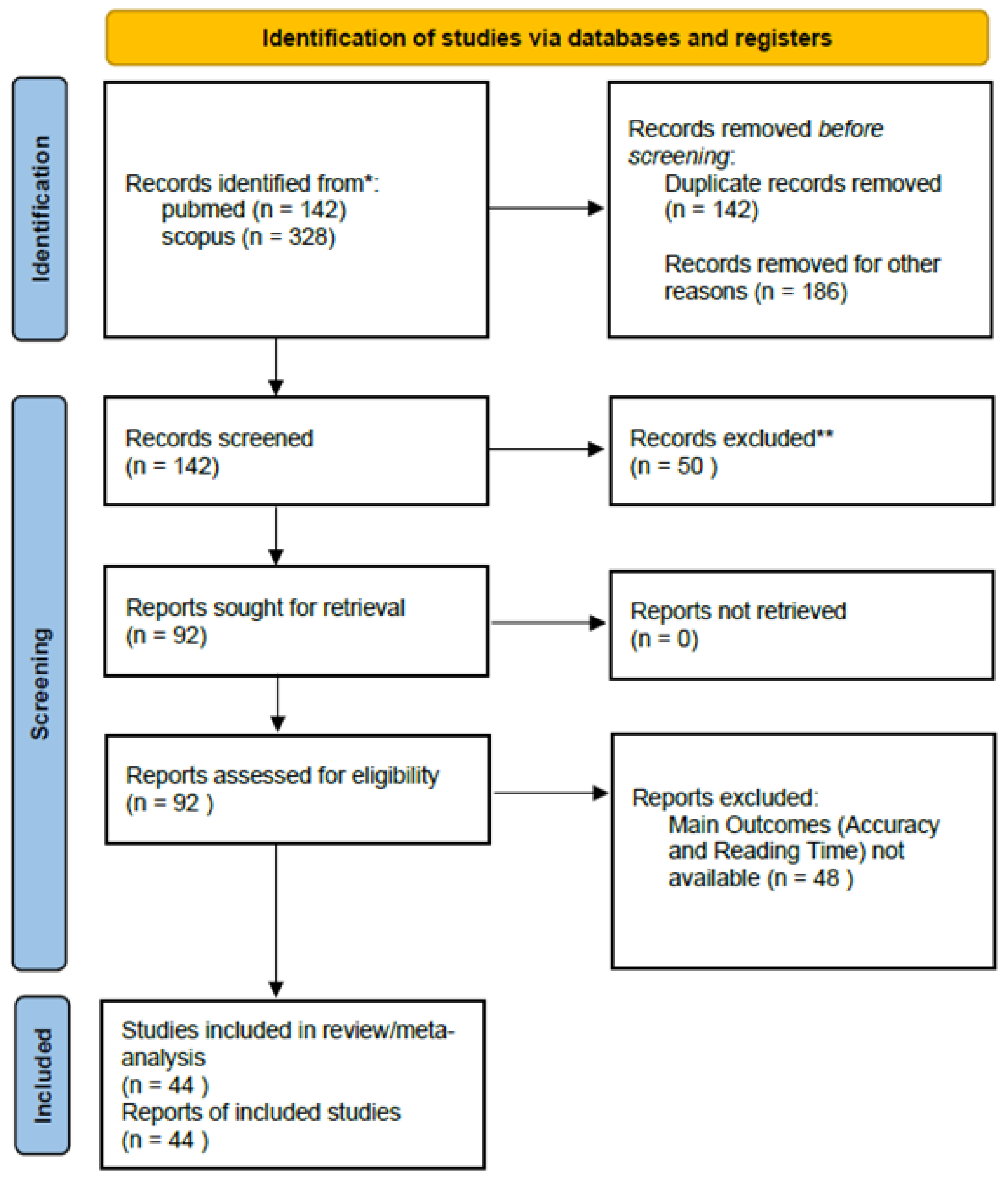
2.3. Study Selection and Data Extraction
2.4. Outcome Measures
2.5. Data Analysis
2.6. Quality Assessment
3. Results
3.1. Assessment of Quality of Research
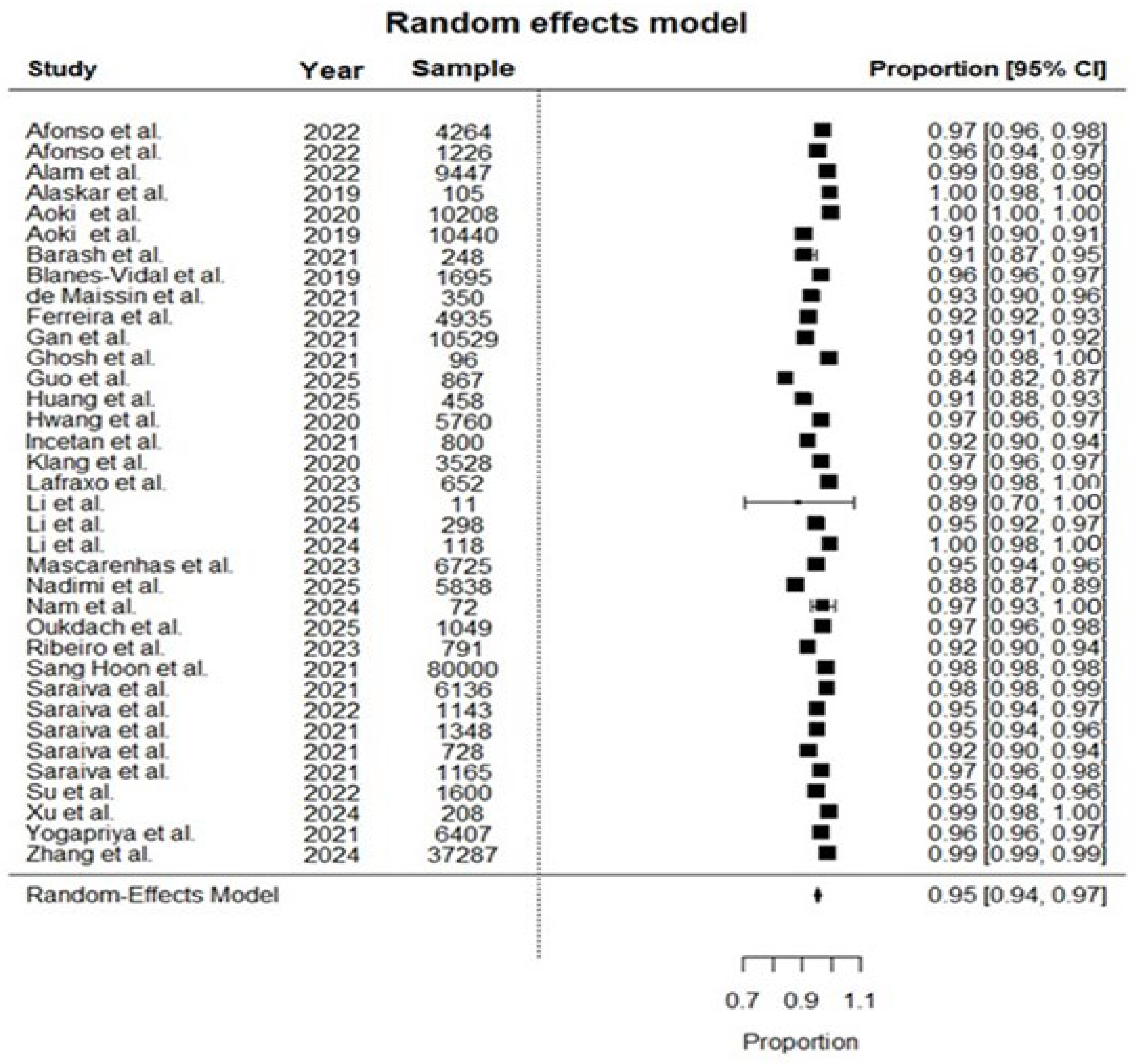
3.2. Meta-Analysis on Accuracy of Neural Networks in Lesion Detection
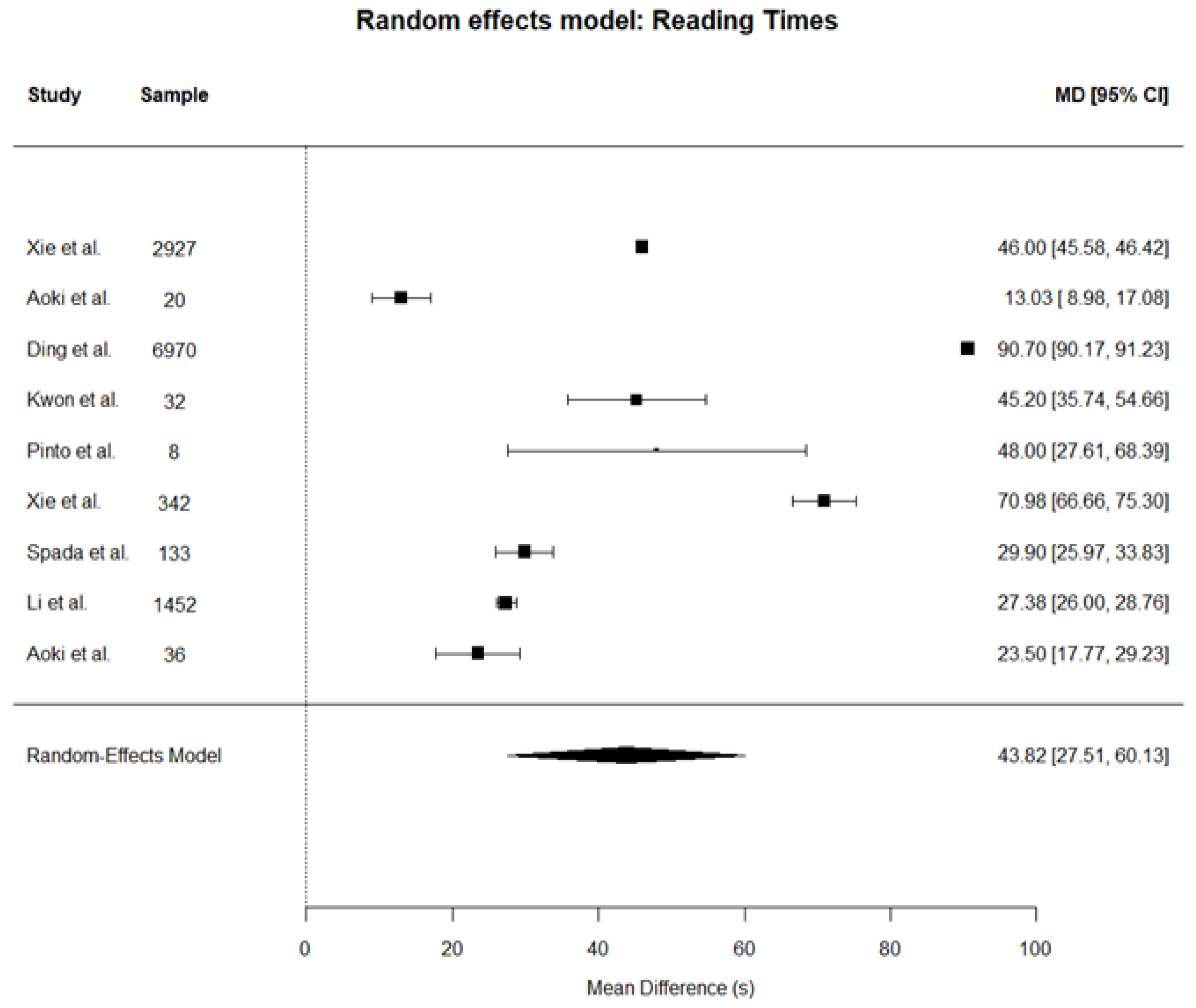
3.3. Meta-Analysis of Video Capsule Endoscopy Reading Time with and Without Neural Network Assistance
4. Discussion
5. Conclusions
Supplementary Materials
Author Contributions
Funding
Institutional Review Board Statement
Informed Consent Statement
Data Availability Statement
Acknowledgments
Conflicts of Interest
Abbreviations
| AI | Artificial intelligence |
| SBCE | Small-bowel capsule endoscopy |
| CNN | Convolutional neural network |
| NN | Neural network |
| PRISMA | Preferred Reporting Items for Systematic Reviews and Meta-Analyses |
| TP | True positive |
| TN | True negative |
| VCE | Video capsule endoscopy |
| CI | Confidence intervals |
| SD | Standard deviation |
| CV | Coefficient of variation |
| V | Validation study |
| C | Clinical study |
| R | Retrospective study |
| P | Prospective study |
| TNN | Transformer neural network |
| RNN | Recurrent neural network |
| GI Tract | Gastrointestinal tract |
| Upper GI | Upper gastrointestinal |
| SR | Standard reading |
| AIR | Artificial intelligence reading |
References
- Schmidhuber, J. Annotated History of Modern AI and Deep Learning. arXiv 2022, arXiv:2212.11279. [Google Scholar] [CrossRef] [Scilit]
- Zhao, X.; Wang, L.; Zhang, Y.; Han, X.; Deveci, M.; Parmar, M. A review of convolutional neural networks in computer vision. Artif. Intell. Rev. 2024, 57, 99. [Google Scholar] [CrossRef] [Scilit]
- LeCun, Y.; Boser, B.; Denker, J.S.; Henderson, D.; Howard, R.E.; Hubbard, W.; Jackel, L.D. Backpropagation Applied to Handwritten Zip Code Recognition. Neural. Comput. 1989, 1, 541–551. [Google Scholar] [CrossRef] [Scilit]
- Alom, M.Z.; Taha, T.M.; Yakopcic, C.; Westberg, S.; Sidike, P.; Nasrin, M.S.; Van Essen, B.; Awwal, A.A.S.; Asari, V.K. The History Began from AlexNet: A Comprehensive Survey on Deep Learning Approaches. arXiv 2018, arXiv:1803.01164. [Google Scholar] [CrossRef] [Scilit]
- Sengupta, A.; Ye, Y.; Wang, R.; Liu, C.; Roy, K. Going Deeper in Spiking Neural Networks: VGG and Residual Architectures. Front. Neurosci. 2019, 13, 425055. [Google Scholar] [CrossRef] [Scilit]
- Ronneberger, O.; Fischer, P.; Brox, T. U-Net: Convolutional Networks for Biomedical Image Segmentation. In Lecture Notes in Computer Science; Springer: Berlin/Heidelberg, Germany, 2015; Volume 9351, pp. 234–241. [Google Scholar] [CrossRef] [Scilit]
- Wightman, R.; Touvron, H.; Jégou, H.; Ai, F. ResNet Strikes Back: An Improved Training Procedure in Timm. arXiv 2021, arXiv:2110.00476. [Google Scholar] [CrossRef] [Scilit]
- Tan, M.; Le, Q.V. EfficientNet: Rethinking Model Scaling for Convolutional Neural Networks. In Proceedings of the 36th International Conference on Machine Learning, ICML 2019, Long Beach, CA, USA, 9–15 June 2019; pp. 10691–10700. [Google Scholar]
- Mienye, I.D.; Swart, T.G.; Obaido, G.; Jordan, M.; Ilono, P. Deep Convolutional Neural Networks in Medical Image Analysis: A Review. Information 2025, 16, 195. [Google Scholar] [CrossRef] [Scilit]
- Spada, C.; Salvi, D.; Ferrari, C.; Hassan, C.; Barbaro, F.; Belluardo, N.; Grazioli, L.M.; Milluzzo, S.; Olivari, N.; Papparella, L.; et al. A comprehensive RCT in screening, surveillance, and diagnostic AI-assisted colonoscopies (ACCENDO-Colo study). Dig. Liver Dis. 2025, 57, 762–769. [Google Scholar] [CrossRef] [Scilit]
- Shaukat, A.; Lichtenstein, D.R.; Somers, S.C.; Chung, D.C.; Perdue, D.G.; Gopal, M.; Colucci, D.R.; Phillips, S.A.; Marka, N.A.; Church, T.R.; et al. Computer-Aided Detection Improves Adenomas per Colonoscopy for Screening and Surveillance Colonoscopy: A Randomized Trial. Gastroenterology 2022, 163, 732–741. [Google Scholar] [CrossRef] [Scilit]
- Soleymanjahi, S.; Huebner, J.; Elmansy, L.; Rajashekar, N.; Lüdtke, N.; Paracha, R.; Thompson, R.; Grimshaw, A.A.; Foroutan, F.; Sultan, S.; et al. Artificial Intelligence–Assisted Colonoscopy for Polyp Detection. Ann. Intern. Med. 2024, 177, 1652–1663. [Google Scholar] [CrossRef] [Scilit] [PubMed]
- Repici, A.; Spadaccini, M.; Antonelli, G.; Correale, L.; Maselli, R.; Galtieri, P.A.; Pellegatta, G.; Capogreco, A.; Milluzzo, S.M.; Lollo, G.; et al. Artificial intelligence and colonoscopy experience: Lessons from two randomised trials. Gut 2022, 71, 757–765. [Google Scholar] [CrossRef] [Scilit]
- Hassan, C.; Rizkala, T.; Mori, Y.; Spadaccini, M.; Misawa, M.; Antonelli, G.; Rondonotti, E.; Dekker, E.; Houwen, B.B.S.L.; Pech, O.; et al. Computer-aided diagnosis for the resect-and-discard strategy for colorectal polyps: A systematic review and meta-analysis. Lancet Gastroenterol. Hepatol. 2024, 9, 1010–1019. [Google Scholar] [CrossRef] [Scilit]
- Gui, X.; Bazarova, A.; del Amor, R.; Vieth, M.; de Hertogh, G.; Villanacci, V.; Zardo, D.; Parigi, T.L.; Røyset, E.S.; Shivaji, U.N.; et al. PICaSSO Histologic Remission Index (PHRI) in ulcerative colitis: Development of a novel simplified histological score for monitoring mucosal healing and predicting clinical outcomes and its applicability in an artificial intelligence system. Gut 2022, 71, 889–898. [Google Scholar] [CrossRef] [Scilit]
- Cannatelli, R.; Parigi, T.L.; Iacucci, M.; Nardone, O.M.; Tontini, G.E.; Labarile, N.; Buda, A.; Rimondi, A.; Bazarova, A.; Bisschops, R.; et al. A virtual chromoendoscopy artificial intelligence system to detect endoscopic and histologic activity/remission and predict clinical outcomes in ulcerative colitis. Endoscopy 2022, 55, 332–341. [Google Scholar] [CrossRef] [Scilit]
- Iacucci, M.; Parigi, T.L.; Del Amor, R.; Meseguer, P.; Mandelli, G.; Bozzola, A.; Bazarova, A.; Bhandari, P.; Bisschops, R.; Danese, S.; et al. Artificial Intelligence Enabled Histological Prediction of Remission or Activity and Clinical Outcomes in Ulcerative Colitis. Gastroenterology 2023, 164, 1180–1188.e2. [Google Scholar] [CrossRef] [Scilit]
- Maeda, Y.; Kudo, S.-E.; Ogata, N.; Misawa, M.; Iacucci, M.; Homma, M.; Nemoto, T.; Takishima, K.; Mochida, K.; Miyachi, H.; et al. Evaluation in real-time use of artificial intelligence during colonoscopy to predict relapse of ulcerative colitis: A prospective study. Gastrointest. Endosc. 2022, 95, 747–756.e2. [Google Scholar] [CrossRef] [Scilit]
- Labarile, N.; Vitello, A.; Sinagra, E.; Nardone, O.M.; Calabrese, G.; Bonomo, F.; Maida, M.; Iacucci, M. Artificial Intelligence in Advancing Inflammatory Bowel Disease Management: Setting New Standards. Cancers 2025, 17, 2337. [Google Scholar] [CrossRef] [Scilit] [PubMed]
- Piccirelli, S.; Salvi, D.; Pugliano, C.L.; Tettoni, E.; Facciorusso, A.; Rondonotti, E.; Mussetto, A.; Fuccio, L.; Cesaro, P.; Spada, C. Unmet Needs of Artificial Intelligence in Small Bowel Capsule Endoscopy. Diagnostics 2025, 15, 1092. [Google Scholar] [CrossRef] [Scilit] [PubMed]
- Viechtbauer, W. Conducting Meta-Analyses in R with the metafor Package. J. Stat. Softw. 2010, 36, 1–48. [Google Scholar] [CrossRef] [Scilit]
- Higgins, J.P.T.; Thompson, S.G.; Deeks, J.J.; Altman, D.G. Measuring inconsistency in meta-analyses. BMJ 2003, 327, 557–560. [Google Scholar] [CrossRef] [Scilit]
- Whiting, P.F.; Rutjes, A.W.S.; Westwood, M.E.; Mallett, S.; Deeks, J.J.; Reitsma, J.B.; Leeflang, M.M.G.; Sterne, J.A.C.; Bossuyt, P.M.M.; QUADAS-2 Group. QUADAS-2: A revised tool for the quality assessment of diagnostic accuracy studies. Ann. Intern. Med. 2011, 155, 529–536. [Google Scholar] [CrossRef] [Scilit]
- Afonso, J.; Mascarenhas, M.; Ribeiro, T.; Cardoso, H.; Andrade, P.; Ferreira, J.P.; Saraiva, M.M.; Macedo, G. Deep Learning for Automatic Identification and Characterization of the Bleeding Potential of Enteric Protruding Lesions in Capsule Endoscopy. Gastro Hep Adv. 2022, 1, 835–843. [Google Scholar] [CrossRef] [Scilit]
- Afonso, J.; Saraiva, M.M.; Ferreira, J.P.S.; Cardoso, H.; Ribeiro, T.; Andrade, P.; Parente, M.; Jorge, R.N.; Macedo, G. Automated detection of ulcers and erosions in capsule endoscopy images using a convolutional neural network. Med. Biol. Eng. Comput. 2022, 60, 719–725. [Google Scholar] [CrossRef] [Scilit]
- Alam, M.J.; Rashid RBin Fattah, S.A.; Saquib, M. RAt-CapsNet: A Deep Learning Network Utilizing Attention and Regional Information for Abnormality Detection in Wireless Capsule Endoscopy. IEEE J. Transl. Eng. Health Med. 2022, 10, 3300108. [Google Scholar] [CrossRef] [Scilit]
- Alaskar, H.; Hussain, A.; Al-Aseem, N.; Liatsis, P.; Al-Jumeily, D. Application of Convolutional Neural Networks for Automated Ulcer Detection in Wireless Capsule Endoscopy Images. Sensors 2019, 19, 1265. [Google Scholar] [CrossRef] [Scilit]
- Aoki, T.; Yamada, A.; Aoyama, K.; Saito, H.; Fujisawa, G.; Odawara, N.; Kondo, R.; Tsuboi, A.; Ishibashi, R.; Nakada, A.; et al. Clinical usefulness of a deep learning-based system as the first screening on small-bowel capsule endoscopy reading. Dig. Endosc. 2020, 32, 585–591. [Google Scholar] [CrossRef] [Scilit]
- Aoki, T.; Yamada, A.; Aoyama, K.; Saito, H.; Tsuboi, A.; Nakada, A.; Niikura, R.; Fujishiro, M.; Oka, S.; Ishihara, S.; et al. Automatic detection of erosions and ulcerations in wireless capsule endoscopy images based on a deep convolutional neural network. Gastrointest. Endosc. 2019, 89, 357–363.e2. [Google Scholar] [CrossRef] [Scilit]
- Aoki, T.; Yamada, A.; Kato, Y.; Saito, H.; Tsuboi, A.; Nakada, A.; Niikura, R.; Fujishiro, M.; Oka, S.; Ishihara, S.; et al. Automatic detection of blood content in capsule endoscopy images based on a deep convolutional neural network. J. Gastroenterol. Hepatol. 2020, 35, 1196–1200. [Google Scholar] [CrossRef] [Scilit] [PubMed]
- Aoki, T.; Yamada, A.; Oka, S.; Tsuboi, M.; Kurokawa, K.; Togo, D.; Tanino, F.; Teshima, H.; Saito, H.; Suzuki, R.; et al. Comparison of clinical utility of deep learning-based systems for small-bowel capsule endoscopy reading. J. Gastroenterol. Hepatol. 2024, 39, 157–164. [Google Scholar] [CrossRef] [Scilit] [PubMed]
- Barash, Y.; Azaria, L.; Soffer, S.; Yehuda, R.M.; Shlomi, O.; Ben-Horin, S.; Eliakim, R.; Klang, E.; Kopylov, U. Ulcer severity grading in video capsule images of patients with Crohn’s disease: An ordinal neural network solution. Gastrointest. Endosc. 2021, 93, 187–192. [Google Scholar] [CrossRef] [Scilit] [PubMed]
- Blanes-Vidal, V.; Baatrup, G.; Nadimi, E.S. Addressing priority challenges in the detection and assessment of colorectal polyps from capsule endoscopy and colonoscopy in colorectal cancer screening using machine learning. Acta Oncol. 2019, 58, S29–S36. [Google Scholar] [CrossRef] [Scilit]
- de Maissin, A.; Vallée, R.; Flamant, M.; Fondain-Bossiere, M.; Le Berre, C.; Coutrot, A.; Normand, N.; Mouchère, H.; Coudol, S.; Trang, C.; et al. Multi-expert annotation of Crohn’s disease images of the small bowel for automatic detection using a convolutional recurrent attention neural network. Endosc. Int. Open 2021, 9, E1136–E1144. [Google Scholar] [CrossRef] [Scilit] [PubMed]
- Ding, Z.; Shi, H.; Zhang, H.; Meng, L.; Fan, M.; Han, C.; Zhang, K.; Ming, F.; Xie, X.; Liu, H.; et al. Gastroenterologist-Level Identification of Small-Bowel Diseases and Normal Variants by Capsule Endoscopy Using a Deep-Learning Model. Gastroenterology 2019, 157, 1044–1054.e5. [Google Scholar] [CrossRef] [Scilit]
- Ferreira, J.P.S.; De Mascarenhas Saraiva, M.J.d.Q.e.C.; Afonso, J.P.L.; Ribeiro, T.F.C.; Cardoso, H.M.C.; Andrade, A.P.R.; Parente, M.P.L.; Jorge, R.N.; Lopes, S.I.O.; de Macedo, G.M.G. Identification of Ulcers and Erosions by the Novel PillcamTM Crohn’s Capsule Using a Convolutional Neural Network: A Multicentre Pilot Study. J. Crohns Colitis 2022, 16, 169–172. [Google Scholar] [CrossRef] [Scilit]
- Gan, T.; Yang, Y.; Liu, S.; Zeng, B.; Yang, J.; Deng, K.; Wu, J.; Yang, L. Automatic Detection of Small Intestinal Hookworms in Capsule Endoscopy Images Based on a Convolutional Neural Network. Gastroenterol. Res. Pract. 2021, 2021, 5682288. [Google Scholar] [CrossRef] [Scilit]
- Ghosh, T.; Chakareski, J. Deep Transfer Learning for Automated Intestinal Bleeding Detection in Capsule Endoscopy Imaging. J. Digit. Imaging 2021, 34, 404–417. [Google Scholar] [CrossRef] [Scilit] [PubMed]
- Guo, X.; Pang, L.; Chen, P.; Jiang, Q.; Zhong, Y. Deep ensemble framework with Bayesian optimization for multi-lesion recognition in capsule endoscopy images. Med. Biol. Eng. Comput. 2025, 63, 3037–3052. [Google Scholar] [CrossRef] [Scilit]
- Huang, Y.-H.; Lin, Q.; Jin, X.-Y.; Chou, C.-Y.; Wei, J.-J.; Xing, J.; Guo, H.-M.; Liu, Z.-F.; Lu, Y. Classification of pediatric video capsule endoscopy images for small bowel abnormalities using deep learning models. World J. Gastroenterol. 2025, 31, 107601. [Google Scholar] [CrossRef] [Scilit]
- Hwang, Y.; Lee, H.H.; Park, C.; Tama, B.A.; Kim, J.S.; Cheung, D.Y.; Chung, W.C.; Cho, Y.; Lee, K.; Choi, M.; et al. Improved classification and localization approach to small bowel capsule endoscopy using convolutional neural network. Dig. Endosc. 2021, 33, 598–607. [Google Scholar] [CrossRef] [Scilit]
- İncetan, K.; Celik, I.O.; Obeid, A.; Gokceler, G.I.; Ozyoruk, K.B.; Almalioglu, Y.; Chen, R.J.; Mahmood, F.; Gilbert, H.; Durr, N.J.; et al. VR-Caps: A Virtual Environment for Capsule Endoscopy. Med. Image Anal. 2021, 70, 101990. [Google Scholar] [CrossRef] [Scilit] [PubMed]
- Klang, E.; Barash, Y.; Margalit, R.Y.; Soffer, S.; Shimon, O.; Albshesh, A.; Ben-Horin, S.; Amitai, M.M.; Eliakim, R.; Kopylov, U. Deep learning algorithms for automated detection of Crohn’s disease ulcers by video capsule endoscopy. Gastrointest. Endosc. 2020, 91, 606–613.e2. [Google Scholar] [CrossRef] [Scilit]
- Kwon, Y.S.; Park, T.Y.; Kim, S.E.; Park, Y.; Lee, J.G.; Lee, S.P.; Kim, K.O.; Jang, H.J.; Yang, Y.J.; Cho, B.-J. Deep learning-based localization and lesion detection in capsule endoscopy for patients with suspected small-bowel bleeding. World J. Gastroenterol. 2025, 31, 106819. [Google Scholar] [CrossRef] [Scilit]
- Lafraxo, S.; Souaidi, M.; El Ansari, M.; Koutti, L. Semantic Segmentation of Digestive Abnormalities from WCE Images by Using AttResU-Net Architecture. Life 2023, 13, 719. [Google Scholar] [CrossRef] [Scilit] [PubMed]
- Li, L.; Yang, L.; Zhang, B.; Yan, G.; Bao, Y.; Zhu, R.; Li, S.; Wang, H.; Chen, M.; Jin, C.; et al. Automated detection of small bowel lesions based on capsule endoscopy using deep learning algorithm. Clin. Res. Hepatol. Gastroenterol. 2024, 48, 102334. [Google Scholar] [CrossRef] [Scilit] [PubMed]
- Li, Q.; Xie, W.M.; Wang, Y.; Qin, K.; Huang, M.; Liu, T.M.; Chen, Z.M.; Chen, L.; Teng, L.; Fang, Y.; et al. A Deep Learning Application of Capsule Endoscopic Gastric Structure Recognition Based on a Transformer Model. J. Clin. Gastroenterol. 2024, 58, 937–943. [Google Scholar] [CrossRef] [Scilit] [PubMed]
- Li, X.; Gan, Y.; Duan, D.; Yang, X. Toward automatic and reliable evaluation of human gastric motility using magnetically controlled capsule endoscope and deep learning. Sci. Rep. 2025, 15, 25955. [Google Scholar] [CrossRef] [Scilit]
- Nadimi, E.S.; Braun, J.-M.; Schelde-Olesen, B.; Khare, S.; Gogineni, V.C.; Blanes-Vidal, V.; Baatrup, G. Towards full integration of explainable artificial intelligence in colon capsule endoscopy’s pathway. Sci. Rep. 2025, 15, 5960. [Google Scholar] [CrossRef] [Scilit]
- Nam, S.J.; Moon, G.; Park, J.H.; Kim, Y.; Lim, Y.J.; Choi, H.S. Deep Learning-Based Real-Time Organ Localization and Transit Time Estimation in Wireless Capsule Endoscopy. Biomedicines 2024, 12, 1704. [Google Scholar] [CrossRef] [Scilit]
- Oukdach, Y.; Garbaz, A.; Kerkaou, Z.; El Ansari, M.; Koutti, L.; Papachrysos, N.; El Ouafdi, A.F.; de Lange, T.; Distante, C. Vision transformer distillation for enhanced gastrointestinal abnormality recognition in wireless capsule endoscopy images. J. Med. Imaging 2025, 12, 014505. [Google Scholar] [CrossRef] [Scilit]
- Pinto, L.; Figueiredo, I.N.; Figueiredo, P.N. Reducing reading time and assessing disease in capsule endoscopy videos: A deep learning approach. Int. J. Med. Inform. 2025, 195, 105792. [Google Scholar] [CrossRef] [Scilit]
- Ribeiro, T.; Saraiva, M.J.M.; Afonso, J.; Cardoso, P.; Mendes, F.; Martins, M.; Andrade, A.P.; Cardoso, H.; Saraiva, M.M.; Ferreira, J.; et al. Design of a Convolutional Neural Network as a Deep Learning Tool for the Automatic Classification of Small-Bowel Cleansing in Capsule Endoscopy. Medicina 2023, 59, 810. [Google Scholar] [CrossRef] [Scilit]
- Saraiva, M.J.M.; Afonso, J.; Ribeiro, T.; Ferreira, J.; Cardoso, H.; Andrade, A.P.; Parente, M.; Natal, R.; Saraiva, M.M.; Macedo, G. Deep learning and capsule endoscopy: Automatic identification and differentiation of small bowel lesions with distinct haemorrhagic potential using a convolutional neural network. BMJ Open Gastroenterol. 2021, 8, e000753. [Google Scholar] [CrossRef] [Scilit] [PubMed]
- Saraiva, M.J.M.; Afonso, J.; Ribeiro, T.; Cardoso, P.; Mendes, F.; Martins, M.; Andrade, A.P.; Cardoso, H.; Saraiva, M.M.; Ferreira, J.; et al. AI-Driven Colon Cleansing Evaluation in Capsule Endoscopy: A Deep Learning Approach. Diagnostics 2023, 13, 3494. [Google Scholar] [CrossRef] [Scilit] [PubMed]
- Saraiva, M.M.; Ribeiro, T.; Afonso, J.; Ferreira, J.P.; Cardoso, H.; Andrade, P.; Parente, M.P.; Jorge, R.N.; Macedo, G. Artificial Intelligence and Capsule Endoscopy: Automatic Detection of Small Bowel Blood Content Using a Convolutional Neural Network. GE Port J. Gastroenterol. 2021, 29, 331–338. [Google Scholar] [CrossRef] [Scilit]
- Saraiva, M.M.; Ribeiro, T.; Afonso, J.; Andrade, P.; Cardoso, P.; Ferreira, J.; Cardoso, H.; Macedo, G. Deep Learning and Device-Assisted Enteroscopy: Automatic Detection of Gastrointestinal Angioectasia. Medicina 2021, 57, 1378. [Google Scholar] [CrossRef] [Scilit]
- Saraiva, M.M.; Ferreira, J.P.S.; Cardoso, H.; Afonso, J.; Ribeiro, T.; Andrade, P.; Parente, M.P.L.; Jorge, R.N.; Macedo, G. Artificial intelligence and colon capsule endoscopy: Automatic detection of blood in colon capsule endoscopy using a convolutional neural network. Endosc. Int. Open 2021, 9, E1264–E1268. [Google Scholar] [CrossRef] [Scilit]
- Mascarenhas, M.; Ribeiro, T.; Afonso, J.; Ferreira, J.P.; Cardoso, H.; Andrade, P.; Parente, M.P.; Jorge, R.N.; Saraiva, M.M.; Macedo, G. Deep learning and colon capsule endoscopy: Automatic detection of blood and colonic mucosal lesions using a convolutional neural network. Endosc. Int. Open 2022, 10, E171–E177. [Google Scholar] [CrossRef] [Scilit]
- Spada, C.; Piccirelli, S.; Hassan, C.; Ferrari, C.; Toth, E.; González-Suárez, B.; Keuchel, M.; McAlindon, M.; Finta, Á.; Rosztóczy, A.; et al. AI-assisted capsule endoscopy reading in suspected small bowel bleeding: A multicentre prospective study. Lancet Digit. Health 2024, 6, e345–e353. [Google Scholar] [CrossRef] [Scilit]
- Su, Q.; Wang, F.; Chen, D.; Chen, G.; Li, C.; Wei, L. Deep convolutional neural networks with ensemble learning and transfer learning for automated detection of gastrointestinal diseases. Comput. Biol. Med. 2022, 150, 106054. [Google Scholar] [CrossRef] [Scilit]
- Xie, X.; Xiao, Y.-F.; Yang, H.; Peng, X.; Li, J.-J.; Zhou, Y.-Y.; Fan, C.-Q.; Meng, R.-P.; Huang, B.-B.; Liao, X.-P.; et al. A new artificial intelligence system for both stomach and small-bowel capsule endoscopy. Gastrointest. Endosc. 2024, 100, 878.e1–878.e14. [Google Scholar] [CrossRef] [Scilit] [PubMed]
- Xie, X.; Xiao, Y.-F.; Zhao, X.-Y.; Li, J.-J.; Yang, Q.-Q.; Peng, X.; Nie, X.-B.; Zhou, J.-Y.; Zhao, Y.-B.; Yang, H.; et al. Development and Validation of an Artificial Intelligence Model for Small Bowel Capsule Endoscopy Video Review. JAMA Netw. Open 2022, 5, E2221992. [Google Scholar] [CrossRef] [Scilit]
- Xu, T.; Li, Y.-Y.; Huang, F.; Gao, M.; Cai, C.; He, S.; Wu, Z.-X. A Multi-task Neural Network for Image Recognition in Magnetically Controlled Capsule Endoscopy. Dig. Dis. Sci. 2024, 69, 4231–4239. [Google Scholar] [CrossRef] [Scilit]
- Yogapriya, J.; Chandran, V.; Sumithra, M.G.; Anitha, P.; Jenopaul, P.; Dhas, C.S.G. Gastrointestinal Tract Disease Classification from Wireless Endoscopy Images Using Pretrained Deep Learning Model. Comput. Math. Methods Med. 2021, 2021, 5940433. [Google Scholar] [CrossRef] [Scilit]
- Zhang, R.-Y.; Qiang, P.-P.; Cai, L.-J.; Li, T.; Qin, Y.; Zhang, Y.; Zhao, Y.-Q.; Wang, J.-P. Automatic detection of small bowel lesions with different bleeding risks based on deep learning models. World J. Gastroenterol. 2024, 30, 170–183. [Google Scholar] [CrossRef] [Scilit] [PubMed]
- Islam, K. Recent Advances in Vision Transformer: A Survey and Outlook of Recent Work. arXiv 2022, arXiv:2203.01536. [Google Scholar]
- Arnab, A.; Dehghani, M.; Heigold, G.; Sun, C.; Lucic, M.; Schmid, C. ViViT: A Video Vision Transformer. In Proceedings of the IEEE International Conference on Computer Vision 2021, Montreal, BC, Canada, 10–17 October 2021; pp. 6816–6826. [Google Scholar] [CrossRef] [Scilit]
- Salvi, D.; Piccirelli, S.; Parmigiani, M.; Cesaro, P.; Spada, C. How to measure quality in capsule endoscopy. Best Pract. Res. Clin. Gastroenterol. 2025, 76, 102012. [Google Scholar] [CrossRef] [Scilit] [PubMed]
- Page, M.J.; McKenzie, J.E.; Bossuyt, P.M.; Boutron, I.; Hoffmann, T.C.; Mulrow, C.D.; Shamseer, L.; Tetzlaff, J.M.; Akl, E.A.; Brennan, S.E.; et al. The PRISMA 2020 statement: An updated guideline for reporting systematic reviews. BMJ 2021, 372, n71. [Google Scholar] [CrossRef] [Scilit]



| Author | Year | Country | Validation (V) or Clinical Study (C) | Study Design | Multicenter Study | Type of Neural Network | Type of Architecture | Purpose of NN | Application [Upper GI, Small Bowel, Colon] | Type of Lesion | Sample Size Patients | Sample Test £ | Mean SR | SD SR | Mean AIR | SD AIR | TP + TN |
|---|---|---|---|---|---|---|---|---|---|---|---|---|---|---|---|---|---|
| Afonso et al. [24] | 2022 | Portugal | V | P | yes | Xception | CNN | classification | small bowel | Other lesion & | 4264 | 4136 | |||||
| Afonso et al. [25] | 2022 | Portugal | V | R | no | Xception | CNN | classification | small bowel | 1226 | 1172 | ||||||
| Alam et al. [26] | 2022 | Bangladesh | V | RAt-CapsNet | Capsule network | classification | GI tract | 9447 | 9306 | ||||||||
| Alaskar et al. [27] | 2019 | Saudi Arabia | V | AlexNet | CNN | classification | GI tract | Ulcers | 105 | 105 | |||||||
| Aoki et al. [28] | 2020 | Japan | V | R | no | ResNet | CNN | classification | colon | Other lesion & | 10,208 | 10,197 | |||||
| Aoki et al. [29] | 2019 | Japan | V | R | yes | small bowel | Ulcers | 10,440 | 9480 | ||||||||
| Aoki et al. [30] | 2024 | Japan | C | R | yes | ResNet | CNN | classification | small bowel | 36 | 10.1 | 5.0 | 33.6 | 16.8 | |||
| Aoki et al. [31] | 2020 | Japan | C | R | yes | small bowel | 20 | 4.8 | 2.4 | 17.8 | 8.9 | ||||||
| Barash et al. [32] | 2021 | Israel | V | R | no | small bowel | Ulcers | 49 | 248 | 226 | |||||||
| Blanes-Vidal et al. [33] | 2019 | Denmark | V | R | no | AlexNet | CNN | classification | colon | 1695 | 1634 | ||||||
| de Maissin et al. [34] | 2021 | France | V | R | yes | small bowel | Ulcers | 350 | 326 | ||||||||
| Ding et al. [35] | 2019 | China | C | R | yes | small bowel | 6970 | 4206 | 5.9 | 2.2 | 96.6 | 22.5 | |||||
| Ferreira et al. [36] | 2022 | Portugal | V | R | yes | Xception | CNN | classification | small bowel | Ulcers | 4935 | 4560 | |||||
| Gan et al. [37] | 2021 | China | V | R | no | YOLO | CNN | object detection | small bowel | 10,529 | 9602 | ||||||
| Ghosh et al. [38] | 2021 | USA | V | AlexNet | CNN | classification | small bowel | Other lesion & | 96 | 95 | |||||||
| Guo et al. [39] | 2025 | China | V | EfficientNet | CNN | classification | GI tract | 867 | 731 | ||||||||
| Huang et al. [40] | 2025 | China | V | no | VGGNet | CNN | classification | small bowel | 458 | 415 | |||||||
| Hwang et al. [41] | 2020 | Korea | V | R | no | VGGNet | CNN | classification | small bowel | 526 | 5760 | 5577 | |||||
| Incetan et al. [42] | 2021 | Turkey | V | ResNet | CNN | classification | GI tract | 800 | 736 | ||||||||
| Klang et al. [43] | 2020 | Israel | V | R | no | small bowel | Ulcers | 3528 | 3412 | ||||||||
| Kwon et al. [44] | 2025 | South Korea | C | R | yes | DenseNet | CNN | classification | GI tract | Other lesion & | 32 | 32 | 8.7 | 4.3 | 53.9 | 26.9 | |
| Lafraxo et al. [45] | 2023 | Morocco | V | U-Net | CNN | segmentation | GI tract | Other lesion & | 652 | 647 | |||||||
| Li et al. [46] | 2024 | China | V | R | yes | ViT | TNN | classification | upper GI | 118 | 118 | ||||||
| Li et al. [47] | 2025 | China | V | upper GI | 11 | 10 | |||||||||||
| Li et al. [48] | 2024 | China | V | R | yes | YOLO | CNN | object detection | small bowel | 298 | 5.6 | 2.8 | 33.0 | 26.7 | 264 | ||
| Nadimi et al. [49] | 2025 | Denmark | V | CartoonX23 | CNN | classification/object detection | colon | Other lesion & | 5838 | 5137 | |||||||
| Nam et al. [50] | 2024 | South Korea | V | ResNet | CNN | classification | GI tract | 72 | 70 | ||||||||
| Oukdach et al. [51] | 2025 | Morocco | V | ViT | TNN | classification | GI tract | 1049 | 1018 | ||||||||
| Pinto et al. [52] | 2025 | Portugal | V | AlexNet | CNN | classification | GI tract | 8 | 70 | 10.0 | 5.0 | 58.0 | 29.0 | ||||
| Ribeiro et al. [53] | 2023 | Portugal | V | R | RegNet Y | CNN | classification | small bowel | 791 | 729 | |||||||
| Saraiva et al. [54] | 2021 | Portugal | V | R | no | Xception | CNN | classification | colon | Other lesion & | 728 | 671 | |||||
| Saraiva et al. [55] | 2023 | Portugal | V | yes | ResNet | CNN | classification | colon | 6725 | 6389 | |||||||
| Saraiva et al. [56] | 2022 | Portugal | V | P | yes | Xception | CNN | classification | colon | Other lesion & | 1143 | 1089 | |||||
| Saraiva et al. [57] | 2021 | Portugal | V | R | no | Xception | CNN | classification | small bowel | Other lesion & | 6136 | 6044 | |||||
| Saraiva et al. [58] | 2021 | Portugal | V | P | no | small bowel | 1348 | 1285 | |||||||||
| Saraiva et al. [59] | 2021 | Portugal | V | R | no | Xception | CNN | classification | colon | Other lesion & | 1165 | 1125 | |||||
| Spada et al. [60] | 2024 | Italy | C | P | yes | small bowel | 133 | 3.8 | 3.3 | 33.7 | 22.9 | ||||||
| Su et al. [61] | 2022 | China | V | Xception | CNN | classification | GI tract | 1600 | 1517 | ||||||||
| Xie et al. [62] | 2022 | China | C | P | yes | small bowel | Other lesion & | 2927 | 5.4 | 1.5 | 51.4 | 11.6 | |||||
| Xie et al. [63] | 2024 | China | V | R | yes | GI tract | 342 | 9.9 | 4.9 | 80.8 | 40.4 | ||||||
| Xu et al. [64] | 2024 | China | V | YOLO | CNN | object detection | upper GI | Other lesion & | 208 | 207 | |||||||
| Yogapriya et al. [65] | 2021 | India | V | VGGNet | CNN | classification | GI tract | 6407 | 6174 | ||||||||
| Zhang et al. [66] | 2024 | China | V | no | ResNet | CNN | classification | small bowel | 701 | 37,287 | 36,899 |
| Study Subgroups | N of Studies | Accuracy Estimate | 95% IC | p-Value | Heterogeneity | Publication Bias | ||||
|---|---|---|---|---|---|---|---|---|---|---|
| Group Heterogeneity | Begg’s Test | |||||||||
| I2 | Q | df (Q) | p-Value | Tau | p-Value | |||||
| Total | 36 | 0.95 | [0.94, 0.96] | 99.80 | 4887.06 | 35 | <0.001 | 0.117 | 0.322 | |
| Country Group | ||||||||||
| Europe | 13 | 0.94 | [0.92, 0.96] | 0.254 | 98.08 | 768.83 | 12 | <0.001 | −0.230 | 0.306 |
| Non-Europe | 23 | 0.95 | [0.94, 0.97] | 99.89 | 3368.04 | 22 | <0.001 | 0.146 | 0.345 | |
| Year | ||||||||||
| 2019–2022 | 23 | 0.95 | [0.94, 0.96] | 0.503 | 99.69 | 3758.41 | 22 | <0.001 | 0.201 | 0.188 |
| 2023–2025 | 13 | 0.94 | [0.91, 0.97] | 99.34 | 1092.73 | 12 | <0.001 | −0.025 | 0.952 | |
| Site Comparison 1 | ||||||||||
| Colon | 7 | 0.94 | [0.91, 0.97] | 0.912 | 99.32 | 1296.69 | 6 | <0.001 | −0.047 | 1.000 |
| Small Bowel | 17 | 0.94 | [0.93, 0.96] | 99.69 | 2013.52 | 16 | <0.001 | −0.058 | 0.776 | |
| Site Comparison 2 | ||||||||||
| Colon | 7 | 0.94 | [0.91, 0.97] | <0.001 | 99.32 | 1296.69 | 6 | <0.001 | −0.047 | 1.000 |
| Upper GI | 3 | 0.99 | [0.98, 1.00] | 100.00 | 1.32 | 2 | 0.515 | −0.333 | 1.000 | |
| Type of Lesion | ||||||||||
| Ulcers | 6 | 0.94 | [0.91, 0.96] | 0.175 | 98.24 | 295.64 | 5 | <0.001 | 0.066 | 1.000 |
| No Ulcers | 10 | 0.96 | [0.94, 0.98] | 99.60 | 1099.91 | 9 | <0.001 | 0.022 | 1000 | |
| Type of Architecture | ||||||||||
| CNN | 26 | 0.93 | [0.89, 0.97] | 0.015 | 99.97 | 3919.29 | 25 | <0.001 | −0.021 | 0.895 |
| Other | 3 | 0.98 | [0.97, 0.99] | 88.00 | 11.02 | 2 | 0.004 | 0.333 | 1000 | |
| Purpose of NN | ||||||||||
| Classification | 24 | 0.96 | [0.94, 0.97] | 0.265 | 99.76 | 1974.65 | 23 | <0.001 | 0.123 | 0.4172 |
| Other | 5 | 0.93 | [0.88, 0.98] | 99.44 | 677.99 | 4 | <0.001 | 0.000 | 1.000 | |
Disclaimer/Publisher’s Note: The statements, opinions and data contained in all publications are solely those of the individual author(s) and contributor(s) and not of MDPI and/or the editor(s). MDPI and/or the editor(s) disclaim responsibility for any injury to people or property resulting from any ideas, methods, instructions or products referred to in the content. |
© 2026 by the authors. Licensee MDPI, Basel, Switzerland. This article is an open access article distributed under the terms and conditions of the Creative Commons Attribution (CC BY) license.
Share and Cite
Salvi, D.; Zani, C.; Spada, C.; Piccirelli, S.; Zileri Dal Verme, L.; Tripodi, G.; Gualtieri, L.; Cesaro, P.; Ferrari, C. Neural Network Architectures in Video Capsule Endoscopy: A Systematic Review and Meta-Analysis on Accuracy and Reading Time Performances. Appl. Sci. 2026, 16, 1134. https://doi.org/10.3390/app16021134
Salvi D, Zani C, Spada C, Piccirelli S, Zileri Dal Verme L, Tripodi G, Gualtieri L, Cesaro P, Ferrari C. Neural Network Architectures in Video Capsule Endoscopy: A Systematic Review and Meta-Analysis on Accuracy and Reading Time Performances. Applied Sciences. 2026; 16(2):1134. https://doi.org/10.3390/app16021134
Chicago/Turabian StyleSalvi, Daniele, Chiara Zani, Cristiano Spada, Stefania Piccirelli, Lorenzo Zileri Dal Verme, Giulia Tripodi, Loredana Gualtieri, Paola Cesaro, and Clarissa Ferrari. 2026. "Neural Network Architectures in Video Capsule Endoscopy: A Systematic Review and Meta-Analysis on Accuracy and Reading Time Performances" Applied Sciences 16, no. 2: 1134. https://doi.org/10.3390/app16021134
APA StyleSalvi, D., Zani, C., Spada, C., Piccirelli, S., Zileri Dal Verme, L., Tripodi, G., Gualtieri, L., Cesaro, P., & Ferrari, C. (2026). Neural Network Architectures in Video Capsule Endoscopy: A Systematic Review and Meta-Analysis on Accuracy and Reading Time Performances. Applied Sciences, 16(2), 1134. https://doi.org/10.3390/app16021134