Review

Modern Cancer Diagnostics in the Era of Precision Oncology: From Liquid Biopsy and Molecular Biomarkers to Artificial Intelligence and Multi-Omics Approaches. A Scoping Review


Poznan University of Medical Sciences, Department of Bromatology, Rokietnicka 3, 60-806 Poznan, Poland
*
Author to whom correspondence should be addressed.
Appl. Sci. 2026, 16(18), 9311; https://doi.org/10.3390/app16189311
Submission received: 20 August 2026 / Revised: 16 September 2026 / Accepted: 17 September 2026 / Published: 19 September 2026
(This article belongs to the Special Issue Artificial Intelligence in Cancer Diagnosis and Prognosis)

Abstract

Modern oncological diagnostics increasingly integrates conventional imaging and histopathological techniques with molecular analyses, liquid biopsy, multi-omics technologies, and artificial intelligence (AI). This scoping review aimed to synthesise current knowledge on modern cancer diagnostic approaches and their role in the development of precision oncology. A literature search conducted in PubMed, Web of Science, Embase, the Cochrane Library, and Google Scholar up to 16 August 2026 identified 143 sources of evidence that met the eligibility criteria and were included in this scoping review. Unlike reviews focused on individual diagnostic technologies, this review integrates imaging diagnostics, liquid biopsy, molecular biomarkers, AI, multi-omics approaches, and patient phenotyping within a precision oncology framework. The literature indicates that cancer diagnostics is evolving from the assessment of isolated parameters towards the integration of clinical, imaging, histopathological, and molecular data. Liquid biopsy, particularly circulating tumour DNA (ctDNA) analysis, has emerged as a promising tool for disease monitoring, identifying mechanisms of treatment resistance, and detecting minimal residual disease. Genomic and multi-omics technologies provide deeper insight into tumour biology, while AI, radiomics, and digital pathology facilitate the analysis of complex diagnostic data. Nutritional status and body composition assessment also contribute valuable prognostic and predictive information. The integration of these approaches may improve risk stratification, support earlier cancer detection, enhance treatment monitoring, and facilitate personalised therapeutic decision-making. However, broader clinical implementation requires further standardisation, biomarker validation, assessment of reproducibility, and confirmation of clinical utility.

1. Introduction

1.1. The Epidemiology of Cancer and the Significance of the Problem

Cancer represents one of the greatest challenges to modern public health and remains one of the leading causes of morbidity and mortality worldwide. In the United States, approximately 2,114,850 new cancer cases and 626,140 cancer deaths are projected to occur in 2026. Although cancer mortality rates have continued to decline, with approximately 4.8 million deaths averted since 1991, substantial disparities in survival remain according to cancer type and stage. Overall, 5-year relative survival reached 70% for cancers diagnosed during 2015–2021, compared with 63% in the mid-1990s. Nevertheless, lung cancer remains the leading cause of cancer-related mortality in the United States and is projected to cause more deaths in 2026 than colorectal and pancreatic cancers combined [1].
Gender-specific cancers also remain a significant problem. Breast cancer is the most common invasive cancer in women worldwide and affects approximately one in seven women during their lifetime. The development of screening programmes and advances in diagnosis and treatment have enabled earlier detection of the disease and improved patient survival [2]. At the same time, breast cancer remains an example of a tumour with high biological heterogeneity, in which molecular differences can influence the course of the disease and the response to treatment [3]. Among men, one of the most commonly diagnosed cancers is prostate cancer, the development of which is associated with both genetic and environmental factors [4].
Despite advances in diagnosis and treatment, many cancers are still associated with a poor prognosis, particularly in cases of late diagnosis or the presence of metastases. Metastatic disease accounts for over 90 per cent of deaths related to solid tumours and remains one of the greatest challenges in modern oncology [5]. An example of a cancer that continues to be of significant clinical importance is gastric cancer, which, despite a decline in incidence in many regions of the world, remains the fifth most common cancer and the fourth leading cause of cancer-related deaths [6].
The burden of cancer also affects the paediatric population. Although cancers in children are much less common than in adults, they remain the second most common cause of death among children in Europe and the United States, after injuries. At the same time, advances in diagnostic and therapeutic methods have significantly improved the prognosis—the average five-year survival rate for children with cancer in Europe now exceeds 80% [7]. Assessing the scale of the cancer problem and monitoring epidemiological trends are made possible by population-based cancer registries, which enable the systematic collection and analysis of data on incidence, mortality and patient survival. This information forms the basis for planning health strategies, evaluating the effectiveness of prevention programmes and organising cancer care [8].
One of the key challenges facing modern oncology remains the biological complexity of cancers. Tumours are dynamic populations of cells in which, during successive divisions, genetic and epigenetic changes accumulate, leading to the formation of subclones with distinct biological properties. This process, referred to as clonal tumour evolution, influences disease progression and treatment response [9].
The epidemiological and biological complexity of cancer highlights the need for further development of methods that enable earlier detection of tumour changes, more accurate characterisation of disease, and the personalisation of therapeutic management [10]. This forms the basis for the development of modern diagnostic strategies utilising new biomarkers, advanced analytical technologies, and the integration of clinical and biological data [8].

1.2. The Role of Early Cancer Detection in Improving Patient Prognosis

Early detection of cancer is one of the most important factors influencing treatment effectiveness and patient prognosis [11]. Diagnosing the disease at a stage where it is confined to the primary organ increases the likelihood of radical treatment and improves patients’ long-term survival. As the disease progresses, the effectiveness of surgical treatment decreases, the need for more aggressive therapeutic methods increases, and healthcare costs rise significantly [12]. For this reason, early diagnosis is a fundamental element of modern cancer control strategies [13].
The importance of early diagnosis is reflected in screening programmes, which include, amongst others, mammography, HPV testing or cervical smear tests, colorectal cancer screening, and low-dose computed tomography in individuals at high risk of lung cancer. These programmes have been shown to reduce mortality from selected cancers. However, their effectiveness is limited by low patient uptake, organisational barriers, restricted access to services and insufficient public health awareness, particularly in low- and middle-income countries. Educational initiatives and appropriately conducted interventions by healthcare professionals play a significant role in increasing participation in screening programmes [14].
Advances in molecular biology and analytical technologies offer the possibility of detecting cancers even before clinical symptoms appear [15]. Particularly promising are tests utilising circulating tumour DNA analysis and multi-organ cancer early detection (MCED) tests, which have demonstrated high specificity and increasingly improved diagnostic sensitivity in studies [16,17]. These solutions are also used in individuals at high risk of hereditary cancer syndromes, where they can complement conventional cancer screening programmes [18]. At the same time, it is emphasised that the implementation of new methods into routine clinical practice requires further validation, cost-effectiveness assessment and confirmation of their efficacy in population-based studies [19]. However, the development of precision medicine and related technologies offers the prospect of further improvements in the effectiveness of early cancer diagnosis [20].

1.3. Limitations of the Currently Used Diagnostic Methods

Currently used diagnostic methods play a key role in detecting and diagnosing cancer; however, each has specific limitations that affect the effectiveness of early diagnosis. Imaging tests, such as computed tomography, offer high diagnostic value but expose patients to ionising radiation, the cumulative exposure to which may increase the risk of developing cancer, particularly when tests are performed frequently [21]. In turn, screening programmes, despite their proven impact on reducing mortality, are not without drawbacks—they can lead to overdiagnosis and, consequently, to the unnecessary treatment of some lesions that would not have posed a threat to the patient’s life [22].
A further problem lies in the limited sensitivity and specificity of many diagnostic methods, as well as their dependence on medical staff experience. For example, the effectiveness of endoscopy, ultrasound, or assessment of skin lesions may depend on the examiner’s skills, thereby increasing the risk of missing lesions in the early stages of the disease [23,24,25]. For some cancers, diagnosis still relies on histopathological examination of material obtained from a biopsy, an invasive procedure that does not always capture the full heterogeneity of the tumour [26].
Diagnostic delays resulting from both the organisation of the healthcare system and socio-economic factors also remain a significant challenge; these can lead to the disease being diagnosed at a more advanced stage and to a poorer prognosis [27,28,29]. These limitations highlight the need to develop new methods that are more sensitive, less invasive, and capable of detecting cancer at the earliest possible stage [30].

1.4. The Need to Develop New Diagnostic Technologies

The high proportion of cancers diagnosed at advanced stages, together with the limitations of currently used diagnostic methods, highlights the need to develop new tools enabling earlier detection and more precise diagnosis of neoplastic changes [31,32,33]. Methods characterised by high sensitivity and specificity, minimal invasiveness, rapid turnaround times and the potential for widespread application in clinical practice are particularly desirable [31,34]. Advances in molecular biology, imaging techniques, and biomarker analysis have created new opportunities to identify neoplastic changes at very early stages of development, even before the onset of clinical symptoms [35]. At the same time, these new approaches require careful clinical validation, standardisation and confirmation of their efficacy in large-scale trials before they can be introduced into routine diagnostic practice [36,37,38].

1.5. Aim and Scope of the Article

This article aims to provide an overview of current methods for cancer diagnosis and to present the potential of liquid biopsy as a complementary diagnostic tool. The limitations of conventional diagnostic methods, the characteristics of the most important biomarkers used in liquid biopsy, and their potential applications in the early detection of cancer, monitoring disease progression and assessing treatment efficacy are discussed. The current challenges in implementing this technology in clinical practice, as well as the prospects for its further development, are also presented.
Unlike previous reviews focusing on individual diagnostic technologies, this paper presents an integrated approach encompassing imaging diagnostics, liquid biopsy, molecular biomarkers, artificial intelligence, multi-omic approaches and nutritional status assessment within the framework of precision oncology. This approach not only enables assessment of the current potential of individual methods but also highlights opportunities to integrate them into multidimensional diagnostic and prognostic models that support personalised care for cancer patients. Unlike previous reviews that focused on individual diagnostic domains such as liquid biopsy, artificial intelligence, radiomics, or multi-omics technologies, the present review provides an integrated overview of modern cancer diagnostics with precision.

2. Materials and Methods

This work was conducted as a scoping review to comprehensively present and synthesise the current state of knowledge regarding contemporary cancer diagnostic methods and their importance in the development of precision medicine. The scope of the review included classical diagnostic methods, including imaging and histopathology, as well as liquid biopsy, molecular biomarkers, genomic and multi-omic diagnostics, digital pathology, artificial intelligence, and assessment of nutritional status and body composition in cancer patients.
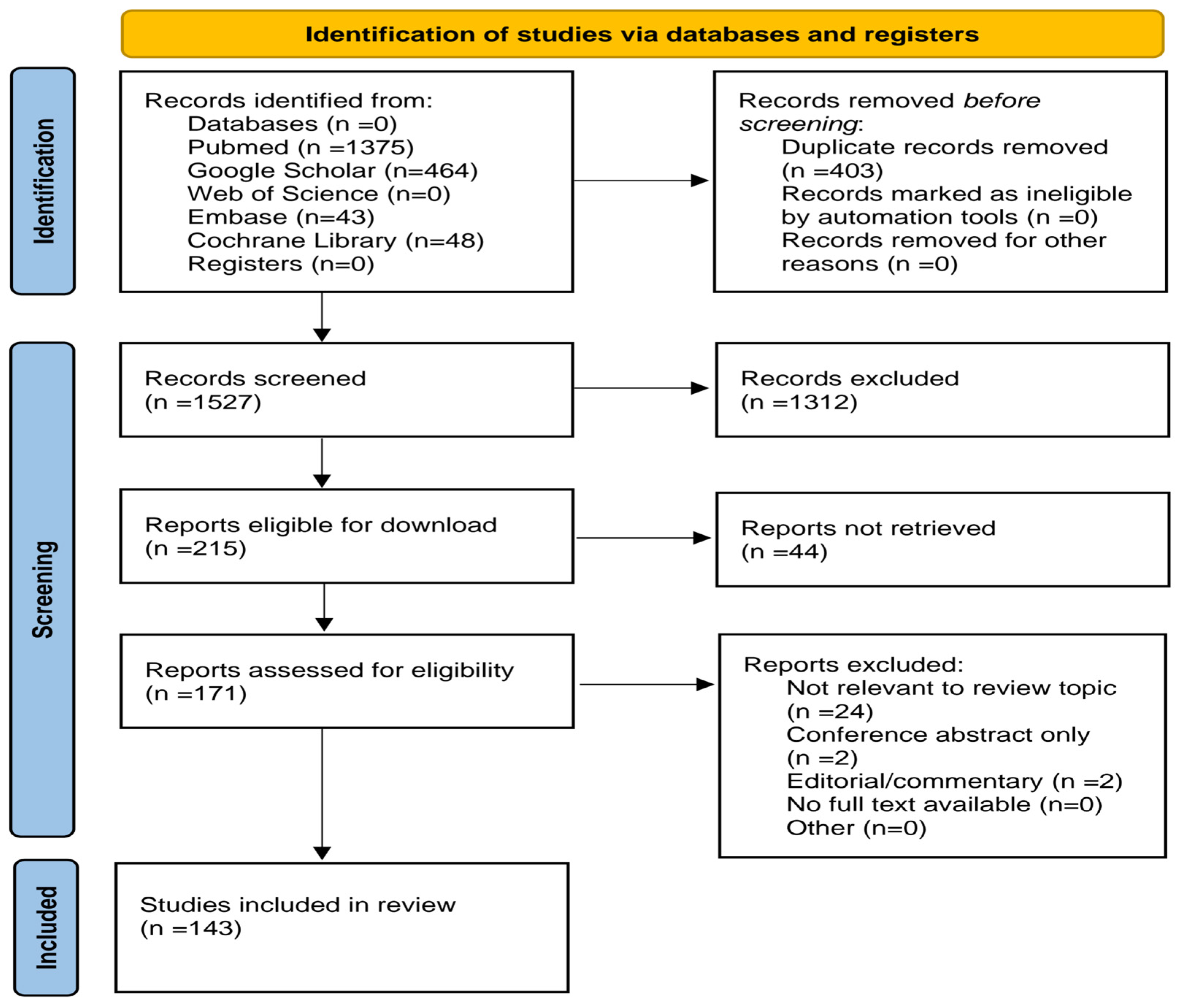
The review was conducted in accordance with the PRISMA Extension for Scoping Reviews (PRISMA-ScR) guidelines, and the process of identifying, selecting, and qualifying publications is presented in the PRISMA flowchart (Figure 1). The completed PRISMA-ScR checklist is provided in the Supplementary Materials (Table S1), whereas the characteristics and key findings of all studies included in the scoping review are presented in Supplementary Materials (Table S2). The review was guided by the Population–Concept–Context (PCC) framework recommended for scoping reviews, presented in Supplementary Table S3.
The protocol for this scoping review was registered on the Open Science Framework (OSF; Center for Open Science) and is publicly available at https://osf.io/dpfbe/ (accessed on 19 August 2026). The registration was completed after the initial literature screening had commenced and therefore represents retrospective registration. No amendments or methodological changes were introduced following registration.
Literature searches were conducted in PubMed, Web of Science, Embase, the Cochrane Library, and Google Scholar. The final search was conducted on 16 August 2026. Detailed database-specific search strategies are provided in Supplementary Table S4. The search strategy was based on keyword combinations related to cancer diagnostics, including: “cancer diagnosis,” “early cancer detection,” “diagnostic imaging,” “liquid biopsy,” “circulating tumor DNA,” “ctDNA,” “circulating tumor cells,” “molecular biomarkers,” “next-generation sequencing,” “multi-omics,” “artificial intelligence,” “machine learning,” “radiomics,” “digital pathology,” “precision oncology,” “nutritional status,” “sarcopenia,” “cancer cachexia,” and “body composition.” Individual terms were combined using the Boolean operators AND and OR, according to the analysed topic. Full-text publications on cancer diagnostics, molecular biomarkers, liquid biopsy, imaging methods, artificial intelligence, multi-omics technologies, and the assessment of nutritional status and body composition in cancer patients were eligible for review. Conference abstracts, editorials, letters to the editor, publications without full-text access, and works not directly related to the topic of this review were excluded. English-language publications were eligible for review. Additionally, a limited number of highly relevant non-English publications identified through reference screening were included. Google Scholar was used as an additional source to identify difficult-to-find and recent publications that may not have been indexed in other databases. The first 464 records ranked by relevance were screened. No restrictions were placed on the year of publication to ensure the most complete representation of current knowledge. The publication selection process was conducted in stages. All retrieved records were exported to Zotero, and duplicate records were removed automatically and subsequently verified manually. After removing duplicates, titles and abstracts were assessed, and full-text analyses were conducted for publications that met the eligibility criteria. Publication selection was conducted independently by two authors, according to previously established inclusion and exclusion criteria. In case of disagreement, a third researcher was consulted.
Data from eligible publications were qualitatively synthesised and organised into the following thematic areas: imaging diagnostics, histopathology and tissue biopsy, liquid biopsy, molecular biomarkers, artificial intelligence, multi-omics diagnostics, assessment of nutritional status and body composition, and clinical applications of modern diagnostic methods. The analysis focused on the diagnostic capabilities of individual technologies, their limitations, clinical potential, and their role in the development of precision medicine.
Due to the limited scope of the review and the significant heterogeneity of the technologies, patient populations, cancer types, and endpoints analysed, no meta-analysis or formal quality assessment of the studies was conducted. The results are presented in a descriptive qualitative synthesis, with particular emphasis on current developments in cancer diagnostics, research gaps, and future clinical applications.
As this scoping review aimed to map the available evidence rather than evaluate intervention effectiveness, no formal risk-of-bias assessment was conducted. Consequently, the findings should be interpreted as a synthesis of available evidence rather than a formal comparative assessment of diagnostic performance or clinical effectiveness.

3. Conventional Methods of Cancer Diagnosis

Despite the rapid development of new diagnostic technologies, conventional imaging methods, histopathological examinations, and tissue biopsies remain the cornerstones of cancer diagnosis. They enable the detection of neoplastic lesions, the assessment of their stage and the determination of suitability for treatment; however, their diagnostic and organisational limitations provide a significant impetus for the development of more sensitive, less invasive and faster diagnostic methods [39,40].

3.1. Diagnostic Imaging (CT, MRI, PET/CT, Ultrasound)

Advances in medical imaging techniques have significantly transformed modern oncological diagnostics. Today, the aim of imaging studies is not merely to localise the tumour, but also to obtain information about its biology, metabolic activity, vascularisation, microstructure and response to treatment. Functional magnetic resonance imaging (MRI) techniques and modern ultrasound methods have become particularly important, as they provide quantitative biomarkers that aid diagnosis, treatment planning and monitoring of treatment efficacy [41,42].

3.1.1. Computed Tomography (CT)

Computed tomography remains one of the fundamental imaging methods in oncology due to its high availability, short scan time, and high spatial resolution. It is widely used to detect tumour lesions, assess their location, determine disease stage, and monitor treatment effects [43,44]. The rapid development of computed tomography technology includes, amongst other things, dual-energy CT, perfusion tomography and modern photon-counting CT detectors, which enhance image quality and enable the acquisition of additional functional information [45,46,47]. At the same time, the primary limitations of CT remain the use of ionising radiation and the need to administer iodinated contrast agents in some patients [48].

3.1.2. Magnetic Resonance Imaging (MRI)

Magnetic resonance imaging is one of the most accurate methods for imaging soft tissues, whilst not utilising ionising radiation. In addition to conventional morphological assessment, MRI enables functional techniques, such as diffusion-weighted imaging (DWI) and dynamic contrast-enhanced MRI (DCE-MRI), which provide information on tissue microstructure, perfusion, and tumour angiogenesis [41]. The parameters obtained during these examinations are increasingly being used as quantitative imaging biomarkers to aid diagnosis, assess prognosis and monitor treatment efficacy [42]. Whole-body imaging (WB-MRI) is also gaining in importance, as it enables a comprehensive assessment of the body in a single scan, without exposing the patient to ionising radiation [49]. Radiomic techniques and new contrast agents are also being developed, which may enhance MRI’s diagnostic capabilities in personalised medicine [41,42,48].

3.1.3. Positron Emission Tomography Combined with Computed Tomography (PET/CT)

Positron emission tomography combined with computed tomography (PET/CT) is a hybrid method that combines metabolic information with a detailed anatomical image [50]. The most commonly used radiotracer—^18F-FDG—enables assessment of the increased glucose metabolism characteristic of many cancers, making PET/CT useful for detecting tumour foci, determining disease stage, planning treatment, and monitoring response to therapy [44]. In recent years, there has been rapid development of new radiopharmaceuticals and hybrid PET/MR systems, which enable even more precise characterisation of tumour lesions whilst reducing radiation exposure [51,52,53]. The limitations of PET/CT remain its relatively high cost and limited availability compared with CT and ultrasound [54].

3.1.4. Ultrasonography (USG)

Ultrasonography is one of the most commonly performed imaging methods in oncological diagnostics. Its main advantages are wide availability, safety, the absence of ionising radiation, and the ability to provide real-time imaging, which makes it particularly useful during image-guided biopsies [55]. The standard examination is complemented by Doppler techniques and elastography, which enable assessment of vascularisation and tissue mechanical properties [56]. Contrast-enhanced ultrasonography (CEUS), which uses gas microbubbles to assess tumour perfusion, is also becoming increasingly important [57]. At the same time, technologies utilising microbubbles as carriers of drugs, genes and siRNA molecules are being developed, opening prospects for the use of ultrasound not only in diagnosis but also in targeted cancer therapy [58,59]. The main characteristics, advantages, limitations, and clinical applications of the most commonly used oncological imaging techniques are summarised in Table 1.

3.2. Histopathological and Immunohistochemical Diagnosis

Histopathological examination remains the gold standard for the diagnosis of most cancers, enabling assessment of the histological type, grade of differentiation, and biological characteristics of the tumour, which are crucial for prognosis and treatment selection [60]. In many cancers, the results of histopathological examination form the basis for further molecular diagnostics and eligibility for targeted therapies [61]. Immunohistochemistry (IHC), which uses specific antibodies to identify the expression of proteins characteristic of particular tumour types, is an important complement to morphological assessment. This method enhances diagnostic accuracy, facilitates the differentiation of lesions with similar histological features, and enables the assessment of predictive and prognostic biomarkers—such as hormone receptors, HER2, PD-L1 and MMR proteins—which are crucial for the selection of personalised treatment [61,62,63]. Despite their high diagnostic value, histopathological and immunohistochemical examinations are not without limitations. Results may depend on the quality of the tissue sample, tumour heterogeneity, the antibodies used, and the pathologist’s experience, which in some cases makes unambiguous interpretation difficult [62,64]. For this reason, the integration of conventional microscopic assessment with molecular methods, as well as digital pathology tools and artificial intelligence, is becoming increasingly important; these can enhance the accuracy and reproducibility of diagnosis, but require further validation before routine use in clinical practice [65,66].

3.3. Tissue Biopsy as the Gold Standard

Tissue biopsy remains the gold standard in cancer diagnosis, as it provides material for histopathological, immunohistochemical, and molecular testing, which form the basis for diagnosis, prognosis assessment, and eligibility for targeted therapy. Modern sampling techniques, including core needle, endoscopic, and image-guided biopsies, improve diagnostic accuracy and enable the more precise collection of representative tissue. At the same time, this method remains invasive and may not capture the full heterogeneity of the tumour, particularly with small tissue samples or lesions with varied structure. Limitations associated with sampling errors, the need to repeat biopsies and difficulties in monitoring dynamic molecular changes during treatment are among the main drivers behind the development of less invasive diagnostic methods, such as liquid biopsy [67,68,69].

3.4. Screening Programmes and Their Effectiveness

Screening programmes are among the most important elements of secondary cancer prevention, enabling the detection of precancerous lesions or cancers at an early stage, thereby improving prognosis and reducing mortality from selected cancers [70]. In the case of, amongst others, colorectal, breast and cervical cancer, it has been shown that population-based screening programmes are cost-effective and yield long-term health benefits, particularly when there is a high participation rate [71,72]. However, the effectiveness of screening programmes depends not only on the characteristics of the diagnostic methods used, but also on the organisation of the healthcare system, the availability of tests and the active participation of the target population, which remains one of the main challenges of modern cancer prevention [70,73].

4. Liquid Biopsy as a Modern Diagnostic Tool

4.1. The Concept and Principle of Liquid Biopsy

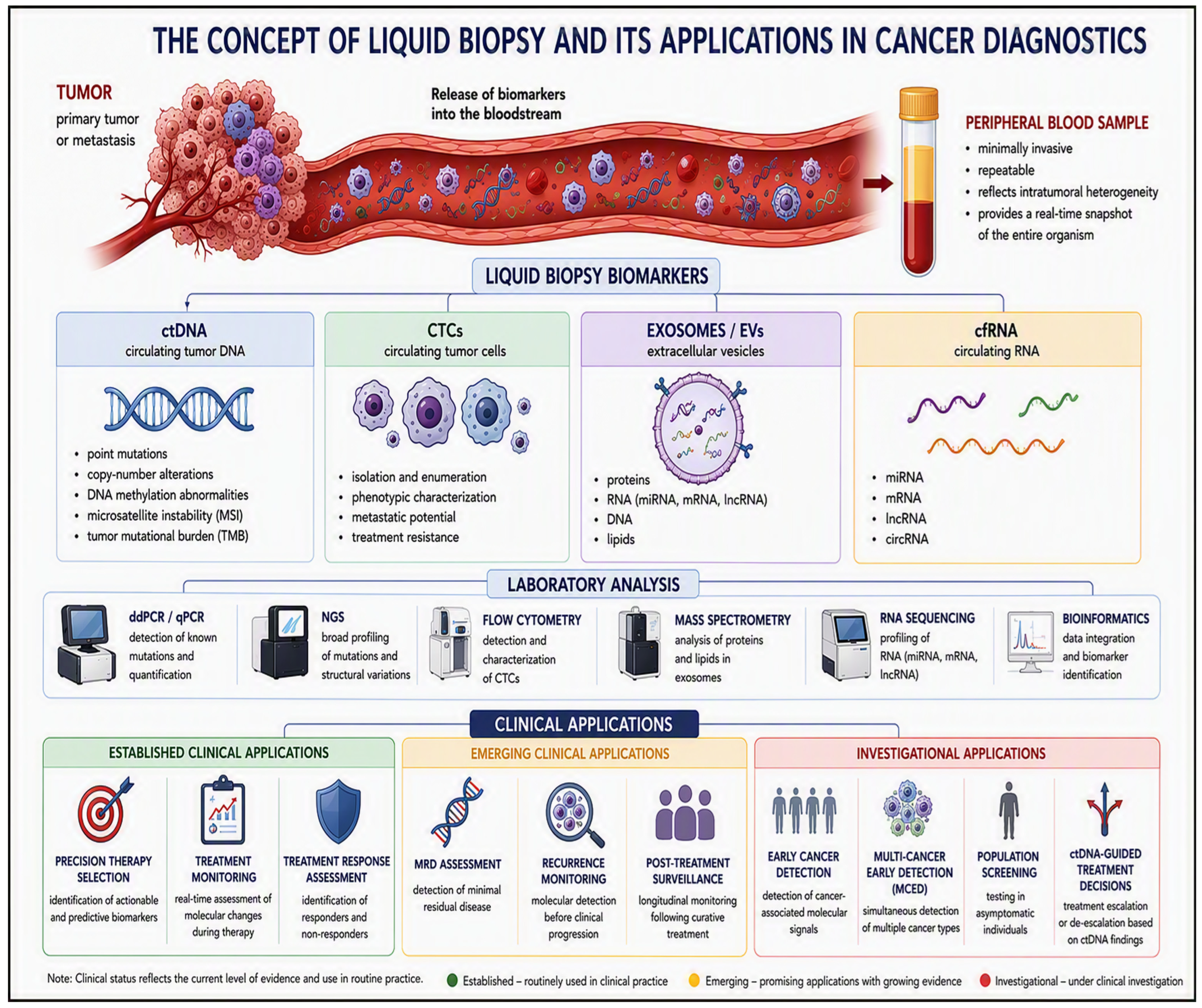
Liquid biopsy is a modern, minimally invasive diagnostic method that involves the analysis of tumour biomarkers present in body fluids, primarily blood, but also urine, saliva, cerebrospinal fluid, and body cavity fluids [74,75]. Unlike a conventional tissue biopsy, it allows repeated sampling, thereby enabling dynamic monitoring of molecular changes during disease progression and treatment [16]. The most commonly analysed components of liquid biopsy are circulating tumour DNA (ctDNA), circulating tumour cells (CTCs), circulating RNA (cfRNA, miRNA, lncRNA, circRNA) and extracellular vesicles, particularly exosomes [74,75,76]. These biomarkers reflect the current biological status of the tumour, enabling assessment of tumour heterogeneity, identification of genetic alterations, and monitoring of treatment response and the development of therapeutic resistance [16,74]. The most important advantages of liquid biopsy include its non-invasive nature, the possibility of serial testing, and a better reflection of tumour heterogeneity compared with a single tissue biopsy [16,74]. Despite its great diagnostic potential, this method still requires further standardisation and improvements in the sensitivity and specificity of certain analytical techniques; consequently, it is currently most often used to complement conventional histopathological diagnosis [74,75]. A diagram illustrating the sources of liquid biopsy biomarkers, their types and the most important clinical applications is shown in Figure 2.
The clinical applications of liquid biopsy should be interpreted according to the intended use of the assay. Evidence supporting ctDNA detection in patients with established cancer differs substantially from evidence required for population screening in asymptomatic individuals [16,74]. Likewise, the prognostic value of postoperative ctDNA positivity should not be equated with evidence that ctDNA-guided treatment decisions improve patient outcomes [74,76]. While ctDNA detection is strongly associated with recurrence risk across several tumour types, prospective interventional studies evaluating ctDNA-guided management remain emerging [74]. Furthermore, earlier molecular detection of disease does not necessarily translate into improved survival or other clinically meaningful outcomes [16]. The clinical utility of liquid biopsy, therefore, depends not only on analytical performance but also on evidence demonstrating that test-guided interventions improve patient management and outcomes [16,76]. Consequently, some applications of liquid biopsy are already incorporated into clinical practice, whereas others remain investigational and require further validation in prospective studies [16,74]. In particular, applications intended for population-level cancer screening require substantially higher levels of evidence than applications performed in patients with known malignancy.

4.2. Circulating Tumour DNA (ctDNA)

Circulating tumour DNA (ctDNA) constitutes a small fraction of cell-free DNA (cfDNA), which is released into the bloodstream mainly during apoptosis and necrosis of tumour cells [74]. It contains tumour-specific mutations, copy-number alterations, and DNA methylation abnormalities, and can therefore reflect the tumour’s molecular profile without the need to collect tissue samples [77]. The short half-life of ctDNA means that its concentration rapidly reflects changes occurring in the patient’s body, enabling the monitoring of treatment efficacy, the detection of residual disease and the early diagnosis of relapses [74,78]. In clinical practice, ctDNA analysis is already used, amongst other things, to identify EGFR mutations in non-small cell lung cancer and to monitor treatment response in many solid tumours [77,78]. Advances in analytical methods, such as digital PCR (ddPCR) and next-generation sequencing (NGS), have significantly increased the sensitivity of ctDNA detection; however, the low concentration of this biomarker in the early stages of the disease remains one of the main limitations of this method [72,77]. An important limitation of ctDNA analysis is the potential for false-positive results due to clonal haematopoiesis of indeterminate potential (CHIP) [74,76]. Somatic mutations originating from haematopoietic cells may be incorrectly interpreted as tumour-derived alterations [16,74]. Therefore, careful assay design, matched leukocyte sequencing, and appropriate bioinformatic filtering strategies are important for accurate interpretation of ctDNA results [74,76].

4.3. Circulating Tumour Cells (CTCs)

Circulating tumour cells (CTCs) are cells that detach from the primary tumour or metastatic sites and enter the bloodstream, where they contribute to the formation of distant metastases. Their number is usually very small—often a single cell per million leukocytes—which is why their isolation requires highly sensitive analytical methods [74,79]. The presence and number of CTCs correlate with disease stage, the risk of metastasis, and shorter overall and progression-free survival. For this reason, CTCs are regarded as valuable prognostic biomarkers and tools for monitoring the efficacy of anticancer treatment [74,78]. Methods for detecting CTCs include both those based on the physical properties of the cells and immunological techniques utilising surface markers, such as EpCAM. Currently, the only system approved by the FDA for determining CTC counts is CellSearch®; however, the development of new microfluidic technologies and molecular methods is continually increasing detection sensitivity and the potential for clinical use [74].

4.4. Exosomes and Extracellular Vesicles

Exosomes constitute a small subgroup of extracellular vesicles (EVs), secreted by most cells in the body, including cancer cells. They contain proteins, lipids and nucleic acids that reflect the properties of the cell of origin, enabling them to play a significant role in intercellular communication and in modulating the tumour microenvironment [74,80]. Due to their high stability in body fluids and their ability to protect the genetic material they carry from degradation, exosomes are considered promising biomarkers for liquid biopsy [81]. Their analysis may provide information regarding the presence of a tumour, its stage, metastatic potential and response to treatment. However, the main limitations remain the lack of standardisation in methods for isolating and characterising vesicles, and the difficulty in obtaining homogeneous exosome populations [74,82].

4.5. Circulating RNA (miRNA, lncRNA, circRNA)

Circulating RNA (cell-free RNA, cfRNA) comprises a variety of molecules, including microRNA (miRNA), long non-coding RNA (lncRNA) and circular RNA (circRNA), which can be released into the bloodstream both in free form and within extracellular vesicles [76,83]. Unlike ctDNA, many RNA molecules are secreted by living tumour cells, enabling them to reflect the biological activity of the tumour even at very early stages of its development [83]. The best-characterised group of biomarkers consists of miRNAs, whose altered expression has been described in many types of cancer and is associated with processes such as proliferation, angiogenesis, invasion and metastasis [84,85]. There is also growing interest in lncRNAs and circRNAs, which exhibit high stability in body fluids and possess diagnostic and prognostic potential [83]. Despite promising results from clinical trials, the use of RNA biomarkers still requires standardisation of assay methods and validation in large patient populations [76,83].

4.6. The Use of Liquid Biopsy in Diagnosis, Treatment Monitoring and the Detection of Relapses

Liquid biopsy is finding increasingly widespread application at all stages of cancer care—from the early detection of tumours, through the selection of therapy, to the monitoring of treatment efficacy and the detection of residual disease and relapses [16,74]. Thanks to the ability to collect biological samples serially, it is possible to assess molecular changes occurring in real time, which is difficult to achieve using a single tissue biopsy [74,86]. Analysis of ctDNA enables identification of mutations that may guide targeted therapy selection and can facilitate the detection of molecular residual disease (MRD), in some cases months before changes become visible on conventional imaging [16,86]. In turn, the measurement of CTCs, exosomes and circulating RNA can provide additional information regarding prognosis, response to therapy and the risk of disease progression [80,81,83]. Despite rapid technological development, liquid biopsy still primarily plays a complementary role to conventional histopathological diagnosis; however, as methods become further standardised, its importance in precision medicine is predicted to steadily increase [16,74]. Earlier molecular detection of disease does not necessarily translate into improved survival outcomes. Demonstrating clinical benefit requires prospective interventional studies that show that management decisions guided by liquid biopsy results improve patient outcomes. Therefore, the ability of liquid biopsy to detect molecular residual disease or recurrence should be distinguished from evidence demonstrating a survival benefit. Although ctDNA-based minimal residual disease (MRD) assessment demonstrates substantial prognostic value, most studies to date primarily report associations between ctDNA positivity and recurrence risk. Evidence supporting ctDNA-guided treatment escalation or de-escalation remains limited, and results from prospective interventional trials evaluating treatment decisions based on ctDNA findings are still emerging [74,75].

5. Modern Molecular Biomarkers

Advances in molecular biology and high-throughput technologies have led to rapid developments in the biomarkers used in modern oncology. Molecular biomarkers are utilised not only in the diagnosis of cancers but also in assessing prognosis, monitoring treatment efficacy, and selecting patients for targeted therapies and immunotherapy [87,88]. Increasing importance is also being attached to their role in realising the principles of precision medicine, which enables the selection of treatment based on the tumour’s individual molecular characteristics [87,89]. Among the most important groups of biomarkers are genetic, epigenetic, proteomic, and metabolomic biomarkers, as well as changes in microbiome composition, which are increasingly analysed as potential indicators of cancer development and progression [87,88,89,90,91].

5.1. Genetic Biomarkers

Genetic biomarkers are among the best-understood and most widely used molecular markers in modern oncology. They include gene mutations, amplifications, deletions, chromosomal rearrangements, and gene expression abnormalities that contribute to tumour transformation, disease progression, and treatment response [87,88]. Thanks to the development of next-generation sequencing (NGS) methods, it has become possible to detect multiple genetic alterations simultaneously, significantly enhancing the capabilities of molecular diagnostics and patient selection for molecularly targeted therapies [89,91]. The importance of genetic biomarkers has been confirmed in many types of cancer. In gastric cancer, HER2 gene abnormalities, TP53 mutations, CDH1 gene alterations and microsatellite instability (MSI) are particularly significant, as they have diagnostic, prognostic and predictive value [87,90]. The molecular classifications developed have enabled the identification of cancer subtypes with distinct prognoses and varying sensitivities to treatment, particularly immunotherapy [87]. In ovarian cancer, mutations in the BRCA1 and BRCA2 genes—which are responsible for DNA repair—are of key importance; their presence is associated with an increased risk of developing the disease and with the potential for PARP inhibitor use [88]. In colorectal cancer, different molecular alterations provide distinct types of clinical information. MMR/MSI status has established prognostic significance and may influence treatment decision-making in specific clinical settings. KRAS mutations are primarily predictive biomarkers associated with resistance to anti-EGFR therapies, whereas APC and TP53 alterations contribute to the molecular characterisation of tumour biology and disease progression. Therefore, these biomarkers should not be considered interchangeable, as their clinical roles differ in diagnosis, prognosis, and prediction of treatment benefit [91]. The importance of comprehensive molecular profiles is also increasingly emphasised, as these allow simultaneous assessment of multiple genetic alterations and even more precise tailoring of therapy to the individual patient’s needs [89,90].

5.2. Epigenetic Biomarkers

Epigenetic biomarkers encompass changes that influence gene expression without altering the DNA sequence. The most important of these include DNA methylation, histone modifications and the expression of non-coding RNAs, particularly microRNAs (miRNAs). Epigenetic changes occur as early as the initial stages of carcinogenesis, meaning they can be utilised for diagnosis, prognostic assessment, and monitoring treatment efficacy. An additional advantage of epigenetic biomarkers is their reversibility and the possibility of detecting them in samples obtained via minimally invasive methods, such as blood or other body fluids, thereby enhancing their clinical potential [92]. Studies on colorectal cancer have shown that DNA methylation patterns and miRNA expression profiles may be associated with response to neoadjuvant and adjuvant treatment; however, their routine use requires further validation in clinical trials [93].

5.3. Proteomic Biomarkers

Proteomic biomarkers are based on the analysis of proteins and their expression changes that occur during tumour development. Thanks to modern proteomic techniques, particularly mass spectrometry, it is possible to simultaneously analyse thousands of proteins and identify characteristic molecular profiles associated with specific cancer types. This enables not only earlier detection of the disease, but also the assessment of prognosis, monitoring of treatment outcomes and the identification of new therapeutic targets. Increasing attention is also being paid to the analysis of proteins present in extracellular vesicles (EVs), which may constitute a stable and readily available source of biomarkers for cancer diagnosis via liquid biopsy [94,95].

5.4. Metabolomic Biomarkers

Metabolomic biomarkers comprise small molecules produced by metabolic processes in cells and tissues, whose profiles undergo characteristic changes during tumour development [96]. Using high-resolution mass spectrometry and chromatography, it is possible to analyse metabolites in readily available biological samples, such as plasma, serum, faeces, or saliva [96,97]. In colorectal cancer, metabolite sets have been identified that allow differentiation between healthy individuals, patients with adenomas, and patients with cancer, indicating their usefulness in the early diagnosis of the disease [98]. Similar results have been obtained in studies of gastric and lung cancer, where changes in the metabolism of amino acids, lipids, and other small-molecule compounds have been shown to serve as sensitive biomarkers of tumour development and to aid in assessing prognosis and treatment response [97,99,100].

5.5. Structural Biomarkers and Extracellular Matrix Alterations

Recent studies have highlighted structural biomarkers as a complementary source of information for cancer detection, particularly through analysis of cancer-associated alterations in the extracellular matrix (ECM). X-ray scattering can capture molecular and supramolecular structural features of tissue components that may differ between cancerous and non-cancerous tissues. In a study of 107 patients and 2958 tissue measurements, Denisov et al. demonstrated that X-ray scattering combined with machine learning could distinguish cancerous from non-cancerous breast tissue based on structural signatures, with sensitivity and specificity reaching 86% and 83%, respectively, depending on the measurement configuration [101]. More recent research further demonstrated that X-ray diffraction of collagen and collagen-structured water can distinguish cancerous breast tissue from fibroadenoma, suggesting that alterations in collagen organisation may provide additional structural biomarkers for cancer detection [102]. These findings indicate that structural information obtained from X-ray-based techniques may complement conventional molecular biomarkers by providing an additional layer of information on tissue architecture and molecular organisation. Integration of such structural biomarkers with genomic, transcriptomic, proteomic, and metabolomic data may therefore contribute to a more comprehensive characterisation of tumour biology and support the development of integrated approaches in precision oncology [101,102].

5.6. The Microbiome as a Potential Tumour Biomarker

The human microbiome plays a significant role in regulating the immune response, inflammatory processes and the maintenance of the body’s homeostasis; consequently, disturbances in the microbiome are increasingly associated with the initiation and progression of tumours [103,104]. Analysis of microbiota composition using 16S rRNA gene sequencing and metagenomic methods enables identification of characteristic microbial signatures associated with specific cancer types [103]. In colorectal cancer, the presence of specific bacteria and microbiological markers has been demonstrated, enabling non-invasive detection of the disease from stool samples, even in the early stages of tumour development [105]. A growing body of evidence also suggests that the microbiome profile can predict immunotherapy efficacy and support diagnosis and treatment monitoring; it is therefore considered one of the most promising biomarkers of the future in oncology [104,106].

6. Artificial Intelligence in Cancer Diagnostics

6.1. Machine Learning and Deep Learning

Machine learning (ML) and deep learning (DL) form the basis of modern artificial intelligence systems used in oncological diagnostics. ML algorithms enable the identification of relationships among clinical, imaging, and molecular features, whilst deep learning models, particularly convolutional neural networks (CNNs), allow the automatic recognition of complex patterns in histopathological and radiological images without the need to define diagnostic features manually [107]. This makes it possible to assist in the classification of neoplastic lesions, the assessment of malignancy grade and the prediction of patient prognosis [108]. In recent years, advances in deep learning architectures have enabled the use of multimodal models that integrate imaging, histopathological and molecular data. Transformer models are also becoming increasingly important, as they allow for the simultaneous analysis of multiple data types and the development of more precise diagnostic systems. Despite the very high effectiveness of these algorithms, their implementation in clinical practice requires sufficiently large, well-documented datasets and independent model validation before routine use [109].

6.2. Radiomics

Radiomics is one of the most rapidly developing fields of artificial intelligence-assisted diagnostics. It involves the automatic extraction of numerous quantitative features from images obtained using computed tomography, magnetic resonance imaging, positron emission tomography, or ultrasound. Both the morphological features of the tumour and parameters relating to texture and heterogeneity are analysed; these often remain invisible during a standard image assessment by a doctor [110]. This data can then be analysed using machine learning algorithms to distinguish between benign and malignant lesions, determine disease stage, and predict treatment response [111]. An important area of development in radiomics is radiogenomics, which enables the correlation of imaging features with a tumour’s molecular profile. The integration of radiomic data with clinical and genomic information enables the development of models that support precision medicine and personalised therapy [112]. At the same time, limitations include a lack of full standardisation of the feature-extraction process and differences in imaging protocols across centres, which hinder the widespread implementation of AI models in everyday clinical practice [113].

6.3. Digital Pathology

Digital pathology utilises images of histopathological slides obtained with Whole Slide Imaging (WSI) scanners, enabling computerised analysis with artificial intelligence algorithms. These solutions assist pathologists in detecting cancer cells, assessing immunohistochemical biomarkers (including Ki-67, HER2 and PD-L1), classifying the grade of malignancy, and standardising histopathological assessment [114]. In many applications, AI achieves accuracy comparable to that of experts, whilst reducing analysis time and minimising inter-observer variability [105]. Modern digital pathology systems are increasingly integrating image analysis with molecular and clinical data, enabling the identification of predictive and prognostic biomarkers and supporting the development of precision medicine [115]. Despite very promising results, it remains necessary to standardise digital image quality, validate algorithms across diverse populations, and ensure the transparency of AI models to enable their safe use in routine oncological diagnostics [109,116].

6.4. Integration of Clinical and Molecular Data

One of the most important areas of development for artificial intelligence in oncology is the integration of data from multiple sources, including radiological and histopathological images, genomic, transcriptomic and proteomic profiles, and clinical information regarding the course of the disease. The use of machine learning models enables the analysis of multidimensional datasets, the identification of relationships not apparent during conventional assessment, and the development of more precise diagnostic and prognostic models [117]. Algorithms analysing data from liquid biopsies, including cell-free DNA (cfDNA), are also becoming increasingly important, as they aid in the early detection of cancer and the monitoring of treatment response [118]. The development of multimodal models enables the simultaneous use of imaging, molecular and clinical information, thereby increasing the accuracy of tumour classification and supporting the selection of therapies tailored to the individual patient’s profile [119]. The integration of multi-omic data using artificial intelligence is one of the cornerstones of precision medicine; however, the effectiveness of such models still depends on data quality, appropriate standardisation and the availability of large, representative datasets used for their training [120].

6.5. Limitations and Ethical Considerations Regarding the Use of AI

Despite the rapid development of artificial intelligence, its implementation in clinical practice presents numerous technical, organisational and ethical challenges. The most significant limitations include insufficient data standardisation, the risk of overfitting models to the training data, limited interpretability of algorithmic decisions, and difficulties in validating them across diverse patient populations [109,121]. An additional problem is the variation in diagnostic image quality and the potential bias in models arising from non-representative training datasets [114]. The protection of personal data, accountability for AI-assisted decisions, and the need to ensure transparency in how algorithms operate also remain significant issues. Currently, artificial intelligence is viewed as a tool to support doctors, rather than a replacement for their clinical expertise [107]. Further development of the technology will require establishing uniform regulations, ensuring data security, and conducting prospective clinical trials to confirm the efficacy and safety of new solutions before their routine implementation [116].

7. Multi-Omic Diagnostics as the Basis for Precision Medicine in Oncology

7.1. Genomics

Genomics is a fundamental component of modern molecular cancer diagnostics. Next-generation sequencing enables the simultaneous assessment of multiple genes and the detection of single-nucleotide variants, small insertions and deletions, copy-number variations, and gene rearrangements and fusions. In clinical practice, targeted panels, whole-exome sequencing and—in selected situations—whole-genome sequencing are used. The results of genomic profiling may identify targets for molecular therapies, support eligibility for immunotherapy and reveal germline variants requiring genetic counselling [122]. A limitation of genomics remains the ambiguity in interpreting certain detected alterations. Variants of undetermined clinical significance should not be treated as the sole basis for a therapeutic decision. The test result requires interpretation in the context of the histopathological findings, the stage of the disease, previous treatment and current knowledge bases. The analysis of tumour heterogeneity and single-cell technologies, which enable the separation of signals originating from different tumour cell populations and the microenvironment, are also becoming increasingly important [123].

7.2. Transcriptomics

Transcriptomics describes the set of RNA molecules present in a cell or tissue at a given point in time. RNA-seq enables the assessment of gene expression levels, alternative splicing, gene fusions, and coding and non-coding transcripts. In oncology, transcriptome analysis supports the molecular classification of tumours, the identification of biologically active pathways and the search for predictive biomarkers [124,125]. Unlike genomic studies, which identify potential genetic alterations, transcriptome analysis reflects the actual activity of genes within the cell. This makes it possible to assess the biological processes occurring within the tumour and to identify differences between tumours with similar histopathological appearances but different molecular profiles. Information obtained from gene expression analysis can facilitate the assessment of prognosis and support treatment selection in specific patient groups [124,125]. A clinical example of the application of expression profiles is the 21-gene test used in selected patients with hormone-dependent breast cancer. The TAILORx trial demonstrated that the test result can inform decisions about the appropriateness of adjuvant chemotherapy and limit its use in some patients without compromising treatment outcomes [126].

7.3. Proteomics and Proteogenomics

Proteomics provides information on the quantity, activity and modifications of proteins, and thus on the functional effects of genomic and transcriptomic changes. High-resolution mass spectrometry enables the analysis of thousands of proteins and the phosphoproteome. Proteogenomic studies of lung, breast and colorectal cancer have shown that integrating DNA, RNA and protein data can reveal active signalling pathways and potential therapeutic targets that cannot be reliably predicted from DNA sequence alone [127,128,129]. Combining proteomic data with genomic and transcriptomic results enables a better understanding of the mechanisms underlying tumour development and the emergence of treatment resistance. Protein analysis also allows the assessment of signalling pathway activity, which does not always result directly from the presence of specific mutations; as such, it can provide additional information useful for identifying new biomarkers and potential therapeutic targets [127,128,129]. The routine use of proteomics remains limited by the complexity of sample preparation, equipment costs, platform differences, and the need to validate biomarkers in independent cohorts. Therefore, proteomic results should be regarded as a complement to, rather than a substitute for, validated clinical trials [127,128,129].

7.4. Metabolomics

Metabolomics involves the analysis of low-molecular-weight products of biochemical reactions. As metabolites reflect the combined influence of the genome, gene expression, protein activity, diet, the microbiota and treatment, they can provide information on the current biological phenotype of a tumour. The most commonly used techniques are mass spectrometry coupled with chromatography and magnetic resonance spectroscopy [130]. Analysis of the metabolite profile allows assessment of changes in the metabolism of cancer cells and their microenvironment. In experimental studies, metabolomics is used to identify diagnostic and prognostic biomarkers and to assess treatment response. As metabolites reflect the results of numerous biological processes, their analysis can provide complementary information to genomic and proteomic studies [130]. A significant limitation is the metabolome’s susceptibility to pre-analytical factors, including the time of sample collection, diet, medication, physical activity and hydration status. For this reason, the clinical implementation of metabolomic biomarkers requires rigorous standardisation and multi-centre validation [130].

7.5. Integration of Multi-Omic Data

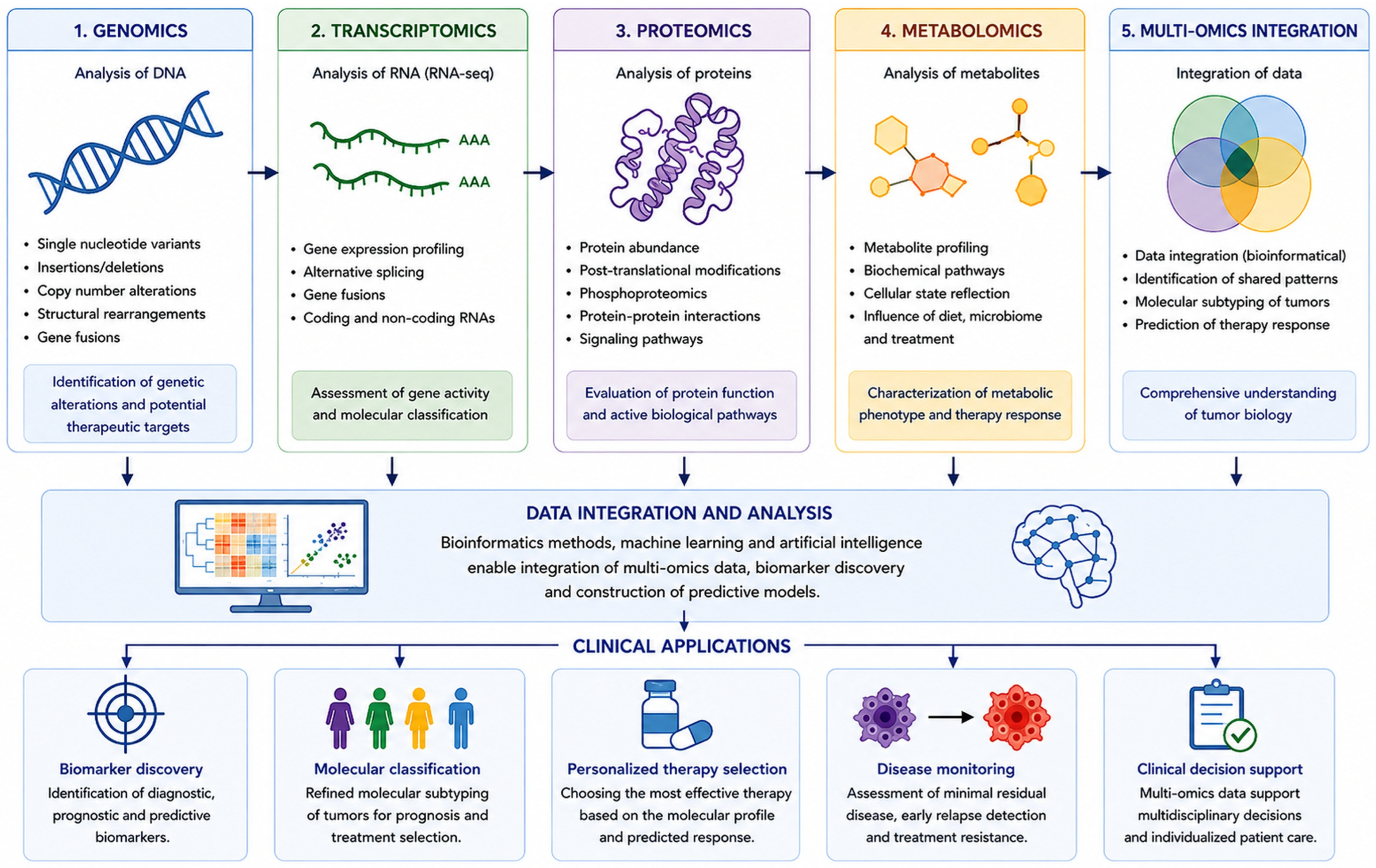
No single omics layer describes the full biology of a tumour. The integration of genomics, transcriptomics, proteomics and metabolomics allows molecular changes to be linked to their functional consequences. Tools such as Multi-Omics Factor Analysis identify common sources of variability and biological subgroups that are not apparent in analyses of individual datasets [131]. The multi-omics approach enables information from different levels of a cell’s biological organisation to be considered simultaneously, thereby providing a more comprehensive characterisation of the tumour. The integration of multiple data types can facilitate the identification of new biomarkers, improve molecular classification, and support the development of predictive models for precision medicine [131]. However, multi-omic models require an adequate sample size, independent validation, control for multiple comparisons, and an assessment of clinical utility. Artificial intelligence algorithms can support biomarker discovery and the development of predictive models. Still, their value depends on data quality, population representativeness and a transparent assessment of the risk of error [132,133,134]. A diagram illustrating the integration of multi-omic data and its applications in precision medicine is shown in Figure 3.

7.6. Clinical Importance

Multi-omics diagnostics can support therapy selection, elucidation of resistance mechanisms, and patient selection for clinical trials [122,131]. In clinical practice, its greatest value is observed in situations where information from different molecular analyses complements one another, allowing for better characterisation of the tumour’s biological characteristics [122,131]. However, its use should focus on clinical questions for which validated interpretation procedures are available. Due to the large amount of data generated, proper interpretation requires collaboration between specialists from various fields, including oncologists, pathologists, laboratory diagnosticians, molecular biologists, and bioinformaticians. The greatest value is achieved when molecular data are discussed in a multidisciplinary team and integrated with pathology, imaging, and the patient’s clinical condition [122,132,133,134].

8. The Importance of Nutritional Status and Body Composition in the Diagnosis and Prognosis of Cancer Patients

In recent years, increasing attention has been paid to the importance of nutritional status and body composition in the care of cancer patients. These parameters not only influence the course of treatment and prognosis but can also complement information obtained from imaging and molecular tests. Therefore, they are increasingly being viewed as part of a comprehensive patient assessment within the framework of precision medicine.

8.1. Nutritional Status Assessment

Eating disorders are common in oncological patients and may worsen treatment tolerance, increase the risk of complications, prolong hospitalisation, and reduce quality of life. Early diagnosis of malnutrition is important because it enables the implementation of appropriate nutritional interventions before irreversible changes in the patient’s body composition and fitness occur [135,136]. The Global Leadership Initiative on Malnutrition (GLIM) criteria base the diagnosis of malnutrition on the co-occurrence of at least one phenotypic criterion and one etiological criterion. Phenotypic criteria include unintentional weight loss, low body mass index (BMI) or decreased muscle mass, while etiological criteria include limited food intake or assimilation and the presence of an inflammatory process associated with the disease [135]. In oncological practice, regular nutritional screening and a comprehensive assessment of nutritional status, including analysis of food intake, body weight changes, body composition, inflammation, and physical fitness, are recommended. The guidelines of the European Society for Clinical Nutrition and Metabolism (ESPEN) emphasise that nutritional assessment should be carried out at all stages of cancer treatment and periodically repeated, because the nutritional status may change during anticancer therapy [136].

8.2. Sarcopenia

Sarcopenia is a muscle disease characterised by reduced muscle strength. According to the recommendations of the European Working Group on Sarcopenia in Older People 2 (EWGSOP2), low muscle strength indicates probable sarcopenia, low muscle quantity or quality confirms the diagnosis, and deterioration of physical function indicates a severe form [137]. In oncological patients, sarcopenia may occur regardless of body mass index (BMI) and may be masked by overweight or obesity [137]. Assessment should include at least a simple functional test, such as handgrip strength or a chair rise test, and a method for assessing muscle counts. In oncology, the analysis of routine computed tomography (CT) images is particularly useful, while dual-energy X-ray absorptiometry (DXA) and bioelectrical impedance analysis (BIA) may be a valuable addition. The result should be interpreted together with body weight dynamics, energy and protein intake, and physical activity [137,138].

8.3. Cancer Cachexia

Cancer cachexia is a multifactorial syndrome characterised by progressive muscle loss that cannot be fully reversed by conventional nutritional support alone. Consensus criteria include weight loss of more than 5% or more than 2% with a body mass index (BMI) less than 20 kg/m2 or concomitant sarcopenia. Cachexia is divided into pre-cachexia, cachexia, and refractory cachexia [139]. Management should be multifaceted and include cancer treatment, control of restrictive symptoms, nutritional support, adapted physical activity, and psychosocial care. The European Society for Medical Oncology (ESMO) guidelines emphasise the importance of early assessment and tailoring the intensity of intervention to the patient’s prognosis, performance status, and preferences [140].

8.4. Bioimpedance and Phase Angle

Bioelectrical impedance analysis (BIA) is a non-invasive method for assessing body composition by measuring tissue resistance and reactance to a low-intensity current. It allows for the estimation of lean body mass, adipose tissue mass, and body fluid distribution. Due to its simplicity, short test time, and lack of exposure to ionising radiation, BIA is used both in clinical practice and in monitoring body composition changes in cancer patients. However, its limitations remain: dependence on hydration status, the device used, the prediction equations employed, and the measurement conditions. Therefore, the test should be performed under repeatable conditions and should not be the sole basis for diagnosing sarcopenia or malnutrition [141].
Phase angle is a parameter calculated from resistance and reactance values, reflecting the integrity of cell membranes and the proportion of metabolically active cell mass. Lower phase angle values often coincide with poor nutritional status, reduced muscle mass and function, and a poorer prognosis in cancer patients. However, it should be emphasised that reference values and cutoff points vary with age, gender, study population, and analyser type, limiting their universal use in clinical practice [141]. A meta-analysis published in 2024 demonstrated a moderate diagnostic value of phase angle in screening for sarcopenia, while also highlighting significant heterogeneity between studies and the need for further standardisation of measurement methods and the establishment of uniform threshold values for different patient groups [141].

8.5. Computed Tomography and DXA

Body composition analysis of routine computed tomography (CT) images, most often at the level of the third lumbar vertebra (L3), allows assessment of muscle area, skeletal muscle index, and muscle quality based on radiographic muscle weakness. It also allows for the assessment of visceral and subcutaneous fat. Because CT scans are widely available for cancer diagnosis and treatment monitoring, this method does not require additional imaging studies, thereby increasing its usefulness in everyday clinical practice [138].
Dual-energy X-ray absorptiometry (DXA) provides data on lean limb mass, total fat mass, and bone mineral density, but does not directly assess myosteatosis. Compared to CT, this method involves lower radiation exposure and is widely used to assess body composition in clinical trials. However, its use in oncology may be limited by lower availability and the need for a separate examination [138]. Interpretation of results should take into account the patient’s clinical condition, cancer stage, and the purpose of the assessment. In studies of cachexia and body composition changes, it is recommended to use standardised measurement methods, clearly define endpoints, and provide detailed reporting of the techniques and analytical methods used, thereby facilitating comparison of results between studies and increasing their clinical value [138].
Assessment of nutritional status and body composition is an important complement to clinical, imaging, and molecular data used in modern precision medicine. A comparison of selected methods and technologies used in modern oncology diagnostics is presented in Table 2.

9. Clinical Applications of Modern Diagnostic Methods

Modern oncology diagnostics combine morphology, imaging, molecular biomarkers, and clinical data. Early detection can be based on imaging methods and screening tests and, in the future, on validated multi-tumour tests. In the diagnosis of established cancer, molecular profiling enables the identification of predictive changes and the selection of targeted therapies [122]. Liquid biopsy enables the analysis of circulating tumour DNA, tumour cells, and other blood components. Its most important current and developing applications include detecting resistance mechanisms, monitoring disease dynamics, and assessing molecular residual disease. In solid tumours, the detection of circulating tumour DNA (ctDNA) after radical treatment is usually associated with a high risk of relapse; however, the use of this finding for routine escalation or de-escalation of treatment still requires data from prospective studies [142,143].
In lung cancer, molecular assays support the selection of inhibitors of epidermal growth factor receptor (EGFR), anaplastic lymphoma kinase (ALK), ROS proto-oncogene 1 receptor tyrosine kinase (ROS1), B-Raf proto-oncogene (serine/threonine kinase) (BRAF), MET proto-oncogene (receptor tyrosine kinase) (MET), RET (rearranged during transfection) receptor tyrosine kinase (RET), KRAS G12C mutation (Kirsten rat sarcoma viral oncogene homolog, KRAS G12C), and neurotrophic tyrosine receptor kinase (NTRK) gene fusions. In breast cancer, hormone receptor status, human epidermal growth factor receptor 2 (HER2), selected expression profiles, and germline or somatic mutations related to deoxyribonucleic acid (DNA) repair are used. In colorectal cancer, mutations in the RAS (rat sarcoma virus) (RAS) family, BRAF, microsatellite instability and mismatch repair (MSI/MMR) disorders, and HER2 are important. In pancreatic cancer, assessing hereditary predispositions and selected DNA repair disorders is crucial. The scope of testing should be consistent with current therapeutic indications and the material’s quality [122]. Digital pathology and artificial intelligence (AI) algorithms can support lesion detection, image classification, and quantitative biomarker assessment. A meta-analysis of studies on diagnostic accuracy reported high sensitivity and specificity but also frequent errors and problems with external validation. Therefore, AI should support, not replace, the pathologist’s diagnostic responsibility [132,133,134].
The greatest potential of precision oncology lies not in the isolated use of individual diagnostic technologies but in their integration. Imaging biomarkers, liquid biopsy, molecular profiling, multi-omics analyses, artificial intelligence, and patient phenotyping may provide complementary information that supports more comprehensive tumour characterisation and more individualised therapeutic decision-making.

10. Current Challenges and Future Directions for Oncology Diagnostics

Modern oncology diagnostics is evolving towards the increasing integration of data from various sources. At the same time, the growing number of available technologies poses challenges for their standardisation, validation, interpretation, and use in clinical decision-making. Therefore, a key challenge remains not only the development of new biomarkers and diagnostic tools, but, above all, demonstrating their clinical value, reproducibility, and safety in various patient populations and across oncology centres [122,131].
In addition to analytical and clinical validation, successful implementation of these technologies requires consideration of cost-effectiveness, accessibility, regulatory approval pathways, data interoperability, and healthcare infrastructure requirements. These factors may substantially influence the real-world adoption of innovative diagnostic technologies and remain important barriers to broader clinical translation.
One of the most promising directions is the integration of multimodal data, including histopathology, radiology, genomics, transcriptomics, proteomics, laboratory test results, and patient-monitoring data. This approach can enable a more comprehensive characterisation of both the tumour itself and the disease course. Multimodal models can potentially improve risk stratification and support more precise clinical decision-making. However, demonstrating that complex models integrating multiple types of data offer a real advantage over simpler and easier-to-use tools remains a challenge. Therefore, their usefulness should be assessed primarily in properly designed, prospective clinical trials [122,131,132,133,134].
Liquid biopsy will also play a crucial role in future oncology diagnostics, particularly for detecting minimal residual disease, monitoring treatment response, and identifying emerging resistance. The ability to serially measure circulating tumour DNA (ctDNA) offers the prospect of a more dynamic assessment of the disease course than diagnostics performed only at selected time points. However, significant challenges remain in ensuring adequate method sensitivity, standardising procedures, and reducing the risk of false positive results. Demonstrating that therapeutic decisions based on changes in ctDNA concentration or profile actually translate into improved survival, quality of life, or other clinically relevant endpoints is also becoming increasingly important [142,143]. Another rapidly developing area is digital pathology and the use of artificial intelligence in medical image analysis. Automatic image segmentation and quantitative assessment of morphological features can support the analysis of diagnostic material and improve workflow. At the same time, the use of AI algorithms in clinical practice requires special caution. Models should be validated using data from various centres and populations, as their effectiveness in the conditions in which they were developed does not necessarily imply similar effectiveness in other clinical environments. Human supervision and systematic evaluation of tool performance after implementation are also necessary. Previous experience with digital pathology indicates that potential diagnostic and organisational benefits are accompanied by technical requirements and ethical issues that must be considered when implementing these technologies [132,133,134].
The development of oncology diagnostics also includes remote monitoring of patient health. Regular collection of information on symptoms, activity, and selected physiological parameters can support earlier recognition of patient deterioration and enable a faster clinical response. However, the implementation of such solutions should consider their usefulness from the patient’s perspective. Monitoring technologies should not overburden patients with the need for continuous data reporting or replace direct contact with medical personnel [122,131]. Therefore, a significant challenge remains in translating technological progress into genuine improvements in cancer patient care. The growing number of available biomarkers, molecular data, and AI-based tools is not, in itself, a sufficient indicator of the development of precision medicine. The value of new methods will depend on their reliable, repeatable application, the correct interpretation of results, and the use of the information obtained to make treatment-relevant decisions [122,131].
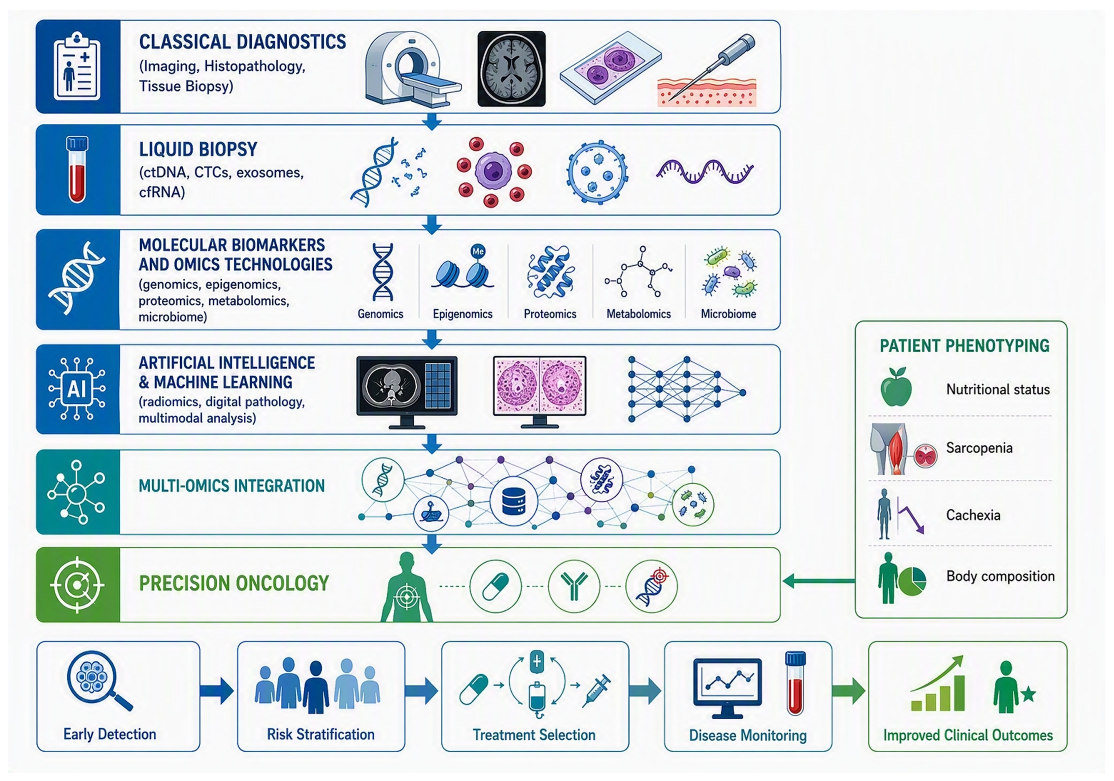
To provide a concise summary of the topics discussed in this review, Table 3 presents the current state of knowledge, key challenges, research gaps, and future directions in key areas of modern precision medicine. The table highlights the importance of diagnostic imaging, liquid biopsy, molecular biomarkers, artificial intelligence, multi-omics approaches, and the assessment of nutritional status and body composition in the diagnostic and therapeutic process. Figure 4, in turn, illustrates the evolution of modern cancer diagnostics from classical methods to integrated precision medicine strategies. The diagram illustrates how imaging, histopathological, molecular, and clinical data can complement each other, supporting more accurate diagnosis, individualised treatment, and disease monitoring.
Several limitations of the present review should be acknowledged. The included evidence comprised original studies, reviews, clinical guidelines, consensus documents, methodological papers, and other evidence sources. These publication types provide different levels and types of evidence and should not be interpreted as equivalent. As the primary objective of this scoping review was to map and synthesise the available literature rather than to evaluate comparative effectiveness, no formal risk-of-bias assessment was conducted. Consequently, the findings should be interpreted as descriptive and exploratory rather than confirmatory, and they should not be considered a formal comparison of diagnostic performance or clinical effectiveness across technologies.

11. Conclusions

The issues presented in this paper indicate that the future of oncology diagnostics will be based on a comprehensive patient assessment, taking into account clinical, imaging, and histopathological data, as well as molecular and multi-omic information, and assessment of nutritional status and body composition. Integrating this data using modern bioinformatics methods and artificial intelligence tools enables more accurate risk stratification, earlier disease detection, monitoring treatment response, and more precise therapy selection. Effective implementation of these solutions requires further standardisation of diagnostic methods, validation of new biomarkers in large clinical trials, and the availability of these biomarkers in everyday practice.
In recent years, there has been a clear shift from diagnostics based on single parameters to integrated analysis of multiple sources of information. The development of next-generation sequencing, multi-omic diagnostics, liquid biopsy, and digital pathology enables increasingly accurate biological characterisation of tumours. Available evidence suggests that these technologies may improve diagnostic accuracy and support precision oncology; however, further validation and comparative studies are required. These technologies constitute one of the pillars of the evolving precision medicine, which aims to individualise diagnostic and therapeutic procedures.
An equally important element of modern oncology care is the comprehensive assessment of nutritional status and body composition. A growing body of research indicates that malnutrition, sarcopenia, and cachexia affect not only treatment tolerance but also the risk of complications, length of hospitalisation, quality of life, and patient survival. The inclusion of systematic nutritional assessment, body composition analysis, and early implementation of nutritional and rehabilitation interventions should be integral to comprehensive oncology treatment.
Despite the rapid development of these technologies, numerous challenges remain related to their implementation in everyday clinical practice. Among the most important are the need to standardise laboratory and bioinformatics procedures, to ensure high-quality data, to validate new biomarkers in large multi-centre studies, and to assess their actual impact on treatment outcomes. At the same time, it is essential to address issues related to medical data protection, the ethical aspects of using artificial intelligence, and the need to ensure equal access to modern diagnostic methods.
Regardless of rapid technological developments, the fundamental goal of diagnostics remains to improve treatment outcomes and patients’ quality of life. Achieving this goal requires close collaboration between specialists from multiple fields, including oncologists, pathologists, radiologists, laboratory diagnosticians, molecular biologists, dietitians, and bioinformaticians. A multidisciplinary approach, based on reliable data interpretation and individualised treatment, is currently one of the most important directions in the development of precision medicine and could significantly improve the effectiveness of cancer diagnosis and treatment in the coming years.

Supplementary Materials

The following supporting information can be downloaded at https://www.mdpi.com/article/10.3390/app16189311/s1. Table S1. Completed PRISMA 2020 Checklist. Table S2. Characteristics and key findings of studies included in the scoping review. Table S3. PCC Framework. Table S4. Database-Specific Search Strategies Used in the Scoping Review. Ref. [144] is cited in the Supplementary Materials.

Author Contributions

Conceptualisation, B.M., D.K. and I.G.; writing—original draft preparation, B.M., D.K. and I.G.; writing—review and editing, D.K., B.M., I.G., M.K., A.B.-O. and S.D.-C.; visualisation, D.K., B.M. and I.G.; Supervision, S.D.-C. All authors have read and agreed to the published version of the manuscript.

Funding

This research received no external funding.

Institutional Review Board Statement

Not applicable.

Informed Consent Statement

Not applicable.

Data Availability Statement

No new data were created or analysed in this study. Data sharing is not applicable to this article.

Acknowledgments

During the preparation of this manuscript, the authors used Canva (Canva Pty Ltd., Sydney, Australia) for the preparation of the Figures. Canva AI was used only as an auxiliary tool during the development of selected graphical concepts. The authors have reviewed and edited the output and take full responsibility for the content of this publication.

Conflicts of Interest

The authors declare no conflicts of interest.

Abbreviations

The following abbreviations are used in this manuscript:
^18F-FDG[^18F]Fluorodeoxyglucose
AIArtificial Intelligence
ALKAnaplastic Lymphoma Kinase
APCAdenomatous Polyposis Coli
BIABioelectrical Impedance Analysis
BMIBody Mass Index
BRAFB-Raf Proto-Oncogene
BRCA1Breast Cancer gene 1
BRCA2Breast Cancer gene 2
CDH1Cadherin-1
CEUSContrast-Enhanced Ultrasound
cfDNACell-Free Deoxyribonucleic Acid
cfRNACell-Free Ribonucleic Acid
circRNACircular Ribonucleic Acid
CNNConvolutional Neural Network
CTComputed Tomography
CTCsCirculating Tumour Cells
ctDNACirculating Tumour Deoxyribonucleic Acid
DCE-MRIDynamic Contrast-Enhanced Magnetic Resonance Imaging
ddPCRDroplet Digital Polymerase Chain Reaction
DLDeep Learning
DNADeoxyribonucleic Acid
DWIDiffusion-Weighted Imaging
DXADual-Energy X-ray Absorptiometry
EGFREpidermal Growth Factor Receptor
EpCAMEpithelial Cell Adhesion Molecule
ESMOEuropean Society for Medical Oncology
ESPENEuropean Society for Clinical Nutrition and Metabolism
EWGSOP2European Working Group on Sarcopenia in Older People 2
EVsExtracellular Vesicles
FDAFood and Drug Administration
GLIMGlobal Leadership Initiative on Malnutrition
HER2Human Epidermal Growth Factor Receptor 2
IHCImmunohistochemistry
lncRNALong Non-Coding Ribonucleic Acid
KRASKirsten Rat Sarcoma Viral Oncogene Homolog
L3Third Lumbar Vertebra
MCEDMulti-Cancer Early Detection
METMET Proto-Oncogene
miRNAMicro Ribonucleic Acid
MLMachine Learning
MMRMismatch Repair
MRDMinimal Residual Disease
MRIMagnetic Resonance Imaging
MSIMicrosatellite Instability
NGSNext-Generation Sequencing
NTRKNeurotrophic Tyrosine Receptor Kinase
PARPPoly(ADP-ribose) Polymerase
PCRPolymerase Chain Reaction
PD-L1Programmed Death-Ligand 1
PET/CTPositron Emission Tomography–Computed Tomography
PET/MRPositron Emission Tomography–Magnetic Resonance Imaging
PRISMA-ScRPreferred Reporting Items for Systematic Reviews and Meta-Analyses Extension for Scoping Reviews
RASRat Sarcoma Virus
RETRearranged During Transfection
RNARibonucleic Acid
RNA-seqRibonucleic Acid Sequencing
ROS1ROS Proto-oncogene 1 Receptor Tyrosine Kinase
rRNARibosomal Ribonucleic Acid
siRNASmall Interfering Ribonucleic Acid
TAILORxTrial Assigning Individualised Options for Treatment
TP53Tumour Protein P53
USGUltrasonography
WB-MRIWhole-Body Magnetic Resonance Imaging
WSIWhole Slide Imaging

References

  1. Siegel, R.L.; Kratzer, T.B.; Wagle, N.S.; Sung, H.; Jemal, A. Cancer statistics, 2026. CA Cancer J. Clin. 2026, 76, e70043. [Google Scholar] [CrossRef] [Scilit]
  2. Katsura, C.; Ogunmwonyi, I.; Kankam, H.K.; Saha, S. Breast cancer: Presentation, investigation and management. Br. J. Hosp. Med. 2022, 83, 1–7. [Google Scholar] [CrossRef] [Scilit] [PubMed]
  3. Karim, A.M.; Kwon, J.E.; Ali, T.; Jang, J.; Ullah, I.; Lee, Y.-G.; Park, D.W.; Park, J.; Jeang, J.W.; Kang, S.C. Triple-negative breast cancer: Epidemiology, molecular mechanisms, and modern vaccine-based treatment strategies. Biochem. Pharmacol. 2023, 212, 115545. [Google Scholar] [CrossRef] [Scilit] [PubMed]
  4. Hsing, A.W.; Chokkalingam, A.P. Prostate cancer epidemiology. Front. Biosci. 2006, 11, 1388–1413. [Google Scholar] [CrossRef] [Scilit] [PubMed]
  5. Horn, S.R.; Stoltzfus, K.C.; Lehrer, E.J.; Dawson, L.A.; Tchelebi, L.; Gusani, N.J.; Sharma, N.K.; Chen, H.; Trifiletti, D.M.; Zaorsky, N.G. Epidemiology of liver metastases. Cancer Epidemiol. 2020, 67, 101760. [Google Scholar] [CrossRef] [Scilit] [PubMed]
  6. Yang, W.J.; Zhao, H.P.; Yu, Y.; Wang, J.H.; Guo, L.; Liu, J.Y.; Pu, J.; Lv, J. Updates on global epidemiology, risk and prognostic factors of gastric cancer. World J. Gastroenterol. 2023, 29, 2452–2468. [Google Scholar] [CrossRef] [Scilit] [PubMed] [PubMed Central]
  7. Krejci, D.; Zapletalova, M.; Svobodova, I.; Bajciova, V.; Mudry, P.; Smelhaus, V.; Sterba, J.; Stary, J.; Capocaccia, R.; Dusek, L. Childhood cancer epidemiology in the Czech Republic (1994–2016). Cancer Epidemiol. 2020, 69, 101848. [Google Scholar] [CrossRef] [Scilit] [PubMed]
  8. Stracci, F.; Ferrante, M.; Caldarella, A.; Francisci, S.; Fusco, M.; Gatta, G.; Serraino, D.; Mantovani, W.; Mazzucco, W. Challenges of cancer registration and epidemiology in Italy. Cancer Epidemiol. 2025, 96, 102804. [Google Scholar] [CrossRef] [Scilit] [PubMed]
  9. Graham, T.A.; Sottoriva, A. Measuring cancer evolution from the genome. J. Pathol. 2017, 241, 183–191. [Google Scholar] [CrossRef] [Scilit] [PubMed]
  10. AbdulMajeed, J.; Khatib, M.; Dulli, M.; Sioufi, S.; Al-Khulaifi, A.; Stone, J.; Furuya-Kanamori, L.; Onitilo, A.A.; Doi, S.A. Use of conditional estimates of effect in cancer epidemiology: An application to lung cancer treatment. Cancer Epidemiol. 2024, 88, 102521. [Google Scholar] [CrossRef] [Scilit] [PubMed]
  11. Fitzgerald, R.C.; Antoniou, A.C.; Fruk, L.; Rosenfeld, N. The future of early cancer detection. Nat. Med. 2022, 28, 666–677. [Google Scholar] [CrossRef] [Scilit] [PubMed]
  12. Connal, S.; Cameron, J.M.; Sala, A.; Brennan, P.M.; Palmer, D.S.; Palmer, J.D.; Perlow, H.; Baker, M.J. Liquid biopsies: The future of cancer early detection. J. Transl. Med. 2023, 21, 118. [Google Scholar] [CrossRef] [Scilit] [PubMed] [PubMed Central]
  13. Vineis, P.; Wild, C.P. Global cancer patterns: Causes and prevention. Lancet 2014, 383, 549–557. [Google Scholar] [CrossRef] [Scilit] [PubMed]
  14. Li, C.; Liu, Y.; Xue, D.; Chan, C.W.H. Effects of nurse-led interventions on early detection of cancer: A systematic review and meta-analysis. Int. J. Nurs. Stud. 2020, 110, 103684. [Google Scholar] [CrossRef] [Scilit] [PubMed]
  15. Zhang, K.; Fu, R.; Liu, R.; Su, Z. Circulating cell-free DNA-based multi-cancer early detection. Trends Cancer 2024, 10, 161–174. [Google Scholar] [CrossRef] [Scilit] [PubMed]
  16. Wan, J.C.M.; Sasieni, P.; Rosenfeld, N. Promises and pitfalls of multi-cancer early detection using liquid biopsy tests. Nat. Rev. Clin. Oncol. 2025, 22, 566–580. [Google Scholar] [CrossRef] [Scilit] [PubMed]
  17. Bao, H.; Yang, S.; Chen, X.; Dong, G.; Mao, Y.; Wu, S.; Cheng, X.; Wu, X.; Tang, W.; Wu, M. Early detection of multiple cancer types using multidimensional cell-free DNA fragmentomics. Nat. Med. 2025, 31, 2737–2745. [Google Scholar] [CrossRef] [Scilit] [PubMed]
  18. Wong, D.; Luo, P.; Oldfield, L.E.; Gong, H.; Brunga, L.; Rabinowicz, R.; Subasri, V.; Chan, C.; Downs, T.; Farncombe, K.M.; et al. Early Cancer Detection in Li-Fraumeni Syndrome with Cell-Free DNA. Cancer Discov. 2024, 14, 104–119. [Google Scholar] [CrossRef] [Scilit] [PubMed] [PubMed Central]
  19. Bruhm, D.C.; Vulpescu, N.A.; Foda, Z.H.; Phallen, J.; Scharpf, R.B.; Velculescu, V.E. Genomic and fragmentomic landscapes of cell-free DNA for early cancer detection. Nat. Rev. Cancer 2025, 25, 341–358. [Google Scholar] [CrossRef] [Scilit] [PubMed] [PubMed Central]
  20. Paleari, L. Personalized Assessment for Cancer Prevention, Detection, and Treatment. Int. J. Mol. Sci. 2024, 25, 8140. [Google Scholar] [CrossRef] [Scilit] [PubMed] [PubMed Central]
  21. Smith-Bindman, R.; Chu, P.W.; Azman Firdaus, H.; Stewart, C.; Malekhedayat, M.; Alber, S.; Bolch, W.E.; Mahendra, M.; de González, A.B.; Miglioretti, D.L. Projected Lifetime Cancer Risks From Current Computed Tomography Imaging. JAMA Intern. Med. 2025, 185, 710–719. [Google Scholar] [CrossRef] [Scilit] [PubMed] [PubMed Central]
  22. Independent UK Panel on Breast Cancer Screening. The benefits and harms of breast cancer screening: An independent review. Lancet 2012, 380, 1778–1786. [Google Scholar] [CrossRef] [Scilit] [PubMed]
  23. Lei, C.; Sun, W.; Wang, K.; Weng, R.; Kan, X.; Li, R. Artificial intelligence-assisted diagnosis of early gastric cancer: Present practice and future prospects. Ann. Med. 2025, 57, 2461679. [Google Scholar] [CrossRef] [Scilit] [PubMed] [PubMed Central]
  24. Sano, F.; Uemura, H. The Utility and Limitations of Contrast-Enhanced Ultrasound for the Diagnosis and Treatment of Prostate Cancer. Sensors 2015, 15, 4947–4957. [Google Scholar] [CrossRef] [Scilit]
  25. Dorrell, D.N.; Strowd, L.C. Skin Cancer Detection Technology. Dermatol. Clin. 2019, 37, 527–536. [Google Scholar] [CrossRef] [Scilit] [PubMed]
  26. Nataren, N.; Yamada, M.; Prow, T. Molecular Skin Cancer Diagnosis: Promise and Limitations. J. Mol. Diagn. 2023, 25, 17–35. [Google Scholar] [CrossRef] [Scilit] [PubMed]
  27. Barata, F.; Fidalgo, P.; Figueiredo, S.; Tonin, F.S.; Duarte-Ramos, F. Limitations and perceived delays for diagnosis and staging of lung cancer in Portugal: A nationwide survey analysis. PLoS ONE 2021, 16, e0252529. [Google Scholar] [CrossRef] [Scilit] [PubMed] [PubMed Central]
  28. Martins, T.; Merriel, S.W.D.; Hamilton, W. Routes to diagnosis of symptomatic cancer in sub-Saharan Africa: Systematic review. BMJ Open 2020, 10, e038605. [Google Scholar] [CrossRef] [Scilit] [PubMed] [PubMed Central]
  29. Pollack, C.E.; Blackford, A.L.; Craig, T.K.; Fan, Q.; Hussaini, S.M.Q.; Chen, K.L.; Polsky, D.; Ogongo, M.K.; Warren, J.L.; Gross, C.P.; et al. Federal Housing Assistance and Stage at Cancer Diagnosis Among Older Adults in the US. JAMA Netw. Open 2025, 8, e2536281. [Google Scholar] [CrossRef] [Scilit] [PubMed] [PubMed Central]
  30. Gurney, J.; Stanley, J.; Jackson, C.; Sarfati, D. Stage at diagnosis for Māori cancer patients: Disparities, similarities and data limitations. N. Z. Med. J. 2020, 133, 43–64. [Google Scholar] [PubMed]
  31. Nooreldeen, R.; Bach, H. Current and Future Development in Lung Cancer Diagnosis. Int. J. Mol. Sci. 2021, 22, 8661. [Google Scholar] [CrossRef] [Scilit] [PubMed] [PubMed Central]
  32. Harðardottir, H.; Jonsson, S.; Gunnarsson, O.; Hilmarsdottir, B.; Asmundsson, J.; Gudmundsdottir, I.; Sævarsdóttir, V.Ý.; Hansdóttir, S.; Hannesson, P.; Guðbjartsson, T. Advances in lung cancer diagnosis and treatment—A review. Laeknabladid 2022, 108, 17–29. (In Icelandic) [Google Scholar] [CrossRef] [Scilit] [PubMed]
  33. Bird-Lieberman, E.L.; Fitzgerald, R.C. Early diagnosis of oesophageal cancer. Br. J. Cancer 2009, 101, 1–6. [Google Scholar] [CrossRef] [Scilit] [PubMed] [PubMed Central]
  34. Wright, T.; McGechan, A. Breast cancer: New technologies for risk assessment and diagnosis. Mol. Diagn. 2003, 7, 49–55. [Google Scholar] [CrossRef] [Scilit] [PubMed]
  35. Kwon, T.; Gunasekaran, S.; Eom, K. Atomic force microscopy-based cancer diagnosis by detecting cancer-specific biomolecules and cells. Biochim. Biophys. Acta Rev. Cancer 2019, 1871, 367–378. [Google Scholar] [CrossRef] [Scilit] [PubMed]
  36. Zhou, M.; Wang, Q.; Lu, X.; Zhang, P.; Yang, R.; Chen, Y.; Xia, J.; Chen, D. Exhaled breath and urinary volatile organic compounds (VOCs) for cancer diagnoses, and microbial-related VOC metabolic pathway analysis: A systematic review and meta-analysis. Int. J. Surg. 2024, 110, 1755–1769. [Google Scholar] [CrossRef] [Scilit] [PubMed] [PubMed Central]
  37. Ishizuki, S.; Nakamura, Y. Extramammary Paget’s Disease: Diagnosis, Pathogenesis, and Treatment with Focus on Recent Developments. Curr. Oncol. 2021, 28, 2969–2986. [Google Scholar] [CrossRef] [Scilit] [PubMed] [PubMed Central]
  38. Waaijer, S.J.H.; Kok, I.C.; Eisses, B.; Schröder, C.P.; Jalving, M.; Brouwers, A.H.; Lub-de Hooge, M.N.; De Vries, E.G. Molecular Imaging in Cancer Drug Development. J. Nucl. Med. 2018, 59, 726–732. [Google Scholar] [CrossRef] [Scilit] [PubMed]
  39. Ferguson, J.L.; Turner, S.P. Bone Cancer: Diagnosis and Treatment Principles. Am. Fam. Physician 2018, 98, 205–213. [Google Scholar] [PubMed]
  40. Cowan, N.C.; Crew, J.P. Imaging bladder cancer. Curr. Opin. Urol. 2010, 20, 409–413. [Google Scholar] [CrossRef] [Scilit] [PubMed]
  41. Benz, M.R.; Vargas, H.A.; Sala, E. Functional MR Imaging Techniques in Oncology in the Era of Personalized Medicine. Magn. Reson. Imaging Clin. N. Am. 2016, 24, 1–10. [Google Scholar] [CrossRef] [Scilit] [PubMed] [PubMed Central]
  42. Smits, M. MRI biomarkers in neuro-oncology. Nat. Rev. Neurol. 2021, 17, 486–500. [Google Scholar] [CrossRef] [Scilit] [PubMed]
  43. Tylski, E.; Goyal, M. Low Dose CT for Lung Cancer Screening: The Background, the Guidelines, and a Tailored Approach to Patient Care. Mo. Med. 2019, 116, 414–419. [Google Scholar] [PubMed] [PubMed Central]
  44. Fonti, R.; Conson, M.; Del Vecchio, S. PET/CT in radiation oncology. Semin. Oncol. 2019, 46, 202–209. [Google Scholar] [CrossRef] [Scilit] [PubMed]
  45. Greffier, J.; Villani, N.; Defez, D.; Dabli, D.; Si-Mohamed, S. Spectral CT imaging: Technical principles of dual-energy CT and multi-energy photon-counting CT. Diagn. Interv. Imaging 2023, 104, 167–177. [Google Scholar] [CrossRef] [Scilit] [PubMed]
  46. Zeng, D.; Zeng, C.; Zeng, Z.; Li, S.; Deng, Z.; Chen, S.; Bian, Z.; Ma, J. Basis and current state of computed tomography perfusion imaging: A review. Phys. Med. Biol. 2022, 67, 18TR01. [Google Scholar] [CrossRef] [Scilit] [PubMed]
  47. Flohr, T.; Petersilka, M.; Henning, A.; Ulzheimer, S.; Ferda, J.; Schmidt, B. Photon-counting CT review. Phys. Med. 2020, 79, 126–136. [Google Scholar] [CrossRef] [Scilit] [PubMed]
  48. Di Cesare, E.; Splendiani, A.; Barile, A.; Squillaci, E.; Di Cesare, A.; Brunese, L.; Masciocchi, C. CT and MR imaging of the thoracic aorta. Open Med. 2016, 11, 143–151. [Google Scholar] [CrossRef] [Scilit] [PubMed] [PubMed Central]
  49. Vulasala, S.S.; Virarkar, M.; Karbasian, N.; Calimano-Ramirez, L.F.; Daoud, T.; Amini, B.; Bhosale, P.; Javadi, S. Whole-body MRI in oncology: A comprehensive review. Clin. Imaging 2024, 108, 110099. [Google Scholar] [CrossRef] [Scilit] [PubMed]
  50. Derlin, T.; Grünwald, V.; Steinbach, J.; Wester, H.J.; Ross, T.L. Molecular Imaging in Oncology Using Positron Emission Tomography. Dtsch. Arztebl. Int. 2018, 115, 175–181. [Google Scholar] [CrossRef] [Scilit] [PubMed] [PubMed Central]
  51. Frankl, J.; Rajamohan, N.; Yang, J.; Sibley, R.C. PET/MRI evaluation of hepatobiliary tumors. Q. J. Nucl. Med. Mol. Imaging 2024, 68, 259–269. [Google Scholar] [CrossRef] [Scilit] [PubMed]
  52. Jayaprakasam, V.S.; Ince, S.; Suman, G.; Nepal, P.; Hope, T.A.; Paspulati, R.M.; Fraum, T.J. PET/MRI in colorectal and anal cancers: An update. Abdom. Radiol. 2023, 48, 3558–3583, Erratum in Abdom. Radiol. 2023, 48, 3584. https://doi.org/10.1007/s00261-023-04120-8. [Google Scholar] [CrossRef] [Scilit] [PubMed]
  53. Duncan, Z.N.; Summerlin, D.; West, J.T.; Packard, A.T.; Morgan, D.E.; Galgano, S.J. PET/MRI for evaluation of patients with pancreatic cancer. Abdom. Radiol. 2023, 48, 3601–3609. [Google Scholar] [CrossRef] [Scilit] [PubMed]
  54. Schöder, H.; Gönen, M. Screening for cancer with PET and PET/CT: Potential and limitations. J. Nucl. Med. 2007, 48, 4S–18S. [Google Scholar] [PubMed]
  55. Moradi, M.; Mousavi, P.; Abolmaesumi, P. Computer-aided diagnosis of prostate cancer with emphasis on ultrasound-based approaches: A review. Ultrasound Med. Biol. 2007, 33, 1010–1028. [Google Scholar] [CrossRef] [Scilit] [PubMed]
  56. Bidkar, P.U.; Kannabiran, N.; Chatterjee, P. Clinical applications of ultrasound in neurosurgery and neurocritical care: A narrative review. Med. J. Armed Forces India 2024, 80, 16–28. [Google Scholar] [CrossRef] [Scilit] [PubMed] [PubMed Central]
  57. Postema, M.; Gilja, O.H. Contrast-enhanced and targeted ultrasound. World J. Gastroenterol. 2011, 17, 28–41. [Google Scholar] [CrossRef] [Scilit] [PubMed] [PubMed Central]
  58. Cavalieri, F.; Zhou, M.; Ashokkumar, M. The design of multifunctional microbubbles for ultrasound image-guided cancer therapy. Curr. Top. Med. Chem. 2010, 10, 1198–1210. [Google Scholar] [CrossRef] [Scilit] [PubMed]
  59. Carson, A.R.; McTiernan, C.F.; Lavery, L.; Grata, M.; Leng, X.; Wang, J.; Chen, X.; Villanueva, F.S. Ultrasound-targeted microbubble destruction to deliver siRNA cancer therapy. Cancer Res. 2012, 72, 6191–6199. [Google Scholar] [CrossRef] [Scilit] [PubMed] [PubMed Central]
  60. Budny, A.; Starosławska, E.; Budny, B.; Wójcik, R.; Hys, M.; Kozłowski, P.; Budny, W.; Brodzik, A.; Burdan, F. Epidemiologia oraz diagnostyka raka piersi [Epidemiology and diagnosis of breast cancer]. Pol. Merkur. Lek. 2019, 46, 195–204. (In Polish) [Google Scholar] [PubMed]
  61. Kowalewski, A.M.; Szylberg, Ł. Immunohistochemical and molecular diagnostics tailoring personalised ovarian cancer therapy. Pol. J. Pathol. 2026, 77, 156–161. [Google Scholar] [CrossRef] [Scilit] [PubMed]
  62. Ericson Lindquist, K.; Gudinaviciene, I.; Mylona, N.; Urdar, R.; Lianou, M.; Darai-Ramqvist, E.; Haglund, F.; Béndek, M.; Bardoczi, E.; Dobra, K.; et al. Real-World Diagnostic Accuracy and Use of Immunohistochemical Markers in Lung Cancer Diagnostics. Biomolecules 2021, 11, 1721. [Google Scholar] [CrossRef] [Scilit] [PubMed] [PubMed Central]
  63. Momeni-Boroujeni, A.; Yousefi, E.; Balakrishnan, R.; Riviere, S.; Kertowidjojo, E.; Hensley, M.L.; Ladanyi, M.; Ellenson, L.H.; Chiang, S. Molecular-Based Immunohistochemical Algorithm for Uterine Leiomyosarcoma Diagnosis. Mod. Pathol. 2023, 36, 100084. [Google Scholar] [CrossRef] [Scilit] [PubMed] [PubMed Central]
  64. Ozeki, Y.; Hirasawa, K.; Kobayashi, R.; Sato, C.; Tateishi, Y.; Sawada, A.; Ikeda, R.; Nishio, M.; Fukuchi, T.; Makazu, M.; et al. Histopathological validation of magnifying endoscopy for diagnosis of mixed-histological-type early gastric cancer. World J. Gastroenterol. 2020, 26, 5450–5462. [Google Scholar] [CrossRef] [Scilit] [PubMed] [PubMed Central]
  65. Li, L.; Geng, Y.; Chen, T.; Lin, K.; Xie, C.; Qi, J.; Wei, H.; Wang, J.; Wang, D.; Yuan, Z.; et al. Deep learning model targeting cancer surrounding tissues for accurate cancer diagnosis based on histopathological images. J. Transl. Med. 2025, 23, 110. [Google Scholar] [CrossRef] [Scilit] [PubMed] [PubMed Central]
  66. Yuan, J.; Zhu, W.; Li, H.; Yan, D.; Shen, S. Neural Network Based Classification of Breast Cancer Histopathological Image from Intraoperative Rapid Frozen Sections. J. Digit. Imaging 2023, 36, 1597–1607. [Google Scholar] [CrossRef] [Scilit] [PubMed] [PubMed Central]
  67. Bjurlin, M.A.; Taneja, S.S. Standards for prostate biopsy. Curr. Opin. Urol. 2014, 24, 155–161. [Google Scholar] [CrossRef] [Scilit] [PubMed] [PubMed Central]
  68. Song, H.; Park, J.Y.; Kim, J.H.; Shin, T.S.; Hong, S.A.; Huda, M.N.; Kim, B.J.; Kim, J.G. Establishment of Patient-Derived Gastric Cancer Organoid Model From Tissue Obtained by Endoscopic Biopsies. J. Korean Med. Sci. 2022, 37, e220. [Google Scholar] [CrossRef] [Scilit] [PubMed] [PubMed Central]
  69. Qian, J.; Peng, S.; Gao, J.; Fu, Y.; Huang, H.; Wu, C.; Gan, W.; Qiu, X.; Guo, H. Patient-derived organoids based on targeted biopsy of primary prostate cancer: Development, identification, and drug screening. Ann. Med. 2025, 57, 2602324. [Google Scholar] [CrossRef] [Scilit] [PubMed] [PubMed Central]
  70. Hamashima, C.; Takahashi, H. Cancer screening programs in Japan: Progress and challenges. J. Med. Screen. 2024, 31, 207–210. [Google Scholar] [CrossRef] [Scilit] [PubMed]
  71. Ran, T.; Cheng, C.Y.; Misselwitz, B.; Brenner, H.; Ubels, J.; Schlander, M. Cost-Effectiveness of Colorectal Cancer Screening Strategies-A Systematic Review. Clin. Gastroenterol. Hepatol. 2019, 17, 1969–1981.e15. [Google Scholar] [CrossRef] [Scilit] [PubMed]
  72. Bedell, S.L.; Goldstein, L.S.; Goldstein, A.R.; Goldstein, A.T. Cervical Cancer Screening: Past, Present, and Future. Sex. Med. Rev. 2020, 8, 28–37. [Google Scholar] [CrossRef] [Scilit] [PubMed]
  73. Mon, H.M.; Robb, K.A.; Demou, E. Effectiveness of workplace cancer screening interventions: A systematic review. BMC Cancer 2024, 24, 999. [Google Scholar] [CrossRef] [Scilit] [PubMed] [PubMed Central]
  74. Ma, L.; Guo, H.; Zhao, Y.; Liu, Z.; Wang, C.; Bu, J.; Sun, T.; Wei, J. Liquid biopsy in cancer current: Status, challenges and future prospects. Signal Transduct. Target. Ther. 2024, 9, 336. [Google Scholar] [CrossRef] [Scilit] [PubMed] [PubMed Central]
  75. Li, L.; Jiang, H.; Zeng, B.; Wang, X.; Bao, Y.; Chen, C.; Ma, L.; Yuan, J. Liquid biopsy in lung cancer. Clin. Chim. Acta 2024, 554, 117757. [Google Scholar] [CrossRef] [Scilit] [PubMed]
  76. Loy, C.; Ahmann, L.; De Vlaminck, I.; Gu, W. Liquid Biopsy Based on Cell-Free DNA and RNA. Annu. Rev. Biomed. Eng. 2024, 26, 169–195. [Google Scholar] [CrossRef] [Scilit] [PubMed]
  77. Zhou, H.; Zhu, L.; Song, J.; Wang, G.; Li, P.; Li, W.; Luo, P.; Sun, X.; Wu, J.; Liu, Y.; et al. Liquid biopsy at the frontier of detection, prognosis and progression monitoring in colorectal cancer. Mol. Cancer 2022, 21, 86. [Google Scholar] [CrossRef] [Scilit] [PubMed] [PubMed Central]
  78. Toland, S.; Ridge, P.; O’Brien, E.; Morgan, R.; Toomey, S.; Hennessy, B.T.; Ryan, D.J. Liquid biopsy in lung cancer. Breathe 2025, 21, 250051. [Google Scholar] [CrossRef] [Scilit] [PubMed] [PubMed Central]
  79. Tay, T.K.Y.; Tan, P.H. Liquid Biopsy in Breast Cancer: A Focused Review. Arch. Pathol. Lab. Med. 2021, 45, 678–686. [Google Scholar] [CrossRef] [Scilit] [PubMed]
  80. Yu, D.; Li, Y.; Wang, M.; Gu, J.; Xu, W.; Cai, H.; Fang, X.; Zhang, X. Exosomes as a new frontier of cancer liquid biopsy. Mol. Cancer 2022, 21, 56. [Google Scholar] [CrossRef] [Scilit] [PubMed] [PubMed Central]
  81. Rayamajhi, S.; Sipes, J.; Tetlow, A.L.; Saha, S.; Bansal, A.; Godwin, A.K. Extracellular Vesicles as Liquid Biopsy Biomarkers across the Cancer Journey: From Early Detection to Recurrence. Clin. Chem. 2024, 70, 206–219. [Google Scholar] [CrossRef] [Scilit] [PubMed] [PubMed Central]
  82. Giannopoulou, L.; Lianidou, E.S. Liquid biopsy in ovarian cancer. Adv. Clin. Chem. 2020, 97, 13–71. [Google Scholar] [CrossRef] [Scilit] [PubMed]
  83. Kabzinski, J.; Kucharska-Lusina, A.; Majsterek, I. RNA-Based Liquid Biopsy in Head and Neck Cancer. Cells 2023, 12, 1916. [Google Scholar] [CrossRef] [Scilit] [PubMed] [PubMed Central]
  84. Xing, S.; Zhu, Y.; You, Y.; Wang, S.; Wang, H.; Ning, M.; Jin, H.; Liu, Z.; Zhang, X.; Yu, C.; et al. Cell-free RNA for the liquid biopsy of gastrointestinal cancer. Wiley Interdiscip. Rev. RNA 2023, 14, e1791. [Google Scholar] [CrossRef] [Scilit] [PubMed]
  85. Borea, R.; Saldanha, E.F.; Maheswaran, S.; Nicolo, E.; Singhal, S.; Pontolillo, L.; Perez, D.d.M.; Venetis, K.; Dipasquale, A.; Ghazali, N.; et al. Cancer in a drop: Advances in liquid biopsy in 2024. Crit. Rev. Oncol. Hematol. 2025, 213, 104776. [Google Scholar] [CrossRef] [Scilit] [PubMed]
  86. Kitagawa, S.; Seike, M. Liquid biopsy in lung cancer. Jpn. J. Clin. Oncol. 2025, 55, 453–458. [Google Scholar] [CrossRef] [Scilit] [PubMed]
  87. Lei, Z.N.; Teng, Q.X.; Tian, Q.; Chen, W.; Xie, Y.; Wu, K.; Zeng, Q.; Zeng, L.; Pan, Y.; Chen, Z.-S.; et al. Signaling pathways and therapeutic interventions in gastric cancer. Signal Transduct. Target. Ther. 2022, 7, 358. [Google Scholar] [CrossRef] [Scilit] [PubMed] [PubMed Central]
  88. Bukłaho, P.A.; Kiśluk, J.; Wasilewska, N.; Nikliński, J. Molecular features as promising biomarkers in ovarian cancer. Adv. Clin. Exp. Med. 2023, 32, 1029–1040. [Google Scholar] [CrossRef] [Scilit] [PubMed]
  89. Röcken, C. Predictive biomarkers in gastric cancer. J. Cancer Res. Clin. Oncol. 2023, 149, 467–481. [Google Scholar] [CrossRef] [Scilit] [PubMed] [PubMed Central]
  90. Taylor, A.S.; Acosta, A.M.; Al-Ahmadie, H.A.; Mehra, R. Precursors of urinary bladder cancer: Molecular alterations and biomarkers. Hum. Pathol. 2023, 133, 5–21. [Google Scholar] [CrossRef] [Scilit] [PubMed]
  91. Luo, X.J.; Zhao, Q.; Liu, J.; Zheng, J.B.; Qiu, M.Z.; Ju, H.Q.; Xu, R.H. Novel Genetic and Epigenetic Biomarkers of Prognostic and Predictive Significance in Stage II/III Colorectal Cancer. Mol. Ther. 2021, 29, 587–596. [Google Scholar] [CrossRef] [Scilit] [PubMed] [PubMed Central]
  92. Verma, M. Epigenetic biomarkers in cancer epidemiology. Methods Mol. Biol. 2012, 863, 467–480. [Google Scholar] [CrossRef] [Scilit] [PubMed]
  93. Barchitta, M.; Maugeri, A.; Li Destri, G.; Basile, G.; Agodi, A. Epigenetic Biomarkers in Colorectal Cancer Patients Receiving Adjuvant or Neoadjuvant Therapy: A Systematic Review of Epidemiological Studies. Int. J. Mol. Sci. 2019, 20, 3842. [Google Scholar] [CrossRef] [Scilit] [PubMed] [PubMed Central]
  94. Gonçalves, E.; Poulos, R.C.; Cai, Z.; Barthorpe, S.; Manda, S.S.; Lucas, N.; Beck, A.; Bucio-Noble, D.; Dausmann, M.; Hall, C.; et al. Pan-cancer proteomic map of 949 human cell lines. Cancer Cell 2022, 40, 835–849.e8. [Google Scholar] [CrossRef] [Scilit] [PubMed] [PubMed Central]
  95. Xu, G.; Huang, R.; Wumaier, R.; Lyu, J.; Huang, M.; Zhang, Y.; Chen, Q.; Liu, W.; Tao, M.; Li, J.; et al. Proteomic Profiling of Serum Extracellular Vesicles Identifies Diagnostic Signatures and Therapeutic Targets in Breast Cancer. Cancer Res. 2024, 84, 3267–3285. [Google Scholar] [CrossRef] [Scilit] [PubMed] [PubMed Central]
  96. Wishart, D.S. Metabolomics for Investigating Physiological and Pathophysiological Processes. Physiol. Rev. 2019, 99, 1819–1875. [Google Scholar] [CrossRef] [Scilit] [PubMed]
  97. Qi, S.A.; Wu, Q.; Chen, Z.; Zhang, W.; Zhou, Y.; Mao, K.; Li, J.; Li, Y.; Chen, J.; Huang, Y.; et al. High-resolution metabolomic biomarkers for lung cancer diagnosis and prognosis. Sci. Rep. 2021, 11, 11805. [Google Scholar] [CrossRef] [Scilit] [PubMed] [PubMed Central]
  98. Sun, Y.; Zhang, X.; Hang, D.; Lau, H.C.; Du, J.; Liu, C.; Xie, M.; Pan, Y.; Wang, L.; Liang, C.; et al. Integrative plasma and fecal metabolomics identify functional metabolites in adenoma-colorectal cancer progression and as early diagnostic biomarkers. Cancer Cell 2024, 42, 1386–1400.e8. [Google Scholar] [CrossRef] [Scilit] [PubMed]
  99. Yang, L.; Wang, J.; Li, P.; Guo, Y.; Li, S.; Liu, S.; Lou, Y.; Qi, J.; Yang, Q. Metabolomic biomarkers discovery across chronic gastritis to gastric cancer progression. Sci. Rep. 2025, 15, 33706. [Google Scholar] [CrossRef] [Scilit] [PubMed] [PubMed Central]
  100. Kajiwara, N.; Kakihana, M.; Maeda, J.; Kaneko, M.; Ota, S.; Enomoto, A.; Ikeda, N.; Sugimoto, M. Salivary metabolomic biomarkers for non-invasive lung cancer detection. Cancer Sci. 2024, 115, 1695–1705. [Google Scholar] [CrossRef] [Scilit] [PubMed] [PubMed Central]
  101. Denisov, S.; Blinchevsky, B.; Friedman, J.; Gerbelli, B.; Ajeer, A.; Adams, L.; Greenwood, C.; Rogers, K.; Mourokh, L.; Lazarev, P. Vitacrystallography: Structural Biomarkers of Breast Cancer Obtained by X-ray Scattering. Cancers 2024, 16, 2499. [Google Scholar] [CrossRef] [Scilit]
  102. Murokh, S.; Alekseev, A.; Kubytskyi, V.; Shcherbakov, V.; Avdieiev, O.; Denisov, S.A.; Ajeer, A.; Adams, L.; Greenwood, C.; Rogers, K.; et al. X-Ray Diffraction of Collagen-Structured Water Molecules for Cancer Detection. Molecules 2026, 31, 650. [Google Scholar] [CrossRef] [Scilit] [PubMed] [PubMed Central]
  103. Pourali, G.; Kazemi, D.; Chadeganipour, A.S.; Arastonejad, M.; Kashani, S.N.; Pourali, R.; Maftooh, M.; Akbarzade, H.; Fiuji, H.; Hassanian, S.M.; et al. Microbiome as a biomarker and therapeutic target in pancreatic cancer. BMC Microbiol. 2024, 24, 16. [Google Scholar] [CrossRef] [Scilit] [PubMed] [PubMed Central]
  104. Kim, J.; Lee, H.K. Potential Role of the Gut Microbiome In Colorectal Cancer Progression. Front. Immunol. 2022, 12, 807648. [Google Scholar] [CrossRef] [Scilit] [PubMed] [PubMed Central]
  105. Yu, J.; Feng, Q.; Wong, S.H.; Zhang, D.; Liang, Q.Y.; Qin, Y.; Tang, L.; Zhao, H.; Stenvang, J.; Li, Y.; et al. Metagenomic analysis of faecal microbiome as a tool towards targeted non-invasive biomarkers for colorectal cancer. Gut 2017, 66, 70–78. [Google Scholar] [CrossRef] [Scilit] [PubMed]
  106. Severino, A.; Marchitto, S.A.; Bisegna, P.; Porcari, S.; Rondinella, D.; Schepis, T.; Barbaro, F.; Pecere, S.; Maida, M.; Spada, C.; et al. Measuring gut microbiome as a colorectal cancer screening tool: Potential and challenges. Expert. Rev. Gastroenterol. Hepatol. 2025, 19, 1285–1298. [Google Scholar] [CrossRef] [Scilit] [PubMed]
  107. Shafi, S.; Parwani, A.V. Artificial intelligence in diagnostic pathology. Diagn. Pathol. 2023, 18, 109. [Google Scholar] [CrossRef] [Scilit] [PubMed] [PubMed Central]
  108. Majumder, A.; Sen, D. Artificial intelligence in cancer diagnostics and therapy: Current perspectives. Indian J. Cancer 2021, 58, 481–492. [Google Scholar] [CrossRef] [Scilit] [PubMed]
  109. Khosravi, P.; Fuchs, T.J.; Ho, D.J. Artificial Intelligence-Driven Cancer Diagnostics: Enhancing Radiology and Pathology through Reproducibility, Explainability, and Multimodality. Cancer Res. 2025, 85, 2356–2367. [Google Scholar] [CrossRef] [Scilit] [PubMed]
  110. Qi, Y.J.; Su, G.H.; You, C.; Zhang, X.; Xiao, Y.; Jiang, Y.Z.; Shao, Z.M. Radiomics in breast cancer: Current advances and future directions. Cell Rep. Med. 2024, 5, 101719. [Google Scholar] [CrossRef] [Scilit] [PubMed] [PubMed Central]
  111. Bo, Z.; Song, J.; He, Q.; Chen, B.; Chen, Z.; Xie, X.; Shu, D.; Chen, K.; Wang, Y.; Chen, G. Application of artificial intelligence radiomics in the diagnosis, treatment, and prognosis of hepatocellular carcinoma. Comput. Biol. Med. 2024, 173, 108337. [Google Scholar] [CrossRef] [Scilit] [PubMed]
  112. Fan, H.; Luo, Y.; Gu, F.; Tian, B.; Xiong, Y.; Wu, G.; Nie, X.; Yu, J.; Tong, J.; Liao, X. Artificial intelligence-based MRI radiomics and radiogenomics in glioma. Cancer Imaging 2024, 24, 36. [Google Scholar] [CrossRef] [Scilit] [PubMed] [PubMed Central]
  113. Alabi, R.O.; Elmusrati, M.; Leivo, I.; Almangush, A.; Mäkitie, A.A. Artificial Intelligence-Driven Radiomics in Head and Neck Cancer: Current Status and Future Prospects. Int. J. Med. Inform. 2024, 188, 105464. [Google Scholar] [CrossRef] [Scilit] [PubMed]
  114. Niazi, M.K.K.; Parwani, A.V.; Gurcan, M.N. Digital pathology and artificial intelligence. Lancet Oncol. 2019, 20, e253–e261. [Google Scholar] [CrossRef] [Scilit] [PubMed] [PubMed Central]
  115. Bera, K.; Schalper, K.A.; Rimm, D.L.; Velcheti, V.; Madabhushi, A. Artificial intelligence in digital pathology—New tools for diagnosis and precision oncology. Nat. Rev. Clin. Oncol. 2019, 16, 703–715. [Google Scholar] [CrossRef] [Scilit] [PubMed] [PubMed Central]
  116. Marra, A.; Morganti, S.; Pareja, F.; Campanella, G.; Bibeau, F.; Fuchs, T.; Loda, M.; Parwani, A.; Scarpa, A.; Reis-Filho, J.; et al. Artificial intelligence entering the pathology arena in oncology: Current applications and future perspectives. Ann. Oncol. 2025, 36, 712–725. [Google Scholar] [CrossRef] [Scilit] [PubMed]
  117. He, X.; Liu, X.; Zuo, F.; Shi, H.; Jing, J. Artificial intelligence-based multi-omics analysis fuels cancer precision medicine. Semin. Cancer Biol. 2023, 88, 187–200. [Google Scholar] [CrossRef] [Scilit] [PubMed]
  118. Tsui, W.H.A.; Ding, S.C.; Jiang, P.; Lo, Y.M.D. Artificial intelligence and machine learning in cell-free-DNA-based diagnostics. Genome Res. 2025, 35, 1–19. [Google Scholar] [CrossRef] [Scilit] [PubMed] [PubMed Central]
  119. Prelaj, A.; Miskovic, V.; Zanitti, M.; Trovo, F.; Genova, C.; Viscardi, G.; Rebuzzi, S.; Mazzeo, L.; Provenzano, L.; Kosta, S.; et al. Artificial intelligence for predictive biomarker discovery in immuno-oncology: A systematic review. Ann. Oncol. 2024, 35, 29–65. [Google Scholar] [CrossRef] [Scilit] [PubMed]
  120. Carini, C.; Seyhan, A.A. Tribulations and future opportunities for artificial intelligence in precision medicine. J. Transl. Med. 2024, 22, 411. [Google Scholar] [CrossRef] [Scilit] [PubMed] [PubMed Central]
  121. Karczewski, K.J.; Snyder, M.P. Integrative omics for health and disease. Nat. Rev. Genet. 2018, 19, 299–310. [Google Scholar] [CrossRef] [Scilit]
  122. Chakravarty, D.; Solit, D.B. Clinical cancer genomic profiling. Nat. Rev. Genet. 2021, 22, 483–501. [Google Scholar] [CrossRef] [Scilit]
  123. Jiang, Y.; Qian, F.; Bai, B.; Yin, Z.; Xu, X. Single-cell sequencing in cancer research. Cell 2021, 184, 2127–2152. [Google Scholar] [CrossRef] [Scilit]
  124. Cieślik, M.; Chinnaiyan, A.M. Cancer transcriptome profiling at the juncture of clinical translation. Nat. Rev. Genet. 2018, 19, 93–109. [Google Scholar] [CrossRef] [Scilit]
  125. Byron, S.A.; Van Keuren-Jensen, K.R.; Engelthaler, D.M.; Carpten, J.D.; Craig, D.W. Translating RNA sequencing into clinical diagnostics: Opportunities and challenges. Nat. Rev. Genet. 2016, 17, 257–271. [Google Scholar] [CrossRef] [Scilit]
  126. Sparano, J.A.; Gray, R.J.; Makower, D.F.; Pritchard, K.I.; Albain, K.S.; Hayes, D.F.; Geyer, C.E., Jr.; Dees, E.C.; Goetz, M.P.; Olson, J.A.; et al. Adjuvant chemotherapy guided by a 21-gene expression assay in breast cancer. N. Engl. J. Med. 2018, 379, 111–121. [Google Scholar] [CrossRef] [Scilit]
  127. Gillette, M.A.; Satpathy, S.; Cao, S.; Dhanasekaran, S.M.; Vasaikar, S.V.; Krug, K.; Petralia, F.; Li, Y.; Liang, W.W.; Reva, B.; et al. Proteogenomic characterization reveals therapeutic vulnerabilities in lung adenocarcinoma. Cell 2020, 182, 200–225.e35. [Google Scholar] [CrossRef] [Scilit]
  128. Krug, K.; Jaehnig, E.J.; Satpathy, S.; Blumenberg, L.; Karpova, A.; Anurag, M.; Miles, G.; Mertins, P.; Geffen, Y.; Tang, L.C.; et al. Proteogenomic landscape of breast cancer tumorigenesis and targeted therapy. Cell 2020, 183, 1436–1456.e31. [Google Scholar] [CrossRef] [Scilit]
  129. Vasaikar, S.; Huang, C.; Wang, X.; Petyuk, V.A.; Savage, S.R.; Wen, B.; Dou, Y.; Zhang, Y.; Shi, Z.; Arshad, O.A.; et al. Proteogenomic analysis of human colon cancer reveals new therapeutic opportunities. Cell 2019, 177, 1035–1049.e19. [Google Scholar] [CrossRef] [Scilit]
  130. Nicholson, J.K.; Lindon, J.C. Systems biology: Metabonomics. Nature 2008, 455, 1054–1056. [Google Scholar] [CrossRef] [Scilit]
  131. Argelaguet, R.; Velten, B.; Arnol, D.; Dietrich, S.; Zenz, T.; Marioni, J.C.; Buettner, F.; Huber, W.; Stegle, O. Multi-Omics Factor Analysis—A framework for unsupervised integration of multi-omics data sets. Mol. Syst. Biol. 2018, 14, e8124. [Google Scholar] [CrossRef] [Scilit]
  132. McGenity, C.; Clarke, E.L.; Jennings, C.; Matthews, G.; Cartlidge, C.; Freduah-Agyemang, H.; Stocken, D.D.; Treanor, D. Artificial intelligence in digital pathology: A systematic review and meta-analysis of diagnostic test accuracy. npj Digit. Med. 2024, 7, 114. [Google Scholar] [CrossRef] [Scilit]
  133. Zhang, D.Y.; Venkat, A.; Khasawneh, H.; Sali, R.; Zhang, V.; Pei, Z. Implementation of digital pathology and artificial intelligence in routine pathology practice. Lab. Investig. 2024, 104, 102111. [Google Scholar] [CrossRef] [Scilit]
  134. Sulaieva, O.; Dudin, O.; Koshyk, O.; Panko, M.; Kobyliak, N. Digital pathology implementation in cancer diagnostics: Towards informed decision-making. Front. Digit. Health 2024, 6, 1358305. [Google Scholar] [CrossRef] [Scilit]
  135. Cederholm, T.; Jensen, G.L.; Correia, M.I.T.D.; Gonzalez, M.C.; Fukushima, R.; Higashiguchi, T.; Baptista, G.; Barazzoni, R.; Blaauw, R.; Coats, A.J.; et al. GLIM criteria for the diagnosis of malnutrition: A consensus report from the global clinical nutrition community. Clin. Nutr. 2019, 38, 1–9. [Google Scholar] [CrossRef] [Scilit]
  136. Muscaritoli, M.; Arends, J.; Bachmann, P.; Baracos, V.; Barthelemy, N.; Bertz, H.; Bozzetti, F.; Hütterer, E.; Isenring, E.; Kaasa, S.; et al. ESPEN practical guideline: Clinical Nutrition in cancer. Clin. Nutr. 2021, 40, 2898–2913. [Google Scholar] [CrossRef] [Scilit]
  137. Cruz-Jentoft, A.J.; Bahat, G.; Bauer, J.; Boirie, Y.; Bruyère, O.; Cederholm, T.; Cooper, C.; Landi, F.; Rolland, Y.; Sayer, A.A.; et al. Sarcopenia: Revised European consensus on definition and diagnosis. Age Ageing 2019, 48, 16–31. [Google Scholar] [CrossRef] [Scilit]
  138. Brown, L.R.; Sousa, M.S.; Yule, M.S.; Baracos, V.E.; McMillan, D.C.; Arends, J.; Balstad, T.R.; Bye, A.; Dajani, O.; Dolan, R.D.; et al. Body weight and composition endpoints in cancer cachexia clinical trials: Systematic Review 4 of the cachexia endpoints series. J. Cachexia Sarcopenia Muscle 2024, 15, 816–852. [Google Scholar] [CrossRef] [Scilit]
  139. Fearon, K.; Strasser, F.; Anker, S.D.; Bosaeus, I.; Bruera, E.; Fainsinger, R.L.; Jatoi, A.; Loprinzi, C.; MacDonald, N.; Mantovani, G.; et al. Definition and classification of cancer cachexia: An international consensus. Lancet Oncol. 2011, 12, 489–495. [Google Scholar] [CrossRef] [Scilit]
  140. Arends, J.; Strasser, F.; Gonella, S.; Solheim, T.; Madeddu, C.; Ravasco, P.; Buonaccorso, L.; de van der Schueren, M.; Baldwin, C.; Chasen, M.; et al. Cancer cachexia in adult patients: ESMO Clinical Practice Guidelines. ESMO Open 2021, 6, 100092. [Google Scholar] [CrossRef] [Scilit]
  141. Zhang, J.; Wang, N.; Li, J.; Wang, Y.; Xiao, Y.; Sha, T. The diagnostic accuracy and cutoff value of phase angle for screening sarcopenia: A systematic review and meta-analysis. J. Am. Med. Dir. Assoc. 2024, 25, 105283. [Google Scholar] [CrossRef] [Scilit]
  142. Ma, Y.; Gan, J.; Bai, Y.; Cao, D.; Jiao, Y. Minimal residual disease in solid tumors: An overview. Front. Med. 2023, 17, 649–674. [Google Scholar] [CrossRef] [Scilit]
  143. Heitzer, E.; Haque, I.S.; Roberts, C.E.S.; Speicher, M.R. Current and future perspectives of liquid biopsies in genomics-driven oncology. Nat. Rev. Genet. 2019, 20, 71–88. [Google Scholar] [CrossRef] [Scilit]
  144. Tricco, A.C.; Lillie, E.; Zarin, W.; O’Brien, K.K.; Colquhoun, H.; Levac, D.; Moher, D.; Peters, M.D.J.; Horsley, T.; Weeks, L.; et al. PRISMA Extension for Scoping Reviews (PRISMAScR): Checklist and Explanation. Ann. Intern. Med. 2018, 169, 467–473. [Google Scholar] [CrossRef] [Scilit]
Figure 1. PRISMA 2020 flowchart showing the process of literature identification, screening, eligibility assessment, and final selection of publications included in the review.
Applsci 16 09311 g001
Figure 2. Schematic overview of liquid biopsy biomarkers and their clinical applications, categorised as established, emerging, and investigational uses in oncology.
Applsci 16 09311 g002
Figure 3. Integration of multi-omic data in cancer diagnosis and precision medicine.
Applsci 16 09311 g003
Figure 4. Integration of modern cancer diagnostic approaches in precision oncology.
Applsci 16 09311 g004
Table 1. Compares the basic imaging methods used in modern oncological diagnostics, including their key advantages, limitations, and main clinical applications.
MethodKey AdvantagesLimitationsMain Applications
CTShort examination time, high spatial resolution, whole-body imagingIonising radiation, use of contrast agentsDiagnosis, staging, treatment monitoring
MRIExcellent soft-tissue contrast, functional imaging, no ionising radiationHigh cost, longer examination timeLocal diagnosis, assessment of treatment response, imaging biomarkers
PET/CTSimultaneous assessment of metabolism and anatomy, high sensitivityHigh cost, radiation exposure, limited availabilityStaging, detection of metastases, assessment of recurrence, therapy monitoring
UltrasoundRapid, widely available, low-cost, real-time imagingOperator dependence, limited penetrationDiagnosis of superficial lesions, biopsy guidance, assessment of vascularisation
Table 2. Comparison of selected methods and technologies used in modern oncological diagnostics.
Method/TechnologyMain ApplicationKey AdvantagesMain LimitationsRelevance to Precision Medicine
Imaging diagnostics (CT, MRI, PET/CT, ultrasound)Detection and localisation of lesions, assessment of disease stage, and monitoring of treatment response.Non-invasive assessment of disease location and extent; broad availability of some modalities; possibility of repeated examinations.Ionising radiation exposure in some modalities, costs and limited availability of selected techniques, and dependence on image quality and evaluator experience.Integration of imaging phenotype with clinical and molecular data; development of radiomics and radiogenomics.
Histopathology and tissue biopsyConfirmation of diagnosis, histological classification, biomarker assessment, and qualification for treatment.Direct assessment of tumour morphology; possibility of immunohistochemical and molecular testing; a fundamental diagnostic method for many cancers.Invasiveness, sampling error, limited representation of tumour heterogeneity, and difficulty in frequent repeat sampling.Foundation for integrating morphological data with molecular profiling, digital pathology, and AI algorithms.
Liquid biopsy—ctDNAMolecular profiling, monitoring disease dynamics, detection of minimal residual disease, and identification of resistance mechanisms.Minimally invasive, enables serial measurements, and provides a rapid biological response to changes in disease course.Low ctDNA concentration in early-stage disease, limited sensitivity of some assays, and the possibility of false-positive results.Dynamic monitoring of molecular changes and potential support for therapeutic decision-making.
Liquid biopsy—CTCsAssessment of disease dissemination, prognosis, and monitoring of treatment response.Enables analysis of whole tumour cells and their phenotypic and molecular characteristics.Very low numbers of CTCs in blood, isolation difficulties, and a lack of full methodological standardisation.Complementary characterisation of tumour heterogeneity and monitoring of disease course.
NGS and molecular biomarkersIdentification of genetic alterations with diagnostic, prognostic, and predictive significance; qualification for targeted therapies.Simultaneous analysis of multiple genes and molecular alterations; potential identification of therapeutic targets.Costs, laboratory and bioinformatics requirements, complexity of interpretation, and the presence of variants of uncertain significance.Treatment selection according to the molecular profile of the tumour and the development of personalised oncology.
Multi-omicsIntegration of genomics, transcriptomics, proteomics, and metabolomics for comprehensive tumour characterisation.Multidimensional assessment of disease biology; potential identification of new biomarkers and molecular subtypes.High data complexity, high costs, need for advanced bioinformatics analysis and independent validation.Development of integrated diagnostic, prognostic, and predictive models.
Artificial intelligence and machine learningAnalysis of images and large clinical, histopathological, and molecular datasets.Automation of analysis, ability to detect complex patterns, and integration of multiple data types.Risk of bias, limited interpretability of some models, dependence on data quality, and need for external validation.Support for multimodal models and decision-making in precision medicine.
RadiomicsQuantitative analysis of tumour imaging features and development of diagnostic or prognostic models.Extraction of additional information from routine imaging without additional procedures.Lack of full standardisation of feature extraction, differences in imaging protocols, and risk of limited reproducibility.Integration of imaging features with the patient’s molecular and clinical profile.
Digital pathologyDigital analysis of histopathological slides, lesion segmentation, and quantitative assessment of morphological features and biomarkers.Potential for automation and standardisation of selected analyses and support for pathologists.Infrastructure requirements, system interoperability, cybersecurity, and the need for algorithm validation.Integration of histopathological images with AI, molecular data, and clinical data.
Assessment of nutritional status and body compositionIdentification of malnutrition, sarcopenia, cachexia, and body-composition changes affecting prognosis and treatment tolerance.Complements clinical assessment; may use data from routine CT, BIA, and DXA; supports early intervention planning.Differences between measurement methods, cut-off values, and testing conditions; some results depend on hydration status and population characteristics.Extends precision medicine to patient-related characteristics that influence treatment tolerance and disease course.
Table 3. Current state of knowledge, key challenges, research gaps, and future directions in precision oncology.
Research AreaCurrent State of KnowledgeKey ChallengesResearch GapsFuture Directions
Imaging DiagnosticsCT, MRI, PET/CT, and ultrasound remain fundamental tools for cancer detection, staging, and treatment monitoring. Functional imaging increasingly provides quantitative biomarkers.Radiation exposure, cost, accessibility, and inter-observer variability.Limited integration of imaging with molecular and clinical data.Radiomics, radiogenomics, and AI-assisted image analysis.
Liquid BiopsyctDNA, CTCs, extracellular vesicles, and circulating RNA enable minimally invasive tumour monitoring and molecular profiling.Limited sensitivity in early-stage disease and lack of methodological standardisation.Validation of liquid biopsy for screening and routine clinical decision-making.Minimal residual disease detection, longitudinal monitoring, and early cancer detection.
Molecular BiomarkersGenetic, epigenetic, proteomic, and metabolomic biomarkers support diagnosis, prognosis, and treatment selection.Tumour heterogeneity, biomarker variability, and complex interpretation of results.Limited validation across tumour types and patient populations.Integrated multi-biomarker panels and personalised diagnostic algorithms.
Artificial IntelligenceAI supports medical imaging, digital pathology, biomarker discovery, and predictive modelling.Data quality, algorithm transparency, external validation, and model bias.Limited evidence of real-world clinical benefit.Explainable AI, multimodal decision-support systems, and prospective clinical validation.
Multi-Omics ApproachesIntegration of genomic, transcriptomic, proteomic, metabolomic, and microbiome data enables comprehensive tumour characterisation.Data complexity, bioinformatic requirements, and implementation costs.Lack of standardised analytical and computational workflows.Integrated multi-omics platforms supporting precision oncology.
Nutritional Status and Body Composition AssessmentMalnutrition, sarcopenia, cachexia, and body composition significantly influence treatment tolerance, toxicity, and clinical outcomes.Heterogeneity of assessment methods and lack of unified diagnostic thresholds.Limited incorporation of nutritional and body composition parameters into diagnostic and predictive models.Integration of patient phenotyping into precision oncology and personalised supportive care.
Disclaimer/Publisher’s Note: The statements, opinions and data contained in all publications are solely those of the individual author(s) and contributor(s) and not of MDPI and/or the editor(s). MDPI and/or the editor(s) disclaim responsibility for any injury to people or property resulting from any ideas, methods, instructions or products referred to in the content.