Article

Clinical Outcomes Associated with Intra-Articular Adipose-Derived Mesenchymal Stem Cells in Arthroscopic Repair of Rotator Cuff Tears with Concomitant Chondropathy: A Retrospective Non-Randomized Comparative Cohort Study with Repeated-Measures Analysis


1
Department of Aging, Neurological, Orthopaedic and Head-Neck Sciences, Fondazione Policlinico Universitario Agostino Gemelli IRCCS, Largo A. Gemelli, 8, 00168 Rome, Italy
2
Institute of Biomechanics, Paracelsus Medical University (PMU) Salzburg, 5020 Salzburg, Austria
3
Mater Olbia Hospital, SS 125 Orientale Sarda, 07026 Olbia, Italy
4
Department of Orthopedic, San Carlo di Nancy Hospital, Via Aurelia, 275, 00165 Rome, Italy
*
Author to whom correspondence should be addressed.
Appl. Sci. 2026, 16(12), 6243; https://doi.org/10.3390/app16126243
Submission received: 22 April 2026 / Revised: 14 May 2026 / Accepted: 17 June 2026 / Published: 22 June 2026

Abstract

Background: Osteoarthritis involves the degeneration of cartilage, subchondral bone, and the synovial membrane, often associated with rotator cuff (RC) tears, causing pain and functional limitations. While non-surgical treatments can provide relief, surgery is sometimes necessary. Autologous adipose-derived mesenchymal stem cells (ADMSCs) have shown promise in tissue repair. Objective: This study compared clinical outcomes between patients treated with arthroscopic RCR alone and those treated with RCR combined with intra-articular AdMSC injection. Methods: This retrospective study included 61 patients. Group A (n = 30) underwent standard RCR, while Group B (n = 31) received RCR combined with intra-articular ADMSC injections. Participants had comparable baseline age, BMI, height, CMS, and VAS scores. Shoulder function was assessed using the Constant–Murley Score, and pain intensity was assessed using the visual analog scale at baseline, 3, 6, and 12 months. Statistical significance was set at p < 0.05. Results: At 3 months, Group B showed lower VAS scores than Group A (13.09 ± 8.34 vs. 25.14 ± 13.57, p < 0.001), while CMSs did not differ significantly (70.55 ± 23.46 vs. 63.01 ± 24.33, p = 0.223). At 6 months, Group B showed better VAS and CMSs than Group A (VAS: 5.31 ± 4.38 vs. 23.74 ± 15.72, p < 0.001; CMS: 83.29 ± 18.98 vs. 65.66 ± 11.58, p < 0.001). At 12 months, Group B maintained better VAS and CMSs than Group A (VAS: 4.45 ± 5.67 vs. 18.34 ± 12.65, p < 0.001; CMS: 85.55 ± 13.12 vs. 66.36 ± 9.38, p < 0.001). Conclusions: In this preliminary retrospective non-randomized cohort, AdMSC use as an adjunct to arthroscopic rotator cuff repair was associated with better pain and functional scores over 12 months. Because of the retrospective design and lack of imaging follow-up, these findings should be interpreted as clinical associations and require confirmation in randomized studies.

1. Introduction

Osteoarthritis (OA) is a complex joint disorder involving progressive alterations of several intra-articular structures, including articular cartilage, subchondral bone, and synovial tissue, with subchondral bone changes being closely linked to cartilage deterioration and disease progression [1,2]. OA is therefore no longer considered only a cartilage-wear condition, but rather a disorder of the entire joint, in which inflammatory activity, abnormal mechanical loading, and structural remodeling interact to compromise joint homeostasis and function [1,2]. In the shoulder, degenerative joint changes may coexist with rotator cuff tears, which represent a common source of pain, limited mobility, and reduced functional capacity.
Rotator cuff pathology and glenohumeral cartilage degeneration frequently coexist, particularly in middle-aged and older patients, contributing to persistent symptoms and delayed recovery. The presence of chondral lesions has been associated with worse clinical outcomes following rotator cuff repair, likely due to the compromised biological environment within the joint [3,4]. Degenerative cartilage changes may contribute to a pro-inflammatory intra-articular environment through synovial activation, release of inflammatory mediators, and altered cartilage–subchondral bone crosstalk. In addition, chondropathy may modify glenohumeral biomechanics and increase mechanical stress on the repaired cuff, potentially reducing the biological quality of the tendon-to-bone healing environment. Conservative management may involve lifestyle and activity adjustments, supervised physiotherapy, muscle-strengthening programs, oral pain medications, non-steroidal anti-inflammatory drugs, and intra-articular treatments such as corticosteroids, hyaluronic acid, or orthobiologic products [3,4]. These options are generally aimed at reducing symptoms while offering the advantages of limited invasiveness, relatively low cost, and a favorable safety profile. When pain and functional limitation persist despite adequate non-operative care, surgery may be indicated, particularly in patients with reparable rotator cuff tears [3,4].
Given the favorable clinical experience reported with autologous orthobiologic treatments in other joints, particularly the knee, increasing interest has been directed toward their possible application in the glenohumeral joint [4]. Among these approaches, adipose-derived cellular products have attracted attention because of their potential biological activity and encouraging preliminary clinical results [5,6].
Adipose tissue represents an appealing source for regenerative applications because it is readily available and can be harvested less invasively than bone marrow [7,8,9]. Evidence from systematic reviews and randomized trials in knee osteoarthritis has shown that intra-articular adipose-derived stem cells or stromal vascular fraction may improve pain and functional outcomes, suggesting possible relevance for other synovial joints [8,9]. In addition, experimental and translational studies indicate that mesenchymal stem cells may support tendon-to-bone healing mainly through paracrine effects, inflammatory regulation, and extracellular matrix remodeling [10,11,12]. These mechanisms provide a rationale for using AdMSCs as a biological adjunct during rotator cuff repair, especially because tendon healing may be impaired by degeneration, limited vascularity, and an unfavorable intra-articular environment [11,12,13].
Given the increasing use of ADMSCs in the conservative treatment of OA, the aim of this study was to examine preliminary functional outcomes and pain in a cohort of patients with chondropathy and reparable cuff tear treated with arthroscopic cuff repair and intra-articular injection of autologous adipose tissue over a 12-month follow-up period. We aimed to compare pain and functional outcomes between patients treated with arthroscopic rotator cuff repair alone and those treated with rotator cuff repair combined with intra-articular AdMSC injection. Given the retrospective and non-randomized design, the study was intended to identify associations rather than determine treatment efficacy. This study contributes to the current literature by providing preliminary comparative clinical data on the use of intra-articular AdMSCs as an adjunct to arthroscopic rotator cuff repair in patients with concomitant chondropathy, a population for which clinical evidence remains limited.

2. Materials and Methods

2.1. Study Design

This retrospective single-center study was based on data collected between July 2023 and December 2023. Informed consent, including consent to the use of medical and personal data for research purposes, was obtained from all patients prior to treatment. According to institutional policy, formal Ethics Committee approval and an approval number were not required for this retrospective observational study. The study was authorized by the Health Directorate of Mater Olbia Hospital.

2.2. Inclusion and Exclusion Criteria

Patients were included in the study if they underwent arthroscopic rotator cuff repair (RCR) and met the following conditions at the time of surgery: presence of a repairable rotator cuff (RC) tear and chondropathy (Outerbridge grade 2 and 3) [14], absence of shoulder instability, absence of shoulder fractures, MRI evidence of full- thickness RC tear, symptoms lasting for at least 3 months, and inadequate response to non-operative management. Patients with pathology of the tendon of the long head of the biceps were also included. Exclusion criteria were: irreparable RC tear at surgery, inflammatory joint disease, previous surgery on the affected shoulder, degenerative arthritis of the glenohumeral joint (Kellegren-Lawrence grade 4) [15], RC arthropathy, obesity and pregnancy.
This was a retrospective, non-randomized comparative cohort study. Patient allocation to Group A or Group B was not based on a predefined randomization procedure. Treatment decisions were made at the time of surgery according to surgeon preference and availability of the AdMSC procedure. Baseline characteristics, including age, gender, and disease severity, were similar between groups, suggesting that no major baseline differences were present between the two groups.

2.3. Surgical Technique and Postoperative Care

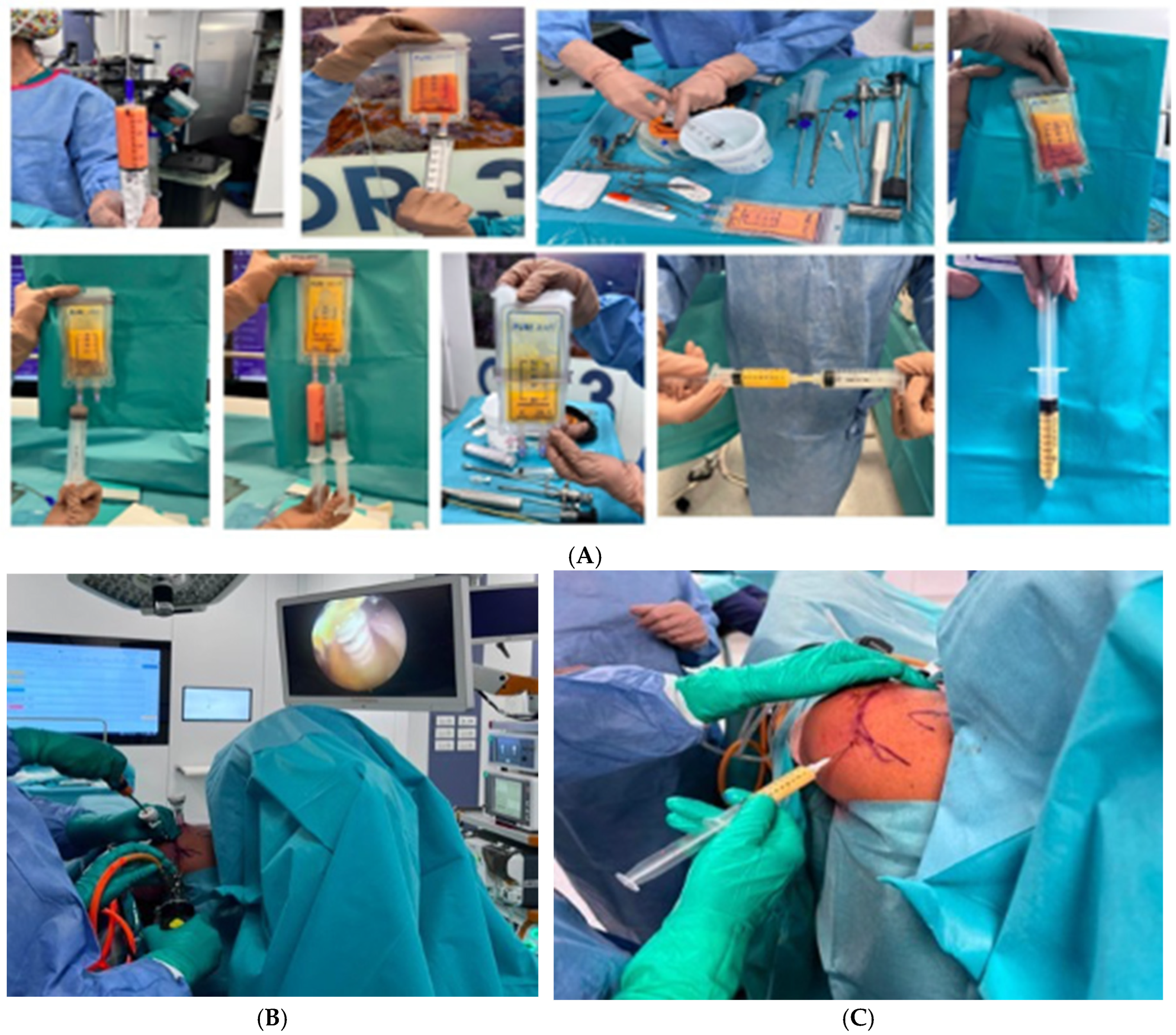
Patients were placed in the supine position in a dedicated procedure room. After local anesthesia, a small infra-umbilical abdominal incision was made to access the subcutaneous adipose tissue. A tumescent solution containing 500 mL of saline and 1 mL of epinephrine diluted 1:1000 was then infiltrated percutaneously into the abdominal fat. Local anesthetics were avoided because of their reported cytotoxic effects on human mesenchymal stem cells [16]. After a waiting period of 9 min, approximately 60 mL of adipose tissue was manually harvested. The lipoaspirate was subsequently processed with the PureGraft system (Cytori Therapeutics, San Diego, CA, USA) according to the manufacturer’s instructions and then mechanically agitated to obtain tissue fragmentation [Figure 1A].
Rotator cuff repair was carried out arthroscopically using either a single-row or double-row configuration with Arthrex Knotless SwiveLock® anchors (Arthrex, Inc., Naples, FL, USA) [Figure 1B]. At the completion of the repair, the processed adipose tissue was injected into the glenohumeral joint through a posterior approach using a 22 G needle [Figure 1C]. Gentle passive shoulder movements were performed immediately after injection. Postoperatively, patients followed the standard RCR rehabilitation protocol, including sling immobilization for 4 weeks, followed by progressive shoulder mobilization and strengthening exercises for at least 2 weeks. Return to light daily activities and sport-specific movements was allowed gradually according to patient tolerance.

2.4. Preoperative Evaluation and Follow-Up

Preoperative data were obtained from the patients’ medical records and included sex, age, body mass index (BMI), American Society of Anesthesiologists (ASA) classification, operated side, and history of previous arthroscopic procedures. Plain radiographs were reviewed to evaluate chondropathy, while magnetic resonance imaging was used to confirm the diagnosis of rotator cuff tear. Clinical outcomes were assessed using validated scoring tools. Shoulder function was evaluated with the Constant–Murley Score (CMS) before surgery and during postoperative follow-up [17]. Pain intensity was recorded preoperatively and postoperatively using the visual analog scale (VAS) [18].
When available, further clinical, radiological, and surgical information was extracted from the retrospective records. These variables included hand dominance, smoking status, diabetes, preoperative activity level, adherence to rehabilitation, tear size, involved tendons, fatty infiltration according to the Goutallier classification, muscle atrophy, long head of the biceps involvement, Outerbridge grade of chondral damage, repair configuration, and the number and type of anchors implanted. Owing to the retrospective nature of the study, not all variables were consistently documented for every patient; therefore, missing data were reported where appropriate.

2.5. Statistical Analysis

Data distribution was assessed using the Shapiro–Wilk test. Continuous variables are reported as mean ± standard deviation or median and interquartile range according to distribution. Categorical variables are reported as frequencies and percentages. Between-group comparisons for continuous variables were performed using the independent-samples t-test or Mann–Whitney U test, as appropriate. Categorical variables were compared using the chi-square test or Fisher’s exact test, as appropriate. Longitudinal changes in CMS and VAS scores were assessed using repeated-measures ANOVA, including time, group, and the group × time interaction. Post hoc comparisons were performed between groups at each timepoint when appropriate. Statistical significance was set at p < 0.05. p-values lower than 0.001 were reported as p < 0.001. No a priori sample size calculation or power analysis was performed because of the retrospective and preliminary nature of the study. The sample size was determined by the number of eligible patients treated during the study period.

3. Results

A total of 61 patients were included in the study, with 30 assigned to Group A and 31 to Group B. Baseline demographic and anthropometric characteristics, including age, BMI, and height, were compared between the two groups to assess their comparability. The mean age in Group A was 53.49 ± 7.20 years, while in Group B it was 54.04 ± 6.34 years. The difference in mean age between the groups was not statistically significant (p = 0.753). Similarly, the mean BMI in Group A was 26.64 ± 2.79, compared to 26.16 ± 3.50 in Group B, with no significant difference observed between the groups (p = 0.556) [Table 1]. In terms of height, Group A had a mean height of 170.13 ± 9.92 cm, while Group B had a mean height of 173.88 ± 8.86 cm. Although Group B had a slightly higher mean height, the difference was not statistically significant (p = 0.124) [Table 1]. These results indicate that there were no significant differences between the two groups in terms of age, BMI, or height, confirming that the groups were comparable at baseline.
Because of the retrospective nature of the study, several clinical and imaging variables requested by the reviewer, including dominant arm, smoking status, diabetes, activity level, rehabilitation adherence, detailed tear size, tendon involvement, fatty infiltration, muscle atrophy, biceps pathology distribution, and exact number of anchors, were not consistently available for all patients and therefore could not be included in the baseline comparison table. This limitation has been acknowledged in the Discussion.
The p-value for the comparison of baseline CMS values between Group A (53.39 ± 17.78) and Group B (48.22 ± 14.44) is 0.216. The difference between the two groups at baseline is not statistically significant. This indicates that no statistically significant baseline difference in CMS was detected between the two groups [Table 2]. Repeated-measures analysis was used to assess the group × time interaction for CMS.
The p-value for the comparison of baseline VAS scores between Group A (49.17 ± 21.35) and Group B (53.15 ± 15.88) is 0.411. This value indicates that there is no statistically significant difference between the two groups at baseline in terms of pain scores [Table 3]. Repeated-measures analysis was used to assess the group × time interaction for VAS.
At 3 months, Group B demonstrated significantly lower pain scores (p < 0.001) compared to Group A (VAS 25.14 ± 13.57 in group A vs. 13.09 ± 8.34 in group B), and this finding suggests an association between AdMSC use and lower postoperative pain scores; however, causality cannot be inferred because of the retrospective, non-randomized study design [Table 3].
Afterward, at 3 months, Group B exhibited higher functional scores; the Constant–Murley Score was 63.01 ± 24.33 in group A vs. 70.55 ± 23.46 in group B but the p-value for the comparison of CMSs at 3 months between the groups is 0.222. The difference at 3 months is not statistically significant. At 6 months the p-value for the comparison of CMSs between Group A (65.66 ± 11.58) and Group B (83.29 ± 18.98) is < 0.001 [Table 2]. The p-value for the comparison of VAS scores at 6 months between Group A (23.74 ± 15.72) and Group B (5.31 ± 4.38) is < 0.001.
At 12 months, Group B maintained significantly higher CMS values compared to Group A (85.55 ± 13.12 vs. 66.36 ± 9.38). Similarly, pain scores remained significantly lower in Group B (VAS 4.45 ± 5.67) compared to Group A (18.34 ± 12.65). These findings confirm the persistence of the clinical advantage observed at earlier follow-up timepoints. [Table 3].

4. Discussion

The main finding of this preliminary retrospective, non-randomized comparative cohort study was that patients treated with arthroscopic rotator cuff repair combined with intra-articular AdMSC injection showed greater improvement in pain and shoulder function during the 12-month follow-up compared with patients treated with rotator cuff repair alone. Because treatment allocation was not randomized, these findings should be interpreted as associations rather than evidence of a causal treatment effect.
Our results suggest that the combination of RCR with AdMSC use was associated with improved clinical outcomes, particularly in terms of pain reduction, as demonstrated by significantly lower visual analog scale (VAS) scores in Group B (RCR + AdMSCs) compared to Group A (RCR alone) at both 3 and 6 months. The observed clinical trend suggests that patients receiving the adipose-derived product experienced earlier pain reduction and greater functional improvement during follow-up. However, given the retrospective and non-randomized design, these findings should be interpreted as associations rather than evidence of a causal treatment effect. The persistence of superior CMS and VAS outcomes at 12 months suggests that the observed differences persisted during follow-up, although the underlying mechanisms remain uncertain. The maintenance of functional improvement in Group B, together with the plateau observed in Group A, suggests that the biological augmentation may reflect clinical improvement; however, structural healing was not assessed.
These findings align with previous research highlighting the regenerative and anti- inflammatory properties of AdMSCs in treating joint degeneration and soft tissue injuries [10,11,12,13]. The AdMSCs’ immunomodulatory effects and ability to promote tissue repair may have played a significant role in the improved pain outcomes in this group. Recent clinical evidence has reinforced the role of adipose-derived orthobiologics in shoulder disorders, particularly in glenohumeral osteoarthritis, where micro-fragmented adipose tissue has shown sustained improvements in pain and function up to 24–36 months, supporting a durable biological effect beyond short-term symptomatic relief [19]. Similarly, emerging data from biologic augmentation strategies in rotator cuff repair indicate that mesenchymal stromal cell–based therapies may enhance tendon healing quality and potentially reduce structural failure rates, although heterogeneity in preparation protocols and delivery techniques remains a major limitation in the current literature [20,21].
The Constant-Murley Score (CMS), a key measure of shoulder function, also reflected the benefits of AdMSC treatment, particularly at the 12-month follow-up. While CMSs at baseline and 3 months did not show significant differences between the groups (p-values of 0.216 and 0.222, respectively), the scores at 6 months were significantly higher in Group B (83.29 ± 18.98) compared to Group A (65.66 ± 11.58), with a p < 0.001. This suggests that the benefits of AdMSCs become more evident over time, with patients in Group B experiencing better functional recovery compared to those in Group A. These findings are consistent with the known regenerative potential of AdMSCs, which, due to their high proliferation rate, multipotency, and ease of collection from adipose tissue, have become an attractive option for enhancing tendon healing [11,12,13,21].
The mechanisms through which AdMSCs exert their effects are varied and complex. Their paracrine activity, which includes the secretion of growth factors and cytokines, supports tissue healing by reducing inflammation, promoting angiogenesis, and inhibiting apoptosis [12,13,21]. These biological processes may explain the enhanced functional outcomes and pain relief observed in the patients treated with AdMSCs. Recent translational research has shifted this paradigm toward a predominantly paracrine mechanism of action. Extracellular vesicles derived from adipose MSCs appear to regulate macrophage polarization, reduce synovial inflammation, stimulate angiogenesis, and promote extracellular matrix remodeling at the tendon–bone interface [22,23]. This “medicinal signaling cell” model provides a plausible explanation for the progressive functional improvement observed at 12 months in our cohort, suggesting that clinical gains may be driven more by biological modulation of the joint environment than by direct structural cell replacement.
However, it is important to acknowledge that not all patients benefited equally from the combination of RCR and AdMSCs. Recent shoulder-focused investigations have reported superior outcomes following MSC-based injections in patients with early to moderate degenerative changes compared to those with advanced osteoarthritis, indicating that severe structural damage may exceed the reparative threshold of biologic therapies [19,24]. This underscores the importance of patient selection in future clinical applications of AdMSCs, as those with less severe joint degeneration may derive the most benefit from this treatment.
One of the limitations of this study is the relatively short follow-up period of 12 months. While the early improvements in pain and function are encouraging, longer follow-up periods are necessary to determine whether the observed benefits are sustained over time. Future research should focus on extending the follow-up to 12 and 24 months to evaluate the long-term efficacy of AdMSC treatment. Additionally, the use of magnetic resonance imaging (MRI) in future studies will provide a more detailed assessment of the structural integrity of the repaired rotator cuff and help verify whether the functional improvements observed correlate with better tendon healing. Third, although a standardized processing system (PureGraft) was used, no quantitative cellular characterization or dose standardization was performed. Recent consensus statements and systematic reviews have emphasized that lack of standardization in cell processing, quantification, and reporting represents one of the main barriers to reproducibility in orthobiologic research [21,25]. Variability in cellular concentration, viability, and biological potency may partially explain inconsistencies observed across published studies and should be addressed in future investigations. Another limitation is the retrospective nature of the study. Although the two groups were comparable at baseline, the retrospective and non-randomized design of the study is a limitation and may have introduced selection bias and confounding factors. Furthermore, no a priori power analysis was performed; therefore, the study may be underpowered for some secondary outcomes. Another important limitation is that the injected adipose-derived product was not formally characterized. No assessment of cellular phenotype, viability, surface marker expression, or stem cell content was performed. Therefore, the injected material should be interpreted as a minimally manipulated adipose-derived product rather than a fully characterized mesenchymal stem cell preparation. Moreover, while the current study focused on pain and functional outcomes, future studies should also assess patient-reported outcome measures (PROMs), structured rehabilitation pathways, and remote postoperative monitoring strategies to capture the subjective experience of patients, adherence to rehabilitation, and overall satisfaction with treatment [26,27].

5. Conclusions

AdMSC use as an adjunct to arthroscopic rotator cuff repair was associated with better pain and functional outcomes in this preliminary retrospective, non-randomized comparative cohort. However, because of the retrospective design, absence of randomization, limited sample size, and lack of imaging follow-up, causal conclusions cannot be drawn. Randomized controlled studies with MRI-based assessment of tendon integrity and cartilage status are needed to confirm these preliminary findings.

Author Contributions

Conceptualization, G.B. and V.C.; methodology, R.T. and G.B.; validation, V.C., D.M., V.D.S. and F.Q.; formal analysis, R.T.; investigation, G.B., D.M., C.B., A.E.M. and G.C.; resources, V.D.S.; data curation, C.B., G.M. and A.E.M.; writing—original draft preparation, G.B. and R.T.; writing—review and editing, V.C. and V.D.S.; visualization, F.Q. and G.M.; supervision, V.C. and V.D.S.; project administration, V.C. All authors have read and agreed to the published version of the manuscript.

Funding

This research received no external funding.

Institutional Review Board Statement

According to institutional policy, formal Ethics Committee approval was not required for this retrospective observational study. The Health Directorate of Mater Olbia authorized the use of anonymized clinical data for research purposes. The study was conducted in accordance with the Declaration of Helsinki, and informed consent for treatment and data use was obtained from all patients.

Informed Consent Statement

Informed consent was obtained from all the subjects involved in this study.

Data Availability Statement

The raw data supporting the conclusions of this article will be made available by the authors on request.

Acknowledgments

We are grateful to the Support Center for Medical Research and Education at Mater Olbia Hospital for clinical and radiological technical support.

Conflicts of Interest

None of the authors has declared any conflicts of interest regarding this manuscript.

Abbreviations

The following abbreviations are used in this manuscript:
AdMSCsAdipose-derived Mesenchymal Stem Cells
ASAAmerican Society of Anesthesiologists
BMIBody Mass Index
CMSConstant-Murley Score
ERExternal Rotation
IRInternal Rotation
MRIMagnetic Resonance Imaging
OAOsteoarthritis
PROMsPatient-Reported Outcome Measures
RCRotator Cuff
RCRRotator Cuff Repair
ROMRange of Motion
VASVisual Analog Scale

References

  1. Glyn-Jones, S.; Palmer, A.J.; Agricola, R.; Price, A.J.; Vincent, T.L.; Weinans, H.; Carr, A.J. Osteoarthritis. Lancet 2015, 386, 376–387. [Google Scholar] [CrossRef] [PubMed]
  2. Li, G.; Yin, J.; Gao, J.; Cheng, T.S.; Pavlos, N.J.; Zhang, C.; Zheng, M.H. Subchondral bone in osteoarthritis: Insight into risk factors and microstructural changes. Arthritis Res. Ther. 2013, 15, 223. [Google Scholar] [CrossRef] [PubMed]
  3. Saltzman, B.M.; Leroux, T.S.; Verma, N.N.; Romeo, A.A. Glenohumeral Osteoarthritis in the Young Patient. J. Am. Acad. Orthop. Surg. 2018, 26, e361–e370. [Google Scholar] [CrossRef] [PubMed]
  4. Ansok, C.B.; Muh, S.J. Optimal management of glenohumeral osteoarthritis. Orthop. Res. Rev. 2018, 10, 9–18. [Google Scholar] [CrossRef] [PubMed] [PubMed Central]
  5. Francis, S.L.; Duchi, S.; Onofrillo, C.; Di Bella, C.; Choong, P.F.M. Adipose-Derived Mesenchymal Stem Cells in the Use of Cartilage Tissue Engineering: The Need for a Rapid Isolation Procedure. Stem Cells Int. 2018, 2018, 8947548. [Google Scholar] [CrossRef] [PubMed]
  6. De Francesco, F.; Gravina, P.; Busato, A.; Farinelli, L.; Soranzo, C.; Vidal, L.; Zingaretti, N.; Zavan, B.; Sbarbati, A.; Riccio, M.; et al. Stem Cells in Autologous Microfragmented Adipose Tissue: Current Perspectives in Osteoarthritis Disease. Int. J. Mol. Sci. 2021, 22, 10197. [Google Scholar] [CrossRef] [PubMed] [PubMed Central]
  7. Kim, K.I.; Kim, M.S.; Kim, J.H. Intra-articular Injection of Autologous Adipose-Derived Stem Cells or Stromal Vascular Fractions: Are They Effective for Patients With Knee Osteoarthritis? A Systematic Review With Meta-analysis of Randomized Controlled Trials. Am. J. Sports Med. 2023, 51, 837–848. [Google Scholar] [CrossRef] [PubMed]
  8. Hagmeijer, M.H.; Korpershoek, J.V.; Crispim, J.F.; Chen, L.T.; Jonkheijm, P.; Krych, A.J.; Saris, D.B.F.; Vonk, L.A. The regenerative effect of different growth factors and platelet lysate on meniscus cells and mesenchymal stromal cells and proof of concept with a functionalized meniscus implant. J. Tissue Eng. Regen. Med. 2021, 15, 648–659. [Google Scholar] [CrossRef] [PubMed]
  9. Garza, J.R.; Campbell, R.E.; Tjoumakaris, F.P.; Freedman, K.B.; Miller, L.S.; Santa Maria, D.; Tucker, B.S. Clinical Efficacy of Intra-articular Mesenchymal Stromal Cells for the Treatment of Knee Osteoarthritis: A Double-Blinded Prospective Randomized Controlled Clinical Trial. Am. J. Sports Med. 2020, 48, 588–598. [Google Scholar] [CrossRef] [PubMed]
  10. Xu, Y.; Zhang, W.X.; Wang, L.N.; Ming, Y.Q.; Li, Y.L.; Ni, G.X. Stem cell therapies in tendon-bone healing. World J. Stem Cells 2021, 13, 753–775. [Google Scholar] [CrossRef] [PubMed]
  11. Kunze, K.N.; Burnett, R.A.; Wright-Chisem, J.; Frank, R.M.; Chahla, J. Adipose-Derived Mesenchymal Stem Cell Treatments and Available Formulations. Curr. Rev. Musculoskelet. Med. 2020, 13, 264–280. [Google Scholar] [CrossRef] [PubMed] [PubMed Central]
  12. Veronesi, F.; Salamanna, F.; Tschon, M.; Maglio, M.; Nicoli Aldini, N.; Fini, M. Mesenchymal stem cells for tendon healing: What is on the horizon? J. Tissue Eng. Regen. Med. 2017, 11, 3202–3219. [Google Scholar] [CrossRef] [PubMed]
  13. Natali, S.; Screpis, D.; Patania, E.; De Berardinis, L.; Benoni, A.; Piovan, G.; Iacono, V.; Magnan, B.; Gigante, A.P.; Zorzi, C. Efficacy and Long-Term Outcomes of Intra-Articular Autologous Micro-Fragmented Adipose Tissue in Individuals with Glenohumeral Osteoarthritis: A 36-Month Follow-Up Study. J. Pers. Med. 2023, 13, 1309. [Google Scholar] [CrossRef] [PubMed] [PubMed Central]
  14. Slattery, C.; Kweon, C.Y. Classifications in Brief: Outerbridge Classification of Chondral Lesions. Clin. Orthop. Relat. Res. 2018, 476, 2101–2104. [Google Scholar] [CrossRef] [PubMed]
  15. Schumaier, A.; Abboud, J.; Grawe, B.; Horneff, J.G.; Ged, C.; Romeo, A.; Keener, J.; Friedman, R.; Yian, E.; Muh, S.; et al. Evaluating Glenohumeral Osteoarthritis: The Relative Impact of Patient Age, Activity Level, Symptoms, and Kellgren- Lawrence Grade on Treatment. Arch. Bone Jt. Surg. 2019, 7, 151–160. [Google Scholar] [PubMed] [PubMed Central]
  16. Rahnama, R.; Wang, M.; Dang, A.C.; Kim, H.T.; Kuo, A.C. Cytotoxicity of local anesthetics on human mesenchymal stem cells. J. Bone Jt. Surg. Am. 2013, 95, 132–137. [Google Scholar] [CrossRef] [PubMed]
  17. Constant, C.R.; Murley, A.H. A clinical method of functional assessment of the shoulder. Clin. Orthop. Relat. Res. 1987, 214, 160–164. [Google Scholar] [CrossRef] [PubMed]
  18. Langley, G.B.; Sheppeard, H. The visual analogue scale: Its use in pain measurement. Rheumatol. Int. 1985, 5, 145–148. [Google Scholar] [CrossRef] [PubMed]
  19. Natali, S.; Screpis, D.; Farinelli, L.; Iacono, V.; Vacca, V.; Gigante, A.; Zorzi, C. The use of intra-articular injection of autologous micro-fragmented adipose tissue as pain treatment for ankle osteoarthritis: A prospective not randomized clinical study. Int. Orthop. 2021, 45, 2239–2244. [Google Scholar] [CrossRef] [PubMed]
  20. Jo, C.H.; Chai, J.W.; Jeong, E.C.; Oh, S.; Kim, P.S.; Yoon, J.Y.; Yoon, K.S. Intratendinous Injection of Autologous Adipose Tissue-Derived Mesenchymal Stem Cells for the Treatment of Rotator Cuff Disease: A First-In-Human Trial. Stem Cells 2018, 36, 1441–1450. [Google Scholar] [CrossRef] [PubMed]
  21. Milano, G.; Colosio, A.; Minotta Quebradas, M.J.; Pratobevera, A.; Daffara, V.; Saccomanno, M.F. Biologic augmentation of rotator cuff repair with microfragmented autologous subacromial bursal tissue enveloped in a patch of compressed autologous long head of biceps tendon tissue: The Bio-Ravioli technique. JSES Int. 2024, 8, 1010–1015. [Google Scholar] [CrossRef] [PubMed]
  22. Chen, S.H.; Chen, Z.Y.; Lin, Y.H.; Chen, S.H.; Chou, P.Y.; Kao, H.K.; Lin, F.H. Extracellular Vesicles of Adipose-Derived Stem Cells Promote the Healing of Traumatized Achilles Tendons. Int. J. Mol. Sci. 2021, 22, 12373. [Google Scholar] [CrossRef] [PubMed] [PubMed Central]
  23. Aziziyan, F.; Asl, S.S.; Mahdipour, M.; Fard, R.N.; Sheykhhasan, M. Mesenchymal stem cell-derived extracellular vesicles in musculoskeletal regeneration: Mechanisms, applications, and future prospects. Stem Cell Res. Ther. 2026, 17, 66. [Google Scholar] [CrossRef] [PubMed] [PubMed Central]
  24. Centeno, C.J.; Al-Sayegh, H.; Bashir, J.; Goodyear, S.; Freeman, M.D. A prospective multi-site registry study of a specific protocol of autologous bone marrow concentrate for the treatment of shoulder rotator cuff tears and osteoarthritis. J. Pain Res. 2015, 8, 269–276. [Google Scholar] [CrossRef] [PubMed]
  25. LaPrade, R.F.; Dragoo, J.L.; Koh, J.L.; Murray, I.R.; Geeslin, A.G.; Chu, C.R. AAOS Research Symposium Updates and Consensus: Biologic Treatment of Orthopaedic Injuries. J. Am. Acad. Orthop. Surg. 2016, 24, e62–e78. [Google Scholar] [CrossRef] [PubMed] [PubMed Central]
  26. Bocchino, G.; El Motassime, A.; Maccauro, G.; Vitiello, R.; Bonilla, G.; Andriollo, L.; Indelli, P.F. Remote patient monitoring: A giant step towards same-day arthroplasty. Minerva Orthop. 2025, 76, 36–42. [Google Scholar] [CrossRef]
  27. Comodo, R.M.; Grassa, D.; Motassime, A.E.; Bocchino, G.; Totti, R.; De Fazio, A.; Meschini, C.; Capece, G.; Maccauro, G.; Vitiello, R. Telerehabilitation in Hip and Knee Arthroplasty: A Narrative Review of Clinical Outcomes, Patient-Reported Measures, and Implementation Challenges. J. Funct. Morphol. Kinesiol. 2025, 10, 370. [Google Scholar] [CrossRef] [PubMed] [PubMed Central]
Figure 1. Surgical workflow. (A) Abdominal adipose tissue harvesting, (B) Arthroscopic rotator cuff repair, (C) Intra-articular AdMSC injection through the posterior approach.
Applsci 16 06243 g001
Table 1. Comparison of Baseline Characteristics between Group A and Group B.
VariableGroup A: RCR Alone (n = 30)Group B: RCR + AdMSCs (n = 31)p-Value
Age, years53.49 ± 7.2054.04 ± 6.340.753
BMI, kg/m226.64 ± 2.7926.16 ± 3.500.556
Height, cm170.13 ± 9.92173.88 ± 8.860.124
Baseline CMS53.39 ± 17.7848.22 ± 14.440.216
Baseline VAS49.17 ± 21.3553.15 ± 15.880.411
Full-thickness rotator cuff tear, n (%)30 (100%)31 (100%)
Repairable rotator cuff tear, n (%)30 (100%)31 (100%)
Concomitant chondropathy, n (%)30 (100%)31 (100%)
Single-row repair technique, n (%)20 (67%)24 (77%)
Double-row repair technique, n (%)10 (33%)7 (23%)
Table 2. CMS at T0, T1, T2 and T3. Values are reported as mean ± standard deviation. CMS: Constant–Murley Score, range 0–100 points, with higher scores indicating better shoulder function.
T0 (Baseline)
p 0.216
T1 (3 Months)
p 0.222
T2 (6 Months)
p < 0.001
T3 (12 Months)
p < 0.001
Group A53.39 ± 17.7863.01 ± 24.3365.66 ± 11.5866.36 ± 9.38
Group B48.22 ± 14.4470.55 ± 23.4683.29 ± 18.9885.55 ± 13.12
Table 3. VAS at T0, T1, T2 and T3. Values are reported as mean ± standard deviation. VAS: visual analog scale, range 0–100 mm, with higher scores indicating greater pain.
T0 (Baseline)
p 0.411
T1 (3 Months)
p < 0.001
T2 (6 Months)
p < 0.001
T3 (12 Months)
p < 0.001
Group A49.17 ± 21.3525.14 ± 13.5723.74 ± 15.7218.34 ± 12.65
Group B53.15 ± 15.8813.09 ± 8.345.31 ± 4.384.45 ± 5.67
Disclaimer/Publisher’s Note: The statements, opinions and data contained in all publications are solely those of the individual author(s) and contributor(s) and not of MDPI and/or the editor(s). MDPI and/or the editor(s) disclaim responsibility for any injury to people or property resulting from any ideas, methods, instructions or products referred to in the content.