Article

Far-Field Terahertz Spectroscopy of a Subwavelength Single Planar Meta-Atom

by
Surya Revanth Ayyagari
1,
Simonas Indrišiūnas
2,
Guillaume Ducournau
3,
Vytautas Janonis
1 and
Irmantas Kašalynas
1,*
1
Terahertz Photonics Laboratory, Center for Physical Sciences and Technology (FTMC), Saulėtekio 3, LT-10257 Vilnius, Lithuania
2
Laser Microfabrication Laboratory, Center for Physical Sciences and Technology (FTMC), Savanoriu Ave. 231, LT-02300 Vilnius, Lithuania
3
Institut d’Electronique, Microélectronique et Nanotechnologie, IEMN, CNRS, University Lille, UMR 8520, F-59000 Lille, France
*
Author to whom correspondence should be addressed.
Appl. Sci. 2026, 16(10), 4608; https://doi.org/10.3390/app16104608
Submission received: 7 April 2026 / Revised: 29 April 2026 / Accepted: 5 May 2026 / Published: 7 May 2026

Abstract

Accurate measurements of light–matter interactions at subwavelength scales are critical for advancing nanophotonic and quantum optical technologies. In this paper, we present the far-field terahertz (THz) spectroscopy of a single planar meta-atom of subwavelength dimensions embedded within a square or circular aperture on a thin free-standing metal film. The meta-atom, composed of concentric disk and ring structures interconnected by narrow bridges, was fabricated by a mask-less direct laser ablation (DLA) technique to exhibit a pronounced transmission peak near a resonance frequency of 0.35 THz. We propose a novel spectral analysis framework that accounts for aperture-to-beam area mismatch suppressing non-resonant background contributions originating from edge diffraction and aperture discontinuities which are commonly encountered in subwavelength geometries. This technical analysis yields transmission spectra with improved accuracy providing good agreement with finite-difference time-domain (FDTD) simulations. A foundation for precise optical characterization of a single subwavelength size resonator is demonstrated paving the way for applications in quantum sensing, meta-surface design, and low-dimensional optoelectronic systems.

Graphical Abstract

1. Introduction

Understanding and controlling light–matter interactions at the subwavelength scale is a cornerstone of modern nanophotonic devices [1]. These subwavelength interactions give rise to strongly enhanced electromagnetic (EM) fields, which require precise control for use in emerging photonic applications [2]. To achieve such control, researchers have turned to artificially engineered materials with properties that are not found in nature. Among these, metamaterials, which are three-dimensional (3D) artificially structured media composed of resonant elements, or meta-atoms, smaller than the wavelength of light, have emerged as critical tools to manipulate the properties of an EM wave. This way, physical phenomena such as negative refraction [3], perfect lensing [4,5], near-perfect absorption [6] and invisibility cloaking [7,8] can be achieved. There are also examples of two-dimensional (2D) artificially structured media which are known as meta-surfaces [9]. In simple words, these are a 2D version of metamaterials with negligible layer thickness with respect to the working wavelength in the propagation direction. Meta-surfaces are composed of an array of artificial meta-atoms, occupying much less physical space, which exhibit much lower insertion loss than the bulky 3D metamaterials, and provide superior abilities to manipulate wavefronts of transmitted and reflected EM waves [9,10,11]. Each meta-atom in a meta-surface is typically realized as a subwavelength resonator that interacts with incident electromagnetic waves through scattering, absorption, or redirection of light, depending on its geometry, arrangement, and material composition [12]. Individual meta-atoms are capable of confining electromagnetic fields to deep-subwavelength volumes, achieving extreme field enhancement. Such strong field confinement can, in principle, enable interaction with quantum emitters or nanoscale systems, which is relevant for emerging quantum and nanophotonic applications [13]. Understanding the response of a single meta-atom or an isolated complex resonator is essential for engineering macroscopic functionalities such as cloaking via anapole states, anomalous reflection, perfect absorption, and wavefront control [14,15]. The electromagnetic response of a single resonator arises from its intrinsic modes, such as electric and magnetic dipoles, quadrupoles, or higher-order multipoles [15,16], which are primarily governed by its structural geometry and configuration [9,15,17]. Moreover, the resonance behavior of a single meta-atom differs significantly from that of periodically arranged structures, or the features separated by subwavelength distances due to mutual coupling effects [18,19].
Recent advances in near-field terahertz spectroscopy have enabled the direct probing of individual meta-atoms, revealing strongly confined resonant modes and intrinsic spectral responses that are not accessible in conventional far-field measurements [20,21]. A recent review on the spectroscopy of THz resonators and meta-surfaces emphasizes that understanding the electrodynamics of subwavelength resonators requires access to evanescent fields, which remain beyond the reach of standard far-field approaches [22]. Beyond these considerations, the realization of far-field spectroscopy on an individual meta-atom represents both a technological milestone and a gateway to explore fundamental physical phenomena, such as extreme light confinement, radiative coupling, superradiance quenching, or vacuum-field fluctuations at the level of a single cavity [13].
Despite extensive progress in characterizing ensembles of meta-atoms, direct optical measurements of individual meta-atoms remain challenging. This difficulty arises from the mismatch between the subwavelength dimensions of the meta-atom and the wavelength of free-space light, resulting in inefficient excitation and detection [23]. As a result, most experimental studies have been performed on a large set of resonator arrays, which enhance the signal-to-noise ratio through collective effects, but obscure the intrinsic properties of individual elements [24,25]. While near-field approaches have addressed this limitation, they often require complex instrumentation and are restricted to localized probing, encouraging the development of alternative far-field methodologies capable of accessing single meta-atom intrinsic responses. More experimental research is needed despite previous far-field characterizations of single split-ring resonators. Gay-Balmaz et al. reported a Q-factor of ~383 at 1 GHz [19] while Rajabali, S. et al. reported a Q-factor of ~10.5 at 300 GHz using an asymmetric silicon immersion lens (aSIL) configuration, where a single resonator was sandwiched between optical elements incorporating silicon immersion lenses on their front and back sides [13].
In this work, we demonstrate the measurement technique of the far-field THz characteristics of a single planar meta-atom fabricated on a free-standing metal film, eliminating the need for external optical elements. The single planar meta-atom is composed of subwavelength size concentric disk- and ring-shaped elements interconnected by narrow bridges in the center of a square- or circular-shape aperture. The meta-atom was developed to exhibit a transparency at the resonant frequency of approximately 0.35 THz [15]. The samples were fabricated from thin stainless-steel film using a mask-less DLA technique, which allowed repeatability and precise dimensional control of the fabricated structures. We observed that a surrounding electromagnetic environment modulates the optical response of the meta-atom, resulting in a redshift of the resonance peak position by approximately 10 GHz, accompanied by an increase in the transmission amplitude by approximately 10% and the Q-factor by approximately 17% for the circular aperture configuration relative to the square aperture configuration. We introduced a novel spectral analysis framework that accounts for the mismatch between the incident beam area and the subwavelength geometries, enabling reliable extraction of the intrinsic response of an isolated meta-atom from far-field measurements. The experimental results with the performed spectra analysis were found to be in good agreement with finite-difference time-domain (FDTD) simulations. The developed spectral analysis framework opens new avenues for studying light–matter interactions with few or even single quantum emitters, paving the way for applications in quantum information processing, nanoscale photonic sensing, and the engineering of compact photonic systems.

2. Methods and Measurements

The performance of the meta-atom was evaluated using finite-difference time-domain (FDTD) simulations conducted in CST Studio software. These simulations were carried out under the normal incidence of a plane wave, with perfectly matched layer (PML) boundary conditions applied in all spatial directions to absorb transmitted and reflected waves. S-parameters were calculated under a steady-state energy assumption using a multi-frequency plane wave excitation.
Fabrication of the meta-atoms was carried out using DLA technology, which enabled high-precision patterning with excellent control over shape and dimensions. A femtosecond pulsed laser (Pharos, Light Conversion, Vilnius, Lithuania) was used, operating at a wavelength of 515 nm, with a 5 μm spot size, 300 fs pulse duration, 12 J/cm2 fluence, scan speed of 1 mm/s (corresponding to 5000 pulses/mm), and 7 repeated scans. A focused laser beam was used to selectively remove the metal, thereby defining the shape and size of the subwavelength components.
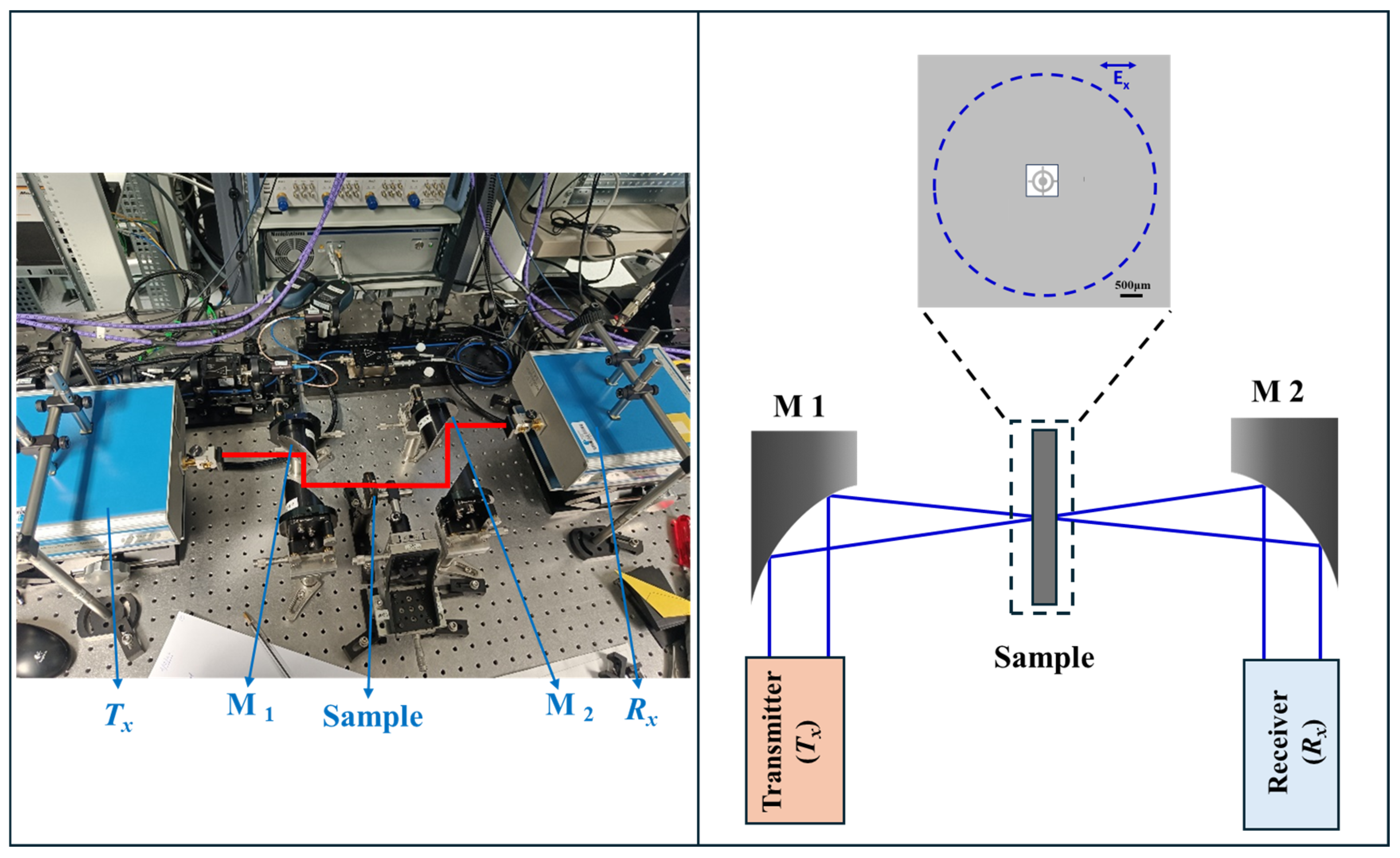
Transmission measurements were performed using a commercial Vector Network Analyzer (ZVA-24, Rohde & Schwarz, Munich, Germany) with two frequency extenders: one covering 220–325 GHz and the other 325–500 GHz. In each case, a hollow-core waveguide delivered the signal to free space via a horn antenna, generating a linearly polarized Gaussian beam along the X-axis. An actual experimental setup including the schematic is shown in Figure 1.
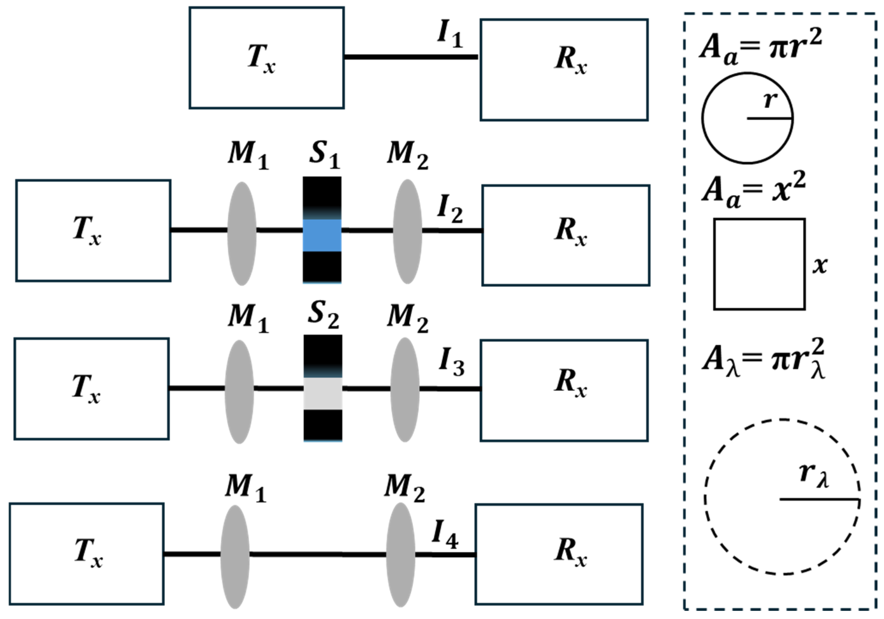
Prior to the measurements, full system calibration was performed using waveguide Thru/Reflect/Match (TRM) standards, between the transmitter (Tx) and receiver (Rx). The measured signal between the transmitter and receiver was taken as a reference signal (I1) by setting the maximum signal to 0 dB. Later, a THz beam was directed via free-space optics using off-axis parabolic mirrors (M1, M2), and the measurements were performed under three conditions: a focused THz beam passing through a sample (S1) meta-atom embedded in an aperture (I2), through a sample (S2) in an empty aperture (I3) with a surrounding metal frame, and without any sample (I4), as shown schematically in Figure 2. A schematic illustrating the areas of the circular and square apertures, along with the THz beam area, is shown in the dashed box in Figure 2 to provide an understanding of the correction factor (A) which will be discussed below.
To extract the intrinsic optical response of a subwavelength meta-atom from far-field measurements, it is essential to account for the mismatch between the spatial extent of the incident beam and the interaction region defined by the meta-atom. At the sample position, the incident terahertz beam has a Gaussian intensity profile with an effective beam area Aλ, which is significantly larger than the aperture area Aa. At the resonance frequency, the THz beam diameter (D) was 4.73 mm, which was about 5.5 times the wavelength size (D > 5.5 ∗ λ0). Therefore, the area of the THz beam at the resonance frequency is 53 times the area of the circular aperture (Aλ > 53 ∗ Aa) and 42 times the area of the square aperture (Aλ > 42 ∗ Aa). As a result, only a small fraction of the incident radiation interacts with the meta-atom, giving rise to the resonance associated with its intrinsic optical properties. The observed resonance arises from near-field scattering and strong field confinement at the meta-atom boundaries, which allows the incident radiation to excite electromagnetic modes such as dipoles and quadrupoles that then radiate into the far field. The remaining portion of the beam impinges on the surrounding metal film and is predominantly reflected. However, due to diffraction at the aperture edges and scattering from the metal boundaries, a fraction of this radiation can couple with the radiation of the meta-atom. As a result, the detected signal consists of a superposition of resonant scattering originating from the meta-atom boundaries, and non-resonant scattering (non-resonant background contributions) arising from the aperture edges and metal boundaries, which can lead to distortions in the extracted spectra.
All measurements were acquired on a logarithmic scale (dB), where all measured signals were normalized to the I1, resulting in the normalized spectra (transmittance) of the overall meta-atom sample Sm = lg(I2)/lg(I1), of the empty aperture sample Sa = lg(I3)/lg(I1), and of the open air without any sample Sair = lg(I4)/lg(I1).

2.1. Conventional Spectral Analysis (T1)

In the conventional approach, the measured signal of the meta-atom sample Sm is normalized by the signal obtained from an empty aperture Sa:
T 1 = S m S a = l g ( I 2 ) l g ( I 3 ) .
This approach assumes that the empty aperture is much larger than the wavelength of used radiation, therefore providing a suitable reference. However, when the aperture is comparable or smaller than the wavelength, it introduces diffraction and scattering effects that must be accounted for in far-field measurements, requiring a different approach.

2.2. Proposed Spectral Analysis (T2)

To overcome the listed limitations, we suggest a new approach for signal analysis:
T 2 = S m S a i r A
Here, A is a correction factor, accounting for the mismatch between the beam size and aperture size, and is defined as:
A = l g ( A a ) lg A λ
Unlike T1, this method does not rely on the aperture as a reference. Instead, it considers free-space transmission and applies a correction that accounts for the mismatch between the beam size and aperture size, allowing for the separation of the intrinsic response of the meta-atom from unwanted background contributions.
Indeed, an intrinsic response from the meta-atom extracted using Equation (2) demonstrated better agreement with numerical calculations, the results of which are discussed in the following sections.
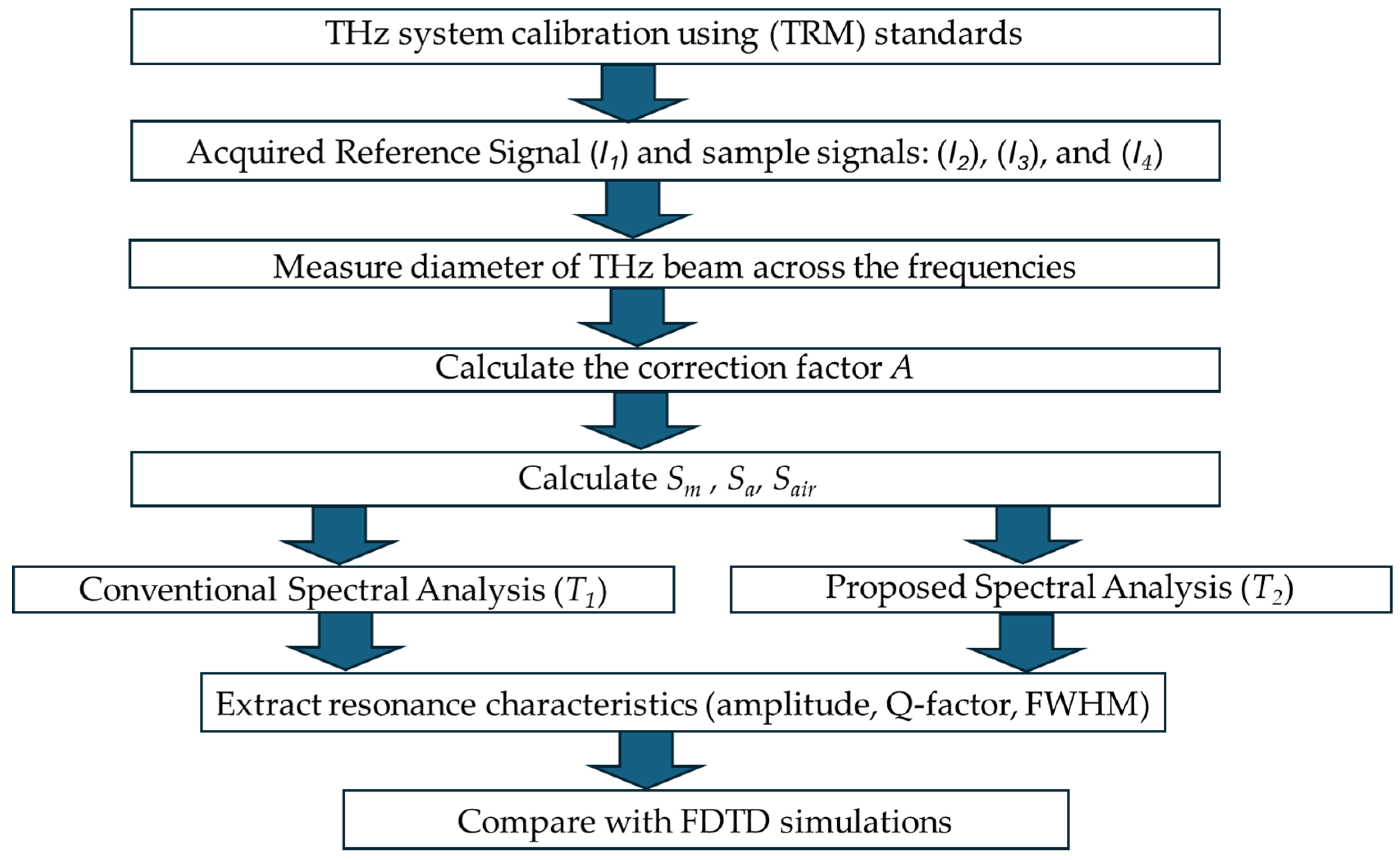
The overall measurement and spectral analysis procedure is summarized in Figure 3. The process begins with system calibration and the acquisition of reference and sample signals under different measurement configurations. These acquired signals are subsequently normalized to account for system response and background contributions. The analysis then proceeds through two parallel approaches: the conventional spectral analysis (T1) and the proposed spectral analysis (T2). While the T1 approach provides a basic normalization, the T2 framework incorporates a correction factor to suppress non-resonant background effects arising from beam–aperture mismatch. This enables a more accurate extraction of the intrinsic transmission characteristics of the meta-atom. Finally, key resonance parameters, including frequency, transmission amplitude, and Q-factor, are determined and compared with simulation results for validation.

3. Results

3.1. Single Planar Meta-Atom Sample

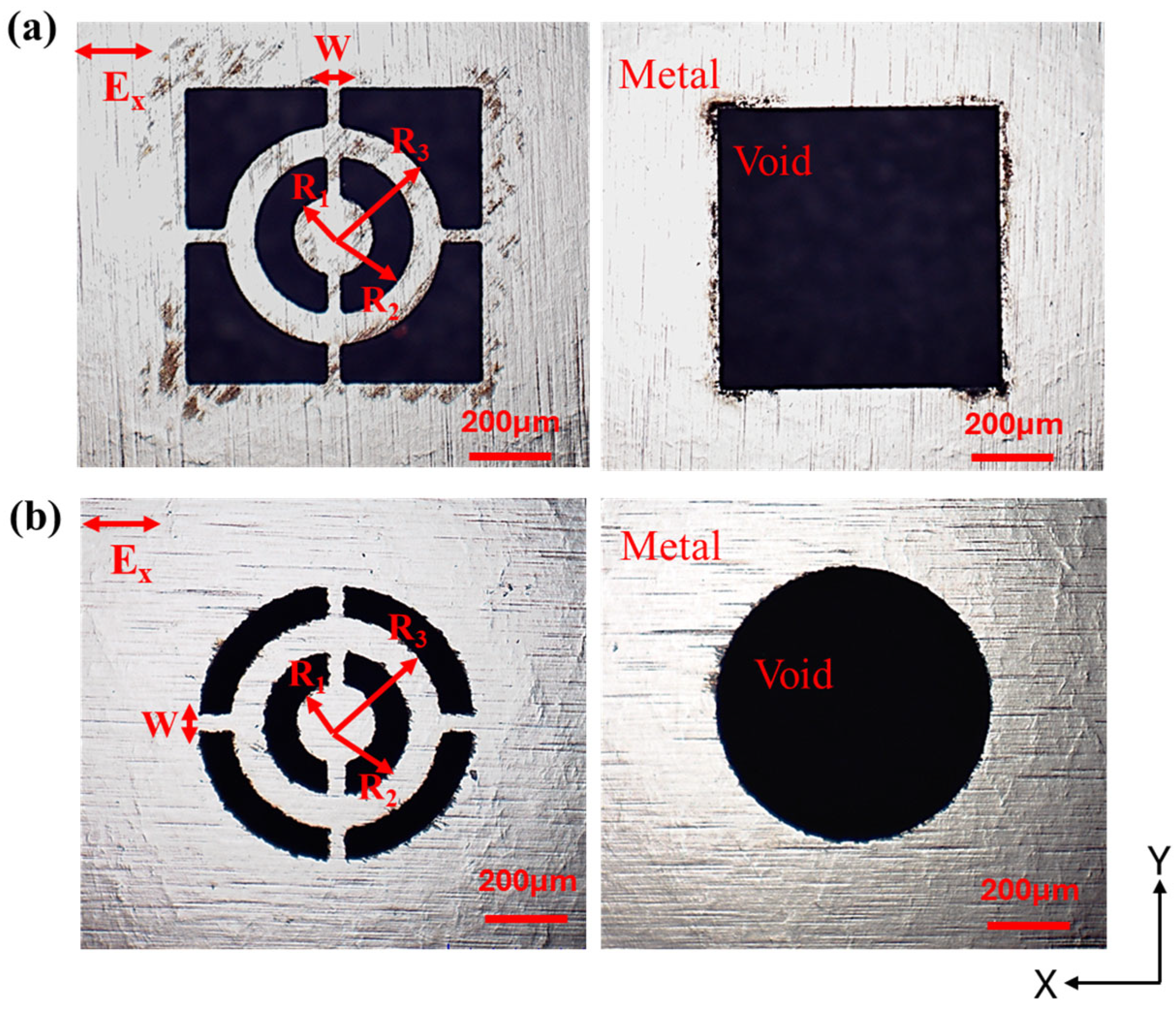
A stainless-steel film with a thickness of 50 μm was selected for fabricating concentric disk- and ring-shaped meta-atoms embedded within either square or circular apertures. The geometrical parameters of a single planar meta-atom were defined as follows: the disk had a radius R1 = 96 μm, while the inner and outer radii of the ring were R2 = 170 μm and R3 = 250 μm, respectively. Narrow metallic bridges with a width of approximately W = 42 μm connected the disk to the ring and further linked the meta-atom to the aperture [18]. The meta-atom was embedded within subwavelength apertures of two geometries: a square aperture with a side length of 650 μm and a circular aperture with a diameter of 650 μm, as illustrated in Figure 4. The meta-atom was designed to demonstrate a resonance at the frequency (f0) of about 0.35 THz or the wavelength (λ0) of 0.856 mm, with a wavelength nearly 1.32 times greater than the lateral size of the meta-atom in the aperture at the resonance frequency. More details on the sample fabrication are provided in the Section 2.

3.2. Measurements of Subwavelength Apertures

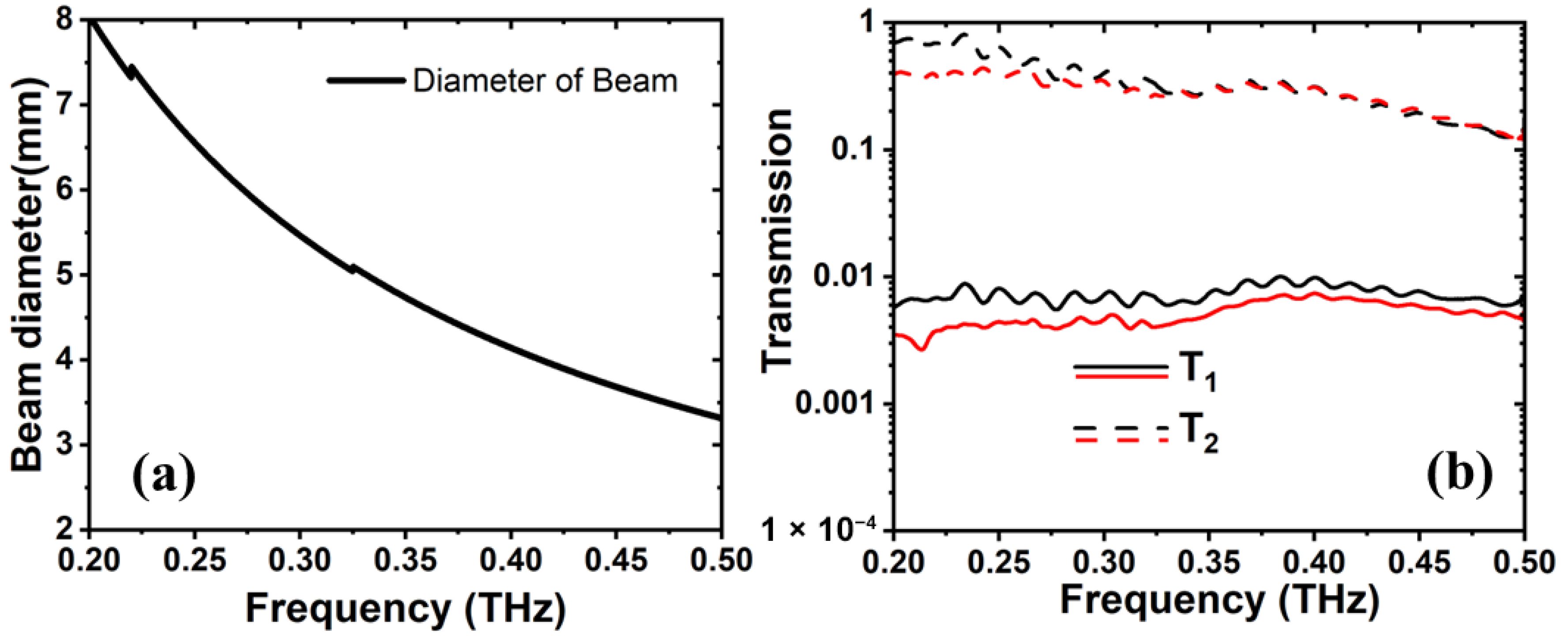
The transmission spectra of the empty subwavelength square and circular apertures were first investigated comparing both Equations (1) and (2). To evaluate the response of the apertures alone, we replaced Sm with Sa in both equations, and Sa with Sair in Equation (1). Figure 5a shows the frequency dependence of the THz beam diameter, which is used in the calculation of the variable A in Equation (2). The spectra of empty apertures were experimentally measured over the frequency range of 0.2 to 0.5 THz. The results are shown in Figure 5b. We compare the transmission spectra T1 (solid lines) with T2 (dashed lines) obtained using Equation (1) and Equation (2), respectively. The latter yields an enhancement in the spectra of up to two orders of magnitude relative to the former for both aperture geometries across the entire frequency range. The results show no distinct resonant features within the studied frequency range, although the aperture dimensions (~650 μm) are comparable to the wavelength (~0.45 THz). Instead, it behaves as a broadband transmission opening, due to the absence of strong field confinement preventing the formation of well-defined resonant modes. A noticeable difference in the spectra between the square and circular apertures was observed at the lower end of the frequency range, which may be attributed to the geometry of the circular aperture. The smooth, continuous boundary of the circular aperture lacking edges likely contributes less to the optical performance. However, at higher frequencies, beginning around 0.325 THz, the difference in the signal ratios between the two aperture types becomes negligible.

3.3. Measurements of Meta-Atom Embedded in Aperture

The optical response of a single meta-atom embedded within square or circular apertures, with the mentioned geometrical parameters at the beginning of the Section 3, was investigated. Transmission spectra of the fabricated samples were experimentally measured in the frequency range of 0.2 to 0.5 THz under normal incidence, with the electric field (E-field) polarized along the x-axis. In both aperture configurations, the meta-atom possessed a resonance centered near 0.35 THz.
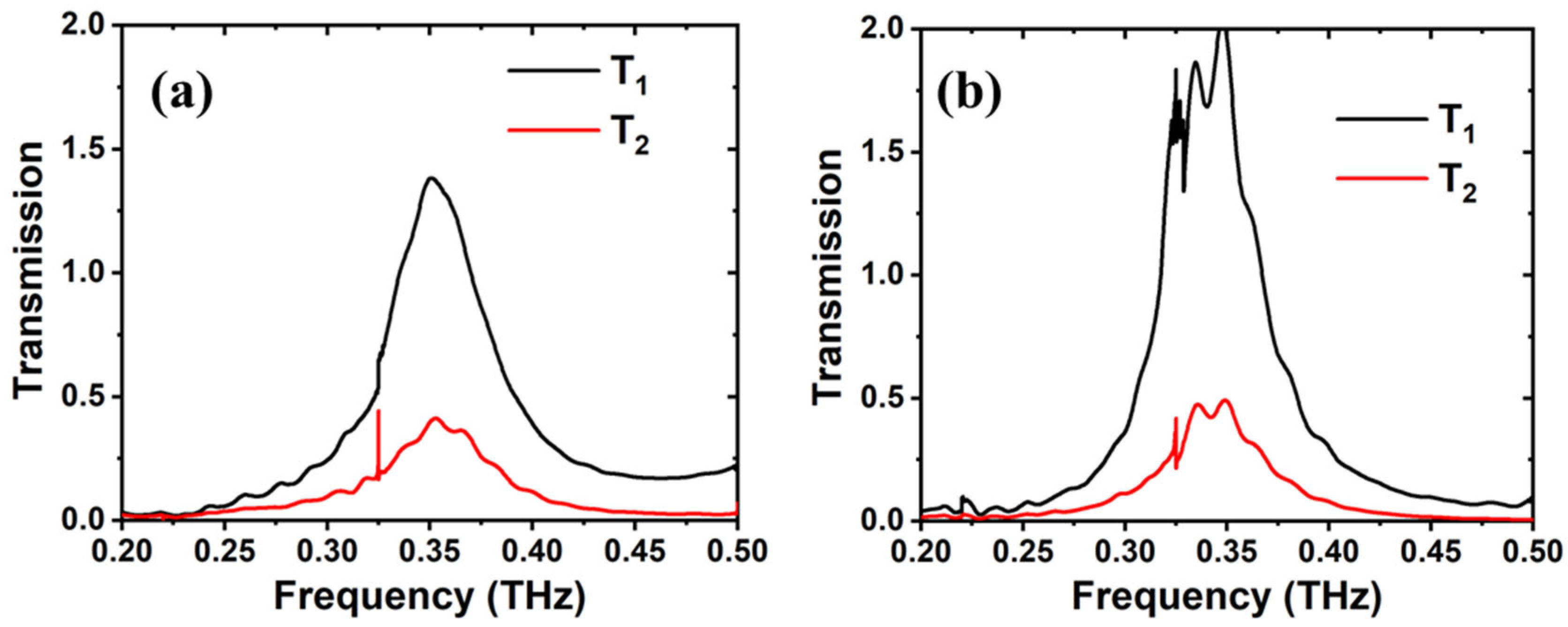
Figure 6a illustrates the transmission spectra corresponding to the square aperture configuration. A significant difference in resonance amplitude is observed between T1 (black curve, Equation (1)) and T2 (red curve, Equation (2)), notably the spectra obtained from T1 with a resonance peak amplitude exceeding unity (>100%). This elevated transmission arises from the combined contribution of resonant scattering and non-resonant scattering, rather than representing the intrinsic response of the meta-atom. In contrast, the spectra obtained from T2 yields a resonance peak amplitude of 0.35 (35%), effectively suppressing the non-resonant scattering effects and isolating the intrinsic optical response of the meta-atom. The resonance peak itself remains well-defined at 0.36 THz without any spectral distortion.
Similarly, Figure 6b shows the transmission spectra for the circular aperture configuration, where a similar difference in the resonance amplitude between T1 (black) and T2 (red) is observed. Here, the resonance peak exhibits a slight redshift to 0.35 THz compared to the square aperture configuration, with a corresponding resonance peak amplitude of 0.45 (45%) for T2. The enhanced signal in the circular aperture is likely due to a stronger electric field coupling between the meta-atom and the curved aperture boundary, which can be supported by the field distribution plots presented in Figure 7.

4. Discussion

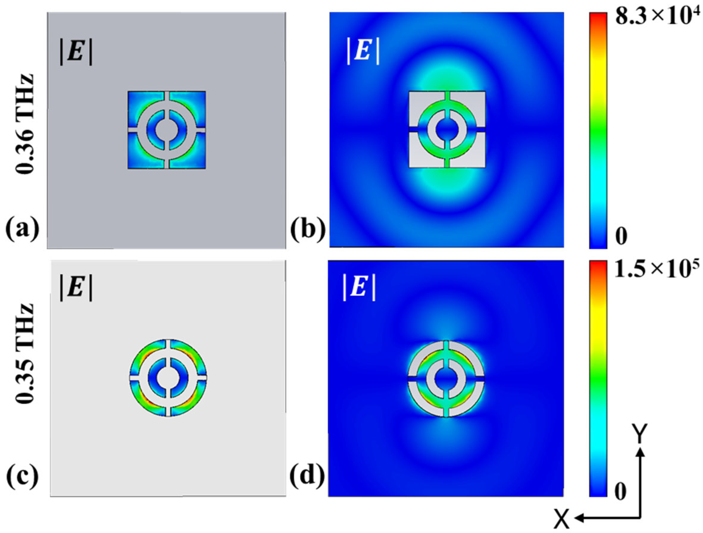
Figure 7 presents the simulated E-field distributions obtained using FDTD for a single meta-atom embedded within two aperture geometries (square and circular) for a given Ex polarization at their respective resonance frequencies of 0.36 THz and 0.35 THz. The field maps in Figure 7a,c, plotted in the xy-plane at z = 1 μm, clearly reveal strong localization of the E-field within the gaps of both the meta-atom and the surrounding aperture structures. Notably, the field enhancement reaches approximately 5 × 10⁴ V/m for the square aperture configuration and up to 1.5 × 10⁵ V/m for the circular aperture configuration, representing an increase of several orders of magnitude compared to the incident field amplitude of 1 V/m. This indicates the circular aperture configuration possess a significantly stronger field confinement, with nearly a threefold enhancement relative to the square aperture configuration. Furthermore, the surface field distributions shown in Figure 7b,d, reveal the strong localization of the E-field on the ring structure and connecting bridges of the meta-atom coupled to the aperture geometrics. This provides insight into the modal characteristics, suggesting an electric dipole-like response for the square aperture configuration and a higher-order electric quadrupole-like response for the circular aperture configuration, both aligned along the x-axis. These dominant electric multipole moments are directly correlated with the observed resonance shape, peak, and amplitude of a meta-atom [18]. E-field distributions for the square and circular aperture configurations highlight the influence of aperture geometry and the surrounding environment on the field enhancement and modal behavior of the meta-atom.
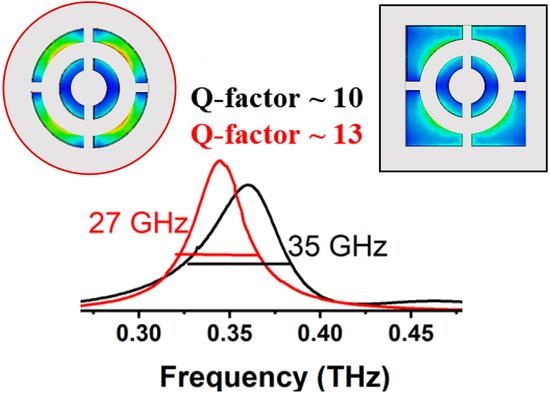
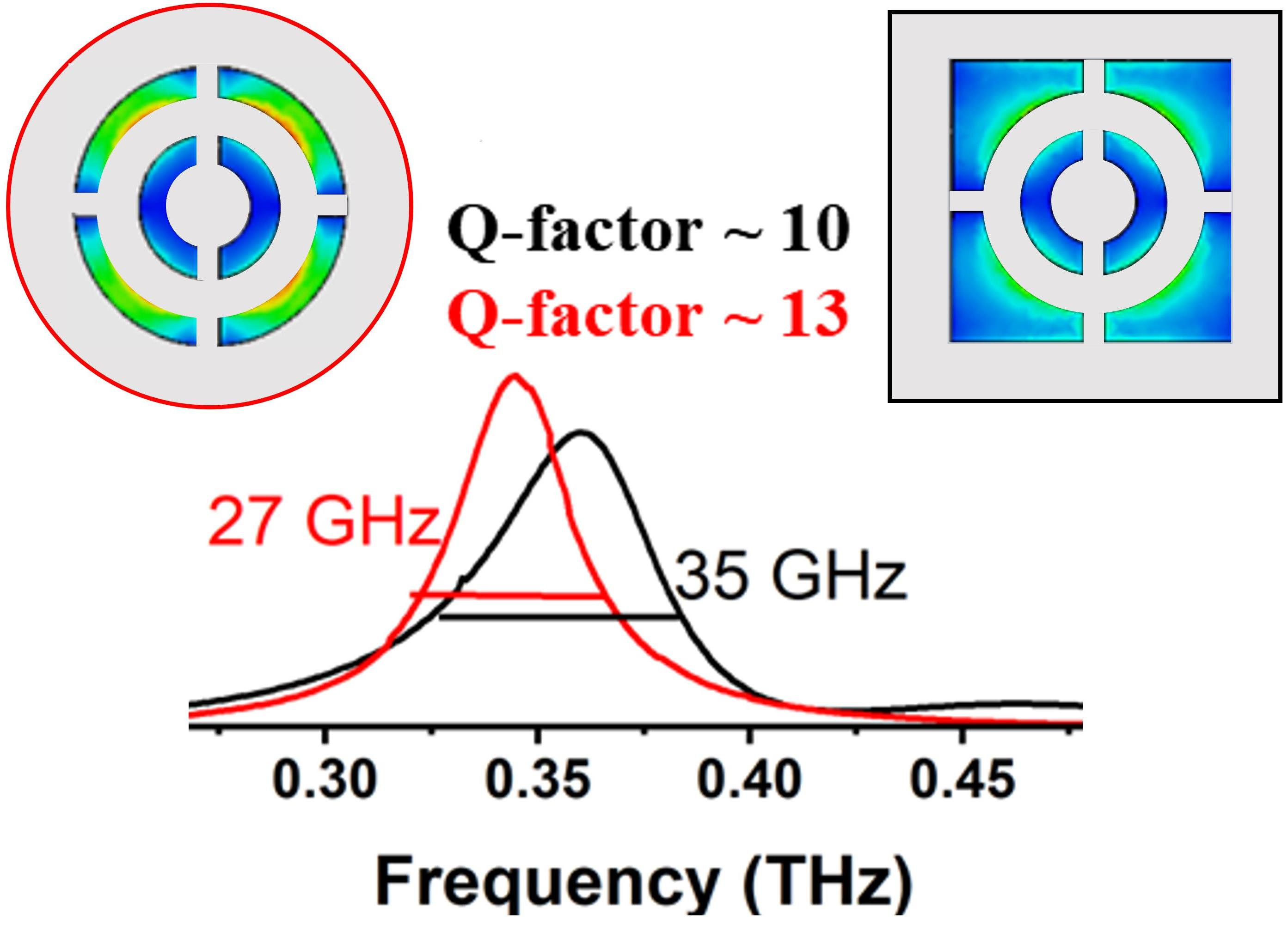
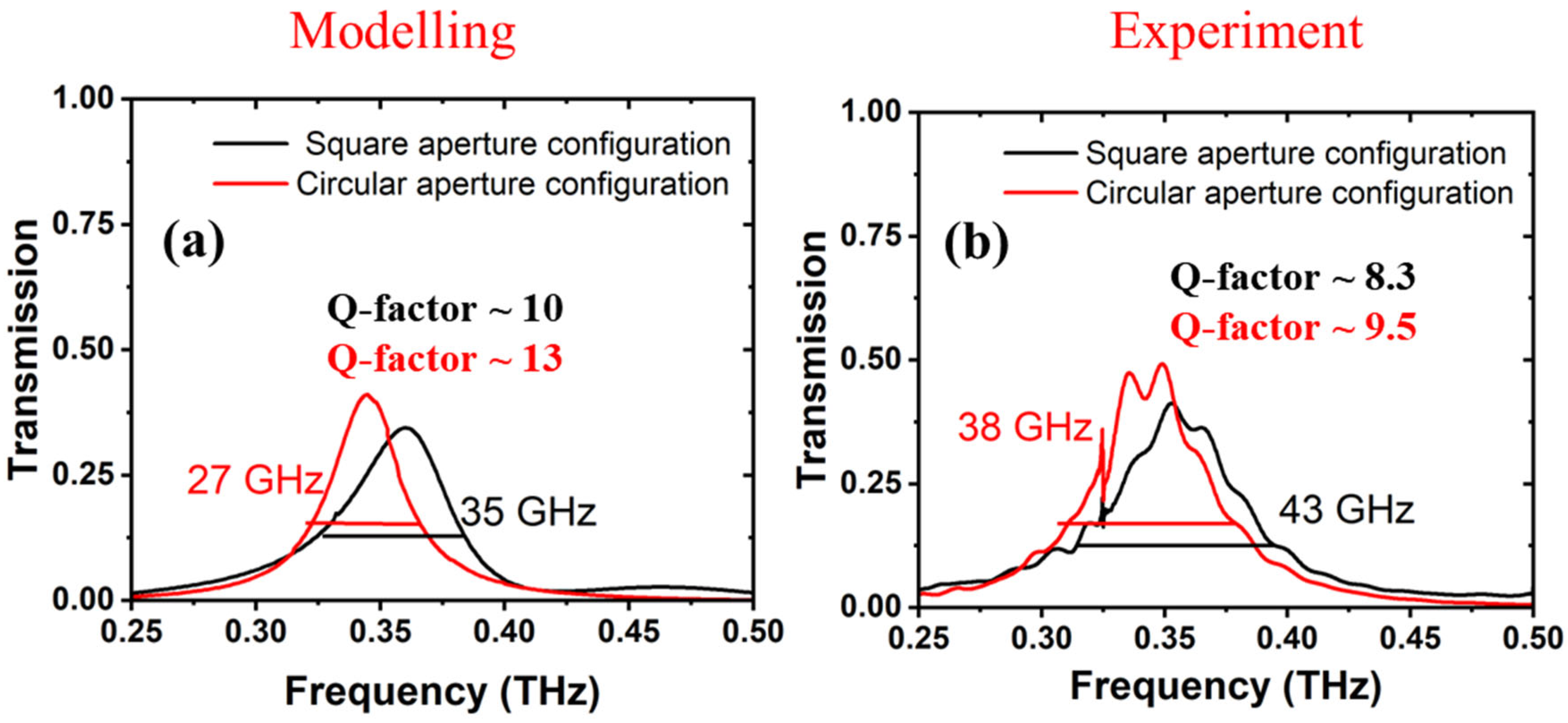
Figure 8a,b present the transmission spectra of the meta-atom with the square aperture configuration (black curves) and circular aperture configuration (red curves), for both simulated and experimental data, over the frequency range of 0.25 to 0.5 THz. For the simulations, the transmission spectra were obtained from FDTD simulations using CST Studio 2025 software, while the transmission spectra for the experimental measurements were processed using Equation (2), namely the T2 spectral analysis. The simulation and experiment results demonstrate excellent agreement in both resonance frequency and transmission amplitude, with minor discrepancies falling within the range of experimental uncertainties. The resonance peak of the circular aperture configuration exhibits a redshift of approximately 10 GHz and an enhancement in transmission amplitude of about 10% compared to the square aperture configuration in both simulations and experiments. This increase in the transmission amplitude can be attributed to a stronger E-field coupling between the meta-atom and the circular aperture boundary, due to the dominance of the electric quadrupole mode, as shown in Figure 7b. Additionally, the circular aperture configuration exhibits a reduction in bandwidth (measured at full width at half maximum (FWHM)) from 35\43 GHz to 27\38 GHz, and a corresponding increase in the quality factor (Q-factor) from 10.2\8.3 to 13.0\9.5 (in modeling\experiment respectively) compared to the square aperture configuration. The results reveal that the electromagnetic environment surrounding the meta-atom significantly modifies its resonance behavior. The observed Q-factor enhancement was up to 21% and 17% in theory and experiment, respectively. Thus, our far-field measurements validate that the T2 spectral analysis is a robust and accurate approach suitable for characterization of the optical properties of single resonator-based samples and devices, particularly when their dimensions fall within the subwavelength range.


5. Conclusions

To conclude, we have successfully characterized the optical response of a single planar meta-atom embedded within either a square or circular aperture developed on a free-standing metal film. We proposed a novel spectral analysis approach for subwavelength size structures, where non-resonant background contributions that typically restrict accurate far-field measurements were suppressed. Comparative analysis of the meta-atoms in the two aperture configurations revealed that a surrounding electromagnetic environment modulates the optical response, resulting in a redshift of the resonance peak position by approximately 10 GHz, accompanied by an increase in the transmission amplitude by approximately 10% and the Q-factor by approximately 17% for the circular aperture configuration relative to the square aperture configuration. These findings lay the groundwork for accurate characterization of individual meta-atoms and resonators, opening a pathway toward new applications in nanoscale quantum optics, photonic sensing, and meta-surface design.

Author Contributions

S.R.A. (Conceptualization: Equal; Data curation: Lead; Investigation: Equal; Methodology: Equal; Software: Lead; Writing—original draft: Equal; Writing—review and editing: Equal). S.I. (Methodology: Equal; Resources: Equal; Writing—review and editing: Equal). G.D. (Conceptualization: Equal; Data curation: Equal; Funding acquisition: Equal; Methodology: Equal; Resources: Equal; Writing—review and editing: Equal). V.J. (Data curation: Equal; Software: Supporting; Writing—review and editing: Equal). I.K. (Conceptualization: Equal; Data curation: Equal; Funding acquisition: Equal; Investigation: Equal; Methodology: Equal; Resources: Equal; Supervision: Lead; Writing—original draft: Equal; Writing—review and editing: Equal). All authors have read and agreed to the published version of the manuscript.

Funding

EU H2020 ITN-MCS project TERAOPTICS under the grant agreement No. 956857 and EU Next Generation Grant under Measure No. #10-038-T-0186. CPER WAVETECH @HdF project and Haut de-France Regional Council, IEMN UHD flagship and PCMP-CHOP and RF Net platforms. CDP C2EMPI, the French State under the France-2030 program, the University of Lille, the Initiative of Excellence of the University of Lille, the European Metropolis of Lille for their funding and support of the R-CDP-24-004-C2EMPI project. PEPR under ANR-22-PEEL-0006 and 22-PEFT-0006 grants, French RENATECH network and the Equipex + Nanofutur (Grant IA-21-ESRE-0012) as well as the EQUIPEX + ADD4P (Grant IA-21-ESRE-0007).

Data Availability Statement

Data is contained within the article.

Acknowledgments

Thise work was supported by the HORIZON-EIC-2025-PATHFINDER project THz-Skin, GA no. 101257203.The author S.R.A. acknowledge the received support of the Research Council of Lithuania (Lietuvos Mokslo Taryba) for a postdoctoral project under the grant agreement No. S-PD-25-44. and Jan Macutkevič for helpful discussions and for reviewing the manuscript in final stage.

Conflicts of Interest

The authors declare no conflicts of interest.

Abbreviations

The following abbreviations are used in this manuscript:
THzTerahertz
DLADirect laser ablation
FDTDFinite-difference time-domain
EMElectromagnetic
3DThree-dimensional
2DTwo-dimensional
PMLPerfectly matched layer
TRMThru/Reflect/Match
E-fieldElectric field
FWHMFull width at half maximum
Q-factorQuality factor
aSILAsymmetric silicon immersion lens

References

  1. González-Tudela, A.; Reiserer, A.; García-Ripoll, J.J.; García-Vidal, F.J. Light–Matter Interactions in Quantum Nanophotonic Devices. Nat. Rev. Phys. 2024, 6, 166–179. [Google Scholar] [CrossRef] [Scilit]
  2. Ebbesen, T.W.; Lezec, H.J.; Ghaemi, H.F.; Thio, T.; Wolff, P.A. Extraordinary Optical Transmission through Sub-Wavelength Hole Arrays. Nature 1998, 391, 667–669. [Google Scholar] [CrossRef] [Scilit]
  3. Shelby, R.A.; Smith, D.R.; Schultz, S. Experimental Verification of a Negative Index of Refraction. Science 2001, 292, 77–79. [Google Scholar] [CrossRef] [Scilit]
  4. Pendry, J.B. Negative Refraction Makes a Perfect Lens. Phys. Rev. Lett. 2000, 85, 3966–3969. [Google Scholar] [CrossRef] [Scilit]
  5. Fang, N.; Lee, H.; Sun, C.; Zhang, X. Sub-Diffraction-Limited Optical Imaging with a Silver Superlens. Science 2005, 308, 534–537. [Google Scholar] [CrossRef] [Scilit] [PubMed]
  6. Hakamada, Y.; Cojocari, M.; Matoba, M.; Kawano, S.; Sakurai, H.; Konishi, K.; Pashnev, D.; Revanth Ayyagari, S.; Janonis, V.; Urbanowicz, A.; et al. Carbon-Coated Moth-Eye Structure: An Ultrabroadband THz-DUV Near-Perfect Absorber. Adv. Opt. Mater. 2025, 13, 2500948. [Google Scholar] [CrossRef] [Scilit]
  7. Pendry, J.B.; Schurig, D.; Smith, D.R. Controlling Electromagnetic Fields. Science 2006, 312, 1780–1782. [Google Scholar] [CrossRef] [Scilit]
  8. Schurig, D.; Mock, J.J.; Justice, B.J.; Cummer, S.A.; Pendry, J.B.; Starr, A.F.; Smith, D.R. Metamaterial Electromagnetic Cloak at Microwave Frequencies. Science 2006, 314, 977–980. [Google Scholar] [CrossRef] [Scilit]
  9. Cui, T.J.; Liu, S.; Zhang, L. Information Metamaterials and Metasurfaces. J. Mater. Chem. C 2017, 5, 3644–3668. [Google Scholar] [CrossRef] [Scilit]
  10. Falcone, F.; Lopetegi, T.; Laso, M.A.G.; Baena, J.D.; Bonache, J.; Beruete, M.; Marqués, R.; Martín, F.; Sorolla, M. Babinet Principle Applied to the Design of Metasurfaces and Metamaterials. Phys. Rev. Lett. 2004, 93, 197401. [Google Scholar] [CrossRef] [Scilit]
  11. Holloway, C.L.; Kuester, E.F.; Gordon, J.A.; O’Hara, J.; Booth, J.; Smith, D.R. An Overview of the Theory and Applications of Metasurfaces: The Two-Dimensional Equivalents of Metamaterials. IEEE Antennas Propag. Mag. 2012, 54, 10–35. [Google Scholar] [CrossRef] [Scilit]
  12. Glybovski, S.B.; Tretyakov, S.A.; Belov, P.A.; Kivshar, Y.S.; Simovski, C.R. Metasurfaces: From Microwaves to Visible. Phys. Rep. 2016, 634, 1–72. [Google Scholar] [CrossRef] [Scilit]
  13. Rajabali, S.; Markmann, S.; Jöchl, E.; Beck, M.; Lehner, C.A.; Wegscheider, W.; Faist, J.; Scalari, G. An Ultrastrongly Coupled Single Terahertz Meta-Atom. Nat. Commun. 2022, 13, 2528. [Google Scholar] [CrossRef] [Scilit]
  14. Otoo, I.A.; Basharin, A.; Matveev, G.; Fedorov, G.; Karvinen, P.; Ayyagari, S.R.; Pashnev, D.; Urbanowicz, A.; Jorudas, J.; Kašalynas, I.; et al. Broadband Transparency in Terahertz Free-Standing Anapole Metasurface. Appl. Phys. Lett. 2024, 125, 261701. [Google Scholar] [CrossRef] [Scilit]
  15. Basharin, A.A.; Zanganeh, E.; Ospanova, A.K.; Kapitanova, P.; Evlyukhin, A.B. Selective Superinvisibility Effect via Compound Anapole. Phys. Rev. B 2023, 107, 155104. [Google Scholar] [CrossRef] [Scilit]
  16. Liu, T.; Xu, R.; Yu, P.; Wang, Z.; Takahara, J. Multipole and Multimode Engineering in Mie Resonance-Based Metastructures. Nanophotonics 2020, 9, 1115–1137. [Google Scholar] [CrossRef] [Scilit]
  17. Liu, B.; Song, K.; Xiao, J. Two-Dimensional Optical Metasurfaces: From Plasmons to Dielectrics. Adv. Condens. Matter Phys. 2019, 2019, 2329168. [Google Scholar] [CrossRef] [Scilit]
  18. Ayyagari, S.R.; Indrišiūnas, S.; Basharin, A.; Janonis, V.; Pashnev, D.; Ducournau, G.; Kuzhir, P.; Kašalynas, I. Experimental Observation of Mutual Coupling in Resonator Array on Thin-Metal-Film. J. Appl. Phys. 2025, 137, 014303. [Google Scholar] [CrossRef] [Scilit]
  19. Gay-Balmaz, P.; Martin, O.J.F. Electromagnetic Resonances in Individual and Coupled Split-Ring Resonators. J. Appl. Phys. 2002, 92, 2929–2936. [Google Scholar] [CrossRef] [Scilit]
  20. Xu, X.; Tang, F.; Zhang, X.; Liu, S. Unveiling the Terahertz Nano-Fingerprint Spectrum of Single Artificial Metallic Resonator. Sensors 2024, 24, 5866. [Google Scholar] [CrossRef] [Scilit]
  21. Lu, Y.; Hale, L.L.; Zaman, A.M.; Addamane, S.J.; Brener, I.; Mitrofanov, O.; Degl’Innocenti, R. Near-Field Spectroscopy of Individual Asymmetric Split-Ring Terahertz Resonators. ACS Photonics 2023, 10, 2832–2838. [Google Scholar] [CrossRef] [Scilit] [PubMed]
  22. Hale, L.L.; Siday, T.; Mitrofanov, O. Near-Field Imaging and Spectroscopy of Terahertz Resonators and Metasurfaces [Invited]. Opt. Mater. Express 2023, 13, 3068. [Google Scholar] [CrossRef] [Scilit]
  23. Rivera, V.A.G.; Ferri, F.A.; Silva, O.B.; Sobreira, F.W.A.; Marega, E., Jr. Light Transmission via Subwavelength Apertures in Metallic Thin Films. In Plasmonics-Principles and Applications; InTech: London, UK, 2012. [Google Scholar] [CrossRef] [Scilit]
  24. Pacifici, D.; Lezec, H.J.; Atwater, H.A.; Weiner, J. Quantitative Determination of Optical Transmission through Subwavelength Slit Arrays in Ag Films: Role of Surface Wave Interference and Local Coupling between Adjacent Slits. Phys. Rev. B 2008, 77, 115411. [Google Scholar] [CrossRef] [Scilit]
  25. Xie, Y.; Zakharian, A.R.; Moloney, J.V.; Mansuripur, M. Transmission of Light through Periodic Arrays of Sub-Wavelength Slits in Metallic Hosts. Opt. Express 2006, 14, 6400. [Google Scholar] [CrossRef] [Scilit] [PubMed]
Figure 1. On the left side, a photograph of the experimental setup, and on the right side, a schematic showing radiation from the transmitter (Tx), focused using off-axis parabolic mirrors (M1, M2), incident onto the sample, and received by the receiver (Rx). Inset on the schematic diagram shows the meta-atom embedded within a square aperture compared to the size of the THz beam diameter (D) at the resonance frequency.
Applsci 16 04608 g001
Figure 2. Measurement configurations used to obtain the reference signal (I1) and sample signals: meta-atom embedded in an aperture (I2), empty aperture with a surrounding metal frame (I3), and without any sample (I4). Inside the dashed box a physical interpretation of the areas of the circular and square apertures along with the area of the THz beam is provided. Note: the areas are not drawn to scale relative to each other.
Applsci 16 04608 g002
Figure 3. Flowchart of the experimental measurements. The process begins with system calibration and acquisition of the reference and sample signals. The results analysis then branches into the conventional (T1) and proposed (T2) approaches, enabling extraction of the resonance frequency, Q-factor, and transmission amplitude, and subsequent comparison with simulation results.
Applsci 16 04608 g003
Figure 4. Microscopic images of the fabricated single planar meta-atom samples: (a) meta-atom embedded within a square aperture alongside a reference empty square aperture; (b) meta-atom embedded within a circular aperture alongside a reference empty circular aperture. Markings represent the relevant geometric dimensions of the fabricated structures.
Applsci 16 04608 g004
Figure 5. (a) THz beam diameter as a function of frequency. (b) Transmission spectra obtained for the empty square (black) and circular (red) apertures, using the T1 (solid lines) and T2 (dashed lines) analysis. Note, data in (b) are presented on a semi-logarithmic scale.
Applsci 16 04608 g005
Figure 6. Transmission spectra of a subwavelength meta-atom with a square aperture configuration (a) and with a circular aperture configuration (b) using T1 (black) and T2 (red) analysis.
Applsci 16 04608 g006
Figure 7. Simulated E-field distributions obtained using finite-difference time-domain (FDTD) for a single meta-atom embedded in (a,b) square and (c,d) circular aperture geometries for a given Ex polarization at their respective resonance frequencies of 0.36 THz and 0.35 THz. (a,c) show the E-field distributions in the xy-plane at z = 1 μm, highlighting strong field localization within the gaps of the meta-atom and aperture. (b,d) present the corresponding surface field distributions, indicating an electric dipole-like response for the square aperture and a quadrupole-like response for the circular aperture, both aligned along the x-axis.
Applsci 16 04608 g007
Figure 8. Transmission spectra of a single meta-atom with a square aperture configuration (black) and with a circular aperture configuration (red): (a) FDTD simulations; (b) experimental measurements using T2 spectral analysis in Equation (2).
Applsci 16 04608 g008
Disclaimer/Publisher’s Note: The statements, opinions and data contained in all publications are solely those of the individual author(s) and contributor(s) and not of MDPI and/or the editor(s). MDPI and/or the editor(s) disclaim responsibility for any injury to people or property resulting from any ideas, methods, instructions or products referred to in the content.