Article

Mechanical Resistance of Implant-Supported Crowns with Abutments Exhibiting Different Margin Designs

by
Daniela Stoeva
1,*,†,
Galena Mateeva
1,†,
Danimir Jevremovic
2,†,
Ana Jevremović
2,†,
Branka Trifkovic
3,† and
Dimitar Filtchev
1,†
1
Department of Prosthetic Dental Medicine, Faculty of Dental Medicine, Medical University, 1431 Sofia, Bulgaria
2
School of Dentistry, University Business Academy, 26000 Pancevo, Serbia
3
Clinic for Prosthodontics, School of Dental Medicine, University of Belgrade, 11000 Belgrade, Serbia
*
Author to whom correspondence should be addressed.
These authors contributed equally to this work.
Appl. Sci. 2025, 15(9), 5193; https://doi.org/10.3390/app15095193
Submission received: 3 March 2025 / Revised: 21 April 2025 / Accepted: 24 April 2025 / Published: 7 May 2025

Abstract

:
Background: Modern dentistry demands accurate finish line designs for abutments. CAD/CAM systems enable the fabrication of thin prosthetic structures to fulfill this requirement. The aim of this study is to research the mechanical resistance of customized implant abutments with different types of marginal design in laboratory environment. The null hypothesis is there is no difference in fatigue loading and compression strength in custom implant abutments with chamfer or vertical marginal design. Methods: The study model includes 60 specimens of implant suprastructures, organized into four test groups, by the margin design and used material: Group A—suprastructures, made of monolithic zirconia implant crown and titanium custom abutment with vertical marginal design; Group B—suprastructures, monolithic lithium disilicate implant crown and titanium custom abutment with vertical marginal design; Group C—suprastructures, made of monolithic zirconia implant crown and titanium custom abutment with chamfer marginal design; and Group D—suprastructures, made of monolithic lithium disilicate implant crown and titanium custom abutment with chamfer marginal design. All samples were subjected to fatigue loading test in chewing Simulator CS-4 (SD-Mechatronik, Westerham, Germany) for 1250,000 cycles, at a frequency of 2 Hz. The specimens, which survived, was conducted to compressive strength test in universal testing machine Instron M 1185 (Instron, Norwood, MA, USA). Results: The results analysis highlighted Group A as the most resistant to compressive forces (4411 MPa). Group D was with lowest values (1864 MPa)—twice than Group A. Group B (3314 MPa) had lower results than Group A, but higher than Groups C (3130 MPa) and D. Conclusion: Compression strength significantly depends on the choice of marginal design of implant abutments. Vertical margin design has better performance, that chamfer one.

1. Introduction

Implant abutments are the chain link between dental implant and prosthetic construction. Their type, design and material have main significance for the mechanical behavior, peri-implant soft tissue support and good treatment prognosis. If the implant abutment is not appropriate, that can affect the gingival aesthetics, unfavorable crown shape and angulation and lead to implant overloading [1]. This makes the abutment selection very important step in treatment planning.
There are many types of implant abutments, which makes the proper choice of implant crown supports difficult. Fatigue overloading and fractures are common prosthetic complications. They might be a result of inappropriate abutments, which could affect the implant or its suprastructure [2].
Metals and their alloys have been the most commonly used materials in dental implant treatment for decades. Titanium is established as an optimal material in fabrication of implant abutments, because of its high strength and biocompatibility. Its mechanical advantages than zirconia abutments are very well described in the literature [3,4,5,6,7,8]. Standard titanium abutments are common treatment options, but in most cases, they cannot respond to each individual clinical need, specifically in designing a stable biologic and aesthetic emergence profile, most favorable prosthetic position of the implant crown, elimination or reduction of unfavorable implant angulation, adequate compensation for the interdental space of the defect, and optimal transfer of masticatory forces to the implant and bone-to-implant interface.
Thirty years ago, Marchak [9] developed the setup for a customized implant abutment as an instrument for predictable management of prosthetic constructions and peri-implant soft tissues. His thesis is endorsed by the concept of prosthetic driven implant placement and number of authors who emphasize the advantages of customized abutments [9,10,11,12]. Our previous laboratory and clinical studies [13,14] proven their strengths in comparison to fabric abutments as well. One of their big values is the possibility to create personalized emergence profile, which models the implant surrounding soft tissue in healthy and stable design [15].
The introduction of CAD/CAM technologies in dental field allows the application of materials with high mechanical resistance, such as zirconium dioxide and lithium disilicate, which are widely used in implantology. Lithium disilicate possesses high optical and mechanical properties. Its flexural strength of 350–400 MPa and fracture toughness of 3.5–4.5 K (MPa/m1/2) make it a suitable material for the fabrication of highly aesthetic monolithic restorations [16,17]. Zirconium dioxide was first introduced by Piconi and Maccauro in 1999, and later, Holst and M. Blatz [18] established the thesis that yttria-stabilized zirconium dioxide exhibits high biocompatibility and excellent mechanical properties—flexural strength around 1000 MPa and fracture toughness of 10–11 K [19]. Combination between titanium abutment and monolithic crown makes the implant suprastructure very resistant to mechanical stress. A precise marginal fit reduces stress on the implant and abutment, preventing micromovements that could lead to screw loosening or fractures. However, fractures of implant suprastructures are still one of the most common complications in dental treatment [20,21].
Contemporary approaches for marginal design of natural teeth, such as the biologically oriented preparation technique (BOPT) of Ignazio Loi [22], have shown durable results under mechanical loads. This concept could be applied in implant treatment, such as integration into abutment’s marginal finish line. They are reliable tools for soft tissue augmentation, but its significance for the mechanical resistance of implant restorations is still not well examined.
The aim of this study is to research the mechanical resistance of customized implant abutments with different types of marginal design in laboratory environment. It is justified in evaluating of mechanical strength of custom abutments, due to the insufficient information into the literature.
The null hypothesis is that there is no difference in fatigue loading and compression strength in custom implant abutments with chamfer or vertical marginal design.

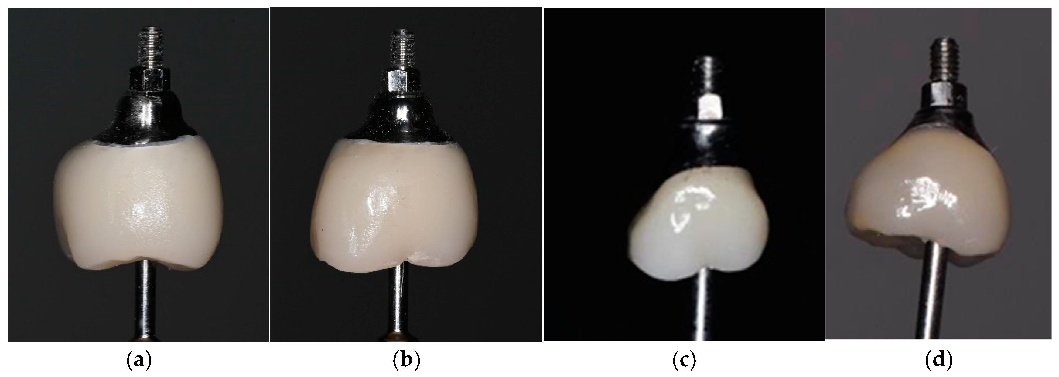
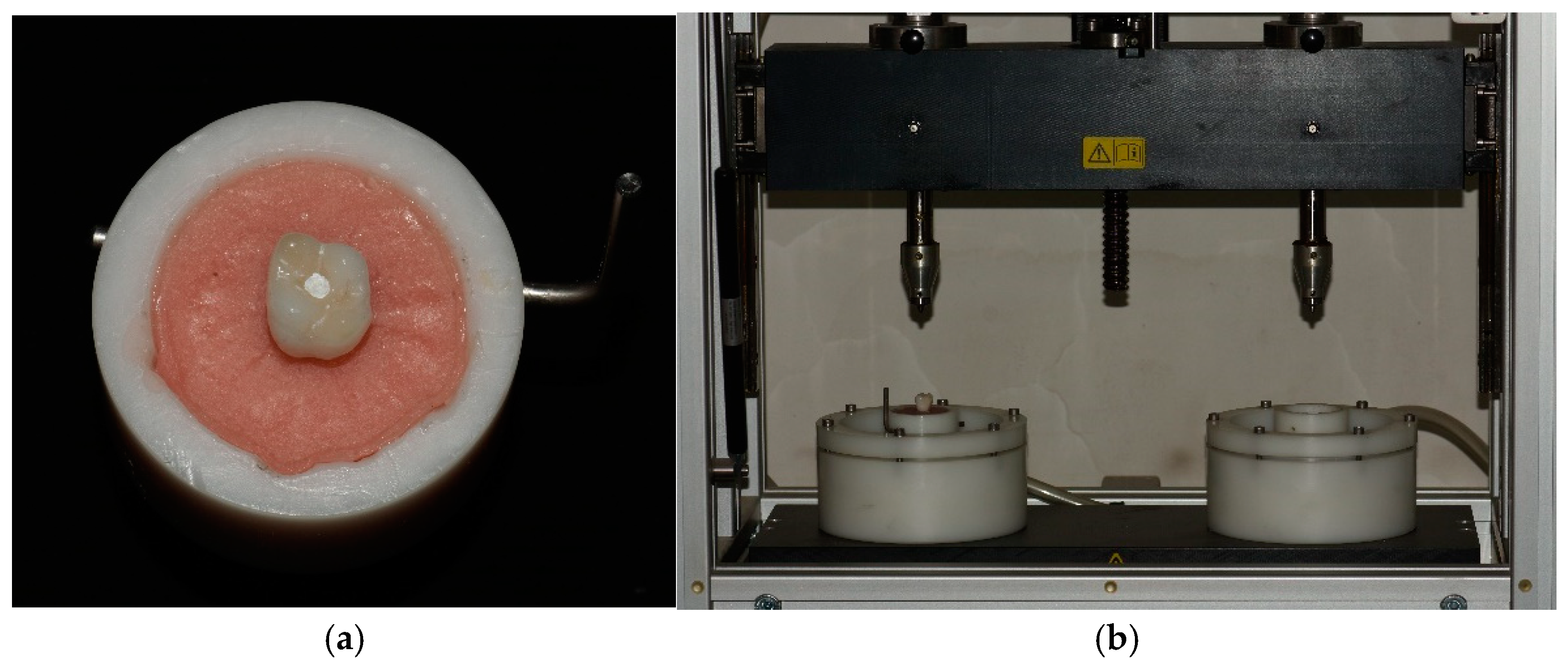
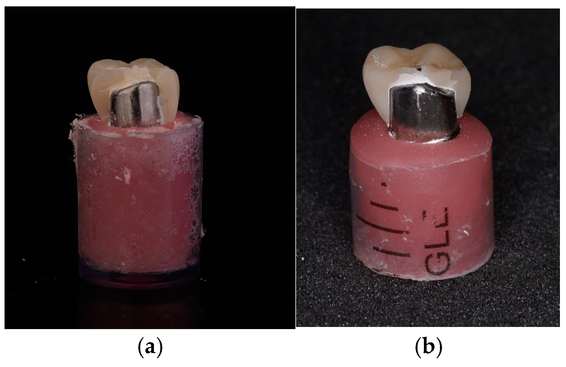
2. Materials and Methods

This laboratory study included 60 specimens, divided into four study groups (A, B, C, D), with 15 samples each. Group A consisted of suprastructures, made of monolithic zirconia implant crown and titanium custom abutment with vertical marginal design. Group B included suprastructures, made of monolithic lithium disilicate implant crown and titanium custom abutment with vertical marginal design. Group C had suprastructures, made of monolithic zirconia implant crown and titanium custom abutment with chamfer marginal design, and Group D was suprastructures, made of monolithic lithium disilicate implant crown and titanium custom abutment with chamfer marginal design. Materials used in this research are displayed in Table 1.
The sample size was computed using SigmaPlot 15.0 software (Systat Software, Inc., San Jose, CA, USA). The sample size was determined in accordance with prior research that investigated the mechanical performance of implant suprastructures under cyclic loading conditions [23,24,25,26].
All samples with chamfer and vertical margin design were fabricated through digital workflow. Study model of maxilla with 4.5 mm diameter implant TSV (Zimmer Biomet, Warsaw, IN, USA), placed in the area upper first right molar, was scanned with intraoral scanner iTero Element intraoral scanner (Align Technology, San Jose, CA, USA). Stereolithographic image was imported in software for virtual design of prosthetic constructions ExoCad software v.3.2 Elefsina (GmbH, Darmstadt, Germany), where monolithic crowns and customized titanium abutments were designed according to their group assignment. Each customized abutment was designed as a cut back of the monolithic crown, which has the same design in each test group. Custom abutments from Groups A and B were planned with vertical margin design and concave emergence profile, following the concept for biologic-oriented preparation technique (BOPT) of Ignazio Loi, and these from Groups C and D—with subgingival chamfer margin design and concave emergence profile.
All crowns were constructed with standard wall thickness, which meets the manufacturer recommendations for the specific material (zirconium dioxide or lithium disilicate)—minimal thickness—1.5 mm, not more than 2.6 mm. Customized abutments were fabricated from titanium blanks GenTek™ Pre-milled Abutment Blanks (Zimmer Biomet, Warsaw, IN, USA) in inLab MC X5 milling machine (Dentsply Sirona, Charlotte, NC, USA). Monolithic zirconia crowns were milled in the same device and these of lithium disilicate were produced in inLab MC XL machine (Dentsply Sirona, Charlotte, NC, USA). Crystallization of crowns from group B and D was conducted in furnace Programat P300 (Ivoclar Vivadent, Shaan, Liechtenstein), and sintering of groups A and C—in Zirconmaster S (VOP Ltd., Sofia, Bulgaria), followed by glazing with IPS Ivocolor Glaze Paste (Ivoclar Vivadent, Schaan, Liechtenstein). Cementation of the crowns to the abutments were performed according to the manufacturers’ prescriptions. The sample groups differed in the type of abutment preparation and the material of the monolithic crown (Figure 1).
The samples were immersed in distilled water at 37 °C for 24 h before undergoing testing.
Each sample was screw-retained to an implant and fixed into the holder of computer controlled 2-axis machine that simulated masticatory movements, Chewing Simulator CS-4 (SD-Mechatronik, Westerham, Germany) [27] by polymethyl methacrylate (Figure 2).
The suprastructures were subjected to fatigue loading test for 1250.000 cycles, representing 5 years of clinical exploitation, at a frequency of 2 Hz. A 300 N load was applied using a standardized steel antagonist with a conical design and a 30° wall inclination, making contact with the sample at the central fissure. The vertical impact was executed in accordance with the manufacturer’s guidelines: upward displacement of 2 mm, downward displacement of 2.5 mm, upward velocity of 60 mm/s, downward velocity of 20 mm/s, horizontal displacement of 0 mm, and horizontal velocity of 20 mm/s. A high precision adjustment tool, developed by the manufacturer, was used to ensure the position of the antagonist relative to the sample [23,24].
The samples, which survived, underwent compression strength test in Instron M1185 (Instron, Norwood, MA, USA) universal testing machine. A steel disk was utilized as an antagonist, applying pressure at a rate of 2 mm/min on each sample at room temperature. The force was maintained until the sample underwent complete failure and defragmentation (Figure 3).
The data were then compiled using Instrument Explorer software v.6.8 and analyzed through descriptive statistics and t-testing. Each sample was thoroughly inspected under a microscope, and a detailed assessment of the fracture type was performed.
Following the laboratory tests, the samples were analyzed by one qualified expert using a magnification with 8:1 zoom range Carl Zeiss SteREO Discovery.V8 microscope (Carl Zeiss Microscopy GmbH, Jena, Germany), and the following criteria were assessed:
  • Deformation of the connecting screw;
  • Deformation of the abutment;
  • Presence of a fracture line or crack in the monolithic crown;
  • Observable failure of the adhesive bond between the monolithic crown and the abutment;
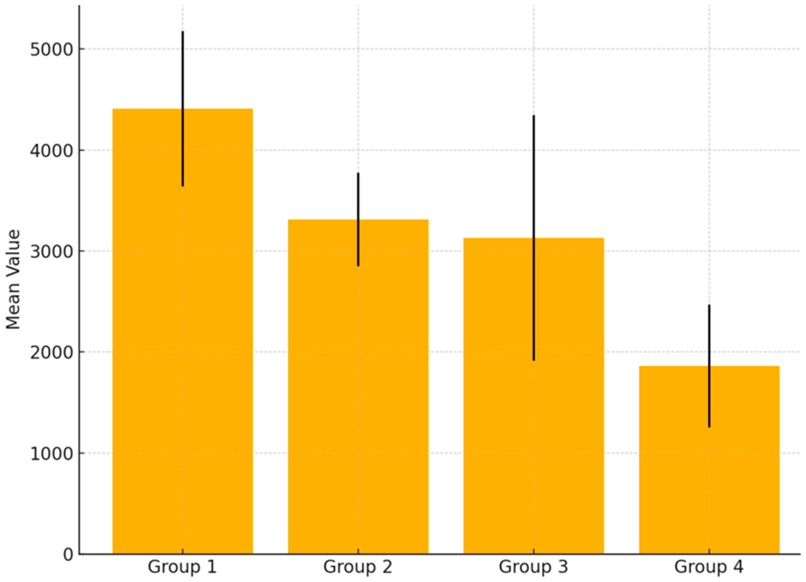
  • Destruction of either the monolithic crown or the titanium abutment. Any fragmentation observed in the sample was considered as destruction.
    Mode of failure was defined due to Burke‘s classification [28]. Type I: minimal fracture or crack in the crown. Type II: less than half of the crown lost. Type III: crown fracture through midline or half of the crown displaced or lost. Type IV: more than half of the crown lost. Type V: severe fracture of the crown and/or tooth. In addition, cracks, chipping, delamination, and catastrophic total failures were noted.
    Statistical analysis was performed with the assistance of Microsoft Excel software (v.2019–2021). Descriptive statistics were used to show the mean values and variations in results. Moreover, analysis of variance (ANOVA) test was applied to compare the mean values of the four groups and measure the existence of statistical significance. More precise results regarding the existence of statistical significance across the specific groups of materials were obtained by using Games-Howell post hoc test. The use of a post hoc test was important to avoid Type I error and false positive differences—a common problem when running multiple ANOVA tests. It is also worth noting that the conventional confidence threshold of 0.05 was applied to confirm the existence of statistical significance.

3. Results

All tested crowns demonstrated a 100% survival rate following chewing simulation. Nonetheless, wear facets were observed at the occlusal contact points on all crowns, with no evidence of abutment fractures or screw loosening.
A compressive strength test evaluates the capacity of a material to withstand deformation and ultimate failure under the application of opposing compressive forces. As the applied compressive load increases, the material initially undergoes deformation, followed by fracturing and eventual structural failure.
Group A resulted as the most resistant to compressive strength (4411 MPa), followed by Group B—3314 MPa, Group C—3130 MPa, and Group D exhibits the lowest value—1864 MPa.
Upon completion of the compressive strength tests, the tested specimens were subjected to repeated microscopic examination in accordance with the same criteria (Table 2).
The microscopic analysis revealed that all test specimens exhibited a vertical trajectory of the fracture line and a compromised adhesive bond. No deformations were observed in the connecting screw or the superstructure (Figure 4).
Mode of failure was defined due to Burke‘s classification as type V.
Figure 5 demonstrates the average compression strength of each group, as well as the observed standard deviation of the compression strength of the observations within each group. Descriptive statistics results indicated that the compression strength of group A (4411 MPa) was the highest and more than twice higher than the performance of group D (1864 MPa). Groups B and C demonstrated a similar performance in terms of compression strengths with values of 3314 MPa and 3130 MPa, respectively; however, the individual measurements in group C demonstrated a high level of discrepancy, as expressed by the high level of standard deviation. In general, it can be concluded that group A outperformed the highest mechanical resistance in terms of compression strength, while maintaining a reasonably low level of variation in results.
The descriptive statistics findings showed that there are variations in the compression strength depending on the group under consideration. Thus, it is important to analyze whether the observed mean strength differences across the analyzed groups were statistically significant. For this purpose, the null hypothesis suggests that the compression strengths remain the same across all groups.
An analysis of variance (ANOVA) test was applied to compare the mean values of the four groups and measure the existence of statistical significance. Results indicated that most differences are between groups, rather than within groups. The F statistic is 24.6—far above the confidence threshold of 2.77, as determined by the applied 0.05 confidence level. As a result, the p-value is 0.000—an indication of very strong statistically significant differences in the compression strength across the four groups (Table 3).
Based on these findings, there is sufficient evidence to reject the null hypothesis.
Moreover, the Games-Howell ad hoc test was applied to thoroughly analyze the existing differences across the specific groups. Results are shown below, thereby evidencing the lack of statistical difference in strength only between Groups B and C (Table 4). Moreover, it can be suggested that Group A demonstrates statistically higher strength level in comparison to other groups under investigation.
In all other cases, p-value is 0.00—which confirms the significance of the observed differences in the strength of the applied materials, as earlier discussed in Figure 5. Hence, it can be suggested that the performance of the materials studied leads to different outcomes in terms of strength.

4. Discussion

The results analysis highlighted Group A—suprastructures made of customized titanium abutments with vertical margin design and monolithic zirconia crowns as the most resistant to compressive forces (4411 MPa). Group D (suprastructures made of customized titanium abutments with chamfer margin design and monolithic lithium disilicate crowns) was with lowest values (1864 MPa)—twice than Group A. Samples were fractured as expected for ceramic materials, with vertical fractures. No abutment or screw fracture was observed. Data revealed better performance of customized abutments with vertical margin design, than the chamfer one. This state was supported by the results from Group B, which had lower results than Group A, but higher than Groups C and D (customized abutments with chamfer design). These findings correspond to the research of García-González et al. [29], who claim that abutments with vertical margin design have better mechanical characteristics.
Comparison between groups, according to the material, used for crown fabrication, points out statistically significant compressive behavior of zirconia monolithic crowns, than lithium disilicate, which share the opinion of Nouh et al. [30], Alessandretti et al. [31], and Elshiyab et al. [32], but it is controversial to the state of Spitznagel et al. for good longevity of lithium disilicate prosthetic constructions [33].
This tendency becomes more pronounced in zirconia crowns, cemented to abutments with vertical margin design. Pries et al. [34] also support the thesis of the higher mechanical resistance of zirconia crowns, but do not analyze the significance of the abutment’s marginal design.
Cocchetto et al. [35] suggest that biologically oriented margin design in implant supported constructions has several advantages, such as better marginal adaptation of the crown after cementation and minimal microgap in comparison with chamfer margin design.
Our study revealed that chamfer margin design provides good peri-implant soft tissue support, but bigger abutment reduces the space for the prosthetic construction, which may lead to its lower mechanical resistance. This proves the statement that crowns are weak points of implant suprastructures and that abutments durability is ensured. All this supports better longevity of crown abutments with vertical preparation.
Studies with same experimental set up, but on natural teeth abutments, report similar results as our findings [36]. This supports the thesis of greater mechanical stability of vertical preparation, but the need for more in-depth studies with a larger number of test samples remains.
The findings have restricted clinical significance due to the sample number and their laboratory characteristics, highlighting the need for further in vivo research. This study was conducted in dry environment. However, the specimens were stored in water until testing, it is essential to determine whether, under the biodynamic balance of the oral cavity, the analyzed materials would maintain similar mechanical properties over the long term.

5. Conclusions

Compression strength significantly depends on the choice of marginal design of implant abutments. Vertical margin design has better performance, that chamfer one. Zirconia implant crowns show two times higher mechanical resistance than lithium disilicate prosthetic constructions. These results can serve as a foundation for further laboratory and clinical research with more specimens.

Supplementary Materials

The following supporting information can be downloaded at: https://www.mdpi.com/article/10.3390/app15095193/s1, Figure S1: Virtual design of the suprastructures: (a) vertical margin design; (b) chamfer margin design; Figure S2: Milling of the prosthetic constructions; Table S1: Results of Compression strength test—Values of all samples.

Author Contributions

Conceptualization, D.S. and D.F.; methodology, G.M.; software, B.T.; validation D.S. and D.F.; formal analysis, D.J.; investigation, D.S.; resources, G.M.; data curation, D.S. and A.J.; writing—original draft preparation, D.S.; writing—review and editing, D.F.; visualization, D.J. and A.J.; supervision, D.S.; project administration, D.F.; funding acquisition, G.M. All authors have read and agreed to the published version of the manuscript.

Funding

This research was funded by GRANT No. 165/03.08.2023 project.

Data Availability Statement

All data of this study is provided into the article and Supplementary Materials. For further information, please contact the corresponding author at stoeva.dani@gmail.com.

Conflicts of Interest

The authors declare no conflicts of interest.

References

  1. Carossa, M.; Alovisi, M.; Crupi, A.; Ambrogio, G.; Pera, F. Full-Arch Rehabilitation Using Trans-Mucosal Tissue-Level Implants with and without Implant-Abutment Units: A Case Report. Dent. J. 2022, 10, 116. [Google Scholar] [CrossRef] [PubMed]
  2. Belser, U.C.; Schmid, B.; Higginbottom, F.; Buser, D. Outcome analysis of implant restorations located in the anterior maxilla: A review of the recent literature. Int. J. Oral Maxillofac. Implant. 2004, 19 (Suppl. 30–42). [Google Scholar] [PubMed]
  3. Fenner, N.; Hämmerle, C.H.; Sailer, I.; Jung, R.E. Long-term clinical, technical, and esthetic outcomes of all-ceramic vs. titanium abutments on implant supporting single-tooth reconstructions after at least 5 years. Clin. Oral Implant. Res. 2016, 27, 716–723. [Google Scholar] [CrossRef]
  4. de Vasconcelos, J.E.L.; de Matos, J.D.M.; Queiroz, D.A.; Lopes, G.d.R.S.; de Lacerda, B.C.G.V.; Bottino, M.A.; Turssi, C.P.; Basting, R.T.; do Amaral, F.L.B.; França, F.M.G. Implant—Abutment Misfit after Cyclic Loading: An In Vitro Experimental Study. Materials 2022, 15, 5341. [Google Scholar] [CrossRef]
  5. Luthy, H. Strength and toughness of dental ceramics. In CAD/CIM in Aesthetic Dentistry: CEREC 10 Year Anniversary Symposium (1994; Zurich); Mormann, W.H., Ed.; Quintessence: Chicago, IL, USA, 1996; pp. 229–240. [Google Scholar]
  6. Glauser, R.; Sailer, I.; Wohlwend, A.; Studer, S.; Schibli, M.; Schärer, P. Experimental zirconia abutments for implant-supported single-tooth restorations in esthetically demanding regions: 4-year results of a prospective clinical study. Int. J. Prosthodont. 2004, 17, 285–290. [Google Scholar] [PubMed]
  7. Canullo, L. Clinical outcome study of customized zirconia abutments for single-implant restorations. Int. J. Prosthodont. 2007, 20, 489–493. [Google Scholar] [PubMed]
  8. Lops, D.; Meneghello, R.; Sbricoli, L.; Savio, G.; Bressan, E.; Stellini, E. Precision of the Connection between Implant and Standard or Computer-Aided Design/Computer-Aided Manufacturing Abutments: A Novel Evaluation Method. Int. J. Oral Maxillofac. Implant. 2018, 33, 23–30. [Google Scholar] [CrossRef]
  9. Marchack, C.B. A custom titanium abutment for the anterior single-tooth implant. J. Prosthet. Dent. 1996, 76, 288–291. [Google Scholar] [CrossRef]
  10. Agustín-Panadero, R.; Serra-Pastor, B.; Roig-Vanaclocha, A.; Román-Rodriguez, J.L.; Fons-Font, A. Mechanical Behavior of Provisional Implant Prosthetic Abutments. Med. Oral Patol. Oral Cir. Buccal 2015, 20, e94–e102. [Google Scholar] [CrossRef]
  11. Azer, S.S. A Simplified Technique for Creating a Customized Gingival Emergence Profile for Implant-Supported Crowns. J. Prosthodont. 2010, 19, 497–501. [Google Scholar] [CrossRef]
  12. Andersson, B.; Taylor, Å.; Lang, B.R.; Scheller, H.; Schärer, P.; Sorensen, J.A.; Tarnow, D. Alumina ceramic implant abutments used for single-tooth replacement: A prospective 1- to 3-year multicenter study. Int. J. Prosthodont. 2001, 14, 432. [Google Scholar] [PubMed]
  13. Iliev, G.; Filtchev, D.; Trifković, B.; Jevremović, D.; Pavlova, Z.; Slavkov, S.; Stoeva, D. Mechanical Resistance of Different Implant Suprastructures: A Laboratory Study. Appl. Sci. 2023, 13, 6100. [Google Scholar] [CrossRef]
  14. Stoeva, D.; Filtchev, D.; Nikolova, N.; Pavlova, Z.; Slavkov, S.; Iliev, G. Clinical Evaluation of Implant Suprastructures Depending on the Biomechanical Characteristics of the Materials Used. Appl. Sci. 2022, 12, 8659. [Google Scholar] [CrossRef]
  15. Khamis, M.M. Prosthetic Role in Peri-Implant Soft Tissue Management: Treatment Planning Phase. In Peri-Implant Soft Tissue Management: A Clinical Guide; Springer International Publishing: Cham, Switzerland, 2024. [Google Scholar] [CrossRef]
  16. Elsayed, A.; Wille, S.; Al-Akhali, M.; Kern, M. Comparison of fracture strength and failure mode of different ceramic implant abutments. J. Prosthet. Dent. 2017, 117, 499–506. [Google Scholar] [CrossRef]
  17. Albakry, M.; Guazzato, M.; Swain, M.V. Biaxial flexural strength, elastic moduli, and x-ray diffraction characterization of three pressable allceramic materials. J. Prosthet. Dent 2003, 89, 374–380. [Google Scholar] [CrossRef] [PubMed]
  18. Holst, S.; Blatz, M.B.; Hegenbarth, E.; Wichmann, M.; Eitner, S. Prosthodontic considerations for predictable single-implant esthetics in the anterior maxilla. J. Oral Maxillofac. Surg. 2005, 63, 89–96. [Google Scholar] [CrossRef]
  19. Lee, S.J.; Betensky, R.A.; Gianneschi, G.E.; Gallucci, G.O. Accuracy of digital versus conventional implant impressions. Clin. Oral Implant. Res. 2015, 26, 715–719. [Google Scholar] [CrossRef]
  20. De Kok, P.; Kleverlaan, C.J.; de Jager, N.; Kuijs, R.; Feilzer, A.J. Mechanical performance of implant-supported posterior crowns. J. Prosthet. Dent. 2015, 114, 59–66. [Google Scholar] [CrossRef]
  21. Sadowsky, S.J. Occlusal overload with dental implants: A review. Int. J. Implant. Dent. 2019, 5, 29. [Google Scholar] [CrossRef]
  22. Loi, I.; Di Felice, A. Biologically oriented preparation technique (BOPT): A new approach for prosthetic restoration of periodontically healthy teeth. Eur. J. Esthet. Dent. 2013, 8, 10–23. [Google Scholar]
  23. Elsayed, A.; Wille, S.; Al-Akhali, M.; Kern, M. Effect of fatigue loading on the fracture strength and failure mode of lithium disilicate and zirconia implant abutments. Clin. Oral Implant. Res. 2018, 29, 20–27. [Google Scholar] [CrossRef] [PubMed]
  24. Verdugo, C.L.; Núñez, G.J.; Avila, A.A.; Martín, C.L.S. Microleakage of the prosthetic abutment/implant interface with internal and external connection: In vitro study. Clin. Oral Implant. Res. 2014, 25, 1078–1083. [Google Scholar] [CrossRef]
  25. Schultheis, S.; Strub, J.R.; Gerds, T.A.; Guess, P.C. Monolithic and bi-layer CAD/CAM lithium–disilicate versus metal–ceramic fixed dental prostheses: Comparison of fracture loads and failure modes after fatigue. Clin. Oral Investig. 2013, 17, 1407–1413. [Google Scholar] [CrossRef] [PubMed]
  26. Dogan, D.O.; Gorler, O.; Mutaf, B.; Ozcan, M.; Eyuboglu, G.B.; Ulgey, M. Fracture Resistance of Molar Crowns Fabricated with Monolithic All-Ceramic CAD/CAM Materials Cemented on Titanium Abutments: An In Vitro Study. J. Prosthodont. 2015, 26, 309–314. [Google Scholar] [CrossRef] [PubMed]
  27. SD Mechatronik. A Guide Through the SD Mechatronik Chewing Simulator; SD Mechatronik: Feldkirchen-Westerham, Germany, 2017. [Google Scholar]
  28. Burke, F.J. Fracture resistance of teeth restored with dentin-bonded crowns: The effect of increased tooth preparation. Quintessence Int. 1996, 27, 115–121. [Google Scholar]
  29. García-González, M.; González-González, I.; García-García, I.; Blasón-González, S.; Lamela-Rey, M.J.; Fernández-Canteli, A.; Álvarez-Arenal, Á. Effect of abutment finish lines on the mechanical behavior and marginal fit of screw-retained implant crowns: An in vitro study. J. Prosthet. Dent. 2022, 127, 318.e1–318.e10. [Google Scholar] [CrossRef]
  30. Nouh, I.; Kern, M.; Sabet, A.E.; AboelFadl, A.K.; Hamdy, A.M.; Chaar, M.S. Mechanical behavior of posterior all-ceramic hybrid abutment-crowns versus hybrid-abutments with separate crowns-A laboratory study. Clin. Oral Implant. Res. 2019, 30, 90–98. [Google Scholar] [CrossRef]
  31. Alessandretti, R.; Borba, M.; Della Bona, A. Cyclic contact fatigue resistance of ceramics for monolithic and multilayer dental restorations. Dent. Mater. 2020, 36, 535–541. [Google Scholar] [CrossRef]
  32. Elshiyab, S.H.; Nawafleh, N.; Öchsner, A.; George, R. Fracture resistance of implant- supported monolithic crowns cemented to zirconia hybrid-abutments: Zirconia-based crowns vs. lithium disilicate crowns. J. Adv. Prosthodont. 2018, 10, 65–72. [Google Scholar] [CrossRef]
  33. Spitznagel, F.A.; Bonfante, E.A.; Vollmer, F.; Gierthmuehlen, P.C. Failure Load of Monolithic Lithium Disilicate Implant-Supported Single Crowns Bonded to Ti-base Abutments versus to Customized Ceramic Abutments after Fatigue. J. Prosthodont. 2022, 31, 136–146. [Google Scholar] [CrossRef]
  34. Preis, V.; Behr, M.; Rosentritt, M. In Vitro Fatigue and Fracture Testing of Implant-Supported Anterior Ceramic Crowns. Int. J. Prosthodont. 2018, 31, 264–266. [Google Scholar] [CrossRef] [PubMed]
  35. Cocchetto, R.; Canullo, L. The “hybrid abutment”: A new design for implant cemented restorations in the esthetic zones. Int. J. Esthet. Dent. 2015, 10, 186–208. [Google Scholar] [PubMed]
  36. Kasem, A.T.; Sakrana, A.A.; Ellayeh, M.; Özcan, M. Evaluation of zirconia and zirconia-reinforced glass ceramic systems fabricated for minimal invasive preparations using a novel standardization method. J. Esthet. Restor. Dent. 2020, 32, 560–568. [Google Scholar] [CrossRef] [PubMed]
Figure 1. Specimens of (a) Group A; (b) Group B; (c) Group C; (d) Group D.
Applsci 15 05193 g001
Figure 2. Fatigue loading test (a) specimen, fixed in the holder; (b) Chewing Simulator CS-4 (SD-Mechatronik, Westerham, Germany).
Applsci 15 05193 g002
Figure 3. Instron M1185 universal testing machine—a compressive strength test set up.
Applsci 15 05193 g003
Figure 4. Vertical fracture model: (a) Group A; (b) Group D.
Applsci 15 05193 g004
Figure 5. Average values with error bars (standard deviation) (MPa).
Applsci 15 05193 g005
Table 1. Materials used for fabrication of implant suprastructures.
MaterialsStudy Groups
Titanium Alloy Blanks GenTek™ Pre-Milled Abutment Blank (Zimmer Biomet, Warsaw, IN, USA), grade 5 titaniumA, B, C, D
Zirconium Dioxide Katana (Kuraray Noritake, Tokyo, Japan)A, C
Lithium Disilicate IPS e.max CAD (IvoclarVivadent, Schaan, Liechtenstein)B, D
Table 2. Results after compressive strength test.
Observed Criteria
GroupsDeformation of the Connecting ScrewDeformation of the AbutmentPresence of a Fracture Line or Crack in the Monolithic CrownFailure of the Adhesive Bond Between the Monolithic Crown and the AbutmentDestruction of Either the Monolithic Crown or the Titanium Abutment
A00151515
B00151515
C00151515
D00151515
Table 3. ANOVA results.
Source of VariationSSdfMSFp-ValueF Crit
Between groups 49,008,821316,336,27424.5970.0002.769
Within groups 37,192,18656664,146
Total 86,201,00759
Table 4. Analysis of the significance of the observed strength differences across the specific groups of materials.
Differences BetweenMean
Difference
p-Value95% CI Lower95% CI Upper
Group AGroup B1096.400.00616.681576.12
Group AGroup C1280.470.00512.462048.47
Group AGroup D2546.800.002026.793066.81
Group BGroup C184.070.59−522.21890.34
Group BGroup D1450.400.001045.371855.43
Group DGroup D1266.330.00535.021997.64
Disclaimer/Publisher’s Note: The statements, opinions and data contained in all publications are solely those of the individual author(s) and contributor(s) and not of MDPI and/or the editor(s). MDPI and/or the editor(s) disclaim responsibility for any injury to people or property resulting from any ideas, methods, instructions or products referred to in the content.

