Review

Antibacterial Ceramics for Dental Applications

by
Lubica Hallmann
1,* and
Mark-Daniel Gerngross
2
1
School of Dentistry, Kiel University, 24118 Kiel, Germany
2
Institute of Material Science, Faculty of Engineering, Kiel University, 24118 Kiel, Germany
*
Author to whom correspondence should be addressed.
Appl. Sci. 2025, 15(8), 4553; https://doi.org/10.3390/app15084553
Submission received: 23 February 2025 / Revised: 7 April 2025 / Accepted: 12 April 2025 / Published: 21 April 2025

Abstract

:
Background: The aim of this review was to evaluate the antibacterial properties of functionalized bioceramics for dental applications. Methods: The electronic databases PubMed, Medline, ProQuest, and Google Scholar were used to search for peer-reviewed scientific publications published between 2020 and 2025 that provide insights to answer research questions related to the role of antibacterial-functionalized bioceramics in combating pathogens in dentistry without triggering immune reactions and inflammation, as well as on their efficacy against various pathogens and whether understanding the antibacterial mechanism can promote the development of glass-ceramic and bioceramic with long-term antibacterial activity. The keywords used to answer the research questions were: bioglass, bioceramic, biocompatible, antibacterial, osseointegration, implant, and bioactive materials. Results: Bacterial infections play a key role in the longevity of medical devices. A crucial problem is drug-resistant bacteria. Antibacterial ceramics have received great attention recently because of their long-term antibacterial activity, good mechanical properties, good biocompatibility, and bioactivity. This review provides a detailed examination of the complex interactions between bacteria, immune cells, and bioceramics from a clinical perspective. The focus of the researchers is on developing new-generation bioceramics with multifunctionality, in particular with antibacterial properties that are independent of conventional antibiotics. The highlight of this review is the exploration of bioceramics with dual functions such as antibacterial and bioactive properties, promoting bone regeneration and antibacterial activity, which have the potential to revolutionize implant technology. Another research focus is modifying the implant surface from hydrophilic to hydrophobic in order to increase the antibacterial activity of bioceramics. Conclusions: The aim of this review is to help researchers understand the current state-of-the-art antibacterial activities of bioceramics, which could promote the development of antibacterial ceramics and their clinical application.

1. Introduction

Bioglasses (BGs) have found wide applications as biomaterials for soft and hard tissue regeneration [1,2,3,4,5,6]. BGs have the ability to bond with bone upon contact with physiological fluids, making them suitable as bone-replacement biomaterials in the form of implants, scaffolds, granules, powders, or coatings. They have excellent biocompatibility, bioactivity, conductivity, and osteoinductivity, a controllable degradation rate, and improved bone healing [1,2,7,8]. These materials have been used to treat cancer since the beginning of the 21st century [2]. However, the applications of BGs as bone and dental implants can induce an immune response even in the absence of immune-stimulating signals. New BGs are being developed that are immunologically inert and also have antibacterial properties [9,10,11].
Microleakages caused by a misfit between the margin of the crown restoration and the dental tissue or the dissolution of adhesive materials lead to plaque accumulation, which can cause secondary caries [12,13]. All-ceramic crowns are an excellent choice for clinical crown restoration due to their good mechanical properties and biological stability, as well as their chemical and optical properties compatible with those of natural teeth, such as translucency, color, fluorescence, opalescence, and aesthetics [14,15,16]. However, due to the lack of antibacterial effect, secondary caries may still develop [17], which shortens the lifespan of dental restorations and leads to early replacement of the crown restoration. Therefore, the development of antibacterial dental ceramics for crown restorations that prevent secondary caries is one of the biggest challenges in prosthodontics.
Another application area for antibacterial ceramics besides crown restorations is dental implants. They are used to replace teeth lost due to severe damage, large area cavities, or gum disease [18,19,20,21,22]. Infections at the implant site can hinder or even completely prevent osseointegration, potentially leading to implant failure or the need for surgical removal of the implant [23,24,25,26].
Bacterial infections play a key role in the development of necrotic processes of the dental pulp and the formation of periapical lesions [27]. In periodontitis, the gums, periodontal ligaments, and supporting alveolar bone are gradually destroyed due to an inflammatory reaction triggered by subgingival pathogenic microorganisms [13,18,19,20,21,22]. For example, chronic apical periodontitis is an inflammatory reaction caused by infection of the pulp. It induces bone resorption in the apical and periapical areas of the teeth [28,29], originating from high bacterial contamination along the root canal system [27]. Staphylococcus aureus, Staphylococcus epidermidis, Pseudomonas aeruginosa, Enterobacter cloacae, and Escherichia coli are the major bacteria that are responsible for implant infections. Gram-positive and Gram-negative bacteria have different cell shapes, biochemistry, and biomolecular mechanisms that allow them to survive and grow on the surface of dental implants. They are difficult to remove, resist the immune system, and frequently cause opportunistic infections [30].
The resistance of microorganisms to antibiotics has required the development of new materials with antibacterial properties to reduce or avoid excessive use of antibiotics [9,24,31,32,33,34]. Most of the current dental materials exhibit no antibacterial functionality against Gram-positive and Gram-negative bacteria. To increase the success rate of dental treatments, dental materials need to be improved to effectively reduce colonization and microbial development [13,35,36,37,38,39,40,41].
Materials with intrinsic antibacterial activity are also in high demand for various clinical applications, such as bone grafts. They are often required to treat open fractures with significant bone loss [24,42]. Contamination of the wound, e.g., from a contaminated implant surface, can cause soft tissue infections and osteomyelitis, which slows down the healing process and hinders bone formation [22]. The growth behavior of bacterial infections is largely controlled by the composition of the biomaterials and their surface properties [25,41,43].
Many attempts have been made to solve these major problems. The antibacterial effect of the materials used is based on three strategies: (a) local release, (b) contact-killing, and (c) the generation of synergetic properties [44,45,46].
However, each method has advantages and disadvantages, such as slow efficacy, toxicity, or the alteration of the physical properties of the sealer. Inorganic antimicrobial materials are typically more stable and exhibit long-term antibacterial properties. New filler additives made of glass and glass-ceramic are being developed to prevent bacterial reinfection of the root canal [27].
The development of bioceramics with good mechanical, bioactive, and antimicrobial properties is a major challenge for medicine [25,26,47,48,49,50,51,52,53]. Zirconia ceramics, a typical dental ceramic material, have excellent biocompatibility, optical properties, and chemical stability [54]. They are often used for tooth repairs, dental implants, and orthopedic artificial joints, but their surface is hydrophilic, which allows for the adhesion of bacteria and the formation of biofilms on the surface when implanted into the body [54,55]. Superhydrophobic surfaces usually exhibit self-cleaning behavior, corrosion, and antibacterial resistance [54,56,57,58,59]. By treating the zirconia surface using femtosecond laser ablation, new surface micro-textures are created, turning it into a hydrophobic surface. Such a treated zirconia surface can enhance biological responses and reduce the bacterial behavior of the interface [58]. The hydrophobic surface created by the laser treatment can be attributed to the topographical modifications of zirconia surfaces at the micro and nano scale. The hydrophobic surface can especially influence the initial stages of biofilm formation. This is very important because early-stage biofilm formation plays a crucial role in bacterial colonization and subsequent infection [60].
Bioceramics for dental applications should not only promote osteocyte formation and osteogenic differentiation but also be able to inhibit bacterial growth. The development of bioceramic materials with inherent antimicrobial properties, such as the incorporation of antimicrobial ions (silver, copper, zinc, cerium, strontium, etc.), is the focus of researchers [38,44,61,62]. Strontium (Sr2+), which is present in bone, muscle, liver, and body fluid offers antibacterial activity, improves osseointegration, and stimulates osteoblastic proliferation. Bioactive glasses doped with strontium have a positive effect on antibacterial efficiency and stimulate bone formation [61]. Zinc (Zn2+) can promote bone growth, proliferation, and the differentiation of bone cells, as well as DNA replication and protein and enzyme production [63]. Zinc ions are used to impact the antibacterial activity of bioactive glass due to their antibacterial and angiogenic properties [63]. Cerium is the only element in the lanthanide group that is stable in the tetravalent state. The easy exchange between the oxidation states Ce3+ and Ce4+ is the foundation for its catalytic activity as a scavenger of reactive oxygen species (ROS) and its antioxidative properties that protect osteoblasts from oxidative stress. Ce-BGs are nontoxic to cells and promote osteoblastic differentiation. Mineralization of primary osteoblasts increases collagen production. Cerium ions play a fundamental and effective role in preventing bacterial adhesion and proliferation on the implant surface. Cerium ions rapidly bind to E. coli and disrupt respiration and other metabolic functions, leading to bacterial death [64,65]. Ag+ ions are known for their toxicity to bacteria, viruses, fungi, and various other organisms, while they have little or no toxicity to human cells. Ag ions can damage the cell membrane, leading to a disruption of the cell functions or intracellular biomolecules and the induction of oxidative stress. The generated ROS result in the formation of free radicals and more extensive cell damage. Charged Ag ions can react with the negatively charged bacterial surface, affecting the zeta potential of the bacterial cell and increasing membrane permeability. Ag+ ions have a high affinity for sulfur groups on the cell membrane and thus inhibit respiratory chains, electron transfer, protein secretion, and lipid biosynthesis, leading to bacteria death [16,24].
Metal-doped bioceramics offer many advantages over other antibacterial methods. They are non-toxic, improve the mechanical properties of the bioglass such as hardness and wear resistance, and provide long-term implant stability, which is very important for the success of the implant.
The most commonly used glass-ceramics and bioceramics for dental applications are listed in Table 1.
This review demonstrates the importance of developing a new generation of antibacterial bioceramics for dental applications by presenting the current state of the art in this field. A comprehensive understanding of antibacterial mechanisms and strategies can help researchers develop innovative solutions to prevent implant infections and improve the lives of patients.

2. Materials and Methods

2.1. Protocols

The aim of this review is to provide an overview of relevant dental bioceramic materials with antibacterial functionalization from a materials science perspective and to provide insights into the following topics. What kind of antibacterial functionalization can be introduced into state-of-the-art bioceramics? Can such functionalized bioceramics combat the pathogens in dentistry without causing immune reactions and inflammation or altering the properties of human cells? Are differences observable in the effectiveness of the antibacterial functionalization for different pathogens? How can the mode of action be understood, and how can it be enhanced?
The PRISMA flow diagram was used to select the studies to answer the above questions (Figure 1).

2.2. Electronic Searches

The electronic databases PubMed, Medline, ProQuest, and Google Scholar were used to search for peer-reviewed scientific publications published between 2020 and 2025 that provide insights to answer the research questions described above.
The keywords used to answer the research questions were bioglass, bioceramics, biocompatible, antibacterial, osseointegration, implant, and bioactive materials.

2.3. Screening Process

The first step was to read titles and abstracts to select the relevant articles that appeared very important in answering the main questions stated above. The inclusion criteria were original and review articles. After selection by title, abstract, and publication year, the full-text publications were examined. Regarding the type of research, they had to be experimental in vitro. Animal and human studies were excluded. Publications that provided insight into answering the questions raised in 2.1 were considered, and their data were extracted and summarized. The exclusion criteria were the use of bioglass and ceramics that were not intended to be incorporated into dental materials or that lacked antimicrobial activity. Articles that were published before the established date (2020), as well as articles in which the study focus did not match the stated criteria, were also excluded.

2.4. Eligibility

Throughout the entire examination process, the publications were assessed for criteria such as antimicrobial activity incorporated into dental materials or a direct connection to dentistry. Biocompatibility, cytotoxicity, bioactive, and mechanical analyses were used as inclusion criteria in this review. Publications that presented bioactivity or biocompatibility but lacked antibacterial activity were excluded.

3. Results and Discussion

Metals such as Ag, Cu, and Zn exhibit a broad spectrum of antibacterial activities against a variety of bacterial strains, including Gram-positive and Gram-negative bacteria [24,46,69]. Both Gram-negative and Gram-positive bacteria have a negatively charged surface that allows interactions with metal cations or positively charged nanoparticles (NPs) due to electrostatic interactions. This interaction disrupts the bacterial cell wall. Metallic or metal oxide nanoparticles can inhibit biofilm formation [26,70]. The cations of these metals are biocompatible and metal-doped bioceramics are a good choice as antibacterial materials for a variety of medical applications.

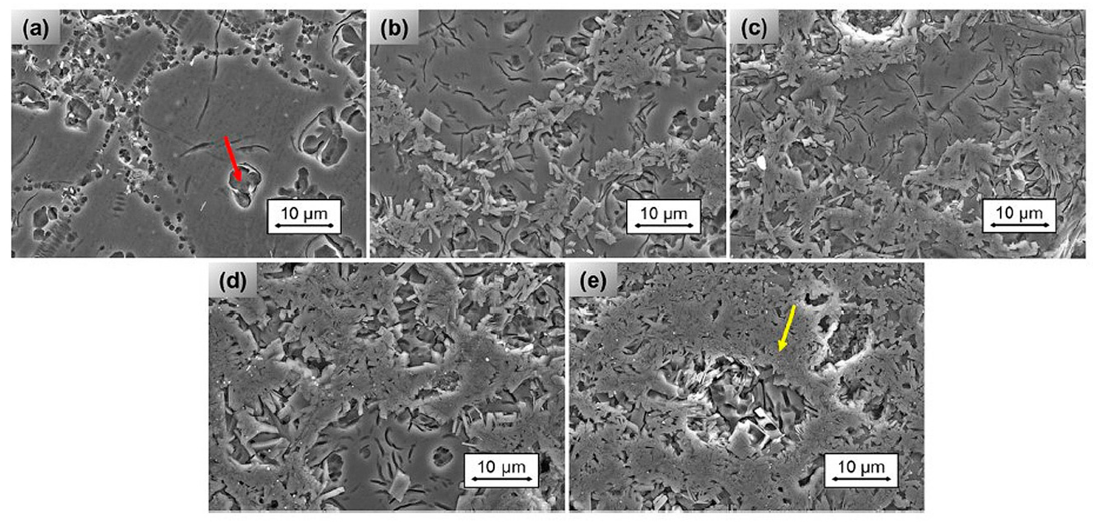
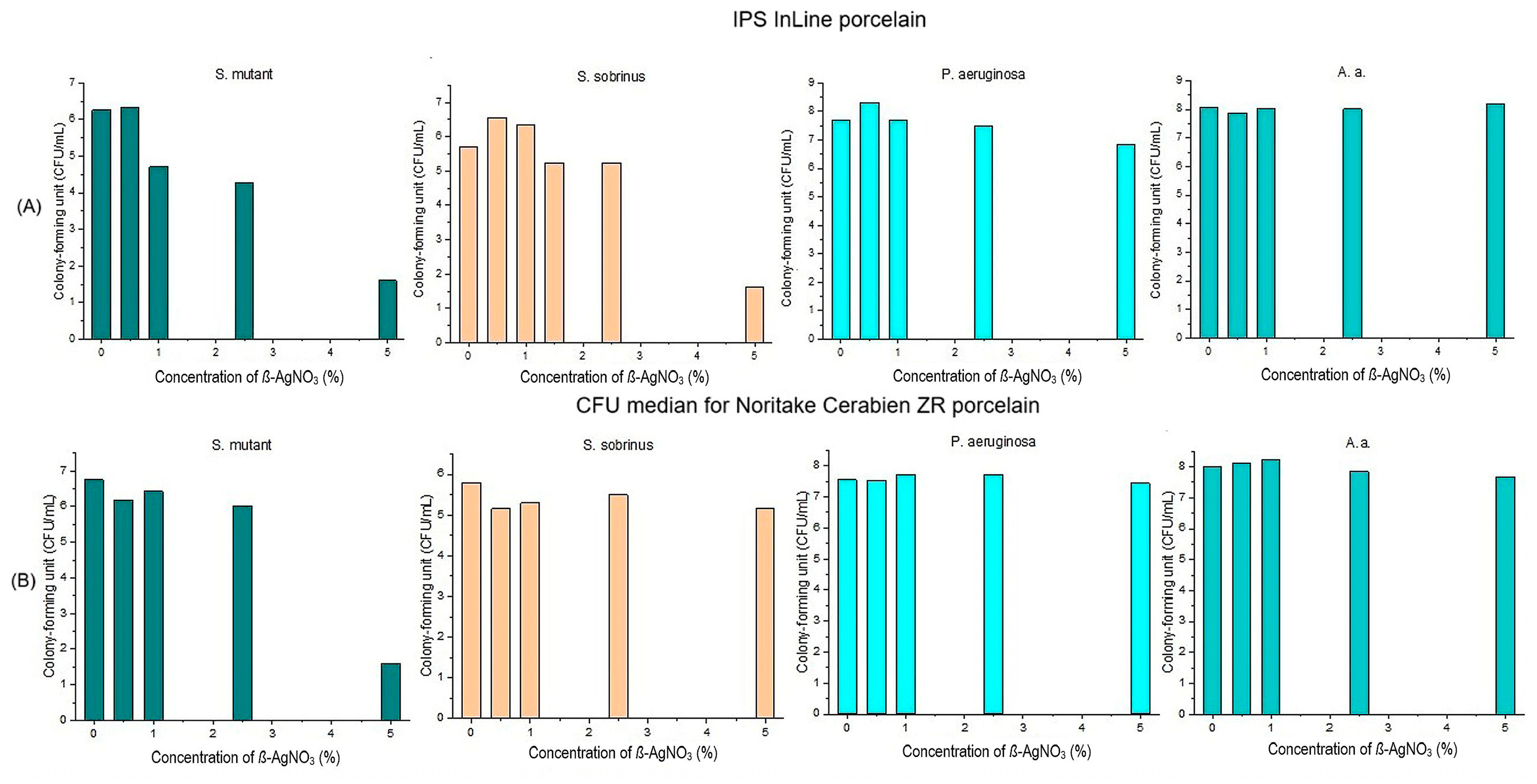
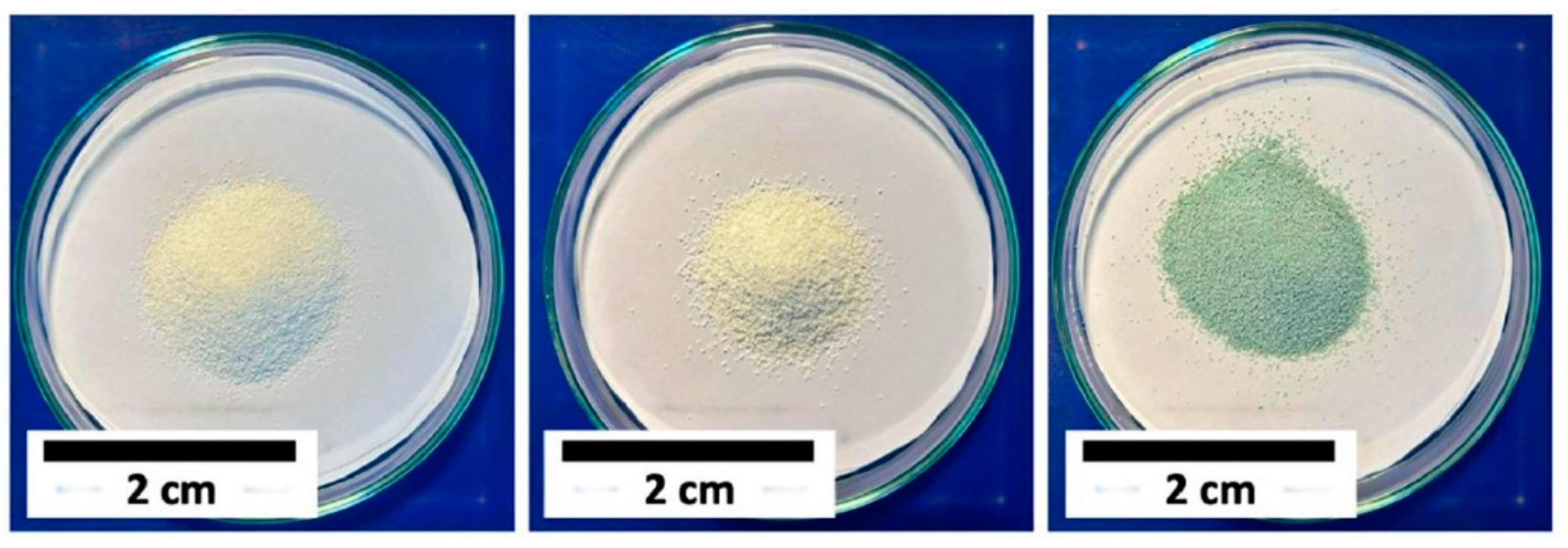
3.1. Antibacterial Properties of Silver and Silver Ions

Silver is known for its chemical stability, superior electric conductivity, and catalytic and antibacterial effects. Silver nanoparticles and their salts are used in the biomedical field as antimicrobial agents [2,37,39,45,46,48,55,70,71]. Ag+ can have both beneficial and harmful effects, as it can eliminate bacteria and induce cytotoxicity. Silver ions have been reported to be toxic to numerous human cells, such as bronchial epithelial cells, umbilical vein endothelial cells, red blood cells, peripheral blood mononuclear cells, keratinocytes, and liver cells. The cytotoxicity of silver compounds depends more on the concentration of released Ag+ ions in the cell medium than on the grain size of the nanoparticles of silver compounds [14].
Baptista et al. [13] used nanostructured ß-AgVO3 to prepare leucite glass-ceramics with antibacterial activity that pure leucite glass-ceramics do not inherently offer, and various attempts are being made to add such functionality. Leucite glass-ceramics are used in dental prostheses as ceramic substructures or as masking metal frameworks. They consist of 1–4 µm sized crystals evenly distributed in the glass matrix [5,13,66]. Leucite (KAlSi2O6) has a tetragonal crystal structure at room temperature, turning into a cubic structure at 673 °C [72]. These glass-ceramics are highly translucent and chemically inert. Their coefficient of thermal expansion (CTE) is approximately 12.0–12.6 × 10⁻⁶ K−1, making them suitable for application in dental crowns and veneer applications [63]. IPS d.SIGN powder with different contents of ß-AgVO3 (1 wt%, 2 wt%, 4 wt%, 6 wt%) was used. The addition of ß-AgVO3 influenced the phase composition of the glass-ceramic. The crystal phase in the control samples consisted only of pure leucite. The addition of AgVO3 reduced the leucite phase, and a new phase of silicate microcline [K0.95AlSi3O8] was formed (Figure 2).
The higher the AgVO3 content, the higher the content of the microcline phase, which is the main phase at a concentration of 6 wt% ß-AgVO3. Baptista et al. investigated the antibacterial activity of leucite glass-ceramics against two bacteria, E. coli and S. aureus, using the agar diffusion method (Figure 3).
As shown in Figure 3, the glass-ceramic modified with 2 wt% or more ß-AgVO3 showed antibacterial activity against Gram-negative and Gram-positive bacteria. The release of Ag+, V4+, and V5+ ions is very important for the observed antibacterial activity. The main problem with such types of modified ceramics is their increasing solubility beyond the limits for dental ceramics used in contact with cells and the oral cavity, as specified in ISO 6872:2024 [12]. At a concentration up to 2 wt% ß-AgVO3, the release of metal ions exceeded the maximum limit. The release of Ag+ ions for 1 wt% ß-AgVO3 was less than 10 µg/L for silver ions and 50 µg/L for vanadium ions. The concentration of released silver and vanadium ions from samples with 6 wt% ß-AgVO3 in the medium was drastically higher. The progressive increase in the concentration of dissolved ions can be explained by the solubility of the ceramic [13]. The investigation of the release kinetics of Ag+, V4+, and V5+ and their influence on peri-implantitis in recent implants needs to be studied.
Furthermore, the leucite phase must be stabilized because the microcline phase has a significantly higher solubility than the leucite phase.
Uehara et al. [73] used IPS InLine and Noritake Cerabien ZR Line for their study to investigate the effect of ß-AgVO3 on the bacteria S. mutans, S. sobrinus, P. aeruginosa, and Aggregatibacter actinomycetemcomitans (A.a.). The concentrations of nano-ß-AgVO3 were 0.5%, 1%, 2.5%, and 5%. Brain Heart Infusion Agar was used for S. mutans and S. sobrinus, cetrimide agar for P. aeruginisa, and blood agar for the preparation of A.a.
As shown in Figure 4A, nano-ß-AgVO3 in IPS InLine at a concentration of 5% had high antibacterial activity against S. mutans and S. sobrinus bacteria, but it was not effective against other bacteria [73]. Nano-ß-AgVO3 at a concentration of 5% was found to have antibacterial activity against S. mutant bacteria in Noritake Cerabian ZR porcelain (Figure 4B) [73]. From these results, it can be concluded that the chemical composition of glass or glass ceramics can also play a significant role in the antibacterial effectiveness of the antibacterial additive used for functionalization. In addition, the antibacterial active ingredients can show pronounced bacterial dependence with regard to their antibacterial effect in ceramics. Further studies are needed to explain the cytotoxicity of these glasses.
Singh et al. used Vita VM9 as a leucite glass-ceramic (LGC) and silver nanoflakes (AgNF: thickness: ~50 nm, width: ~2 µm) to prepare LGC-AgNF and AgNO3 [74]. The concentrations of AgNF and AgNO3 were 2, 5, 10, and 15 wt.%. The addition of Ag to LGC was found to affect the microhardness of LGC-Ag materials, resulting in a decrease from 6.5 GPa for pure LGC to almost 4.6 GPa for LGC + 15 wt.% AgNF and 4.5 GPa for LGC + 15 wt.% AgNO3. When using LGC + Ag in layered dental crowns, the reduction in hardness has a positive effect on preventing wear of the opposing tooth. The addition of Ag to the ceramic as AgNO3 and AgNFs imparts strong antibacterial properties to LGC and prevents the growth of Gram-positive (S. aureus) and Gram-negative (E. coli) bacteria on its surface (Figure 5) [74].
Samples with AgNO3 showed slightly higher antibacterial activity compared to those with added AgNF. The authors explain this difference by the amount of silver ions released, which is higher for samples with added AgNO3. Poor results were obtained for samples with 2 and 5 wt.% additives. The best antibacterial results were achieved with a concentration of 15 wt.% [74]. This means that the concentration of antibacterial ions plays a crucial role in the antibacterial effect of the bioglass. The results of this study provide a promising method for developing durable prostheses for dental restoration and body implants with antibacterial activity against Gram-positive as well as Gram-negative bacteria.
The low release of Ag+ ions between 1 and 2 µg/cm2 in 10 and 15 wt.% LGC + AgNO3 samples and between 3.5 and 4 µg/cm2 in LGC + AgNF is an important result regarding the cytotoxicity of Ag+ ions. It meets the ISO 6872:2024 requirements for dental ceramics. Both doped LGC glasses also show low cytotoxicity on L929 and AW8507 cell lines [74].
The cellular response to glass-ceramics depends on the ionic dissolution of the residual glass phase of LGC. The silver nanoparticles can act as crystallization seeds that accelerate the crystallization of the residual glass phase, thereby reducing the concentration of dissolved silver ions in the cell medium. These results may help researchers reduce the cytotoxicity of silver compounds.

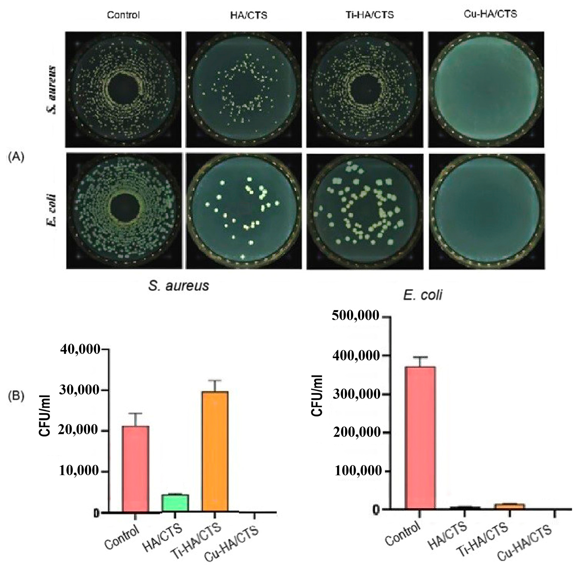
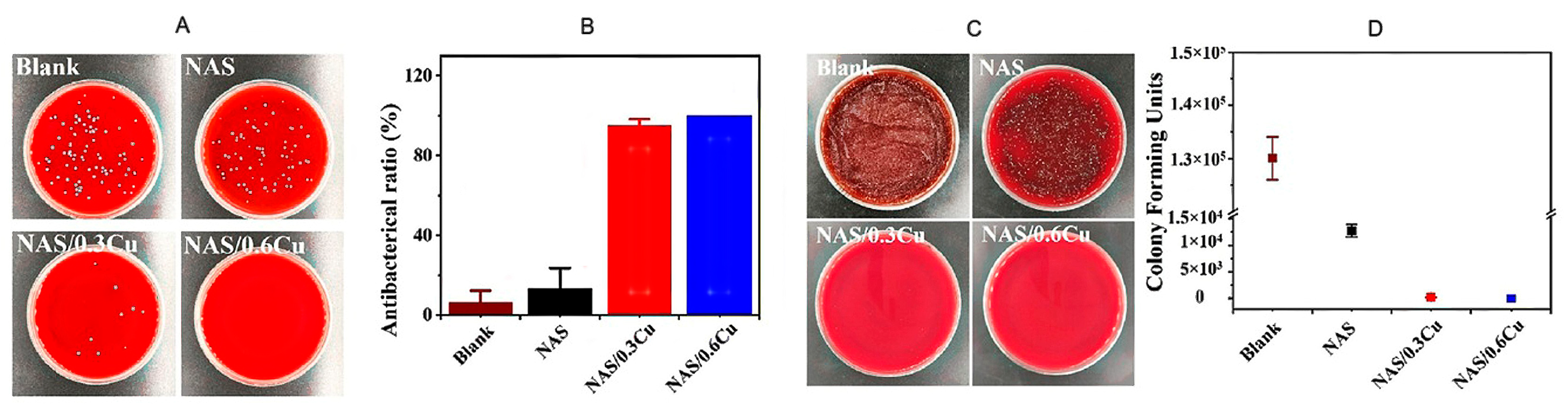
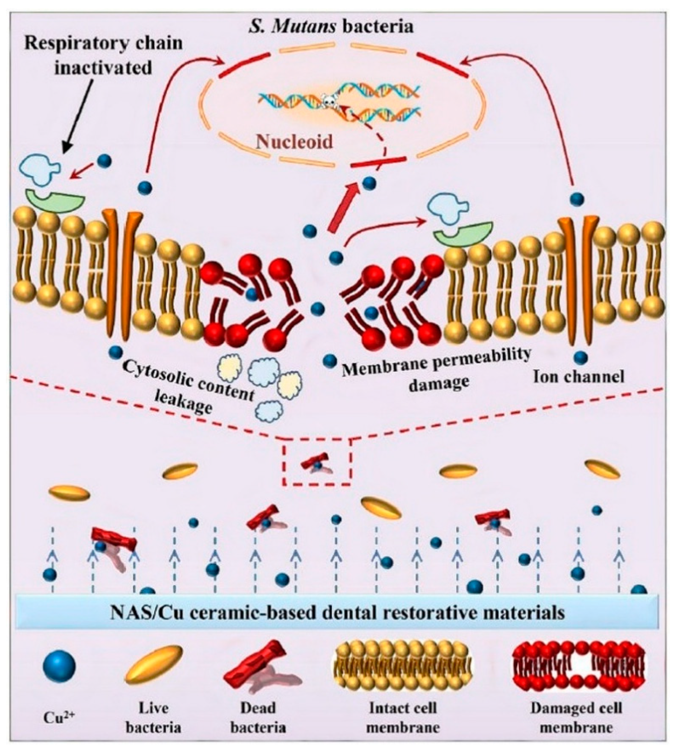
3.2. Antibacterial Properties of Cu and Cu Ions

Copper cations exhibit strong antibacterial activity, which can be explained by the disruption of the cell membrane, the alteration of intracellular biochemical processes, and the damage to bacterial DNA and nucleic acids by chelation. Cu ions can bind to the cell walls of bacteria, and by binding to phospholipids, they impair the integrity and function of cell membranes and related proteins. This can lead to alterations in the physicochemical properties of the membrane and a reduction in membrane fluidity and flexibility. Oxidation of membrane lipids is the fundamental mechanism by which Cu ions destroy bacteria [24,25,75]. The concentration of Cu ions in materials is challenging because Cu is known to have a dose-dependent effect due to its cytotoxicity. An optimal Cu ion concentration has a dual effect: it destroys bacteria and promotes the proliferation of normal eukaryotic cells. Wound healing can also be influenced by Cu ions, as they regulate extracellular matrix remodeling, keratinocyte growth, and integrin production necessary for epithelialization [23].
Calcium phosphate (CaP) ceramics are known for their biocompatibility and bioactivity, which make them superior to other bone substitutes. They can repair broken bones and are thus suitable for use in orthopedics, maxillofacial surgery, and dentistry. Calcium phosphates are available as ceramic blocks, porous sponges, granules, or calcium phosphate-based bone cement [42,76,77]. Calcium phosphates inherently exhibit no antibacterial activity, and infections often occur due to improper implantation procedures or inadequate sterilization, which negatively affect the patient’s treatment. To avoid such risks, many attempts are being made to endow calcium phosphates with antibacterial properties [8,38,78].
Pantak et al. [42] investigated the antibacterial activity of hybrid hydroxyapatite/chitosan (HA/CTS) granules and their modification with titanium (Ti-HA/CTS) or copper (Cu-HA/CTS) against S. aureus and E. coli strains. Chitosan and copper ions are well-known for their antimicrobial properties [8,79]. The concentration of Ti resp. Cu was 5 wt% (Figure 6). TiCl3 and Cu(N03)2 were used as sources of titanium and copper ions [42].
To investigate the antibacterial activity of these doped materials, four cements were prepared: tricalcium phosphate (α-TCP) as a control group, HA/CTS, Ti-HA/CTS, and Cu-HA/CTS. Mueller–Hinton agar plates were used to evaluate the antimicrobial activity. Figure 7 shows the results of the bacteria colonization tests on the prepared cement surfaces based on chitosan [42].
The test results showed that the application of hybrid hydroxyapatite/chitosan granules suppressed bacterial adhesion to the surfaces. Titanium-modified biomaterials reduced the accumulation of S. aureus bacteria, but they did not reduce and rather increased the adhesion of E. coli bacteria. On the other hand, Cu-modified biomaterials showed the highest antibacterial activity and prevented the adhesion of E. coli and S. aureus to the surfaces [42]. The difference between the Ti- and Cu- modified HA/CTS can be explained by their different chemical properties and their ability to form a stable bond to the biomaterials used in dental cements. The release rate of Ti ions from the ceramic composite to the environment is significantly lower than that of Cu ions. This has a positive effect on the treatment of patients over a long period of time, thus improving the safety and effectiveness of bone substitute materials in clinical practice at the expense of antibacterial activity [42]. To prolong the effectiveness of Cu ions, their release rate in body fluids must be reduced.
The different antibacterial activity of Ti and Cu ions cannot be explained only by their different dissolution rates. Ti ions did not exhibit any antibacterial activity on E. coli bacteria, which is due to the different compositions of the cell walls of S. aureus and E. coli bacteria. The enhanced development of E. coli bacteria in the presence of Ti ions can be explained by the catalytic activity of these ions in the intracellular biochemical processes of E. coli bacteria.
The authors did not investigate the cytotoxicity of these materials, which is an important factor in dentistry.
For their study, Fu et al. [35] used a ceramic synthesized from sodium alumino-silicate powder and copper oxide (NAS/Cu); the concentrations of Cu were 0.3% and 0.6%, respectively. S. mutans bacteria prepared using MTT methods were used to evaluate the antimicrobial activity of the NAS/Cu ceramics. As shown in Figure 8, both NAS/Cu ceramics showed significant antimicrobial activity compared to the control and the pure NAS samples [35].
On the NAS/0.3Cu ceramic surface, only minute colonization with S. mutans could be observed, while S. mutans was completely absent on the NAS/0.6Cu ceramic surface [35]. The antibacterial properties of the samples did not change after 3 days, which is a very important result for long-term antibacterial effects on S. mutans (Figure 8C,D).
Fu et al. found that NAS ceramics influenced the DNA replication of S. mutans to some extent, while NAS/Cu severely restricted DNA replication due to the penetration of Cu ions into the bacteria nucleoid of S. mutans (Figure 9). Sodium aluminosilicate ceramics doped with 0.6% copper oxide nanoparticles exhibited stable antibacterial activity against S. mutans and excellent biocompatibility [35].
Figure 9 shows the antibacterial activity of Cu ions. Positively charged Cu ions react with the negative charges of the bacterial cell membrane, causing damage and cell death. Cu ions can penetrate bacteria through ion channels and impair their permeability. This can lead to a flow-out of protein from the bacteria, resulting in a reduction in total protein and disrupting the biomolecular activity of the bacteria. Cu ions impair the respiratory chain and gene replication, which leads to the death of the bacteria [35].
For dental materials, the most important issue is biocompatibility.
Fu et al. also showed that small quantities of copper nanoparticles had no influence on the biocompatibility of sodium aluminosilicate ceramics while providing antibacterial activity. From these results, it can be concluded that these ceramics could be used in dental restoration to prevent secondary caries.

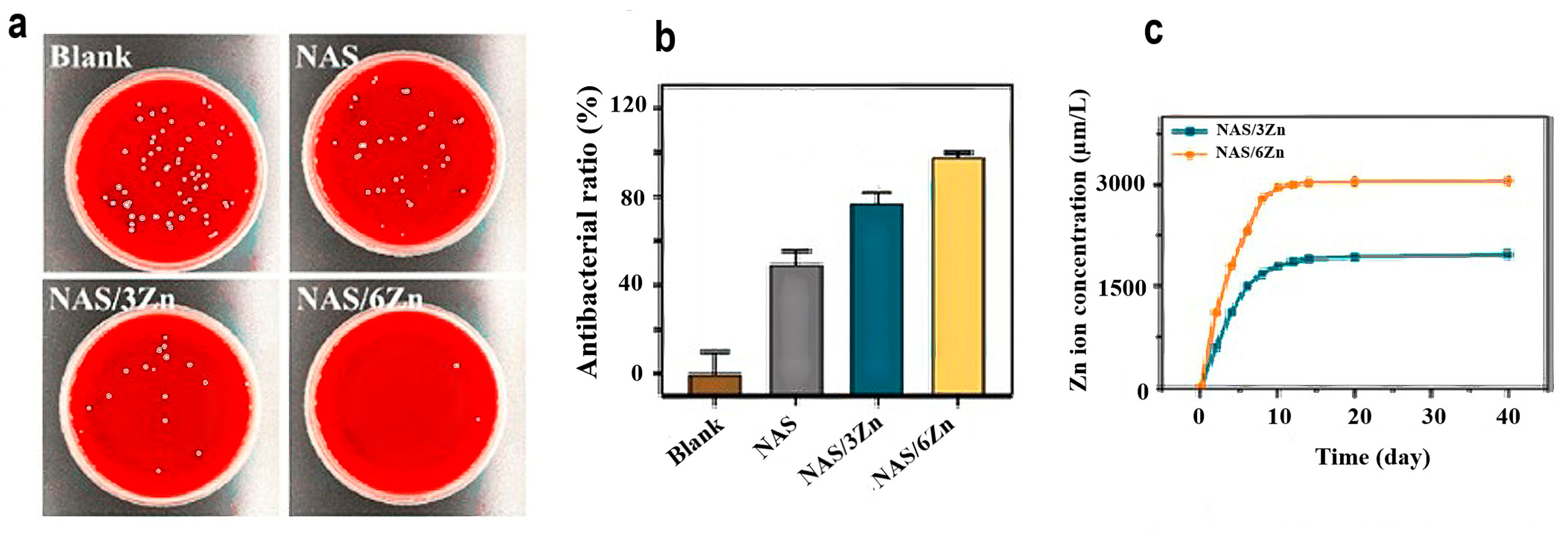
3.3. Antibacterial Properties of Zinc Oxide

Zinc is the second-most abundant trace element after iron and plays an important role in various physiological functions [80,81]. Thirty percent of the zinc accumulated in the body is found in the bone tissue [81,82]. Zinc plays a key role in inhibiting bone resorption as well as in bone formation, development, mineralization, and maintenance. Zinc can be used to prevent bone diseases caused by oxidative stress, such as osteoporosis [81,82,83].
Zinc oxide nanoparticles (ZnO-NPs) are one of the most commonly used inorganic materials with antibacterial activity. Due to their high application safety, they are used in disinfectants, dental materials, cosmetics, and pharmaceuticals [82]. They have been shown to exhibit selective toxicity against both Gram-positive and Gram-negative bacteria, while the effect on human cells is negligible [24,82,84]. A controlled release rate of Zn+ from ceramics plays a crucial role in antibacterial activity. Zn is vital for the survival and development of bacteria. Within an expected concentration range, bacteria can regulate their internal Zn concentrations for their physiological metabolism. However, excess zinc ions lead to some toxic effects, while zinc deficiency leads to the disruption of normal biological activities [84]. This means that the concentration of zinc ions in the cell medium is important for the antibacterial activity of zinc ions. Zn2+ ions interact with the negative charges of the bacteria membrane and cause physical damage to the cell membrane, leading to cell death [24,70]. The antimicrobial effect of ZnO-NPs depends on the particle size and concentration. Smaller particles are more active, but the effect may vary from microorganism to microorganism [70,82]. It has been confirmed that a sufficient number of zinc ions stimulates bone cells and induces a series of in vivo responses, including adhesion, spreading, proliferation, osteogenic processes, differentiation, osteogenesis, and mineralization [21,85].
Chen et al. [21] used ZnO-NPs to impart antibacterial activity to the barium titanate)/hydroxyapatite (BT/HA) piezoelectric ceramic system. BT/HA piezoceramics were used because natural bone also exhibits piezoelectric behavior and generates an induced potential under mechanical loading [21]. The concentration of ZnO-NPs used in this study was 1%. At a concentration below 1%, ZnO-NPs exhibit no pronounced antibacterial activity, while at a concentration of 2%, a detrimental effect on cell survival was observed. The composition of ZnO@BT/HA ceramic was 1% ZnO-NPs, 70% BT, and 30% HA. Chen et al. used S. aureus as Gram-positive bacteria and E. coli as Gram-negative bacteria to evaluate the antibacterial activity of these ceramics.
As shown in Figure 10A, ZnO@BT/HA exhibited high antibacterial activity against S. aureus and E. coli [21]. The counting of viable and dead bacteria data (Figure 10B,C) quantitatively confirmed the antibacterial activity of ZnO-NPs incorporated into BT/HA ceramics. The authors used a CCK-8 assay to evaluate the toxicity profile of ZnO-NPs toward DPSCs (dental pulp stem cells). A significant decline in the cell survival rate was observed at concentrations exceeding 1 wt%, whereas concentrations below 1 wt% showed no detrimental effects on DPSC viability. The authors investigated the bioactivity of these ceramics. ZnO-NP-doped BT/HA ceramic samples exhibited significantly enhanced proliferation capabilities compared to those on HA and BT/HA surfaces. An optimal concentration of ZnO-NPs also promoted MSC (mesenchymal stem cell) osteogenic differentiation through various pathways, which is a powerful therapeutic tool for bone defects. Zinc serves as an important agent for the development of cell structure and functions and acts as a direct or indirect regulator affecting the communication between nociceptor neurons and the host immune systems, modulating the inflammatory response and host defense against bacterial infections [84]. Information on the antibacterial activity over a longer period of time is missing, and this must be investigated.
The development of piezoceramics, known for their superior osteogenic properties with additional antibacterial activity, opens up new possibilities for the regeneration of diseased bones. The development of multifunctional bone tissue engineering ceramics offers promising potential for repairing complex tissue defects.
Xiao et al. [17] investigated the effect of ZnO-doped dental crown restoration materials on the prevention of secondary dental caries. Amorphous sodium alumino-silicate (NAS) doped with 3 resp. 6 wt% ZnO. S. mutans bacteria was used to investigate the antibacterial activity of NAS/3Zn and NAS/6Zn ceramics. As shown in Figure 11, both doped Zn-ceramics possess antibacterial activity, which increases with the ZnO content of the ceramic [17].
The authors also investigated the release of Zn ions into the environment, which was not interrupted during the test period of 0–40 days (Figure 11c). These results are important for the long-term effectiveness of the dental device.
According to Xiao et al., Zn2+ ions destroy the cell membrane structure, induce the leakage of bacterial protein, and lead to the death of bacteria. NAS/Zn ceramics have superior biocompatibility and can be used as dental restoration materials with additional antimicrobial activity to prevent secondary caries [17].
The typical problem with doped bioceramics is maintaining long-term antibacterial activity. When the bactericidal ions responsible for the antibacterial effect of the ceramics are completely released from the ceramic, the antibacterial effect is lost. The results of this study are promising for the development of ZnO-functionalized ceramics with long-term antibacterial effects.

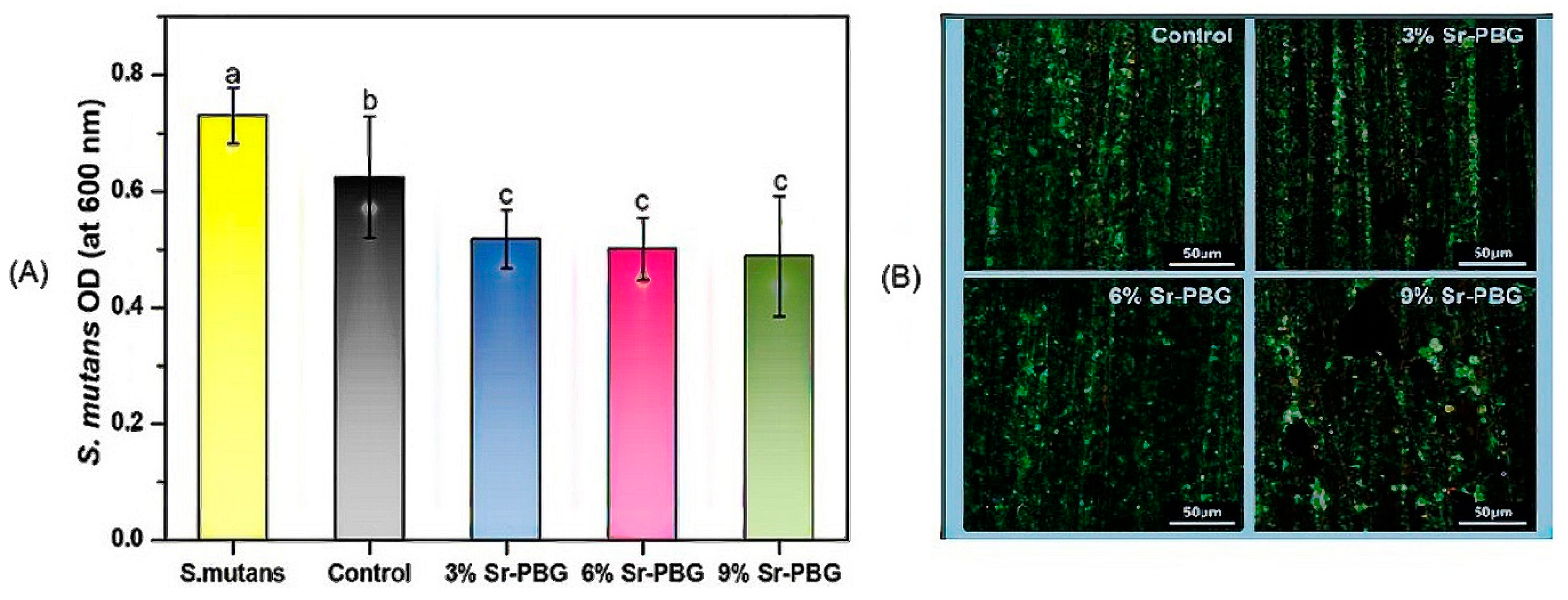
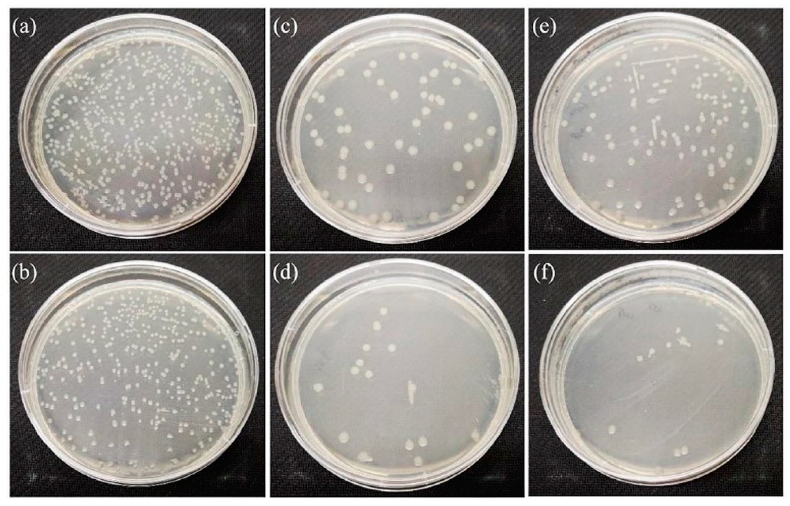
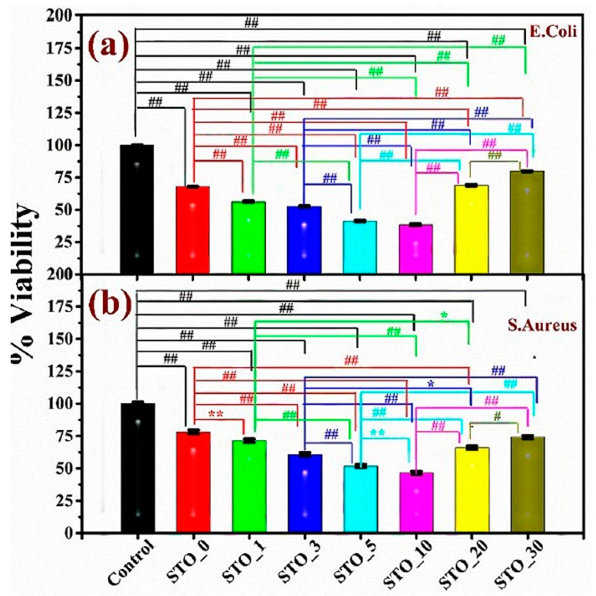
3.4. Antibacterial Properties of Strontium

Strontium cations are known for their role in bone formation and bone resorption due to their osteoinductive and antibacterial properties [86,87,88,89]. These properties are exploited to develop bioceramics for improved bone regeneration and implant integration [90].
Peri-implantitis is a polymicrobial infection that leads to inflammation in the peri-implant mucosa and progressive bone resorption, which may result in implant loss [91,92]. To prevent the loss of the dental or orthopedic implant, its surface can be coated with antibacterial metal ions [93]. In clinical practice, the successful implantation and long-term use of dental and orthopedic implants depend not only on their antibacterial properties but also on their bioactivity, such as promoting osteocyte adhesion, proliferation, differentiation, stability, biocompatibility, and anticoagulation [93,94].
Alshammari et al. [91] investigated the effect of strontium hydroxide on bacteria asso-ciated with peri-implantitis. They used the strains A. actinomycetemcomitans (A.a.), S. mitis, E. coli, S. epidermidi, P. gingivalis, and F. nucleatum. The antibacterial effect of Sr2+ ions at concentrations of 10 mM and 100 mM was investigated using the agar plate method.
The biofilm viability assay test showed that 100 mM Sr(OH)2 solution killed all tested bacterial strains. Complete growth inhibition of S. mitis, S epidermis and F. nucleatum as well as significant growth inhibition of A.a., E. coli and P. gingivalis was observed with a 10 mM Sr(OH)2 solution. Furthermore, 10 mM Sr(OH)2 demonstrated bactericidal activity against all tested strains, with most bacterial cells partially or completely damaged in a bacterial viability assay [91]. No antibacterial effect was found for a concentration below 10 mM or higher than 100 mM [89]. At a concentration higher than 100 mM, the Sr2+ ions no longer dissolved properly in the liquid medium and tended to precipitate, which explains the dependence of antibacte-rial activity on concentration.
The antibacterial activity of Sr ions can be attributed to the higher pH value of the environment, which gradually increases over time due to the release of Sr2+ ions. Most bacteria prefer to grow at a neutral pH value, and a change in it can affect their growth ability as well as their viability and properties. If the concentration of Sr ions in the local medium is too high, mammalian cells can react similarly to bacteria. This effect must be taken into account when using Sr ions to develop new antibacterial bioceramics.
Composite resins are widely used in dentistry due to their aesthetic advantages, high bond strength as a cement, and mechanical reinforcement, but they do not possess any antibacterial properties. If bacteria accumulate at a marginal filling, secondary caries and damage to the composite filling can occur.
Thus, researchers focus on developing antibacterial composite resins for long-term use. The challenge is that adding antibacterial agents to the composite resin commonly affects the mechanical properties of the composite resin in such a way that the properties of the composite become insufficient for long-term applications. As an additional negative effect, the antibacterial activity only lasts for a short period. The development of composite resin with long-term antibacterial properties would be highly beneficial clinically.
Go et al. [95] developed a glass (Sr-PBG) made of P2O5 (50 mol%), CaO (15 mol%), Na2O (20 mol%), and SrO (15 mol%). They used Filtek Z350XT as the composite resin. The concentrations of Sr-PBG in the composite resin were 3, 6, and 9%. They used S. mutans to evaluate the antibacterial properties of the new composite resin.
As shown in Figure 12, the optical density (OD) of S. mutans decreased with increasing Sr-PBG content in the composite resin [95]. The antibacterial effect of the composite resin containing Sr cations can be explained by two mechanisms. Firstly, the release of ions into the environment increases the osmotic pressure and the pH value, which affect the growth of the bacteria. Secondly, the dissolved Sr ions can cause enzyme modification of the bacteria, leading subsequently to the death of the bacteria [61,95,96]. From the results of this study, it can be concluded that Sr-PBG incorporated into the composite resin inhibits the growth of S. mutans. In addition, Sr-PBG can be used as a filler for antibacterial dental materials without deteriorating the mechanical properties of the resin. However, the clinical oral environments are very complex, which may affect the properties of the new cement resin. From this point of view, further tests need to be carried out in such environments to clarify the suitability of such antibacterial composite resin. Antibacterial activity has only been tested on S. mutans bacteria. The antibacterial activity of such composite resin needs to be investigated in long-term studies on different bacteria.
Zhao et al. [61] investigated the antibacterial efficacy of SrO- and ZnO-bioactive glass (BG) scaffolds against E. coli bacteria strains. The composition of the BG was 6 mol% Na2O, 8 mol% K2O, 8 mol% MgO, 22 mol% CaO, 18 mol% SiO2, 36 mol% B2O3, and 2 mol% P2O5. CaO was replaced by SrO and ZnO to prepare the doped BG. The concentrations of SrO and ZnO were 5% and 10%, respectively. Furthermore, Zhao et al. also investigated the effect of BG, Zn-BG, and Sr-BG on cell proliferation. The results showed that 5Sr-BG had a stronger effect on cell proliferation than BG alone. The other samples had no significant effect on cell proliferation. Doping with a small amount of Sr promoted cell proliferation, while doping with Zn inhibited it. The degree of inhibition strongly depended on the amount of zinc oxide as well as the composition of the glass and its rate of degradation [61].
As shown in Figure 13, BG alone already offers an antibacterial effect, but Sr-BG and Zn-BG are more effective against E. coli bacterial strains [61]. Trace metals such as iron, zinc, copper, manganese, molybdenum, tungsten, nickel, cobalt, chromium, vanadium, and several other elements play important roles in a variety of biological and chemical processes when present in the optimum concentrations. Some of these metals are of great importance for enzymatic processes, while others can act as electron donors and facilitate the binding of molecules to corresponding receptors [89,97]. When the concentration of these trace metals increases above the optimal concentration, the bacteria attempt to adapt, and toxic events can occur. Metal ions can impede bacterial proliferation through different mechanisms: (a) enzyme modification, (b) damage to the bacterial cell walls, (c) oxidative stress and intercellular reactive species, and (d) DNA damage and the regulatory roles of DNA and RNA. The antibacterial activity of Sr ions and Zn ions can be explained by two different mechanisms. Sr ions have an effect on bacterial enzymes, which lose their functions and thus lead to the death of the bacteria. Zn cations can also react with electronegative molecules in cell membranes, resulting in the destruction of the cell membrane and subsequently the damage of cell function [69].
Information on the cytotoxicity of the samples and comparisons between Sr ions and Zn ions are missing. The antibacterial effect needs to be investigated in long-term studies.
Yadav et al. [6] investigated the antibacterial and bioactive properties of 85S bioglass (58SiO2-37CaO-5P2O5) and SrTiO3 (STO). This bioglass is biocompatible and exhibits good mechanical properties due to its high silica concentration. In addition to the mechanical properties, the high silica concentration is also responsible for a reduced dissolution rate. Replacing Ca ions with Sr ions was found to be remarkably effective in bone regeneration and angiogenesis [6,60]. The investigated concentrations of SrTiO3 in 85S bioglass were 1, 3, 5, 10, 20, and 30 wt%. In this study, unmodified bioglass showed slight antibacterial activity against E. coli and S. aureus (Figure 14) compared to the control sample.
Antibacterial activity further increased with an increase in the SrTiO3 content in the bioglass up to 10 wt% of STO (STO_10). Interestingly, samples with higher amounts of STO in the bioglass (STO_20 and STO_30) exhibited a significantly lower antibacterial effect compared to STO_10. STO_20 and STO_30 have antibacterial activity compara-ble to that of unmodified or only slightly STO-modified bioglass [6]. One can understand the behavior in terms of the release rate of Sr cations from the bioglass. At an STO content of up to 10 wt%, the Sr cations released from the glass increased the pH value of the environment, resulting in protein denaturation, cell membrane destruction, and DNA as well as RNA disruption. All these processes hinder vital cell functions, including enzyme activity and energy regeneration, ultimately leading to bacterial death. The adhesion of SrTiO3 particles to the cell surface can also hinder the transportation of vital nutrients, which can lead to a rupture in the cell membrane and cell death [6]. At amounts higher than 10 wt%, the Sr cation release rate was significantly reduced due to slower dissolution of the composites, resulting in a decline in the pH value, so that the mechanism leading to cell death described above occurred to a lesser extent.
The antibacterial activity of the samples was demonstrated for 7 days; therefore, long-term measurements are required.
The cytotoxicity investigation showed that the samples STO_20 and STO_30 exhibited toxicity to red blood cells, but more detailed studies on the toxicity and concentration of SrTiO3 are required.

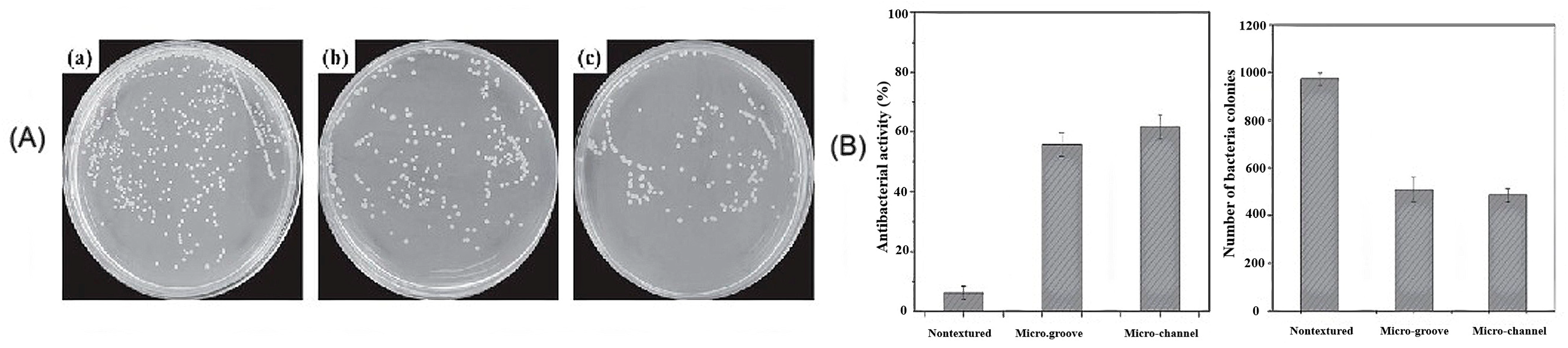
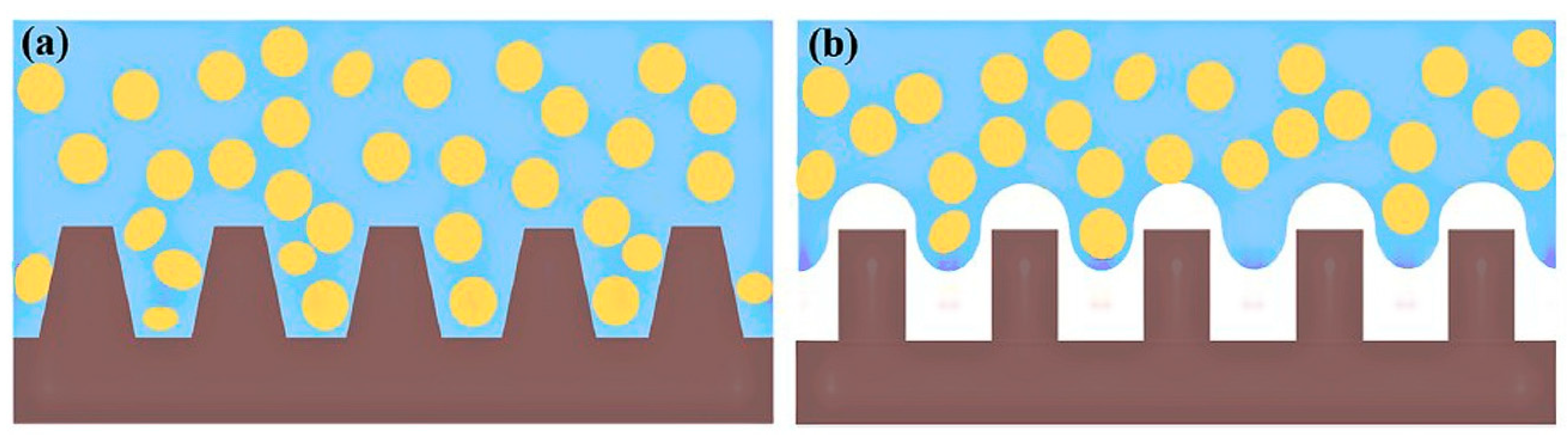
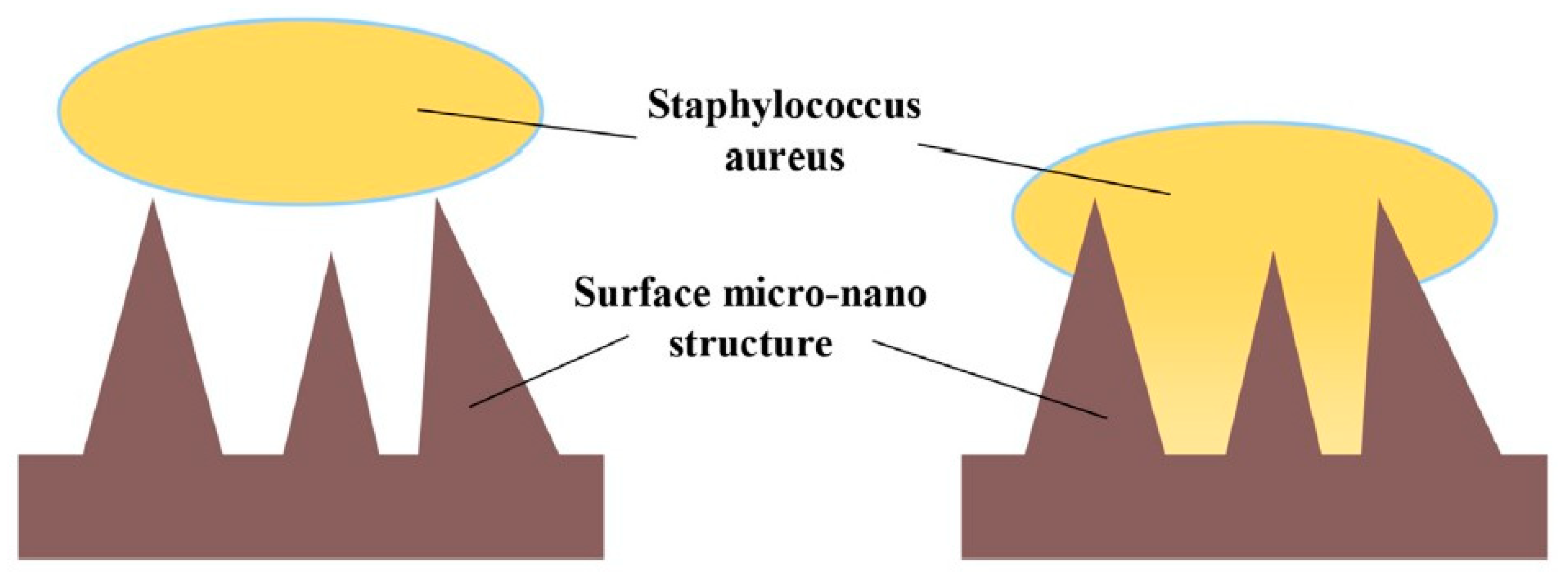
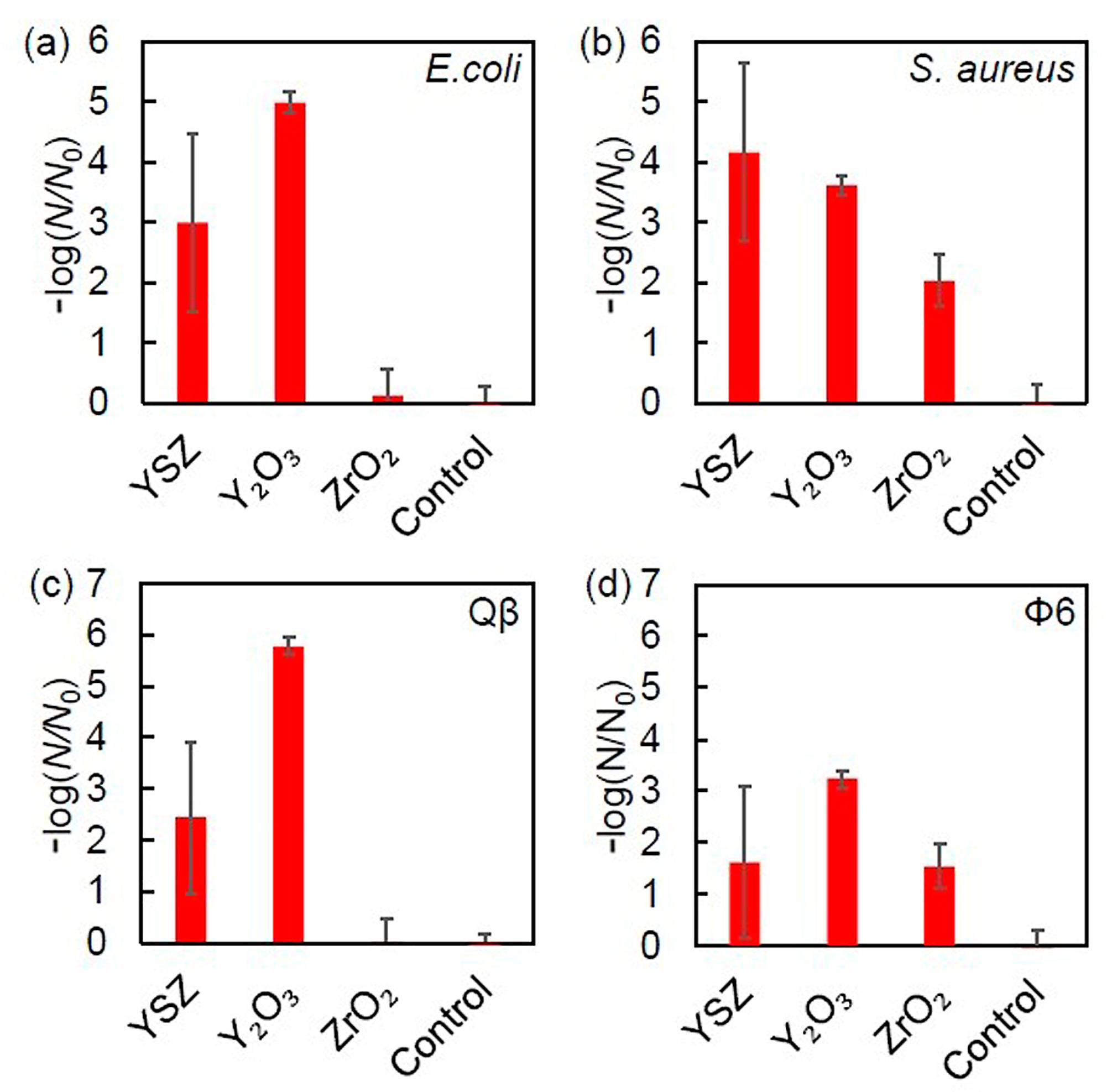
3.5. Influence of Surface Properties on the Antibacterial Activity of Bioceramics

Yttria-stabilized zirconia (YSZ) ceramics are widely used for the fabrication of dental and orthopedic implants due to their excellent mechanical strength and aesthetic and biological properties, and they are an alternative material for patients with metal allergies [59,98]. The largest problem with zirconia restorations is their high hardness, which is responsible for irreversible and irreparable erosion of natural teeth during daily chewing. Zirconia ceramics are biocompatible but not bioactive or antibacterial, which can lead to plaque formation and a high risk of peri-implantitis or even loss of supporting bone [56].
Moghanian et al. [60] investigated the antibacterial activity of ZrO2 in 58S bioactive glass. The composition of 58S bioactive glass (BG) was 60 mol% SiO2, 36 mol% CaO, and 4 mol% P2O5. CaO was replaced by ZrO2 at a concentration of 5 mol% (BG-5Zr) and 10 mol% (BG-10Zr), respectively. Antibacterial activity was tested against S. aureus bacteria for 14 days. In addition to the antibacterial activity, Moghanian et al. also examined the bioactivity of BG-5Zr and BG-10Zr glasses.
As shown in Figure 15, BG-5Zr exhibited higher antibacterial activity than the other glasses. Furthermore, higher cell proliferation and activity of osteoblasts were observed for BG-5Zr glass [60]. An alkaline pH range creates an unfavorable environment for bacterial growth and metabolism, causing morphological changes in bacteria and their death. The release of ions and changes in their concentration in the bacterial environment affect the bacterial cell membrane and cause a pressure drop that alters cell size, shape, and membrane tension. The positive charge of Zr4+ interacts with the negatively charged bacterial cell wall, resulting in the rupture of the walls and, subsequently, cell death [99]. The lower antibacterial activity of BG-10Zr can be explained by the solubility of the BGs in the SBF medium and the crystallization of the BG. The incorporation of Zr into the structure of the BG reduces the solubility of the BG because Zr is less electronegative than Ca, and the Zr-O covalent bonds strengthen the network of the BG, resulting in lower concentrations of Na+, Ca2+, and Zr4+ in the SBF medium [60]. These ions are responsible for the increasing pH value. The increase in Zr4+ ions changes the structure of the BG from amorphous to crystalline, which also affects the dissolution behavior of the BG and the corresponding ion concentration, as amorphous BGs exhibit a higher dissolution rate than crystalline ones. This dissolution behavior affects not only the pH value but also cell proliferation. Compared to other samples, BG-5Zr samples showed a higher increase in cell proliferation and osteoblast-like cell line activity due to the formation of HCA on the BG surface. BG-5Zr glass can be used as a potential candidate for bone tissue engineering and clinical applications based on the results of long-term studies on antibacterial activity, cell proliferation, and in vivo cytotoxicity tests.
The proliferation, growth, and adhesion of bacteria to a surface are influenced by numerous factors, such as chemical, physical, and physicochemical changes. The physicochemical changes of the surface, e.g., the wettability of the surface, can contribute to bacterial adhesion during the first stage, followed by irreversible adhesion (molecularly mediated binding between the cell and the surface) in the second stage [100,101]. The antibacterial and cell adhesion properties of zirconia can be modified by changing its texture [56]. Self-cleaning surfaces are well known in nature, e.g., lotus leaves. The self-cleaning ability of the lotus leaf is based on a hierarchical double structure with hydrophobic wax droplets that reduce or prevent the adhesion of various contaminants. Such behavior inspired researchers to develop a self-cleaning surface for bioceramics for medical applications [102].
Ghalandarzadeh et al. [58] used a carbon dioxide (CO2) laser to create micro-grooves and micro-channel microstructures on zirconia surfaces. The laser texturing performed on the YSZ resulted in a change in the wettability of the surface, changing it from hydrophilic to hydrophobic. It was found that the degree of hydrophobicity was higher in the channel-shaped samples than in the grooved-shaped ones. Both zirconia surface textures showed antibacterial activity, but this was more pronounced for the channel-shaped samples, as shown in Figure 16.
Xu et al. [56] (Figure 17) confirmed that introducing a nature-inspired lotus effect on the YSZ surface with hydrophobic properties can prevent the growth of bacterial colonies, as well as the proliferation and adhesion of S. aureus bacteria, as schematically illustrated in Figure 17. The air gaps of the microtextured surface prevent direct contact with the bacteria, which leads to a reduction of adhering bacteria (Figure 17b).
Besides this effect, the microtextured YSZ surface can also interact with the bacteria in a different manner, as schematically depicted in Figure 18. Here, the part of the bacteria adhering to a textured surface ruptures due to irreversible damage to the cell walls, causing the death of the bacteria. This nicely illustrates that modifications of the surface structure can influence the biological responses of biomaterials used in dental implants, such as protein absorption, cell adhesion, proliferation, and antibacterial activity, without altering the chemical composition of the material. Another advantage of lasers is that they are becoming more affordable while offering high flexibility, reliability, reproducibility, and precise ablation capabilities.
Nevertheless, the problem with such microtextured surfaces is their long-term antibacterial activity. The accumulation of dead bacteria on textured surfaces can trigger an inflammatory reaction due to an immune response and potentially even alter the textured surface. Therefore, the long-term antibacterial activity of such microtextured surfaces needs to be tested for a better clinical approach on tooth-like surfaces.
There are some controversies in the published literature regarding the antibacterial properties of 3YSZ ceramics. Some authors [56,99,103] have reported that dental zirconia ceramics lack antibacterial properties, and peri-implantitis has been observed in zirconia implants.
Kiribayashi et al. [104] investigated the antibacterial properties of YSZ (3YSZ), Y2O3, and Zr2O3 powder. S. aureus (SA, NBRC 12732) was used as Gram-positive bacteria and E. coli (EC, NBRC 3972) as Gram-negative bacteria. Bacteriophage Qß (host bacteria: E. coli) was used as a non-enveloped virus to test the antiviral activity. Bacteriophage Φ6 (host bacteria: P. syringae) was used as an enveloped virus. Figure 19 shows that YSZ possesses antibacterial activity against EC and SA. The order of antibacterial activity against EC was Y2O3 > YSZ ≫ ZrO2 (Figure 19a). The order of antibacterial activity against SA was YSZ ≈ Y2O3 > ZrO2 (Figure 19b). The antiviral activity of YSZ was observed against both Qß and Φ6. The order of antiviral activity against Qß was Y2O3 > YSZ ≫ ZrO2 (Figure 19c), and that against Φ6 was Y2O3 > YSZ ≈ ZrO2 (Figure 19d) [104].
The adsorption of cations on bacterial and virus surfaces can be explained by the electrostatic interactions between the cations and the negatively charged bacteria and viruses. Y2O3 exhibits a high affinity for phosphoric acid, a component of bacteria and viruses, resulting in its high antiviral and antibacterial activity due to strong interactions with phosphoric acid. Due to this interaction, Y3+ can deform the lipid bilayer membrane by mimicking Ca2+. The interaction of Zr4+ with phosphate is weaker than that of Y3+. The inactivation of the proteins resulting from these interactions disrupts bacterial growth [104]. The authors also attributed the death of bacteria and viruses to the denaturation of bacterial proteins. In addition, the surface shapes of the investigated powders could play a role in their antibacterial and antiviral activity [104]. Cytotoxicity was not observed in any sample.
Further studies should be conducted to further investigate the antibacterial and antiviral activities of 3YSZ powder and 3YSZ ceramics to better understand the interaction between YSZ and various bacteria and viruses.

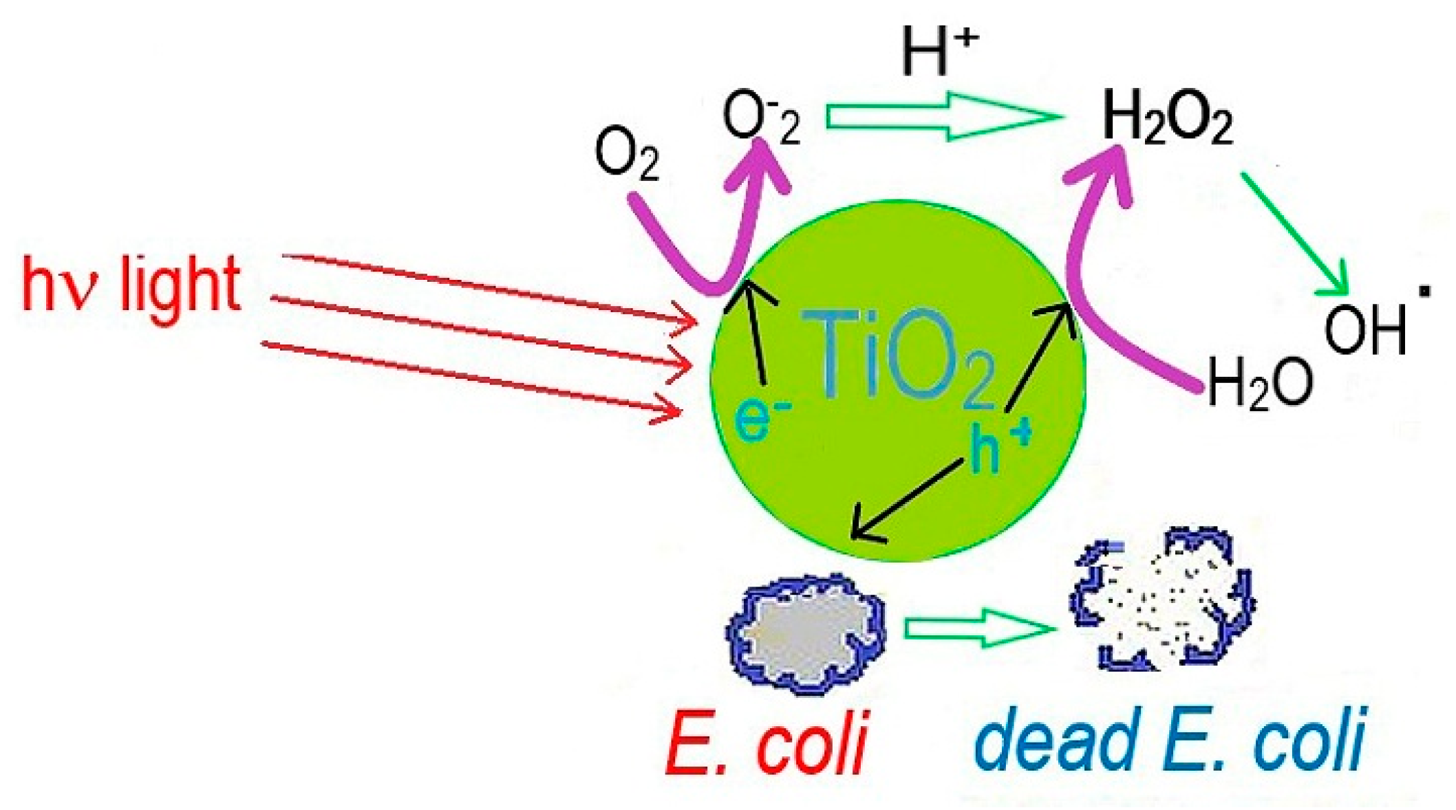
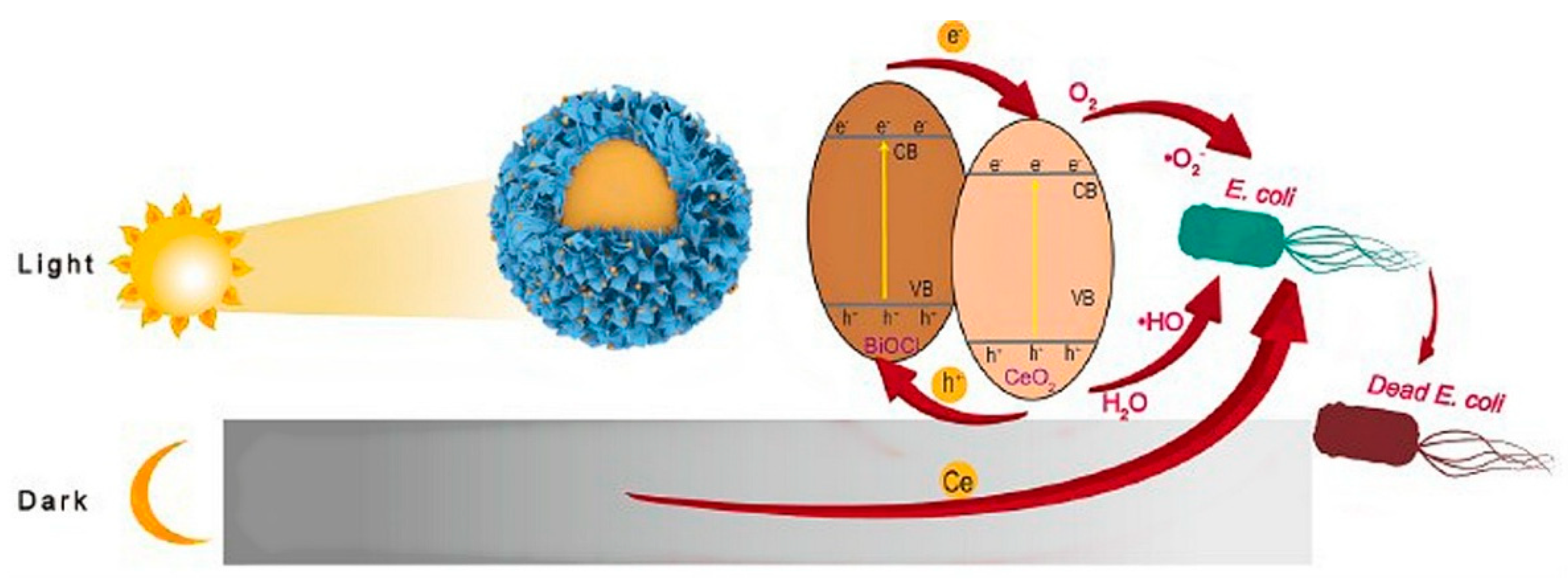
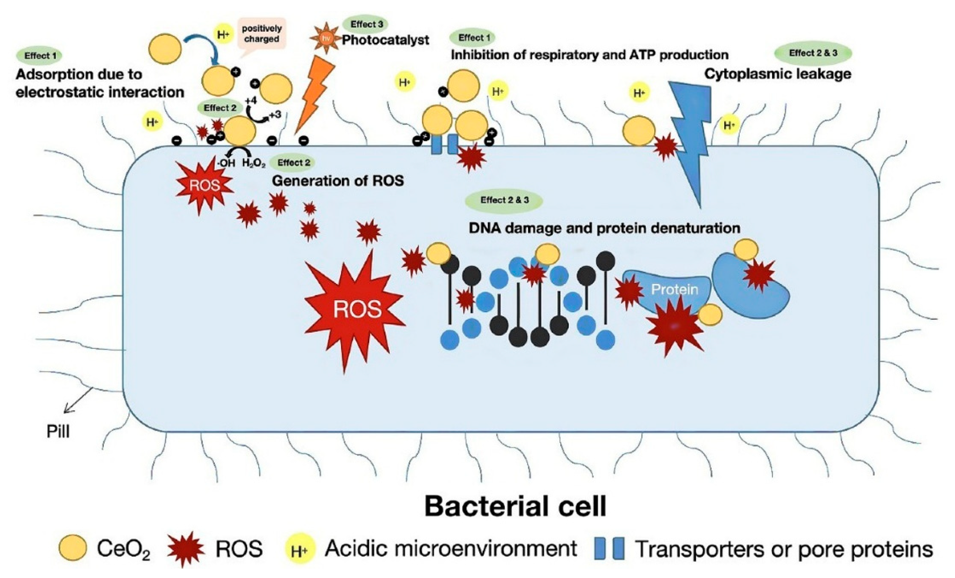
3.6. Antibacterial Activity of Cerium Oxide

Antibacterial ceramics are a focus of research due to their functions, such as self-cleaning and bacterial killing. Adding antibacterial agents to glaze ceramics to generate metal ions or free radicals with antibacterial activity is one of the main methods of preparing antibacterial ceramics. Adding well-known photocatalytic materials such as TiO2 and ZnO to ceramics can generate photogenerated electrons and holes, which can generate free radicals to inactivate bacteria. However, these ceramics often require ultraviolet light for their antibacterial activity, so their application is limited [34,49,78,105,106]. The mechanism of the well-known and described photoactive properties of TiO2 can be explained by the following chemical equations and is graphically summarized in Figure 20.
TiO2 + hv → TiO2* + h+ (hole) + e (electron)
H2O + h+ → OH⠁ + H+
OH + h+ → OH⠁
O2 + e → O2
O2 + 2H+ + 2e → H2O2
H2O2 + e → OH⠁ + OH
Bacteria + OH⠁ + O2 → dead bacteria
Ag, Au, and Cu exhibit antibacterial activity in ceramic glazes in the absence of light, but their activity decreases when their ions are released into the environment, which can be harmful to patients [62]. Ce ions have been shown to stimulate the proliferation, differentiation, and mineralization of osteoblasts; increase collagen production in human mesenchymal cells; and enhance the mechanical properties of bones [107,108,109]. They are known for their antibacterial activity. The reversible conversion of Ce3+ to Ce4+ is the basis of ROS (reactive oxygen species) generation when CeO2 is used as an antibacterial material. The antibacterial activity of CeO2 is not based on the penetration into the cell membrane of bacteria but on the induction of oxidative stress with the production of ROS, which leads to the degradation of DNA and RNA and their proteins [107,108,109,110,111]. CeO2 is known for its low cytotoxicity to mammalian cells and its unique antibacterial mechanism on Gram-negative and Gram-positive bacteria [62]. Almost all bacterial cells are surrounded by a cell wall comprised of peptidoglycans, which protects the cell membrane from rupture due to high intracellular turgor pressure and maintains the cell shape. In contrast to the Gram-positive cell wall, which consists of a thick, porous layer of peptidoglycans with attached teichoic acids, the Gram-negative bacterial cell wall consists of a relatively thin layer of peptidoglycans surrounded by an asymmetric, highly impermeable bilayer with lipopolysaccharides in the external leaflet and phospholipids in the internal leaflet [111]. Bacteria are negatively charged when they are suspended in physiological solutions (pH = 7.4). Gram-negative bacteria display a greater negative potential than Gram-positive bacteria. Cerium ions, which are positively charged, are electrostatically adsorbed on the bacterial surfaces, resulting in high antibacterial toxicity. Furthermore, CeO2 is known for its antibacterial activity as well as for stimulating angiogenesis, osteogenesis, and wound healing.
Bian et al. prepared glass ceramics that exhibit long-lasting antimicrobial activity in both the dark and the light [62]. The compositions of CeO2 glass-ceramics (CGC) were 64 wt% Bi2O3, 22 wt% B2O3, and 14 wt% CeO2. They treated this ceramic with 0.2 M HCl and developed a new ceramic: BiOCL@CeO2 (BCGC) [62]. This BCGC ceramic was tested for antibacterial activity against E. coli in the dark and under illumination. Bien et al. found that BCGC ceramics exhibit antibacterial activity when exposed to darkness and visible light. Figure 21 shows the mechanism of the antibacterial activity of BCGC ceramics.
When irradiated with visible light, BCGC ceramics can generate hydroxyl radicals (OH) and superoxide radicals (O2), which damage the cell membranes of bacteria and lead to their death. In the dark, the release of cerium ions from the BCGC contributed to the antibacterial activity of the BCGC ceramics (Figure 21) [62]. The results of this study show that it is possible to develop durable and effective antibacterial ceramics that work in the dark and under illumination.
Gavinho et al. [64] investigated the effect of cerium oxide on the bioactivity and antibacterial activity of Bioglass 45S5. The concentrations of CeO2 were 0.25 (Ce0.25), 0.5 (Ce0.5), 1 (Ce1), and 2 mol% (Ce2). The antibacterial activity of cerium oxide-doped bioglass was tested against E. coli, S. aureus, and S. mutans. The bioactivity was also investigated. All samples showed antibacterial activity against all bacterial strains, but samples Ce1 and Ce2 were more effective against E. coli. Cerium oxide-doped bioglasses showed less activity against S. aureus. Ce1 showed the highest activity against S. aureus, while Ce2 samples showed no significant effect. The antibacterial activity generally increased with increasing cerium oxide content. The different antibacterial activities of cerium oxide against Gram-positive and Gram-negative bacteria can be explained by the different structures of the cell walls of these bacteria [111]. The results showed an increase in the bioactivity of the BG for the Ce2 samples, which supports the use of Ce2 BG as a material for bone regeneration, the filling of bone defects, or as a coating material for implants [64].
The development of ceramics with multifunctional properties and stable antimicrobial activity is still a major challenge for researchers. An effective method for preventing enamel demineralization and the appearance of cavitated lesions is the use of dental materials that are resistant to bacterial accumulation. Ideally, the material should have bioactive properties for the mineralization of enamel.
Varghese et al. investigated the antibacterial activity of a dental resin with CeO2 nanoparticles (CeO2NP) as an additive [112]. The resin was doped with 3 wt% CeO2. They tested the antibacterial activity of their experimental dental resin against S. mutans, S. mitis, S. aureus, and Lactobacillus spp. It was found that such a dental composite was effective against all tested bacterial strains [112].
The antibacterial activity can be explained by two mechanisms: (a) adsorption on the bacterial surface and (b) oxidative stress. Nanoparticles possess large contact surfaces, which allow positively charged CeO2NPs to adsorb onto the negatively charged bacterial membrane. CeO2NPs pass through the membrane, and the special ion pumps are hindered by the viscosity increase of the membrane. This has a significant impact on the transport exchange between bacterial cells and the fluid, leading to bacterial death. The released cerium ions can disrupt the electron flow and the respiration of the bacteria by reacting with thiol groups (-SH) or by adsorbing onto transporters, disrupting nutrient transport inside the bacteria [112].
Oxidative stress is a significant factor for the antibacterial activity of CeO2 and is caused by the production of reactive oxygen species (ROS) in vivo, whereas ROS produced on bacterial membranes is caused by the reversible conversion of Ce3+ to Ce4+. ROS can damage nucleic acids, proteins, polysaccharides, lipids, and other biological components and even destroy them. Such reactions occur on CeO2NPs when they are irradiated with ultraviolet light (hv) [111]:
CeO2 + hv → CeO2 (e + h+)
e + O2 → O2
e + H2O2 → OH + ⠈OH
h+ + OH → ⠈OH
Figure 22 schematically illustrates the antibacterial activity of CeO2NPS. It should be emphasized that the exact mechanism of the antibacterial activity of CeO2NPs is significantly more complex than depicted. It depends not only on the physical properties of CeO2NPs but also on the bacterial type (Gram-positive or Gram-negative) and the environmental condition for bacterial growth. The interaction of CeO2NPs with bacterial surfaces also depends on their size, shape, surface chemistry, and solubility [112]. The advantages of CeO2NPs have generated increased interest in using this material for various medical applications without compromising biocompatibility, which is especially important in the field of dental materials.
The rapid electron-hole recombination and low production of active species make the use of CeO2NPs as a pure material difficult. The combination of CeO2NPS with other nanomaterials could overcome this single-component problem and create a synergistic effect that separates photoinduced electrons/holes and optimizes the functional properties of both components.

4. Conclusions

Bacterial infections are often responsible for the failure of implants and scaffolds in regenerative dentistry and orthopedics. Drug-resistant bacteria and the long-term efficacy of antibiotics are controversial. Therefore, the need to overcome antibacterial resistance and the consequences of long-term use of antibiotics has increased the research interest in the development of bioceramics with intrinsic antibacterial activity against pathogenic Gram-positive and Gram-negative bacteria, along with significant bone and tissue regenerative potential.
Trace amounts of transition metals in optimum concentrations play an important role in a variety of biological and chemical processes. Some metals are of great importance for enzymatic processes, and others act as electron donors in various redox reactions and facilitate the binding of molecules to corresponding receptors. If the concentration of such trace metal ions rises above the optimal concentration, they can act as antibacterial agents. These processes help researchers develop new antibacterial materials with long-term activity.
Another strategy for improving the antibacterial effect of bioceramics could be the modification of bioceramic surfaces based on, e.g., self-cleaning surfaces from nature, such as the lotus effect, to reduce or prevent the adhesion of various contaminants to the surface of the bioceramic itself.

Author Contributions

Conceptualization L.H.; Methodology, L.H.; Resources L.H. and M.-D.G.; Data Curation L.H. and M.-D.G.; Writing—Original L.H.; Draft Preparation, L.H. and M.-D.G.; Writing—Review and Editing, L.H. and M.-D.G.; Supervision L.H. and M.-D.G. All authors have read and agreed to the published version of the manuscript.

Funding

This research received no external funding.

Conflicts of Interest

The authors declare no conflicts of interest.

References

  1. Kowalska, K.J.; Czechowska, J.P.; Yousef, E.S.; Zima, A. Novel phosphate bioglasses and bioglas.s-ceramics for bone regeneration. J. Ceram. Inter. 2024, 50, 45976–45985. [Google Scholar] [CrossRef]
  2. Moeini, A.; Chinijani, T.H.; Khachatourian, A.M.; Fook, M.V.L.; Baino, F.; Montazerian, M. A critical review of bioactive glasses and glass-ceramics in cancer therapy. Int. J. Appl. Glass. Sci. 2023, 14, 69–87. [Google Scholar] [CrossRef]
  3. Homaeigohar, S.; Li, M.; Boccaccini, A.R. Bioactive glass-based fibrous wound dressings. J. Burn. Trauma 2022, 10, tkac038. [Google Scholar] [CrossRef] [PubMed]
  4. Zheng, K.; Niu, W.; Lei, B.; Bccaccini, A.R. Immunomodulatory bioactive glass for tissue regeneration. J. Acta Biomater. 2021, 133, 168–186. [Google Scholar] [CrossRef]
  5. Fu, L.; Engqvist, H.; Xia, W. Glass-ceramics in dentistry: A review. J. Mater. 2020, 13, 1049. [Google Scholar] [CrossRef] [PubMed]
  6. Yaday, A.K.; Tripathi, H.; Dubey, A.K.; Rath, C. In-vitro assessment of biocompatibility and antimicrobial properties of 85S bio-glass and SiTiO3 composites. J. Mater. Chem. Phys. 2024, 320, 129442. [Google Scholar]
  7. Silva, G.L.; Rodrigues, I.F.; Pereira, S.S.S.; Fontoura, G.M.G.; Reis, A.S.; Pedrochi, F.; Steimacher, A. Bioactive antibacterial borate glass and glass-ceramics. J. Non-Cryst. Solids 2022, 595, 121829. [Google Scholar] [CrossRef]
  8. Stipniece, L.; Skandis, I.; Mosina, M. Silver- and/or titanium-doped ß-tricalcium phosphate bioceramic with antibacterial activity against Staphylococcus aureus. J. Ceram Inter. 2022, 48, 10195–10201. [Google Scholar] [CrossRef]
  9. Zheng, K.; Bider, F.; Monavari, M.; Xu, Z.; Janko, C.; Alexiou, C.; Beltran, A.M.; Boccaccini, A.R. Sol-gel derived B2O3-CaO borate bioactive glasses with hemostatic, antibacterial and pro-angiogenic activities. J. Regener. Biomater. 2024, 11, rbad105. [Google Scholar] [CrossRef]
  10. Majumdar, S.; Hira, S.K.; Tripathi, H.; Kumar, A.S.; Manna, P.P.; Singh, S.P.; Krishnamurthy, S. Synthesis and characterization of barium-doped bioactive glass with potential anti-inflammatory activity. J. Ceram Inter. 2021, 47, 7143–7158. [Google Scholar] [CrossRef]
  11. Naresh, P.; Narsimlu, N.; Srinivas, C.; Shareefuddin, M.; Kumar, K.S. Ag2O doped bioactive glasses: An investigation on the antibacterial, optical, structural and impedance studies. J. Non-Crys. Solids 2020, 549, 120361. [Google Scholar] [CrossRef]
  12. ISO 6872:2024; Dentistry–Ceramic Materials. International Organization for Standardization: Geneva, Switzerland, 2024.
  13. Baptista, I.O.; Alves, M.F.R.P.; Ferreira, S.; Santos, C.; Vieira, S.I.; Fernandes, M.H.V. Antibacterial activity improvement of dental glass-ceramic by incorporation of AgVO3 nanoparticles. J. Dent. Mater. 2022, 38, 1679–1688. [Google Scholar] [CrossRef] [PubMed]
  14. Wie, Y.; Dang, G.; Ren, Z.; Wan, M.; Wang, C.; Li, H.; Zhang, T.; Tay, F.R.; Niu, L. Recent advances in the pathogenesis and prevention strategies of dental calculus. J. Npj Biofilms Microbiomes 2024, 10, 56. [Google Scholar]
  15. Yin, I.X.; Zhang, J.; Zhao, I.S.; Mei, M.L.; Li, Q.; Chu, C.H. The antibacterial mechanism of silber nanoparticles and its application in dentistry. Int. J. Nanomed. 2020, 15, 2555–2562. [Google Scholar] [CrossRef]
  16. Zhang, H.; Yu, H.; Jiang, S.; Dong, H.; Yan, C.; Liu, H.; Li, Q.; Jiang, H. Effect of different removal methods of excess resin adhesive on the microleakage of alumina all-ceramic crowns. BMC Oral Health 2023, 23, 854. [Google Scholar] [CrossRef]
  17. Xiao, C.F.R.; Zhang, Z.; Li, Y.; Yu, P.; Wang, Z.; Ning, C.; Deng, C.; Zhou, Z.; Wang, X.; Tan, G. Sodium aluminum silicate composite ceramics with secondary caries prevention for dental crown restoration. J. Mater. Lett. 2022, 325, 132805. [Google Scholar] [CrossRef]
  18. Ramburrun, P.; Pringle, N.A.; Dube, A.; Adam, R.Z.; D’Souza, S.; Aucamp, M. Recent advances in the development of antimicrobial and antifouling biocompatible materials for dental applications. J. Mater. 2021, 14, 3167. [Google Scholar] [CrossRef]
  19. Guo, W.; Dong, H.; Wang, X. Emerging roles of hydrogel in promoting periodontal tissue regeneration and repairing bone defect. J. Front. Bioengin. Biotech. 2024, 12, 1380528. [Google Scholar] [CrossRef]
  20. Demirel, B.; Taygun, M.E. Antibacterial borosilicate glass and glass ceramics materials doped with ZnO for usage in the pharmaceutical industry. J. ACS Omega 2023, 8, 18735–18742. [Google Scholar] [CrossRef]
  21. Chen, K.; Wang, F.; Sun, X.; Ge, W.; Zhang, M.; Wang, L.; Zheng, H.; Zheng, S.; Tang, H.; Zhou, Z.; et al. 3D-printed zinc oxide nanoparticles modified barium titanate/hydroxyapatite ultrasound-responsive piezoelectric ceramic composite scaffold for treating infected bone defects. J. Bioactiv. Mater. 2025, 45, 479–495. [Google Scholar] [CrossRef]
  22. Reinosa, J.J.; Enriquez, E.; Fuertes, V.; Liu, S.; Menenndez, J.; Fernandez, J.F. The challenge of antimicrobial glazed ceramic surfaces. J. Ceram. Inter. 2022, 48, 7393–7404. [Google Scholar] [CrossRef]
  23. Fadeeva, I.V.; Lazoryak, B.I.; Davidova, G.A.; Murzakhanov, F.F.; Gabbasov, B.F.; Petrakova, N.V.; Fosca, M.; Barinov, S.M.; Vadala, G.; Uskokovic, V.; et al. Antibacterial and cell-friendly copper-substituted tricalcium phosphate ceramics for biomedical implant applications. J. Mater. Sci. Eng. C 2021, 129, 112410. [Google Scholar] [CrossRef] [PubMed]
  24. Nguyen, N.H.; Lu, Z.; Elbourne, A.; Vasilev, K.; Rooohani, I.; Zreiqat, H.; Truong, V.K. Engineering antibacterial bioceramics: Design principles and mechanisms of action. J. Mater. Today Bio. 2024, 26, 101069. [Google Scholar] [CrossRef] [PubMed]
  25. Hayek, J.E.; Belaid, H.; Cyr, L.C.S.; Chawich, G.E.; Coy, E.; Iatsunskyi, I.; Gervais, C.; Elango, J.; Ledezma, C.Z.; Bechalany, M.; et al. 3D printed bioactive calcium silicate ceramics as antibacterial scaffolds for hard tissue engineering. J. Mater. Adv. 2024, 5, 3228–3246. [Google Scholar] [CrossRef]
  26. Vijayakumar, N.; Venkatraman, S.K.; Genasen, K.; Kong, P.; Maria, K.M.N.; Suresh, A.; Abraham, J.; Swamiappan, S. Enhanced bioactivity, antimicrobial efficacy and biocompatibility of silver-doped larnite for orthopedic applications. Arab. J. Chem. 2025, 18, 106055. [Google Scholar] [CrossRef]
  27. Dagna, A.; Colombo, M.; Poggio, C.; Russo, G.; Pellegrini, M.; Pietrocola, G.; Beltrami, R. In vitro antibacterial activity of different bioceramic root canal sealers. J. Ceram. 2022, 5, 901–907. [Google Scholar] [CrossRef]
  28. Arias, Z.; Nizami, M.Z.I.; Chen, X.; Chai, X.; Kuang, C.; Omori, K.; Takashiba, S. Recent advances in apical periodontitis treatment: A narrative review. J. Bioengin. 2023, 10, 488. [Google Scholar] [CrossRef]
  29. Piriz, R.L.; Ball, L.G.; Cabal, B.; Martinez, S.; Moya, J.S.; Bartolome, J.E.; Torrecillas, R. New ceramic multi-unit dental abutments with an antimicrobial glassy coating. J. Mater. 2022, 15, 5422. [Google Scholar] [CrossRef]
  30. Hazavah, M.R.; Mertowska, P.; Mertowski, S.; Skiba, J.; Krawiec, K.; Łobacz, M.; Grywalska, E. How can imbalance in oral microbiota and immune response lead to dental implants problems? Inter. J. Mol. Sci. 2023, 24, 17620. [Google Scholar] [CrossRef]
  31. Kamal, M.M.; Mahmud, S.; Plabon, I.A.; Kader, M.A.; Islam, M.N. Effect of sintering temperature on the physical, structural, mechanical and antimicrobial properties of extracted hydroxyapatite ceramics from Anabas testudineus bone and head scull for biomedical applications. J. Res. Mater. 2024, 22, 100590. [Google Scholar] [CrossRef]
  32. Mirkovic, M.; Filipovic, S.; Kalijadis, A.; Maskovic, P.; Maskovic, J.; Vlahovic, B.; Pavlovic, V. Hydroxyapatite/TiO2 nanomaterial with defined microstructural and good antimicrobial properties. J. Antibiot. 2022, 11, 592. [Google Scholar] [CrossRef] [PubMed]
  33. Vidal, C.L.; Ferreira, I.; Ferreira, P.S.; Valente, M.L.C.; Teixeira, A.B.V.; Reis, A.C. Incorporation of hybrid nanomaterial in dental porcelains: Antimicrobial, chemical, and mechanical properties. Antibiotics 2021, 10, 98. [Google Scholar] [CrossRef] [PubMed]
  34. Chen, Z.; Chu, Z.; Jiang, Y.; Xu, L.; Qian, H.; Wang, Y.; Wang, W. Recent advances on nanomaterials for antibacterial treatment of oral diseases. J. Mater. Today Bio. 2023, 20, 100635. [Google Scholar] [CrossRef] [PubMed]
  35. Fu, R.; Xiao, C.; Zhang, Z.; Ren, K.; Zhou, Z.; Wang, Z.; Ning, C.; Deng, C.; Zhou, L.; Yu, P.; et al. A Nano-CuO doped sodium aluminosilicate composite ceramic with high efficiency against streptococcus mutans for dental restoration materials. J. Ceram. Inter. 2022, 48, 28578–28585. [Google Scholar] [CrossRef]
  36. Goud, S.V.R.; Singamani, R.; Bhaskar, V.; Kumaran, K.M.; Arafat, M.; Duvvuri, S.N.R. Comparative evaluation of antimicrobial effectiveness of titanium oxide coatings on different types of ceramic brackets against Streptococcus mutans. Inter. Dent. J. Stu. Res. 2022, 10, 60–64. [Google Scholar] [CrossRef]
  37. Reyna, P.A.S.; Mejia, O.F.O.; Pedroza, M.G.G.; Bastida, N.M.M.; Plata, B.R.; Luckie, R.A.M. Inhibition of the growth of Escherichia coli and Staphylococcus aureus microorganisms in aesthetic orthodontic brackets through the in-situ synthesis of Ag, TiO2 and Ag/TiO2 Nanoparticles. J. Microorg. 2024, 12, 1583. [Google Scholar] [CrossRef]
  38. El-Hamid, H.K.A.; Fayad, A.M.; Elwan, R.L. Evaluation of bioactivity, biocompatibility, and antibacterial properties of tricalcium silicate bone cement modified with wollastonite/fluorapatite glass and glass-ceramic. J. Ceram. Inter. 2024, 50, 25322–25332. [Google Scholar] [CrossRef]
  39. Zeidan, N.K.; Enany, N.M.; Mohamed, G.G.; Marzouk, E.S. The antibacterial effect of silver, zinc-oxide and combination of silver/zinc oxide nanoparticles coating of orthodontic brackets (an in vitro study). J. BMC Oral/Health 2022, 22, 230. [Google Scholar] [CrossRef]
  40. Gudkov, S.V.; Burmistrov, D.E.; Serov, D.A.; Rebezov, M.B.; Semenova, A.A.; Lisitsyn, A.B. Do iron oxide nanoparticles have significant antibacterial properties? Antibiotics 2021, 10, 884. [Google Scholar] [CrossRef]
  41. Kumar, A.; Mittal, A.; Das, A.; Sen, D.; Mariappan, C.R. Mesoporous electroactive silver doped calcium borosilicate: Structural, antibacterial and myogenic potential relationship of improved bio-ceramics. J. Ceram. Inter. 2021, 47, 3586–3596. [Google Scholar] [CrossRef]
  42. Pantak, P.; Czechowska, J.P.; Belcarz, A.; Zima, A. The influence of titanium and cooper on physiochemical and antibacterial properties of bioceramic-based composites for orthopedic applications. J. Ceram. Inter. 2025, 51, 1214–1226. [Google Scholar] [CrossRef]
  43. Marsh, A.C.; Mellott, N.P.; Crimp, M.; Wren, A.; Hammer, N.; Chatzistavrou, X. Ag-doped bioactive glass-ceramic 3D scaffolds: Microstructural, antibacterial, and biological properties. J. Eur. Ceram. Soc. 2021, 41, 3717–3730. [Google Scholar] [CrossRef]
  44. Yassin, M.T.; Al-Otibi, F.O.; Al-Askar, A.A.; Elmaghrabi, M.M. Synergistic anticandidal effectiveness of greenly synthesized zinc oxide nanoparticles with antifungal agents against nosocomial candida pathogens. J. Microorgan. 2023, 11, 1957. [Google Scholar] [CrossRef]
  45. Tekin, H.O.; Al-Buriahi, M.S.; Issa, S.A.M.; Zakaly, H.M.H.; Issa, B.; Kebaili, I.; Badawi, A.; Karim, M.K.A.; Matori, K.A.; Zaid, M.H.M. Effect Ag2O substituted in bioactive glasses: A synergistic relationship between antibacterial zone and radiation attenuation properties. J. Mater. Res. Tech. 2021, 13, 2194–2201. [Google Scholar] [CrossRef]
  46. Baras, B.H.; Melo, M.A.S.; Math, V.T.; Tay, F.R.; Fouad, A.F.; Oates, T.W.; Weir, M.D.; Cheng, L.; Xu, H.H.K. Novel bioactive and therapeutic root canal sealers with antibacterial and remineralization properties. Materials 2020, 13, 1096. [Google Scholar] [CrossRef]
  47. Jairam, L.S.; Arjun, A.; Premkumar, H.B.; Gali, S.; Deveswaran, R.; Prabhu, T.N. Synthesis, characterization, antibacterial efficacy, and cytotoxicity of monoclinic KMg3(Al0.5B0.5)Si3O10F2: A novel material for anticariogenic applications. J. Ceram. Inter. 2024, 50, 30658–30669. [Google Scholar] [CrossRef]
  48. Lallukka, M.; Miola, M.; Verne, E.; Baino, F. Silver-doped glass-ceramic scaffolds with antibacterial and bioactive properties for bone substitution. J. Ceram. Inter. 2024, 50, 30997–31005. [Google Scholar] [CrossRef]
  49. Kumaravel, V.; Nair, K.M.; Mathew, S.; Bartlett, J.; Kennedy, J.E.; Manning, H.G.; Whelan, B.J.; Leyland, N.S.; Pillai, S.C. Antimicrobial TiO2 nanocomposite coatings for surfaces, dental and orthopedic implants. J. Chem. Engin. 2021, 416, 129071. [Google Scholar] [CrossRef]
  50. Venkatraman, S.K.; Choudhary, R.; Krishnamurithy, G.; Raghavendran, H.R.B.; Murali, M.R.; Kamarul, T.; Suresh, A.; Abraham, J.; Swamiappan, S. Biomineralization, mechanical, antibacterial and biological investigation of larnite and rankinite bioceramics. J. Mater. Sci. Engineer. C 2021, 118, 111466. [Google Scholar] [CrossRef]
  51. Zhou, H.; Engqvist, H.; Dozel-Gargand, O.; Primetzhofer, D.; Xia, W. N-induced antibacterial capability of ZrO2-SiO2 glass ceramics by ion implantation. J. Appli. Surf. Sci. 2025, 683, 161836. [Google Scholar] [CrossRef]
  52. Wong, K.W.; Li, Y.J.; Yang, H.C.; Chien, C.S.; Kao, L.T.; Lin, T.S.; Yang, T.Y.; Shih, C.J. Antimicrobial properties of bimetallic-containing mesoporous bioglass against Enterococcus faecalis. J. Dent. Sci. 2025, 20, 510–521. [Google Scholar] [CrossRef] [PubMed]
  53. Li, C.; Liu, L.; Zhou, Z.; Liu, T.; Zhang, S.; Lu, A. Hydroxyapatite forming ability, ion release and antibacterial activity of the melt-derived SiO2-P2O5-Na2O-Cao-F glasses modified by replacing CaO with SrO and ZnO. J. Ceram. Inter. 2022, 48, 12430–12441. [Google Scholar] [CrossRef]
  54. Liu, C.; Fu, J.; Li, L.; Wang, H.; Pei, X.; Zhang, T.; Wang, Q. Fabrication of antibacterial and anti-corrosive zirconia ceramics with extreme wettability by facile laser-based surface modification. J. Ceram. Inter. 2024, 50, 22733–22747. [Google Scholar] [CrossRef]
  55. Negut, I.; Albu, C.; Bita, B. Advances in antimicrobial coating for preventing infections of head-related implantable medical devices. Coatings 2024, 14, 256. [Google Scholar] [CrossRef]
  56. Xu, J.; Zhang, X.; Dai, J.; Yu, D.; Ji, M.; Chen, M. Biomimetic microtextured surfaces to improve tribological and antibacterial behaviors of 3Y-TZP ceramics. J. Mater. Res. Tech. 2023, 23, 1360–1374. [Google Scholar] [CrossRef]
  57. Xu, J.; Ji, M.; Li, L.; Wu, Y.; Yu, Q.; Chen, M. Improving wettability, antibacterial and tribological behaviors of zirconia ceramics through surface texturing. J. Ceram. Inter. 2022, 48, 3702–3710. [Google Scholar] [CrossRef]
  58. Ghalandarzadeh, A.; Ganjali, M.; Hosseini, M. Effect of surface topography through laser texturing on the surface characteristics of zirconia-based dental materials: Surface hydrophobicity, antibacterial behavior, and cellular response. Surf. Topogr. Metrol. Prop. 2023, 11, 025007. [Google Scholar] [CrossRef]
  59. Sun, X.; Wang, K.; Fan, Z.; Wang, R.; Mei, X.; Lu, Y. Regulation of hydrophobicity on yttria stabilized zirconia surface by femtosecond laser. J. Ceram. Inter. 2021, 47, 9264–9272. [Google Scholar] [CrossRef]
  60. Moghanian, A.; Zohourfazeli, M.; Tajer, M.H.M. The effect of zirconium content on in vitro bioactivity, biological behavior and antibacterial activity of sol-gel derived 58S bioactive glass. J. Non-Crystal. Solid. 2020, 546, 120262. [Google Scholar] [CrossRef]
  61. Zhao, R.; Shi, L.; Gu, L.; Qin, X.; Song, Z.; Fan, X.; Zhao, P.; Li, C.; Zheng, H.; Li, Z.; et al. Evaluation of bioactive glass scaffolds incorporating SrO or ZnO for bone repair: In vitro bioactivity and antibacterial activity. J. Appl. Biomater Funct. Mater 2021, 19, 1–12. [Google Scholar] [CrossRef]
  62. Bian, S.; Li, G.; Cheng, S.; Wang, D.; Cao, J.; Qi, T. Stable and active sites fully exposed antibacterial ceramics obtained from Ce-based glass-ceramics via the method of facile in-situ etching. J. Alloys Compon. 2025, 1010, 177130. [Google Scholar] [CrossRef]
  63. Li, P.; Dai, J.; Li, Y.; Alexander, D.; Capek, J.; Gerstorfer, J.G.; Wan, G.; Han, J.; Yu, Z. Zinc based biodegradable metals for bone repair and regeneration: Bioactivity and molecular mechanisms. J. Mater. Today Bio. 2024, 25, 100932. [Google Scholar] [CrossRef]
  64. Gavinho, S.R.; Padua, A.S.; Nogueira, I.S.; Silva, J.C.; Borges, J.P.; Costa, L.C.; Graca, M.P.F. Biocompatibility, bioactivity and antibacterial behaviour of cerium-containing bioglass. J. Nanomater. 2022, 12, 4479. [Google Scholar] [CrossRef]
  65. Jairam, L.S.; Shri, D.; Chandrashekar, A.; Prabhu, T.; Arjun, A.; Premkumar, H.B. Antibacterial and mechanical properties of cerium oxide nanoparticles modified glass ionomer cement. J. Mater. Chem. Phys. 2024, 315, 129040. [Google Scholar] [CrossRef]
  66. Jiang, Y.; Zhang, C.; Xu, J.; Xu, J.; Zhan, X.Q.; Ma, N.; Tsai, F.C. An overview of dental-glass-ceramics: From material design to the manufacturing process. Int. J. Ceram. Eng. Sci. 2024, 6, e10224. [Google Scholar] [CrossRef]
  67. Kongkiatkamon, S.; Rokaya, D.; Kengtanyakich, S.; Peampring, C. Current classification of zirconia in dentistry: An updated review. PeerJ 2023, 11, e15669. [Google Scholar] [CrossRef]
  68. Gomes, P.L.; Freitas, B.X.; Alves, M.F.R.P.; Olhero, S.; Santos, K.F.; Davila, J.L.; Daguano, J.K.M.B.; dos Santos, C. Development of zirconia based ceramics stabilized with different yttria contents shaped by extrusion 3D-printing. J. Mater. Res. Technol. 2024, 28, 2909–2923. [Google Scholar] [CrossRef]
  69. Makvandi, P.; Wang, C.Y.; Zare, E.N.; Borzacchiello, A.; Niu, L.N.; Tay, F.R. Metal-based nanomaterials in biomedical applications: Antimicrobial activity and cytotoxicity aspects. J. Adv. Funct. Mater. 2020, 30, 1910021. [Google Scholar] [CrossRef]
  70. Lopez, E.S.; Gomes, D.; Esteruelas, G.; Bonilla, L.; Machado, A.L.L.; Galindo, R.; Cano, A.; Espina, M.; Ettcheto, M.; Camins, A.; et al. Metal-based nanoparticles as antimicrobial agents: An overview. J. Nanomater. 2020, 10, 292. [Google Scholar] [CrossRef]
  71. Paryab, A.; Godary, T.; Khalilifard, R.; Khachatourin, A.M.; Abdollahi, F.; Abdollahi, S. The effect of Ag incorporation on the characteristics of the polymer derived bioactive silicate phosphate glass-ceramic scaffolds. Ceram. Y Vidrio 2022, 61, 653–662. [Google Scholar] [CrossRef]
  72. Henderson, C.M.B. Composition, thermal expansion and phase transitions in framework silicates: Revisitation and review of natural and synthetic analogues of nepheline-, feldspar- and leucite-mineral groups. J. Solids 2020, 2, 1–49. [Google Scholar] [CrossRef]
  73. Uehara, L.M.; Ferreira, I.; Botelho, A.L.; Valente, M.L.C.; Reis, C.A. Influence of ß-AgVO3 nanomaterial incorporation on mechanical and microbiological properties of dental porcelain. J. Dent. Mater. 2022, 38, E174–E180. [Google Scholar] [CrossRef] [PubMed]
  74. Singh, S.; Gurjar, A.; Bhatia, E.; Pareek, S.; Krishnatrey, I.; Awasthi, L.; Dani, P.; Srivastava, R.; Kapoor, S.; Bhargava, P. Tailoring antimicrobial characteristic and mechanical behavior with silver in leucite-glass-ceramics for hard tissue engineering. Int. J. Appl. Ceram. Technol. 2024, 21, 2081–2093. [Google Scholar] [CrossRef]
  75. Bisht, N.; Dwivedi, N.; Kumar, P.; Venkatesh, M.; Yadav, A.K.; Mishra, D.; Solanki, P.; Verma, N.K.; Lakshminarayanan, R.; Ramakrishna, S.; et al. Recent advances in copper and copper-derived materials for antimicrobial resistance and infection control. J. Curr. Opin. Biomed. Eng. 2022, 24, 100408. [Google Scholar] [CrossRef]
  76. D’Onofrio, A.; Hill, R.G.; Kent, N.V.; Rawlinson, S.C.F.; Shahdad, S.A. Development of novel formulation of bioactive glass-based calcium phosphate cement for bone grafting. J. Adv. Funct. Mater. 2024, 34, 2401953. [Google Scholar] [CrossRef]
  77. Christie, B.; Musri, N.; Djustiana, N.; Takarini, V.; Tuygunov, N.; Zakaria, M.N.; Cahyanto, A. Advances and challenges in regenerative dentistry: A systematic review of calcium phosphate and silicate-based materials on human dental pulp stem cells. J. Mater. Today Bio. 2023, 23, 100815. [Google Scholar] [CrossRef]
  78. Fatimah, I.; Hidayat, H.; Citradewi, P.W.; Tamyiz, M.; Doong, R.; Sagadevan, S. Hydrothermally synthesized titanium/hydroxyapatite as photoactive and antibacterial biomaterial. Helion 2023, 9, e14434. [Google Scholar] [CrossRef]
  79. Hallmann, L.; Gerngroß, M.-D. Chitosan and its application in dental implantology. J. Stomatol. Oral Maxillofac Surg. 2022, 123, e701–e707. [Google Scholar] [CrossRef]
  80. O’Connor, J.P.; Kanjilai, D.; Teitelbaum, M.; Lin, S.S.; Cottrell, J.A. Zinc as a therapeutic agent in bone regeneration. Materials 2020, 13, 2211. [Google Scholar] [CrossRef]
  81. Kiouri, D.P.; Tsoupra, E.; Peana, M.; Perlepes, S.P.; Stefanidou, M.E.; Chasapis, C.T. Multifunctional role of zinc in human health: An update. J. EXCLI 2023, 22, 809–827. [Google Scholar]
  82. Molenda, M.; Kolmas, J. The role of zinc in bone tissue health and regeneration—A review. J. Bio. Trace Elem. Res. 2023, 201, 5640–5651. [Google Scholar] [CrossRef]
  83. Fernandes, M.H.; Alves, M.M.; Cebotarenco, M.; Ribeiro, I.A.C.; Grenho, L.; Gomes, P.S.; Carmezim, M.J.; Santos, C.F. Citrate zinc hydroxyapatite nanorods with enhanced cytocompatibility and osteogenesis for bone regeneration. J. Mater. Sci. Engin. C 2020, 115, 111147. [Google Scholar] [CrossRef] [PubMed]
  84. Xia, P.; Lian, S.; Wu, Y.; Yan, L.; Quan, G.; Zhu, G. Zinc is an important inter-kingdom signal between the host and microbe. J. Vet. Res. 2021, 52, 39. [Google Scholar] [CrossRef]
  85. Wang, Z.; Wang, X.; Wang, Y.; Zhu, Y.; Liu, X.; Zhou, Q. Nano ZnO-modified titanium implants for enhanced anti-bacterial activity, osteogenesis and corrosion resistance. J. Nanobiotechnol. 2021, 19, 353. [Google Scholar] [CrossRef] [PubMed]
  86. Esposti, L.D.; Ionescu, A.C.; Carella, F.; Adamiano, A.; Brambilla, E.; Iafisco, M. Antimicrobial activity of remineralizing ion-doped amorphous calcium phosphates for preventive dentistry. J. Front. Mater. 2022, 9, 846130. [Google Scholar] [CrossRef]
  87. Sheng, X.; Li, C.; Wang, Z.; Xu, Y.; Sun, Y.; Zhang, W.; Liu, H.; Wang, J. Advanced applications of strontium-containing biomaterials in bone tissue engineering. J. Mater. Today Bio. 2023, 20, 100636. [Google Scholar] [CrossRef]
  88. Ghezzi, D.; Graziani, G.; Cappelleti, M.; Fadeeva, I.V.; Montesissa, M.; Sassoni, E.; Borciani, G.; Barbaro, K.; Boi, M.; Baldini, N.; et al. New strontium-based coatings show activity against pathogenic bacteria in spine infection. J. Front. Bioeng. Biotechnol. 2024, 12, 1347811. [Google Scholar] [CrossRef]
  89. Abdalla, M.M.; Sayed, O.; Lung, C.Y.K.; Rajasekar, V.; Yiu, C.K.Y. Applications of bioactive strontium compounds in dentistry. J. Funct. Biomater. 2024, 15, 216. [Google Scholar] [CrossRef]
  90. Pham, D.Q.; Gangadoo, S.; Lu, Z.; Berndt, C.C.; Newsom, E.T.; Zreiqat, H.; Truong, V.K.; Ang, A.S.M. Strontium-doped hardystonite plasma sprayed coating with robust antimicrobial activity. J. Mater. Today Chem. 2022, 24, 100822. [Google Scholar] [CrossRef]
  91. Alshammari, H.; Neilands, J.; Svensäter, G.; Stavropoulos, A. Antimicrobial potential of strontium hydroxide on bacteria associated with peri-implantitis. Antibiotics 2021, 10, 150. [Google Scholar] [CrossRef]
  92. Kajimoto, N.C.; Buischi, Y.P.; Mohamadzadeh, M.; Loomer, P. The oral microbiome of peri-implant health and disease: A narrative review. J. Dent. 2024, 12, 299. [Google Scholar] [CrossRef]
  93. Chen, X.; Zhou, J.; Qian, Y.; Zhao, L.Z. Antibacterial coating on orthopedic implants. J. Mater. Today Bio. 2023, 19, 100586. [Google Scholar] [CrossRef] [PubMed]
  94. Han, M.; Dong, Z.; Li, J.; Luo, J.; Yin, D.; Sun, L.; Tao, S.; Zhen, L.; Yang, J.; Li, J. Mussel-inspired self-assembly engineered implant coatings for synergistic anti-infection and osteogenesis acceleration. J. Mater. Chem. B 2021, 40, 8501–8511. [Google Scholar] [CrossRef] [PubMed]
  95. Go, H.B.; Lee, M.J.; Seo, J.Y.; Byun, S.Y.; Kwon, J.S. Mechanical properties, sustainable bacterial resistance effect of strontium-modified phosphate-based glass microfiller in dental composite resins. J. Sci. Rep. 2023, 13, 17763. [Google Scholar] [CrossRef] [PubMed]
  96. Dai, L.L.; Mei, M.L.; Chu, C.H.; Lo, E.C.M. Effect of strontium-doped bioactive glass-ceramic containing toothpaste on prevention of artificial dentine caries formation: An in vitro study. J. BMC Oral Health 2022, 22, 288. [Google Scholar] [CrossRef]
  97. Zhang, Y.; Zheng, J. Bioinformatics of metalloproteins and metalloproteomes. Molecules 2020, 25, 3366. [Google Scholar] [CrossRef]
  98. Comisso, I.; Herrera, S.A.; Gupta, S. Zirconium dioxide implants as an alternative to titanium; a systematic review. J. Clin. Exp. Dent. 2021, 13, e511-9. [Google Scholar] [CrossRef]
  99. Hammami, I.; Gavinho, S.R.; Padua, A.S.; Sa-Nogueira, I.; Silva, J.C.; Borges, J.P.; Valente, M.A.; Graça, M.P.F. Bioactive glass modified with zirconium incorporation for dental implant applications: Fabrication, structural electrical, and biological analysis. Inter. J. Mol. Sci. 2023, 24, 10571. [Google Scholar] [CrossRef]
  100. Zheng, S.; Bawazir, M.; Dhall, A.; Kim, H.E.; Heo, J.; Hwang, G. Implication of surface properties, bacterial motility, and hydrodynamic conditions on bacterial surface sensing and their initial adhesion. J. Front. Bioengi. Biotechnol. 2021, 9, 643722. [Google Scholar] [CrossRef]
  101. Krsmanovic, M.; Biswas, D.; Ali, H.; Kumar, A.; Ghosh, R.; Dickerson, A.K. Hydrodynamics and surface properties influence biofilm proliferation. Adv. Colloid Interface Sci. 2021, 288, 102336. [Google Scholar] [CrossRef]
  102. Yu, C.; Sasic, S.; Liu, K.; Salameh, S.; Ras, R.H.A.; Ommen, J.R. Nature-inspired self-cleaning surfaces: Mechanisms, modelling, and manufacturing. J. Chem. Engin. Res. Desig. 2020, 155, 48–65. [Google Scholar] [CrossRef]
  103. Fernandes, B.F.; Silva, N.; Cruz, M.B.D.; Garret, G.; Carvalho, O.; Silva, F.; Mata, A.; Francisco, H.; Marques, J.F. Cell biological and antibacterial evaluation of a new approach to zirconia implant surfaces modified with MTA. Biomimetics 2024, 9, 155. [Google Scholar] [CrossRef] [PubMed]
  104. Kiribayashi, R.; Nakane, R.; Sunada, K.; Mochizuki, Y.; Isobe, T.; Nagai, T.; Ishiguro, H.; Nakajima, A. Antiviral and antibacterial activities of yttria-stabilized zirconia. J. Mater. Lett. 2024, 367, 136658. [Google Scholar] [CrossRef]
  105. Paun, A.G.; Dumitriu, C.; Ungureanu, C.; Popescu, S. Silk fibroin/ZnO coated TiO2 nanotubes for improved antimicrobial effect of Ti dental implants. Materials 2023, 16, 5855. [Google Scholar] [CrossRef]
  106. Tang, J.; Wie, F.; Zhao, L.; Yang, L.; Li, J.; Sun, Z.; Yang, C.; Zhang, W.; Liu, B. Superior antibacterial properties of copper-doped titanium oxide films prepared by micro-arc oxidation. J. Ceram Inter. 2024, 50, 1370–1378. [Google Scholar] [CrossRef]
  107. Chatzimentor, I.; Tsamesidis, I.; Ioannou, M.E.; Pouroutzidou, G.K.; Beketova, A.; Giourieva, V.; Papi, R.; Kontonasaki, E. Study of biological behavior and antimicrobial of cerium oxide nanoparticles. Pharmaceutics 2023, 15, 2509. [Google Scholar] [CrossRef]
  108. Wang, Y.; Zhang, X.; Zheng, H.; Zhou, Z.; Li, S.; Jiang, J.; Li, M.; Fu, B. Remineralization of dentin with cerium oxide and its potential use for root canal disinfection. Inter. J. Nanomed. 2024, 19, 1–17. [Google Scholar] [CrossRef]
  109. Gour, S.; Mukherjee, A.; Balani, K.; Dhami, N.K. Atomic force microscopic investigations of transient early-stage bacterial adhesion and antibacterial activity of silver and ceria modified bioactive glass. J. Mater. Res. 2024, 39, 2415–2430. [Google Scholar] [CrossRef]
  110. Byun, S.Y.; Han, A.R.; Kim, K.M.; Kwon, J.S. Antibacterial properties of mesoporous silica coated with cerium oxide nanoparticles in dental resin composite. J. Sci. Rep. 2024, 14, 18014. [Google Scholar] [CrossRef]
  111. Zheng, H.; Tang, J.; Wei, Y.; Deng, X.; Zhang, Y.; Ma, X.; Jiang, X.; Xu, Z.P.; Liao, H. Antibacterial properties of cerium nanoparticles: Recent progresses and future challenges. Particuology 2024, 93, 264–283. [Google Scholar] [CrossRef]
  112. Varghese, E.J.; Sihivahanan, D.; Vankatesh, V.V. Development of a novel antimicrobial dental composite resin with nano cerium oxide fillers. Inter. J. Biomater. 2022, 2022, 3912290. [Google Scholar] [CrossRef] [PubMed]
Figure 1. Study selection process of the literature search according to the PRISMA flow diagram.
Applsci 15 04553 g001
Figure 2. SEM images of IPS d.SIGN samples mixed with different contents of ß-AgVO3: (a) control, (b) 1 wt%, (c) 2 wt%, (d) 4 wt%, (e) 6 wt%. Leucite is represented by a red arrow and microline phase by a yellow arrow [13]. Reprinted with permission from Elsevier.
Applsci 15 04553 g002
Figure 3. IPS d.SIGN samples mixed with different contents of ß-AgVO3 (1 wt%, 2 wt%, 4 wt%, 6 wt%) to evaluate their antibacterial activity, in Mueller–Hinton agar plate tests with (a) E. coli and (b) S. aureus [13]. Reprinted with permission from Elsevier.
Applsci 15 04553 g003
Figure 4. (A) CFU median for IPS InLine porcelain, (B) CFU median for Noritake Cerabien ZR porcelain. Data are from Uehara et al. [73]. Reprinted with permission from Elsevier.
Applsci 15 04553 g004
Figure 5. Scanning electron microscopy (SEM) images of bacterial activity of (A) E. coli and S. aureus in control, leucite glass-ceramics (LGC) + 10 Ag, and LGC + 15 Ag samples. (B) Digital images of (a) bacterial colonies formed on agar plates colony formation unit method (CFU). (C) Bar graph E. coli and S. aureus for both control leucite glass-ceramics (LGC) and 15 wt.% Ag [74]. Reprinted with permission from Wiley.
Applsci 15 04553 g005
Figure 6. The hybrid inorganic–organic hydroxyapatite-chitosan granules modified with titanium (Ti-HA/CTS) and copper (Cu-HA/CTS) [42]. Reprinted with permission from Elsevier.
Applsci 15 04553 g006
Figure 7. Antibacterial activity of the cements: bacterial growth inhibition as (A) plate images (AATCC Test Method 100-2004) and (B) as graphs [42]. Reprinted with permission from Elsevier.
Applsci 15 04553 g007
Figure 8. (A) Images of antibacterial activity of NAS/Cu ceramics after 24 h. (B) Antibacterial ration of S. mutans after 24 h. (C) Images of antibacterial activity of NAS/Cu ceramics and count (D) after 72 h [35]. Reprinted with permission from Elsevier.
Applsci 15 04553 g008
Figure 9. Possible antibacterial mechanisms on the surface of NAS/Cu ceramic dental restorative materials. Released Cu ions accumulated on the membranes, affecting membrane permeability, disrupting respiratory chain activity, and interfering with gene replication in S. mutans [35]. Reprinted with permission from Elsevier.
Applsci 15 04553 g009
Figure 10. In vitro antibacterial properties of different groups of piezoelectric ceramics samples. (A) The results of plate counting. (B) The quantitative results of CFU. (C) The live/dead bacterial staining [21]. Reprinted with permission from Elsevier.
Applsci 15 04553 g010
Figure 11. Evaluation of antibacterial effectiveness of NAS/Zn ceramics on S. mutans bacteria: (a) bacterial culture plate colony incubated with different samples, (b) antibacterial ratio of S. mutans after incubation with different samples, (c) cumulative release of Zn ions from NAS/Zn [17]. Reprinted with permission from Elsevier.
Applsci 15 04553 g011
Figure 12. (A) Antibacterial activity of composite resin without Sr-PBG and with Sr-PBG; (B) live/dead staining images of bacteria on the surface [95].
Applsci 15 04553 g012
Figure 13. Images of E. coli bacteria grown, (a) blank, (b) BG, (c) 5Sr-BG, (d) 10 Sr-BG, (e) 5Zn-BG, (f) 10Zn-BG [61].
Applsci 15 04553 g013
Figure 14. Antibacterial response of composites (STO_0 to STO_30) against (a) E. coli (MTCC#1673) and (b) S. aureus (MTCC#435) bacteria cell lines. Data presented as mean + SD with triplicate determination, n = 3. Differences were considered significant for “p” value < 0.05 (*) or 0.01 (**), 0.001 (#) and 0.0001 (##) (One-way ANOVA followed by Bonferroni post-hoc test) [6]. Reprinted with permission from Elsevier.
Applsci 15 04553 g014
Figure 15. The bactericidal percentages at 10 mg/mL for BG-0Zr, BG-5Zr, and BG-10Zr (* p < 0.05 and ** p < 0.01) [60]. Reprinted with permission from Elsevier.
Applsci 15 04553 g015
Figure 16. (A) Photographs displaying the distribution of inseminated S. aureus in each 3YSZ sample; (a) nontextured surface; (b) micro-grooved surface: (c) micro-channeled surface. (B) The results of numbers of bacterial colonies and the antibacterial activity of different-textured 3YSZ zirconia ceramics [58].
Applsci 15 04553 g016
Figure 17. Schematic diagram of bacterial adhesion for (a) the hydrophilic surface and (b) the hydrophobic surface [56]. Reprinted with permission from Elsevier.
Applsci 15 04553 g017
Figure 18. The schematic diagram of bacteria rupture on the microtextured surface [56]. Reprinted with permission from Elsevier.
Applsci 15 04553 g018
Figure 19. Results of antiviral and antibacterial activity of samples: (a) E. coli, (b) S. aureus, (c) Qß (non-developed), (d) Φ6 (enveloped) [104]. Reprinted with permission from Elsevier.
Applsci 15 04553 g019
Figure 20. Schematic drawing of the photocatalytic antibacterial process of TiO2.
Applsci 15 04553 g020
Figure 21. Schematic drawing of the photocatalytic antibacterial process of BiOCL@CeO2 under light irradiation and darkness [62].
Applsci 15 04553 g021
Figure 22. The possible mechanisms of antibacterial activity of CeO2 NPs. Note that the bacterial cell and molecular structure are not shown to the scale but rather randomly for symbolic reasons [111]. Reprinted with permission from Elsevier.
Applsci 15 04553 g022
Table 1. Examples of glass-ceramics and bioceramics, their physical properties, and dental applications. σ (flexural strength), Hv (Vickers hardness), KIc (fracture toughness), E (elastic modulus), CTE (coefficient of thermal expansion).
MaterialsCrystalline
Microstructure
Manufacturing
Technique
Mechanical PropertiesClinical ApplicationsRef.
Glass-ceramics
Lithium disilicate (Li2Si2O3)Morphology:
needle-like crystals
(~70 vol%)
length 3–6 µm
Hot press
(CAD/CAM)
σ: 350–450 MPa
Hv: 4–6.5 GPa
KIc: 0.8–1.5 MPa m1/2
E: ~70 GPa
CTE: 10.2 ± 0.4 × 10⁻⁶ K−1
(100–400 °C),
10.6 ± 0.35 × 10⁻⁶ K−1
(100–500 °C)
Crowns, bridges in the anterior regions up to premolars, resin-bonded veneers, inlays, onlays[5,66]
Zirconia-reinforced lithium silicate
(Vita Suprinity®PC)
Morphology:
homogeneous fine
Li2SiO3 crystals
ZrO2 particles (~70 wt%)
CAD/CAMσ: 444 ± 39 MPa
Hv: 6.5 ± 0.5 GPa
KIc: 2.31 ± 0.17 MPa∙m1/2
E: 70 ± 2 GPa
Crowns in the anterior and posterior regions,
inlays, onlays, single-tooth restorations on implant abutments
[5,66]
Leucite -based
(K2O.Al2O3.4SiO2)
Morphology:
lamina-like crystals
(30–50 wt%)
size: 1–4 µm
Hot press
CAD/CAM
σ: 80–120 MPa
Hv: ∼6.5 GPa
KIc: 0.7−1.2 MPa∙m1/2
E: ~70 GPa
CTE: 16.6 × 10⁻⁶ K−1
(100–400 °C)
17.5 × 10⁻⁶ K−1
(100–500 °C)
Crowns, veneers, inlays, onlays, resin-bonded laminates[5,66]
Bioceramics
Zpex (3Y-TZP, 3 mol% Y2O3)Morphology:
ZrO2 particles
size: ~0.40–0.43 µm
CAD/CAMσ: 1100 MPa
Hv: 1023 ± 90
KIc: 5.45 ± 0.9 MPa1/2
CTE: 10.8 × 10⁻⁶ K−1
Custom abutments on titanium bases,
crowns and 4-unit to multi-unit bridge frameworks,
multi-unit screw-retained restorations on titanium bases
[67,68]
Zpex 4 (4Y-PSZ, 4 mol% Y2O3)Morphology:
ZrO2 particles
size: ~0.46 µm
CAD/CAMσ: >900 MPa
Hv: 921 ± 77
KIc: 4.32 ± 0.6 MPa1/2
CTE: 10.8 × 10⁻⁶ K−1
Crowns and 4- to multi-unit bridges,
multi-unit screw-retained constructions on Ti bases
[67,68]
Zpex-Smile
(5Y-PSZ, 5 mol% Y2O3)
Morphology:
ZrO2 particles
size: ~0.53 µm
CAD/CAMσ: 900–1100 MPa
Hv: 896.0 ± 65
KIc: 4.15 ± 0.5 MPa1/2
CTE: 10.2 × 10⁻⁶ K−1
Crowns and bridges (<3 units extending to the molar region), veneers, inlays, onlays[67,68]
Disclaimer/Publisher’s Note: The statements, opinions and data contained in all publications are solely those of the individual author(s) and contributor(s) and not of MDPI and/or the editor(s). MDPI and/or the editor(s) disclaim responsibility for any injury to people or property resulting from any ideas, methods, instructions or products referred to in the content.

