Article

Breaking Barriers: The Detrimental Effects of Combined Ragweed and House Dust Mite Allergen Extract Exposure on the Bronchial Epithelium

by
Răzvan-Ionuț Zimbru
1,2,3,
Manuela Grijincu
1,2,*,
Gabriela Tănasie
1,2,
Elena-Larisa Zimbru
1,2,
Florina-Maria Bojin
1,2,
Roxana-Maria Buzan
1,2,
Tudor-Paul Tamaș
1,
Monica-Daniela Cotarcă
1,2,
Octavia Oana Harich
1,
Raul Pătrașcu
1,
Laura Haidar
1,
Elena Ciurariu
1,
Karina Cristina Marin
3,
Virgil Păunescu
1,2 and
Carmen Panaitescu
1,2
1
Center of Immuno-Physiology and Biotechnologies, Department of Functional Sciences, “Victor Babes” University of Medicine and Pharmacy, 300041 Timisoara, Romania
2
Research Center for Gene and Cellular Therapies in the Treatment of Cancer—OncoGen, Timis County Emergency Clinical Hospital “Pius Brinzeu”, 300723 Timisoara, Romania
3
ENT Department, “Victor Babes” University of Medicine and Pharmacy, 300041 Timisoara, Romania
*
Author to whom correspondence should be addressed.
Appl. Sci. 2025, 15(8), 4113; https://doi.org/10.3390/app15084113
Submission received: 17 March 2025 / Revised: 3 April 2025 / Accepted: 6 April 2025 / Published: 9 April 2025
(This article belongs to the Special Issue Clinical Research on Severe Asthma: Latest Advances and Prospects)

Abstract

:
(1) Background: Respiratory allergens, particularly ragweed (RW) pollen and house dust mites (HDMs), are major triggers of respiratory inflammation and allergic diseases. This study investigated the impact of single- versus combined-allergen exposure on the barrier function of normal human bronchial epithelial (NHBE) cells cultured at the air–liquid interface (ALI). (2) Methods: NHBE cells were exposed to RW pollen extract (200 µg/mL), HDM extract (200 µg/mL) and their combination at varying concentrations (200 µg/mL, 100 µg/mL, 50 µg/mL, 25 µg/mL). Additional groups included a mixture of Amb a 1, Amb a 11 and Amb a 12 (100 mg/mL) and combinations of Der p 1 with the ragweed allergens (50 mg/mL, 100 µg/mL). Transepithelial electrical resistance (TEER) was recorded over 72 hours to assess barrier integrity, and immunofluorescence (IF) staining for zonula occludens-1 (ZO-1) was performed to evaluate tight junction alterations. (3) Results: TEER measurements showed a significant reduction in epithelial barrier integrity following allergen exposure, with the most pronounced disruption observed with the combined exposure to RW and HDM groups. IF staining confirmed extensive tight junction damage, highlighting their synergistic impact. (4) Conclusions: These findings emphasize the importance of assessing cumulative allergen effects, as combined exposure may exacerbate epithelial dysfunction and represent a key aspect in the management of allergic rhinitis and asthma.

1. Introduction

The bronchial epithelial barrier serves as the first line of defense against inhaled allergens, playing a key role in maintaining respiratory homeostasis and preventing the onset of allergic airway diseases [1,2,3,4]. Composed of well-organized intercellular junctions, including tight junctions (TJs) and adherens junctions, the epithelial barrier serves a dual function: it acts as a physical barrier to prevent the penetration of allergens and pathogens while simultaneously regulating immune responses [5,6,7,8].
Disruption of epithelial barrier integrity has emerged as a central feature of asthma pathogenesis, driving inflammation, airway hyper-responsiveness and remodeling. Increasing evidence confirms the important role of airborne allergens in the initiation and exacerbation of respiratory diseases such as asthma and allergic rhinitis. Among these, ragweed (RW) pollen and house dust mites (HDMs) stand out as particularly potent triggers, each capable of independently compromising respiratory health and further disrupting the epithelial barrier [9,10,11,12].
The current literature reveals a considerable gap in our understanding of combined allergen effects on epithelial integrity. Both ragweed pollen and HDMs were found to be important triggers of allergic asthma, yet the role of individual allergenic molecules in triggering these symptoms by potentially affecting the respiratory epithelium has only been partly investigated.
For instance, exposure to ragweed pollen, a seasonal allergen, has been shown to have multiple proteases which could potentially disrupt directly or indirectly the epithelial TJs, thereby facilitating allergen delivery across the epithelia [10,13,14,15,16].
Amb a 1, a major allergen in ragweed pollen, is an enzyme that belongs to the pectate lyase protein family. The high sensitization rate toward this allergen (90–97%), in addition to potential cross-reactivity with pectate lyases from other plant pollen (mugwort, sunflower, Japanese cedar), prompts investigations on whether sensitization could be triggered by damage induced by the allergen to the respiratory epithelium and whether co-exposure to these allergens could further compromise epithelial barrier integrity [13,17].
Amb a 11, along with Amb a 1, is a major allergen in ragweed pollen and functions as a cysteine protease with a reported sensitization rate of up to 66%. Studies on cysteine protease activity suggest that this allergen contributes to airway epithelial disruption, enhances immune cells activation and promotes Th2-type inflammation. Amb a 11 shares 23.4% sequence homology with Der p 1, the major allergen from house dust mites, an allergen previously found to degrade tight junctions in airway epithelial cells [13,18].
Amb a 12, a minor allergen in ragweed pollen, is an enolase that has been shown to be upregulated by elevated NO2 levels alongside other allergenic molecules from ragweed pollen, which can be driven by environmental factors such as high traffic emissions and urbanization [13,19]. Additionally, antibodies against enolases from house dust mites were found to induce neutrophilic airway inflammation, as they form allergen–antibody complexes that activate the complement system in patients with neutrophilic asthma [20].
In parallel, house dust mites, a perennial allergen, contain proteases that directly degrade tight junctions within the bronchial epithelium—the key components of the epithelial paracellular permeability barrier. This degradation contributes to increased permeability and heightened airway hyper-reactivity [13,21,22,23].
Der p 1, a cysteine protease, cleaves key junctional proteins such as occludin and zonula occludens-1 (ZO-1), thereby destabilizing epithelial cell cohesion and facilitating allergen penetration into underlying tissues. This disruption of the epithelial barrier not only enhances the entry of additional allergens but also increases antigen-presenting cell exposure to protein fragments with heightened allergenic potential, thereby amplifying the immune response [18,24].
Moreover, proteolytic HDM allergens (including group 1 allergens and serine peptidase) not only compromise the epithelial barrier but also activate multiple innate immune signaling pathways. This activation triggers the release of proinflammatory cytokines and chemokines, such as interleukin-8 (IL-8) and thymic stromal lymphopoietin (TSLP), which further promote a Th2-skewed immune response [18,25,26,27].
However, relatively few studies have comprehensively evaluated the combined impact of these allergens. Existing studies indicate that dual or multiple exposure may exacerbate respiratory pathology beyond the sum of individual effects, a hypothesis that could explain observed increases in asthma prevalence and severity in individuals exposed and sensitized to both HDM and ragweed allergens [23]. The synergistic effects of ragweed and HDMs may result in compounded inflammatory responses, heightened epithelial permeability and increased risk of chronic respiratory dysfunction [9,10,28]. This reinforces the importance of studying allergen combinations rather than isolated exposures, as real-world environmental conditions often involve concurrent allergen exposure, which may accelerate disease progression.
Emerging research highlights that while each allergen alone has significant effects on the respiratory epithelium, their combination can amplify these disruptions, potentially leading to more severe consequences. This study investigates these effects on NHBE cells cultured at the air–liquid interface (ALI) in a Transwell system, which mimics the bronchial epithelium in vivo and allows for realistic allergen exposure conditions.
The transepithelial electrical resistance (TEER) assay is widely utilized to assess the functionality of epithelial cell culture models grown on a permeable membrane and to provide a quantitative evaluation of paracellular permeability and tight junction integrity [29,30]. In the respiratory tract, the epithelial cells separate inhaled air from underlying tissues and regulate ion and macromolecule transport [31,32]. Respiratory barrier dysfunction is common in various respiratory diseases and can be triggered by exposure to a wide range of inhaled chemicals, particulates and pathogens [21,33,34]. TEER measures the electrical resistance across one or more cell layers grown on a porous membrane. This resistance, reflecting impedance to current flow, serves as a quantitative indicator of monolayer integrity by assessing ionic conductance. A small electrical current is applied between two electrodes placed on opposite sides of the barrier barrier—more precisely, one in the apical chamber (above the epithelial layer) and the other in the basolateral chamber of the Transwell system (below the epithelial layer)—and the voltage is recorded. The recorded TEER values correlate with barrier integrity; higher values indicate tighter cellular junctions, whereas lower values suggest increased permeability and barrier disruption. As a key measure of barrier integrity, TEER assay results can qualitatively correlate with in vivo indicators of barrier dysfunction following respiratory injury.
Complementing these measurements, immunofluorescence (IF) microscopy with ZO-1 staining provides a comprehensive understanding of allergen-induced epithelial changes and a high-resolution view of epithelial architecture. ZO-1, a critical tight junction protein, plays a pivotal role in maintaining epithelial barrier function and its localization can be directly correlated with cell junction integrity. By staining ZO-1 in green and cell nuclei in blue, this study visualizes the extent to which ragweed and HDM allergens—individually and combined—disrupt the epithelial barrier at the structural level [23,35]. Changes in ZO-1 distribution, captured through IF microscopy, are expected to align with fluctuations observed in TEER readings, thereby providing a comprehensive view of allergen impact on both functional and structural aspects of the epithelial barrier.
This study aligns with recent findings reinforcing the role of epithelial barrier dysfunction in allergic diseases while also providing insights into allergen interactions with bronchial epithelial cells. Studies have increasingly pointed to the bronchial epithelium as an active participant in immune signaling and inflammatory regulation, challenging the traditional view of the epithelium as a passive barrier [21,36,37].
Therefore, the present study aims to investigate the individual and combined effects of ragweed pollen and house dust mite exposure on the airway epithelium, as well as their specified allergenic molecules. To assess barrier integrity over time, human bronchial epithelial cells were exposed to these allergens and transepithelial electrical resistance was monitored at defined intervals. Additionally, immunofluorescence imaging was employed to characterize structural alterations in the epithelium induced by allergen exposure.

2. Materials and Methods

2.1. Extraction and Purification of Allergen Preparations

The allergens employed in this study include extracts of ragweed pollen (Ambrosia artemisiifolia, RW) and purified house dust mites (Dermatophagoides pteronyssinus, HDMs), both purchased from Allergon AB (Allergon AB, Thermo Fisher Scientific Inc., Ängelholm, Sweden).
The house dust mite extract was prepared by dissolving 0.3 g of Dermatophagoides pteronyssinus in 5 mL DPBS (Gibco, Thermo Fisher Scientific, Waltham, MA, USA) with a protease inhibitor (SigmaFAST protease inhibitor cocktail tablet, Sigma Aldrich, St. Louis, MO, USA), followed by homogenization. The mixture was stirred at 20 rpm overnight at 4 °C, then centrifuged at 18,000× g for 20 min at 4 °C. The supernatant was dialyzed against PBS and filtered through a 0.22 μm membrane. Ragweed pollen allergen extract was similarly prepared from Ambrosia artemisiifolia pollen, according to established protocols as described by Buzan et al. (2022) [15,19]. Upon extraction, the ragweed pollen extract yielded 5 mg/mL of protein content, whereas the house dust mite extract yielded 4 mg/mL of protein content, as determined using the BCA protein assay (Thermo Fisher Scientific, Waltham, MA, USA).
The ragweed allergens used to test the effect on epithelial cell permeability included natural Amb a 1.01 (Amb a 1), insect cell-expressed Amb a 11 and Amb a 12, which were obtained as previously described (Buzan et al., 2024, Tamaș et al., 2023, Grijincu et al., 2023), whereas natural Der p 1 (INDOOR Biotechnologies, Charlotteville, VA, USA) was used from HDMs [15,19,38,39]. The aforementioned allergens and the allergen extracts were aliquoted and stored at −20 °C until further use.

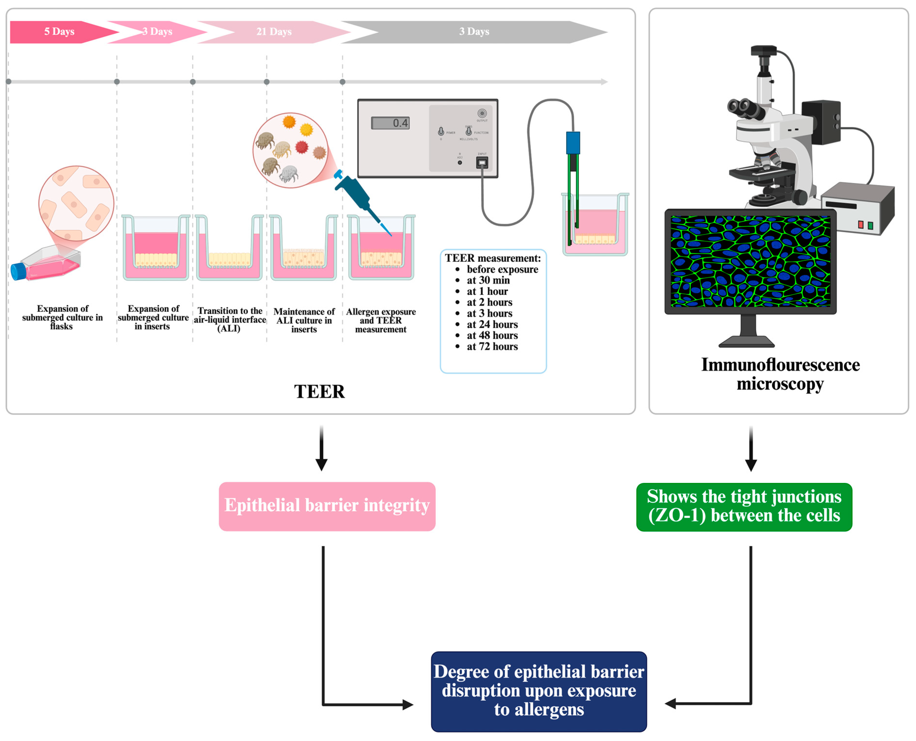
2.2. Culture of Differentiated Bronchial Epithelium at Air–Liquid Interface

Normal human bronchial epithelial cells (NHBE; Bronchial Epi Cells for B-ALI; Lonza Walkersville Inc., Walkersville, MD, USA) were used to establish an in vitro model of a differentiated bronchial epithelium. This model aimed to replicate the properties of a normal bronchial epithelium, including tight junctions, apical microvilli and regulated ion transport. Cells were cultured according to the manufacturer’s protocol with adjustments based on previous studies.
Initially, NHBE cells were expanded in T75-treated cell culture flasks (Corning, New York, NY, USA) and were incubated at 37 °C in a humidified atmosphere containing 5% CO2. The cells were cultured in PneumaCult™-ALI culture media (Stemcell™ Technologies, Vancouver, BC, Canada), supplemented with the necessary growth factors.
Upon reaching 70–90% confluence, cells were trypsinized, counted and seeded in specific plates at densities of 1000 cells/mm2 on 12-well Transwell inserts (Costar®, Corning, New York, NY, USA) with 0.4 μm pore membranes. The inserts were pre-coated with 100 μL of fibronectin (20 μg/mL) before seeding.
In the Transwell system, NHBE cells underwent a three-phase culture process over 24 days: (1) expansion at a liquid–liquid interface to establish a confluent monolayer (the first three days), (2) transition to an air–liquid interface to simulate physiological bronchial conditions and (3) differentiation into a pseudostratified, ciliated epithelium. Medium in the basal chamber was changed every 2 days, while the apical chamber remained exposed to air to mimic in vivo respiratory conditions. From the second week onward, excess mucus was gently removed using periodic PBS washes. This protocol enabled the formation of a structurally and functionally intact respiratory epithelium. Throughout the culture period, cell morphology, confluence and epithelial barrier integrity were carefully monitored to ensure successful differentiation.

2.3. Transepithelial Electrical Resistance (TEER) Measurement for the Assessment of Epithelial Barrier Integrity and Allergen-Induced Responses

The successful establishment of the air–liquid interface facilitated the development of a structurally and functionally differentiated bronchial epithelium which was subsequently exposed to either allergen fragments or allergen extracts. Transepithelial electrical resistance measurements were performed at regular intervals using an epithelial Ohmvoltmeter (Evom2, World Precision Instruments, Sarasota, FL, USA) by placing electrodes in both the apical and basal compartments of the Transwell system. A low-voltage alternating current was applied to determine the resistance across the epithelial monolayer, with values expressed in Ohm·cm2 (Ω·cm2) to account for membrane surface area. This method provided a real-time, non-invasive evaluation of barrier function dynamics under different experimental conditions.
Allergen exposures included different concentrations of HDM and ragweed pollen extracts, as well as mixtures containing purified allergen molecules (Amb a 1, Amb a 11, Amb a 12 and Der p 1). The cell cultures were exposed to the following allergen extracts or molecules and at different concentrations: RW extract 200 µg/mL; HDM extract 200 µg/mL; combined RW + HDM extracts in equal parts at 200 µg/mL (HR200), 100 µg/mL (HR100), 50 µg/mL (HR50) and 25 µg/mL (HR25); combined Amb a allergen mixture corresponding to 100 µg/mL of allergen extract (AM100; Amb a 1.01 at 10 µg/mL, Amb a 11 at 5 µg/mL and Amb a 12 at 2 µg/mL) [40]; combined Der p 1 + Amb a allergen mixture corresponding to 100 µg/mL of allergen extract (DAM100; combined Der p 1 at 10 µg/mL, Amb a 1.01 at 10 µg/mL, Amb a 11 at 5 µg/mL and Amb a 12 at 2 µg/mL) [41]; combined Der p 1 + Amb a allergen mixture corresponding to 50 µg/mL of allergen extract (DAM50). Allergens and allergen extracts were diluted in cell medium and medium alone was used as a negative control, while 5% Triton-X (Sigma Aldrich, St. Louis, MO, USA) served as a positive control.
TEER values were recorded before allergen exposure, 30 min post-exposure, hourly for the first 4 h and daily for up to 72 h (Figure 1). Each condition was tested in triplicate through two sets of experiments, using only technical replicates, as the epithelial cells were sourced from the same batch. The sample size was determined based on standard practices in similar experimental designs to ensure reliable and reproducible measurements. The TEER measurement points prior to allergen addition served as the baseline for normalization. The normalized TEER was set at this point and subsequent changes in cell responses were measured relative to this baseline.
Normalized TEER = (TEERT/TEER0) × 100
where TEERT is the transepithelial electrical resistance measured at each time point for each treatment and TEER0 is the base transepithelial electrical resistance (measured before addition of allergens or control). The data are presented as the mean ± standard deviation (SD).
This approach provided valuable insights into barrier function dynamics and cellular responses under varying experimental conditions. The mechanisms underlying epithelial barrier dysfunction and the immune responses triggered in allergic airway inflammation are illustrated in Figure 1.

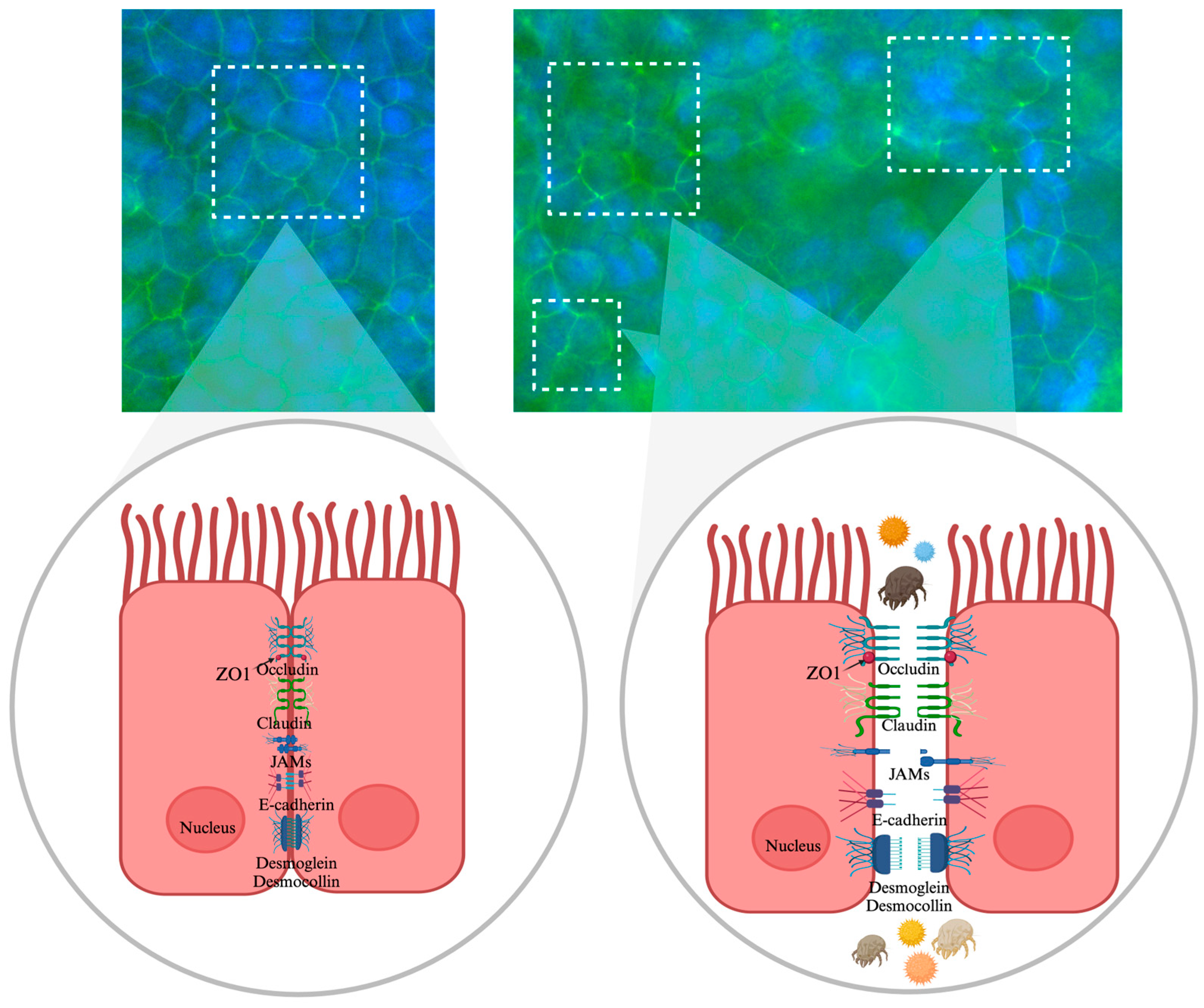
2.4. Assessment of Tight Junction Integrity Using Immunofluorescence with ZO-1

The expression of ZO-1 was assessed using an immunofluorescence assay. NHBE cells were cultured in 4-well chamber slides (NuncTM Lab-Tek, Thermo Fisher Scientific, Waltham, MA, USA) coated with 200 µL fibronectin (20 µg/mL) per well. Cells (25,000 cells/well) were left to adhere and expand in PneumaCultTM-ALI complete basal medium for 48 h. The medium was replaced with PneumaCultTM-ALI maintenance medium and cells were grown for another 48 h. Cells were then exposed to the allergen extracts and allergen combinations at different concentrations (as detailed in Section 2.3) for 72 h. For immunofluorescence staining, NHBE cells were fixed with methanol for 10 min at −20 °C, rinsed with PBS and subsequently blocked with 1% bovine serum albumin for 3 h. The cells were then incubated overnight at 4 °C with anti-ZO-1 monoclonal antibody produced in mouse and labelled with AlexaFluor 488 (Invitrogen, Thermo Fisher Scientific, Waltham, MA, USA). Nuclei were stained with DAPI using SlowFadeTM Gold antifade reagent (Invitrogen, Thermo Fisher Scientific, Waltham, MA, USA). Cell imaging was performed using the Invitrogen EVOSTM FL Auto 2 Imaging System (Thermo Fisher Scientific Inc., Bothell, WA, USA), and image processing was conducted using ImageJ.JS 1.53m software [42,43]. Figure 2 illustrates the methodologies employed, detailing the experimental design, which includes TEER measurements over 72 h and ZO-1 immunofluorescence analysis of NHBE cells following allergen exposure.

2.5. Statistical Analysis

All data were analyzed and are displayed as the mean ± standard deviation. The effects of allergen exposure on cell cultures were evaluated by measuring transepithelial electrical resistance. Statistical analyses were performed using Student’s t-test for pairwise comparisons and one-way ANOVA, as appropriate, followed by Bonferroni’s post hoc test for multiple comparisons. The statistical program used was IBM SPSS Statistics version 29.0. Statistical significance was considered to be p < 0.05.

2.6. Ethical Approval

This study was approved by the Ethics Committee of “Victor Babes” University of Medicine and Pharmacy in Timisoara, Romania (protocol code no.41/20.12.2023).

3. Results

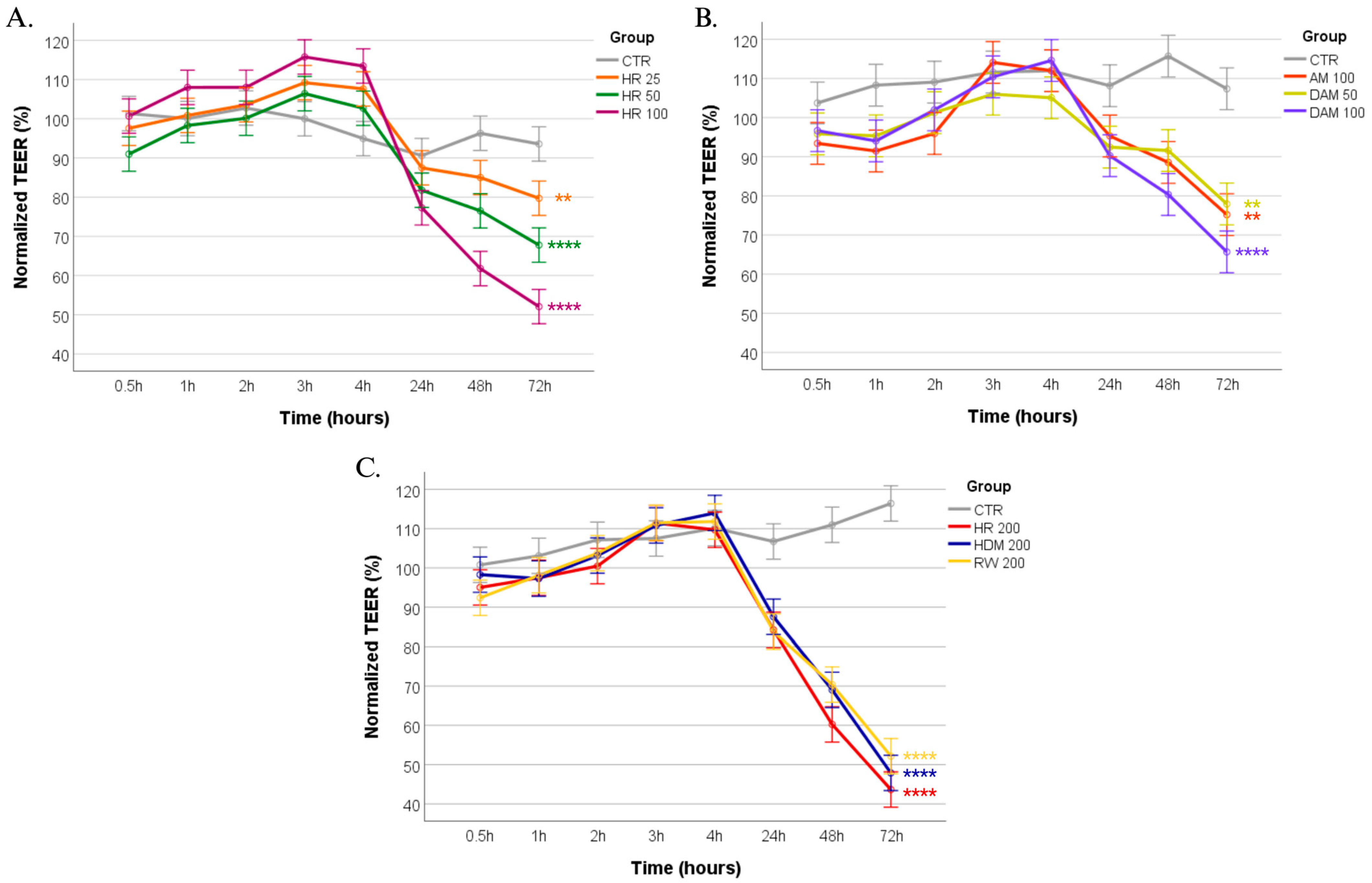
3.1. Revealing the Impact of Allergen Exposure on Epithelial Barrier Integrity Through TEER Monitoring

Transepithelial electrical resistance measurements were employed to assess the integrity of the bronchial epithelial barrier following exposure to various allergen conditions. TEER values, expressed as percentages relative to the pre-exposure baseline, were recorded at multiple time points over a 72 h period (Figure 3) to evaluate the extent of epithelial barrier disruption. At the 72-h mark, the degree of epithelial resistance alteration was quantified as a percentage of the pre-exposure values, providing a comparative measure of barrier integrity loss.
Every experimental group demonstrated statistically significant differences in TEER values when compared to the control group. Furthermore, an interesting remark is that all groups exhibit an initial TEER increase within the first four hours, followed by a clear and progressive decline at later time points.
The dose–response relationship observed among the combined HDM+RW (HR) exposure groups revealed that HR 100 demonstrated a significant decrease of 51.86% ± 4.98% compared to the pre-exposure TEER values (p < 0.0001), while HR 50 exhibited a more modest decline of 68.81% ± 3.27% (p = 0.0001). The HR 25 group showed the least impairment, with TEER values of 79.71% ± 4.42% (p = 0.01). Therefore, higher concentrations of HR (HR100 and HR50) lead to a significant decrease in TEER over time, indicating a loss of epithelial barrier integrity. The data presented are illustrated in Figure 3A. These findings suggest that higher concentrations of RW and HDM allergen extracts lead to a more pronounced reduction in epithelial barrier integrity. Statistically, no significant difference was observed between HR 25 and HR 50. However, significant differences were found between HR 25 and HR 100 (p < 0.0001), as well as between HR 50 and HR 100 (p < 0.05).
To further investigate the impact of specific allergenic proteins, exposures to preparations containing the allergenic molecules from these extracts were assessed. The DAM 100 condition, which included Der p 1, Amb a 1, Amb a 11 and Amb a 12 corresponding to 100 µg/mL allergen extracts, resulted in a significant TEER reduction of 64.95% ± 5.05% (p < 0.0001). In contrast, the DAM 50 condition (corresponding to 50 µg/mL of allergen extract) led to a more modest reduction in TEER values of 76.54% ± 4.17% (p < 0.01). Interestingly, AM 100 containing only the mixture of ragweed allergens (Amb a 1, Amb a 11 and Amb a 12) corresponding to 100 µg/mL of allergen extract exhibited TEER values of 74.15% ± 6.14% (p < 0.01), suggesting that the inclusion of Der p 1 may enhance the barrier-disrupting effects when combined with ragweed allergens. The data are graphically depicted in Figure 3B.
Exposure to RW 200 led to a reduction in TEER, with values decreasing to 52.14% ± 4.88% relative to the baseline (p = 0.0001). In contrast, HDM 200 exposure resulted in a more pronounced decline in TEER values (47.87% ± 3.91%, p = 0.0001), indicating a stronger effect of HDM on epithelial barrier modulation. Notably, the combined HR 200 condition produced the most significant reduction in TEER (44.18% ± 6.19%, p = 0.0001), suggesting that the synergistic interaction between RW and HDM allergens exerts a more substantial impact on epithelial permeability than either allergen alone at the same concentration. The corresponding data are displayed in Figure 3C.
The TEER reduction induced by the positive control, the 5% Triton-X, on NHBE cells was assessed at 30 min post-treatment, revealing an extreme decline in barrier integrity (8.21% ± 2.14%, p < 0.0001).
Although no significant differences were observed between the TEER values in cells exposed to either allergen extract preparation at 200 µg/mL, comparative analysis revealed that single-allergen exposures (RW 200, HDM 200) caused less barrier disruption than the combination of RW and HDM (HR 200). This supports the hypothesis that co-exposure exacerbates epithelial permeability loss. Moreover, the specific allergenic preparations (DAM 100 and DAM 50 groups) demonstrated that the loss of barrier integrity is not solely concentration-dependent but is also influenced by the specific allergenic molecules within the allergen extracts. These findings point out the differential effects of allergen exposure on bronchial epithelial integrity and emphasize the utility of TEER as a quantitative measure of barrier function disruption in allergic airway diseases.

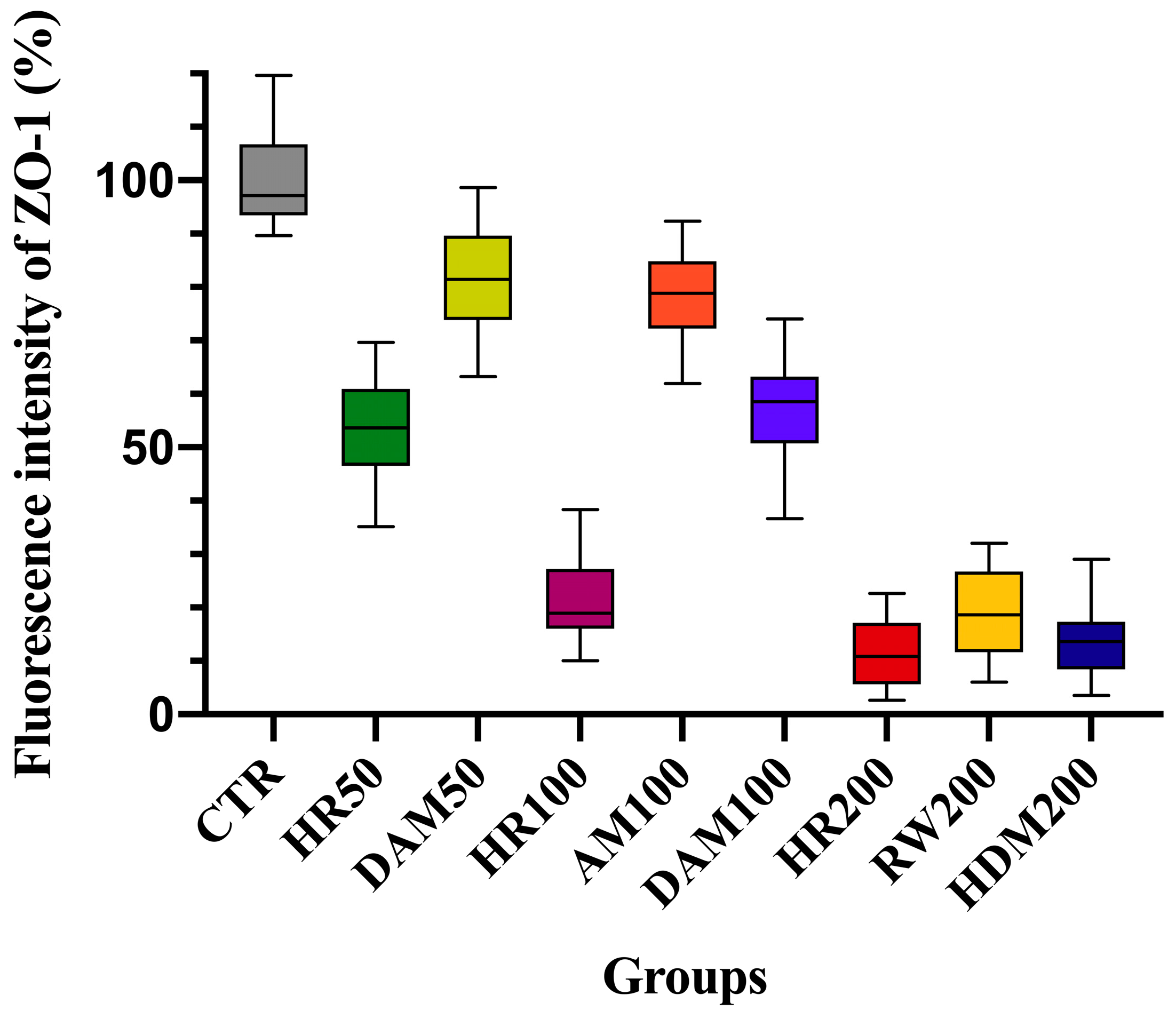
3.2. Visualization of Tight Junction Integrity Through ZO-1 Immunofluorescence Imaging Following Allergen Exposure

Immunofluorescence imaging was utilized to further assess the morphological alterations in bronchial epithelial cells following allergen exposure, with a focus on ZO-1 tight junction (TJ) proteins. Normal human bronchial epithelial cells, cultured at the air–liquid interface in a Transwell system to form a pseudostratified, ciliated epithelium, were exposed to various allergen formulations at different concentrations. Fixed tissue sections were stained for ZO-1 (green) and the cellular morphology was examined under fluorescence microscopy at 60× magnification, as shown in Figure 4, Figure 5 and Figure 6.
Immunofluorescence analysis of ZO-1 expression in NHBE cells reveals distinct patterns of tight junction integrity across different allergen exposures, as exposed in Table 1. In the control group, ZO-1 staining appears well defined and continuous along cell borders, indicating intact epithelial barrier function.
In contrast, exposure to HR at increasing concentrations (50, 100 and 200 µg/mL) results in a dose-dependent disruption of ZO-1, with higher concentrations leading to fragmented staining and reduced fluorescence intensity. HR 100 (100 µg/mL) exhibited pronounced morphological changes. Exposure to the DAM 100 allergenic preparation containing Der p 1, Amb a 1, Amb a 11 and Amb a 12 at 100 µg/mL and HR 50 (50 µg/mL) resulted in a moderate reduction in ZO-1 expression, therefore reflecting partial loss of tight junctions and showing the potential role of Der p 1 upon cell barrier alteration. The DAM 50 condition (50 µg/mL) and AM 100 (100 µg/mL) induced less pronounced barrier dysfunction concerning ZO-1 expression, highlighting the concentration-dependent effects of specific allergenic components on epithelial barrier integrity.
Following allergen exposure, ZO-1 immunofluorescence imaging revealed significant alterations in epithelial morphology that correlated with the TEER measurements. In the RW 200 (200 µg/mL) condition, ZO-1 staining exhibited considerable disruption of the tight junction (TJ) structures, reflecting moderate to severe structural changes in epithelial integrity, even though they seem less severe than high-dose HR or HDM exposure.
In contrast, HDM 200 (200 µg/mL) exposure induced more pronounced changes in the epithelial architecture, causing a marked loss of ZO-1 expression and significant tight junction impairment. This suggests that HDM exposure has a stronger impact on epithelial integrity compared to RW alone, as confirmed by both the imaging and TEER results.
The combined HR 200 condition (RW + HDM at 200 µg/mL) produced the most significant morphological disruption, with ZO-1 staining exhibiting extensive disorganization of tight junctions. The TEER measurements further validated the enhanced epithelial permeability observed in the imaging, supporting the hypothesis that co-exposure to RW and HDM allergens exacerbates epithelial barrier dysfunction.
The immunofluorescence intensity of zonula occludens-1 tight junctions was quantified using ImageJ.JS 1.53m software, with the results presented in Figure 5. The most pronounced reduction in fluorescence intensity compared to the control group was observed in the HR200 (11.47% ± 6.4%) and HDM200 (13.81% ± 6.33%) groups, followed by RW200 (19.18% ± 8.28%) and HR100 (21.05% ± 7.14%). Statistical analysis confirmed a significant difference between all treatment groups and the control (p < 0.0001).

4. Discussion

The bronchial epithelial barrier plays a pivotal role in maintaining respiratory homeostasis by preventing the penetration of inhaled allergens and pathogens while regulating immune responses. Disruption of this barrier is a hallmark of asthma pathogenesis, contributing to inflammation, airway hyperresponsiveness and remodeling [5,37,44]. Enzymes in allergens act as key enhancers of allergic reactions [45]. Studies confirm that protease activity in allergens, including those from pollen and HDMs, disrupts epithelial integrity and activates innate immune responses [46]. HDMs degrade TJs by cleaving occludin and causing intracellular ZO-1 proteolysis, leading to epithelial barrier breakdown [47]. Allergen-derived proteases also activate protease-activated receptor-2, triggering the immune response via thymic stromal lymphopoietin (TSLP) production. Additionally, proteases in sensitized patients may increase epithelial permeability by disrupting adhesion proteins like occludin and claudin-1 [48].
In this study, we investigated the individual and combined effects of RW and HDM extracts on bronchial epithelial barrier integrity using a physiologically relevant air–liquid interface model of normal human bronchial epithelial cells. The use of an ALI culture model of NHBE cells is supported by numerous studies demonstrating the physiological relevance of this system due to the formation of functional TJs and the production of mucus [49,50,51,52,53]. Similarly, a study by Heijink et al. (2020) used ALI cultures to investigate the effects of HDMs on epithelial barrier function and found results consistent with those obtained in animal models [10,54]. These studies validate our experimental approach and support the reliability of our results.
The use of HDM and ragweed allergens is of significant interest due to the rising prevalence of sensitization and the increasing incidence of severe allergic forms of disease. The concentrations employed in this study were in line with those used in previous research, which has demonstrated significant biological effects on respiratory epithelial cells [55]. Moreover, a range of concentrations was utilized to explore the detrimental effects of various allergen exposures on the epithelial barrier. Notably, the 200 µg/mL concentration yielded the most robust response in TEER measurements, allowing for a comprehensive evaluation of the cellular response to these allergens. This aspect is important for understanding their potential damaging effects on bronchial epithelial cell cultures. Lower concentrations may not have induced a sufficiently strong response to reveal substantial disruption of the epithelial barrier, as displayed by ZO-1 architectural alterations demonstrated in this study, particularly in a controlled laboratory setting.
Although the TEER assay is widely used, the TEER readings are highly dependent on the electrode positions and careful handling of the electrodes is required while introducing them into the well under test to avoid any disturbance to the cells. The uniformity of the current density generated by the electrodes across the cell layer has a significant effect on the TEER measurements [18,34,56]. We adhered to the MIRTA (Minimum Information for Reporting on the TEER Assay) recommendations, which provide guidelines for standardizing TEER assay procedures to ensure data consistency and reproducibility across studies. To comply with these standards, we maintained proper calibration of volt–ohm meters and electrodes and recorded the temperature throughout the experiment [56,57].
Our findings demonstrate that combined RW and HDM exposure had the most pronounced impact on barrier disruption, followed by HDMs alone, with RW showing a slightly less severe but still significant effect. These outcomes highlight the importance of studying multi-allergen interactions, as they more accurately reflect real-world exposure scenarios and provide critical insights into asthma pathogenesis.
The central outcome of this study was the synergistic effect of combined RW and HDM exposure on epithelial barrier integrity. While both allergen sources individually disrupted barrier function, their combined exposure resulted in significantly greater TEER reduction and disorganization of TJ proteins, particularly ZO-1, compared to either allergen alone. This synergistic effect aligns with recent studies, for instance, a study demonstrated that co-exposure to multiple allergens significantly exacerbates airway inflammation and barrier dysfunction compared to single-allergen exposure [11]. Similarly, another study emphasized that multi-allergen exposure can lead to more severe asthma phenotypes due to overlapping inflammatory and barrier-disrupting mechanisms [58]. Also, Lambrecht et al. (2012) reinforced the concept that simultaneous exposure to multiple allergens can amplify inflammatory responses and compromise epithelial barrier integrity, potentially through interconnected yet mechanistically distinct pathways, suggesting a multifaceted interplay between immune activation and structural barrier dysfunction [9]. These studies support our observation that combined RW pollen and HDM exposure results in greater TEER reduction and TJ disorganization compared to individual allergens. Another study by Steelant et al. (2020) investigated the effects of combined grass pollen and HDM exposure on nasal epithelial cells and found that combined exposure led to greater barrier disruption and inflammatory cytokine release than either allergen alone [36]. While their study focused on the nasal epithelium, the mechanisms of barrier disruption are similar to those in the bronchial epithelium due to common TJ proteins and immune regulatory pathways. Our findings extend these observations to the bronchial epithelium, emphasizing the broader significance of multi-allergen interactions in allergic airway diseases. Specifically, we investigated the combined exposure to two of the most common allergens implicated in allergic sensitization: house dust mites (HDMs) from indoor environments and ragweed (RW) pollen from outdoor environments.
The significant barrier disruption caused by HDMs alone in our study is consistent with numerous previous investigations. For example, studies demonstrated that HDM proteases, particularly Der p 1, Der p 2 and Der f 1, directly degrade TJ proteins such as ZO-1 and occludin, leading to increased paracellular permeability [24]. Similarly, Heijink et al. (2020) showed that HDM exposure disrupts epithelial barrier function by activating protease-activated receptor 2 (PAR-2), which leads to impaired TJ assembly [10]. Our TEER measurements and immunofluorescence staining for ZO-1 corroborate these findings, confirming that HDM exposure rapidly and severely compromises barrier integrity. HDMs are among the most significant allergens, producing cysteine and serine proteases that may alter the epithelial barrier [16]. The ability of HDM extracts to disrupt epithelial immune and barrier functions is closely linked to allergic sensitization. However, this effect does not appear to be reliant on only serine and cysteine protease activity, suggesting that other components or mechanisms within the extract contribute to epithelial dysfunction. Understanding these alternative pathways may provide new insights into how HDM exposure promotes allergic responses and barrier impairment [44]. Der p1 is the primary allergenic protease, disrupting the barrier by cleaving occludin and claudin, degrading ZO-1, reducing TEER and increasing permeability [18,59,60]. On the other hand, another study found that HDM-induced barrier disruption is partially reversible upon allergen removal, suggesting that epithelial cells retain some capacity for repair [59,61].
Our study found that RW alone caused a smaller TEER reduction compared to HDMs but still significantly impaired barrier integrity. The combination of RW and HDM allergens, however, resulted in the most severe disruption, suggesting that the distinct mechanisms of action of these allergens interact synergistically to exacerbate barrier dysfunction. Unlike HDMs, RW may not directly degrade TJ proteins but induces oxidative stress and inflammatory responses that compromise barrier integrity over time [62]. This is consistent with another study which compared the effects of seasonal (pollen) and perennial (HDM) allergens on epithelial barrier function and found that pollen-induced barrier disruption is generally less severe but more prolonged compared to HDM-induced disruption [63].
RW is known to induce oxidative stress and inflammatory cytokine release, which can weaken TJ integrity indirectly. In contrast, HDMs contain proteases that directly degrade TJ proteins such as ZO-1 and occludin. When combined, these mechanisms likely interact to amplify barrier dysfunction. Our IF staining revealed extensive fragmentation and redistribution of ZO-1 in cells exposed to both allergens, suggesting that the combined exposure not only degrades TJ proteins but also impairs their ability to reorganize and maintain barrier function. This finding is consistent with studies who demonstrated that HDM proteases disrupt TJ assembly, while oxidative stress from RW further inhibits repair mechanisms [36].
The synergistic effects of combined RW and HDM exposure have important implications for asthma pathogenesis. Epithelial barrier dysfunction is a critical early event in asthma development, as it facilitates the penetration of allergens and pathogens, leading to heightened immune activation and chronic inflammation [10,64]. Our findings suggest that individuals exposed to multiple allergens, such as RW pollen and HDMs, may be at greater risk of developing severe asthma due to compounded barrier disruption. This is particularly relevant in urban environments, where individuals are often exposed to a combination of seasonal and perennial allergens, as well as to pollution, which serves as an exacerbating factor that amplifies allergic responses [12].
Moreover, the differential effects of RW and HDMs on barrier integrity may explain the heterogeneity observed in asthma phenotypes. For example, patients with perennial asthma, often associated with HDM sensitivity, may experience more severe barrier dysfunction compared to those with seasonal asthma linked to ragweed pollen exposure [37,64,65]. However, the combined exposure scenario, which is more reflective of real-world conditions, likely contributes to the development of mixed or severe asthma phenotypes characterized by persistent inflammation and remodeling.
Our findings on TJ remodeling following allergen exposure are consistent with several studies investigating the molecular mechanisms of TJ disruption. HDM proteases induce the redistribution of ZO-1 from the cell membrane to the cytoplasm, leading to TJ disassembly [24]. Similarly, oxidative stress from pollen exposure disrupts the cytoskeletal organization necessary for TJ maintenance [36]. IF staining for ZO-1 revealed similar patterns of TJ disorganization following RW and HDM exposure, supporting these mechanisms.
Interestingly, TJ remodeling in response to allergen exposure is partially mediated by the activation of mitogen-activated protein kinase (MAPK) signaling pathways, which regulate TJ protein expression and assembly [62]. This suggests that targeting MAPK signaling may be a potential therapeutic strategy for restoring barrier integrity in asthma. Our findings showing extensive TJ disorganization following combined RW pollen and HDM exposure highlight the need for further research into the signaling pathways underlying TJ remodeling.
Our results highlight the need for therapeutic strategies that target epithelial barrier restoration in asthma. Current treatments, such as corticosteroids and bronchodilators, primarily address inflammation and airway hyperresponsiveness but do not directly repair barrier dysfunction [2,4,66].
The therapeutic implications of our findings are supported by several studies investigating strategies to restore epithelial barrier function in asthma. For example, antioxidants can attenuate allergen-induced oxidative stress and improve barrier integrity [36,67,68]. Protease inhibitors can block HDM-mediated TJ degradation and restore barrier function [16,26,27]. These studies suggest that combination therapies targeting both oxidative stress and protease activity could offer a more effective approach to ameliorating allergen-induced barrier disruption and preventing asthma exacerbations in individuals exposed to multiple allergens [69].
Additionally, the potential of TJ stabilizers, such as synthetic peptides that mimic TJ proteins, has been described for enhancing barrier integrity and preventing allergen penetration [6,62]. Our findings showing severe barrier disruption following combined RW pollen and HDM exposure emphasize the importance of developing such targeted therapies to address the complex mechanisms underlying multi-allergen-induced barrier dysfunction.
While our study provides novel insights into the combined effects of RW and HDM allergens on epithelial barrier integrity, several limitations should be acknowledged. Firstly, the ALI model, while physiologically relevant, does not fully replicate the complexity of the in vivo airway environment, including the presence of immune cells and stromal interactions [49]. Future studies incorporating co-culture systems or animal models may provide a more comprehensive understanding of allergen-induced barrier dysfunction. Secondly, our study focuses on acute allergen exposure, whereas chronic exposure may have different effects on barrier function and repair mechanisms. Longitudinal studies are needed to investigate the long-term impact of combined allergen exposure on epithelial integrity and asthma progression. While our study effectively assesses barrier integrity through TEER measurements and ZO-1 immunostaining, due to technical constraints, it does not include an analysis of downstream cytokine or alarmin responses, such as TSLP, IL-33 or IL-6. The inclusion of cytokine and alarmin profiling could have broadened the scope of our analysis and we plan to address these markers in future studies to better elucidate the relationship between barrier dysfunction and subsequent inflammatory signaling. Additionally, although endotoxin levels were not directly measured, dialysis of the allergen extracts in DPBS using a 3.5 kDa membrane likely facilitated the removal of small endotoxin fragments, thereby reducing the likelihood of endotoxin contamination influencing the results. Despite these limitations, our findings provide important insights into epithelial integrity in response to allergen exposure and lay the groundwork for further mechanistic investigations.
Finally, while we identified synergistic effects between ragweed pollen and HDMs, further studies are needed to advance our understanding of the specific molecular mechanisms and their broader implications, as well as to identify potential therapeutic targets.
The interaction between genotype and environment also shapes the airway epithelial phenotype, which is essential to asthma pathogenesis [70]. Recent evidence suggests that genetic variants linked to asthma risk influence proteins involved in airway epithelial function, including barrier integrity. Advancing our understanding of these regulatory mechanisms, through methods like single-cell RNA sequencing (scRNA-seq), offers potential for identifying patients who may benefit from epithelial-targeted therapies and for discovering novel therapeutic areas aimed at restoring epithelial barrier function [10,48].
Another important factor impacting epithelial barrier integrity is exposure to particulate matter, diesel exhaust particles and ground-level ozone. While these pollutants were not assessed in our study, it is important to note that, in urban environments, asthmatic patients are frequently exposed to the cumulative effects of such environmental cofactors [25,70,71]. The relationship between allergens and air pollution in relation to epithelial barrier integrity is characterized by a complex interplay of environmental and genetic factors. Both allergens and pollutants individually contribute to epithelial damage and their combined effects can exacerbate respiratory diseases [70].
Further studies are needed to elucidate the specific molecular pathways through which ragweed pollen and house dust mites synergistically impair epithelial barrier function, investigate potential therapeutic interventions to restore epithelial barrier integrity and explore whether other common airborne allergens exhibit similar synergistic effects.

5. Conclusions

Our findings highlight the synergistic effects of house dust mite and ragweed allergen co-exposure, revealing a more pronounced disruption of bronchial epithelial integrity compared to single-allergen exposures. This amplified barrier dysfunction likely results from the concurrent activation of degradative pathways that compromise epithelial tight junctions, increase permeability and amplify inflammatory responses beyond the effects of individual allergens. By integrating TEER measurements and immunofluorescence imaging, our study provides a comprehensive assessment of both functional and structural alterations in NHBE cells, reinforcing the bronchial epithelium’s active role in immune modulation and barrier defense. These results emphasize the necessity of a multifaceted approach to allergen research, as real-world exposures frequently involve multiple allergens with potential synergistic effects. Moreover, considering the cumulative impact of multiple allergens is essential for refining respiratory health assessments and improving risk evaluations, particularly in the management of allergic rhinitis and asthma.

Author Contributions

Conceptualization, R.-I.Z., M.G. and C.P.; methodology, R.-I.Z., M.G., G.T., F.-M.B. and R.-M.B.; validation, R.-I.Z., M.G., G.T., F.-M.B. and E.-L.Z.; formal analysis, R.-I.Z., M.G., M.-D.C. and E.-L.Z.; investigation, R.-I.Z., M.G., M.-D.C., O.O.H., R.-M.B. and E.C.; resources, M.G., G.T., R.-M.B., T.-P.T., V.P. and C.P.; data curation, R.-I.Z. and M.G.; writing—R.-I.Z., M.G., E.-L.Z., L.H., E.C. and K.C.M.; writing—review and editing, R.-I.Z., M.G., E.-L.Z., L.H. and R.P.; visualization, E.-L.Z., R.-I.Z. and O.O.H.; supervision, G.T., F.-M.B., V.P. and C.P.; project administration, V.P. and C.P.; funding acquisition, R.-I.Z., M.G., F.-M.B., G.T., V.P. and C.P. All authors have read and agreed to the published version of the manuscript.

Funding

The APC was funded by VICTOR BABES UNIVERSITY OF MEDICINE AND PHARMACY TIMISOARA. This study was supported by the INSPIRED (Innovative Strategies for Prevention, Diagnosis and Therapy of Ragweed Pollen Induced Respiratory Diseases) project, COP 2014–2020 92/09.09.2016, P_37_747, MySMIS 103663.

Institutional Review Board Statement

This study was approved by the Ethics Committee of “Victor Babes” University of Medicine and Pharmacy in Timisoara, Romania (protocol code no.41/20.12.2023, approval date 20 December 2023).

Informed Consent Statement

Not applicable.

Data Availability Statement

The original contributions presented in this study are included in the article; further inquiries can be directed to the corresponding author.

Acknowledgments

We would like to acknowledge Victor Babes University of Medicine and Pharmacy Timisoara for their support in covering the costs of publication for this research paper.

Conflicts of Interest

The authors declare no conflict of interest.

References

  1. Koch, S.; Nusrat, A. The Life and Death of Epithelia During Inflammation: Lessons Learned from the Gut. Annu. Rev. Pathol. Mech. Dis. 2012, 7, 35–60. [Google Scholar] [CrossRef]
  2. Calvén, J.; Ax, E.; Rådinger, M. The Airway Epithelium—A Central Player in Asthma Pathogenesis. IJMS 2020, 21, 8907. [Google Scholar] [CrossRef]
  3. Haidar, L.; Bănărescu, C.F.; Uța, C.; Moldovan, S.I.; Zimbru, E.-L.; Zimbru, R.-I.; Ciurariu, E.; Georgescu, M.; Panaitescu, C. Pollen–Food Allergy Syndrome: Allergens, Clinical Insights, Diagnostic and Therapeutic Challenges. Appl. Sci. 2024, 15, 66. [Google Scholar] [CrossRef]
  4. Beerweiler, C.C.; Masanetz, R.K.; Schaub, B. Asthma and allergic diseases: Cross talk of immune system and environmental factors. Eur. J. Immunol. 2023, 53, 2249981. [Google Scholar] [CrossRef]
  5. Georas, S.N.; Rezaee, F. Epithelial barrier function: At the front line of asthma immunology and allergic airway inflammation. J. Allergy Clin. Immunol. 2014, 134, 509–520. [Google Scholar] [CrossRef] [PubMed]
  6. Citi, S.; Fromm, M.; Furuse, M.; González-Mariscal, L.; Nusrat, A.; Tsukita, S.; Turner, J.R. A short guide to the tight junction. J. Cell Sci. 2024, 137, jcs261776. [Google Scholar] [CrossRef]
  7. Goktas, P.; Damadoglu, E. Future of allergy and immunology. Ann. Allergy Asthma Immunol. 2024, 134, 396–407. [Google Scholar] [CrossRef]
  8. Martens, K.; Hellings, P.W.; Steelant, B. Calu-3 epithelial cells exhibit different immune and epithelial barrier responses from freshly isolated primary nasal epithelial cells in vitro. Clin. Transl. Allergy 2018, 8, 40. [Google Scholar] [CrossRef]
  9. Lambrecht, B.N.; Hammad, H. The airway epithelium in asthma. Nat. Med. 2012, 18, 684–692. [Google Scholar] [CrossRef]
  10. Heijink, I.H.; Kuchibhotla, V.N.S.; Roffel, M.P.; Maes, T.; Knight, D.A.; Sayers, I.; Nawijn, M.C. Epithelial cell dysfunction, a major driver of asthma development. Allergy 2020, 75, 1902–1917. [Google Scholar] [CrossRef] [PubMed]
  11. Myszkowska, D.; Bogawski, P.; Piotrowicz, K.; Bosiacka, B.; Grinn-Gofroń, A.; Berger, U.E.; Bonini, M.; Ceriotti, V.; Charalampopoulos, A.; Galán, C.; et al. Co-exposure to highly allergenic airborne pollen and fungal spores in Europe. Sci. Total Environ. 2023, 905, 167285. [Google Scholar] [CrossRef] [PubMed]
  12. Prodić, I.; Minić, R.; Stojadinović, M. The influence of environmental pollution on the allergenic potential of grass pollen. Aerobiologia 2024, 41, 3–16. [Google Scholar] [CrossRef]
  13. Chen, K.-W.; Marusciac, L.; Tamas, P.T.; Valenta, R.; Panaitescu, C. Ragweed Pollen Allergy: Burden, Characteristics, and Management of an Imported Allergen Source in Europe. Int. Arch. Allergy Immunol. 2018, 176, 163–180. [Google Scholar] [CrossRef] [PubMed]
  14. Zimbru, R.-I.; Zimbru, E.-L.; Ordodi, V.-L.; Bojin, F.-M.; Crîsnic, D.; Grijincu, M.; Mirica, S.-N.; Tănasie, G.; Georgescu, M.; Huțu, I.; et al. The Impact of High-Fructose Diet and Co-Sensitization to House Dust Mites and Ragweed Pollen on the Modulation of Airway Reactivity and Serum Biomarkers in Rats. IJMS 2024, 25, 8868. [Google Scholar] [CrossRef]
  15. Buzan, M.; Zbîrcea, L.; Gattinger, P.; Babaev, E.; Stolz, F.; Valenta, R.; Păunescu, V.; Panaitescu, C.; Chen, K. Complex IgE sensitization patterns in ragweed allergic patients: Implications for diagnosis and specific immunotherapy. Clin. Transl. All. 2022, 12, e12179. [Google Scholar] [CrossRef]
  16. Runswick, S.; Mitchell, T.; Davies, P.; Robinson, C.; Garrod, D.R. Pollen proteolytic enzymes degrade tight junctions. Respirology 2007, 12, 834–842. [Google Scholar] [CrossRef]
  17. Zbîrcea, L.-E.; Buzan, M.-R.; Grijincu, M.; Babaev, E.; Stolz, F.; Valenta, R.; Păunescu, V.; Panaitescu, C.; Chen, K.-W. Relationship between IgE Levels Specific for Ragweed Pollen Extract, Amb a 1 and Cross-Reactive Allergen Molecules. IJMS 2023, 24, 4040. [Google Scholar] [CrossRef]
  18. Ogi, K.; Ramezanpour, M.; Liu, S.; Ferdoush Tuli, J.; Bennett, C.; Suzuki, M.; Fujieda, S.; Psaltis, A.J.; Wormald, P.-J.; Vreugde, S. Der p 1 Disrupts the Epithelial Barrier and Induces IL-6 Production in Patients With House Dust Mite Allergic Rhinitis. Front. Allergy 2021, 2, 692049. [Google Scholar] [CrossRef]
  19. Grijincu, M.; Huțu, I.; Weber, M.; Babaev, E.; Stolz, F.; Valenta, R.; Păunescu, V.; Panaitescu, C.; Chen, K.-W. Physicochemical and immunological characterization of Amb a 12, a novel ragweed (Ambrosia artemisiifolia) pollen allergen. Mol. Immunol. 2023, 157, 18–29. [Google Scholar] [CrossRef]
  20. Lin, J.; Huang, N.; Li, J.; Liu, X.; Xiong, Q.; Hu, C.; Chen, D.; Guan, L.; Chang, K.; Li, D.; et al. Cross-reactive antibodies against dust mite-derived enolase induce neutrophilic airway inflammation. Eur. Respir. J. 2021, 57, 1902375. [Google Scholar] [CrossRef]
  21. Frey, A.; Lunding, L.P.; Ehlers, J.C.; Weckmann, M.; Zissler, U.M.; Wegmann, M. More Than Just a Barrier: The Immune Functions of the Airway Epithelium in Asthma Pathogenesis. Front. Immunol. 2020, 11, 761. [Google Scholar] [CrossRef]
  22. Rezaee, F.; Georas, S.N. Breaking Barriers. New Insights into Airway Epithelial Barrier Function in Health and Disease. Am. J. Respir. Cell Mol. Biol. 2014, 50, 857–869. [Google Scholar] [CrossRef] [PubMed]
  23. Abu Khweek, A.; Kim, E.; Joldrichsen, M.R.; Amer, A.O.; Boyaka, P.N. Insights Into Mucosal Innate Immune Responses in House Dust Mite-Mediated Allergic Asthma. Front. Immunol. 2020, 11, 534501. [Google Scholar] [CrossRef]
  24. Wan, H.; Winton, H.L.; Soeller, C.; Tovey, E.R.; Gruenert, D.C.; Thompson, P.J.; Stewart, G.A.; Taylor, G.W.; Garrod, D.R.; Cannell, M.B.; et al. Der p 1 facilitates transepithelial allergen delivery by disruption of tight junctions. J. Clin. Investig. 1999, 104, 123–133. [Google Scholar] [CrossRef] [PubMed]
  25. De Lagarde, V.M.; Chevalier, L.; Méausoone, C.; Cazier, F.; Dewaele, D.; Cazier-Dennin, F.; Janona, M.; Logie, C.; Achard, S.; André, V.; et al. Acute and repeated exposures of normal human bronchial epithelial (NHBE) cells culture to particles from a coloured pyrotechnic smoke. Environ. Toxicol. Pharmacol. 2024, 105, 104327. [Google Scholar] [CrossRef] [PubMed]
  26. Vinhas, R.; Cortes, L.; Cardoso, I.; Mendes, V.M.; Manadas, B.; Todo-Bom, A.; Pires, E.; Veríssimo, P. Pollen proteases compromise the airway epithelial barrier through degradation of transmembrane adhesion proteins and lung bioactive peptides: Role of pollen proteases on allergic disorders. Allergy 2011, 66, 1088–1098. [Google Scholar] [CrossRef]
  27. Gaspar, R.; De Matos, M.R.; Cortes, L.; Nunes-Correia, I.; Todo-Bom, A.; Pires, E.; Veríssimo, P. Pollen Proteases Play Multiple Roles in Allergic Disorders. IJMS 2020, 21, 3578. [Google Scholar] [CrossRef]
  28. Zimbru, E.-L.; Zimbru, R.-I.; Ordodi, V.-L.; Bojin, F.-M.; Crîsnic, D.; Andor, M.; Mirica, S.-N.; Huțu, I.; Tănasie, G.; Haidar, L.; et al. Rosuvastatin Attenuates Vascular Dysfunction Induced by High-Fructose Diets and Allergic Asthma in Rats. Nutrients 2024, 16, 4104. [Google Scholar] [CrossRef]
  29. Movia, D.; Bruni-Favier, S.; Prina-Mello, A. In vitro Alternatives to Acute Inhalation Toxicity Studies in Animal Models—A Perspective. Front. Bioeng. Biotechnol. 2020, 8, 549. [Google Scholar] [CrossRef]
  30. Blume, C.; Swindle, E.J.; Gilles, S.; Traidl-Hoffmann, C.; Davies, D.E. Low molecular weight components of pollen alter bronchial epithelial barrier functions. Tissue Barriers 2015, 3, e1062316. [Google Scholar] [CrossRef]
  31. Eisenhut, M. Reduction of Alveolar Epithelial Ion and Fluid Transport by Inflammatory Mediators. Am. J. Respir. Cell Mol. Biol. 2007, 36, 388–389. [Google Scholar] [CrossRef]
  32. Hollenhorst, M.I.; Richter, K.; Fronius, M. Ion Transport by Pulmonary Epithelia. BioMed Res. Int. 2011, 2011, 174306. [Google Scholar] [CrossRef]
  33. Radbel, J.; Laskin, D.L.; Laskin, J.D.; Kipen, H.M. Disease-modifying treatment of chemical threat agent–induced acute lung injury. Ann. N. Y. Acad. Sci. 2020, 1480, 14–29. [Google Scholar] [CrossRef]
  34. Sharma, M.; Huber, E.; Arnesdotter, E.; Behrsing, H.P.; Bettmann, A.; Brandwein, D.; Constant, S.; Date, R.; Deshpande, A.; Fabian, E.; et al. Minimum information for reporting on the TEER (trans-epithelial/endothelial electrical resistance) assay (MIRTA). Arch. Toxicol. 2025, 99, 57–66. [Google Scholar] [CrossRef] [PubMed]
  35. Haas, A.J.; Zihni, C.; Krug, S.M.; Maraspini, R.; Otani, T.; Furuse, M.; Honigmann, A.; Balda, M.S.; Matter, K. ZO-1 Guides Tight Junction Assembly and Epithelial Morphogenesis via Cytoskeletal Tension-Dependent and -Independent Functions. Cells 2022, 11, 3775. [Google Scholar] [CrossRef] [PubMed]
  36. Steelant, B.; Seys, S.F.; Boeckxstaens, G.; Akdis, C.A.; Ceuppens, J.L.; Hellings, P.W. Restoring airway epithelial barrier dysfunction: A new therapeutic challenge in allergic airway disease. Rhin 2016, 54, 195–205. [Google Scholar] [CrossRef]
  37. Yazici, D.; Ogulur, I.; Pat, Y.; Babayev, H.; Barletta, E.; Ardicli, S.; Bel Imam, M.; Huang, M.; Koch, J.; Li, M.; et al. The epithelial barrier: The gateway to allergic, autoimmune, and metabolic diseases and chronic neuropsychiatric conditions. Semin. Immunol. 2023, 70, 101846. [Google Scholar] [CrossRef]
  38. Tamaș, T.-P.; Buzan, M.-R.; Zbîrcea, L.-E.; Cotarcă, M.-D.; Grijincu, M.; Păunescu, V.; Panaitescu, C.; Chen, K.-W. Ragweed Major Allergen Amb a 11 Recombinant Production and Clinical Implications. Biomolecules 2023, 13, 182. [Google Scholar] [CrossRef]
  39. Buzan, M.-R.; Grijincu, M.; Zbîrcea, L.-E.; Haidar, L.; Tamaș, T.-P.; Cotarcă, M.-D.; Tănasie, G.; Weber, M.; Babaev, E.; Stolz, F.; et al. Insect Cell-Expressed Major Ragweed Allergen Amb a 1.01 Exhibits Similar Allergenic Properties to Its Natural Counterpart from Common Ragweed Pollen. IJMS 2024, 25, 5175. [Google Scholar] [CrossRef]
  40. Würtzen, P.A.; Hoof, I.; Christensen, L.H.; Váczy, Z.; Henmar, H.; Salamanca, G.; Lundegaard, C.; Lund, L.; Kráľova, T.; Brooks, E.G.; et al. Diverse and highly cross-reactive T-cell responses in ragweed allergic patients independent of geographical region. Allergy 2020, 75, 137–147. [Google Scholar] [CrossRef]
  41. Casset, A.; Mari, A.; Purohit, A.; Resch, Y.; Weghofer, M.; Ferrara, R.; Thomas, W.R.; Alessandri, C.; Chen, K.-W.; De Blay, F.; et al. Varying Allergen Composition and Content Affects the in vivo Allergenic Activity of Commercial Dermatophagoides pteronyssinus Extracts. Int. Arch. Allergy Immunol. 2012, 159, 253–262. [Google Scholar] [CrossRef] [PubMed]
  42. Cao, N.; Wang, J.; Xu, X.; Xiang, M.; Dou, J. PACAP38 improves airway epithelial barrier destruction induced by house dust mites allergen. Immunobiology 2019, 224, 758–764. [Google Scholar] [CrossRef]
  43. Dong, H.; Le, Y.; Wang, Y.; Zhao, H.; Huang, C.; Hu, Y.; Luo, L.; Wan, X.; Wei, Y.; Chu, Z.; et al. Extracellular heat shock protein 90α mediates HDM-induced bronchial epithelial barrier dysfunction by activating RhoA/MLC signaling. Respir. Res. 2017, 18, 111. [Google Scholar] [CrossRef]
  44. Post, S.; Nawijn, M.C.; Hackett, T.L.; Baranowska, M.; Gras, R.; Van Oosterhout, A.J.M.; Heijink, I.H. The composition of house dust mite is critical for mucosal barrier dysfunction and allergic sensitisation. Thorax 2012, 67, 488–495. [Google Scholar] [CrossRef]
  45. Matsumura, Y. Role of Allergen Source-Derived Proteases in Sensitization via Airway Epithelial Cells. J. Allergy 2012, 2012, 903659. [Google Scholar] [CrossRef] [PubMed]
  46. Jacquet, A. Interactions of airway epithelium with protease allergens in the allergic response: Interactions of airway epithelium with protease allergens. Clin. Exp. Allergy 2011, 41, 305–311. [Google Scholar] [CrossRef]
  47. Takai, T.; Ikeda, S. Barrier Dysfunction Caused by Environmental Proteases in the Pathogenesis of Allergic Diseases. Allergol. Int. 2011, 60, 25–35. [Google Scholar] [CrossRef]
  48. Lu, H.-F.; Zhou, Y.-C.; Yang, L.-T.; Zhou, Q.; Wang, X.-J.; Qiu, S.-Q.; Cheng, B.-H.; Zeng, X.-H. Involvement and repair of epithelial barrier dysfunction in allergic diseases. Front. Immunol. 2024, 15, 1348272. [Google Scholar] [CrossRef] [PubMed]
  49. Lee, R.E.; Reidel, B.; Nelson, M.R.; Macdonald, J.K.; Kesimer, M.; Randell, S.H. Air-Liquid interface cultures to model drug delivery through the mucociliary epithelial barrier. Adv. Drug Deliv. Rev. 2023, 198, 114866. [Google Scholar] [CrossRef]
  50. Barron, S.L.; Wyatt, O.; O’Connor, A.; Mansfield, D.; Suzanne Cohen, E.; Witkos, T.M.; Strickson, S.; Owens, R.M. Modelling bronchial epithelial-fibroblast cross-talk in idiopathic pulmonary fibrosis (IPF) using a human-derived in vitro air liquid interface (ALI) culture. Sci. Rep. 2024, 14, 240. [Google Scholar] [CrossRef]
  51. Raby, K.L.; Michaeloudes, C.; Tonkin, J.; Chung, K.F.; Bhavsar, P.K. Mechanisms of airway epithelial injury and abnormal repair in asthma and COPD. Front. Immunol. 2023, 14, 1201658. [Google Scholar] [CrossRef]
  52. Runft, S.; Färber, I.; Krüger, J.; Schöne, K.; Lehmbecker, A.; Baumgärtner, W. In Vitro Characteristics of Canine Primary Tracheal Epithelial Cells Maintained at an Air–Liquid Interface Compared to In Vivo Morphology. IJMS 2023, 24, 4987. [Google Scholar] [CrossRef]
  53. Leach, T.; Gandhi, U.; Reeves, K.D.; Stumpf, K.; Okuda, K.; Marini, F.C.; Walker, S.J.; Boucher, R.; Chan, J.; Cox, L.A.; et al. Development of a novel air–liquid interface airway tissue equivalent model for in vitro respiratory modeling studies. Sci. Rep. 2023, 13, 10137. [Google Scholar] [CrossRef]
  54. Ghio, A.J.; Dailey, L.A.; Soukup, J.M.; Stonehuerner, J.; Richards, J.H.; Devlin, R.B. Growth of human bronchial epithelial cells at an air-liquid interface alters the response to particle exposure. Part. Fibre Toxicol. 2013, 10, 25. [Google Scholar] [CrossRef]
  55. Waltl, E.E.; Selb, R.; Eckl-Dorna, J.; Mueller, C.A.; Cabauatan, C.R.; Eiwegger, T.; Resch-Marat, Y.; Niespodziana, K.; Vrtala, S.; Valenta, R.; et al. Betamethasone prevents human rhinovirus- and cigarette smoke- induced loss of respiratory epithelial barrier function. Sci. Rep. 2018, 8, 9688. [Google Scholar] [CrossRef]
  56. Srinivasan, B.; Kolli, A.R.; Esch, M.B.; Abaci, H.E.; Shuler, M.L.; Hickman, J.J. TEER Measurement Techniques for In Vitro Barrier Model Systems. SLAS Technol. 2015, 20, 107–126. [Google Scholar] [CrossRef]
  57. Braakhuis, H.M.; Gremmer, E.R.; Bannuscher, A.; Drasler, B.; Keshavan, S.; Rothen-Rutishauser, B.; Birk, B.; Verlohner, A.; Landsiedel, R.; Meldrum, K.; et al. Transferability and reproducibility of exposed air-liquid interface co-culture lung models. NanoImpact 2023, 31, 100466. [Google Scholar] [CrossRef]
  58. Agache, I.; Akdis, C.A. Precision medicine and phenotypes, endotypes, genotypes, regiotypes, and theratypes of allergic diseases. J. Clin. Invest. 2019, 129, 1493–1503. [Google Scholar] [CrossRef]
  59. Steelant, B.; Farré, R.; Wawrzyniak, P.; Belmans, J.; Dekimpe, E.; Vanheel, H.; Van Gerven, L.; Kortekaas Krohn, I.; Bullens, D.M.A.; Ceuppens, J.L.; et al. Impaired barrier function in patients with house dust mite–induced allergic rhinitis is accompanied by decreased occludin and zonula occludens-1 expression. J. Allergy Clin. Immunol. 2016, 137, 1043–1053.e5. [Google Scholar] [CrossRef] [PubMed]
  60. Saito, T.; Ichikawa, T.; Numakura, T.; Yamada, M.; Koarai, A.; Fujino, N.; Murakami, K.; Yamanaka, S.; Sasaki, Y.; Kyogoku, Y.; et al. PGC-1α regulates airway epithelial barrier dysfunction induced by house dust mite. Respir. Res. 2021, 22, 63. [Google Scholar] [CrossRef] [PubMed]
  61. Gregory, L.G.; Lloyd, C.M. Orchestrating house dust mite-associated allergy in the lung. Trends Immunol. 2011, 32, 402–411. [Google Scholar] [CrossRef]
  62. Zhang, R.; Zhang, L.; Li, P.; Pang, K.; Liu, H.; Tian, L. Epithelial Barrier in the Nasal Mucosa, Related Risk Factors and Diseases. Int. Arch. Allergy Immunol. 2023, 184, 481–501. [Google Scholar] [CrossRef]
  63. Lambrecht, B.N.; Hammad, H. The immunology of asthma. Nat. Immunol. 2015, 16, 45–56. [Google Scholar] [CrossRef]
  64. Gon, Y.; Hashimoto, S. Role of airway epithelial barrier dysfunction in pathogenesis of asthma. Allergol. Int. 2018, 67, 12–17. [Google Scholar] [CrossRef]
  65. Huang, Z.; Liu, J.; Sun, L.; Ong, H.H.; Ye, J.; Xu, Y.; Wang, D. Updated epithelial barrier dysfunction in chronic rhinosinusitis: Targeting pathophysiology and treatment response of tight junctions. Allergy 2024, 79, 1146–1165. [Google Scholar] [CrossRef]
  66. Amison, R.T.; Page, C.P. Novel pharmacological therapies for the treatment of bronchial asthma. Minerva Med. 2022, 113, 31–50. [Google Scholar] [CrossRef]
  67. Chauhan, B.; Gupta, M.; Chauhan, K. Role of Antioxidants on the Clinical Outcome of Patients with Perennial Allergic Rhinitis. Allergy Rhinol. 2016, 7, ar.2016.7.0163. [Google Scholar] [CrossRef]
  68. Al-Harbi, N.O.; Nadeem, A.; Al-Harbi, M.M.; Imam, F.; Al-Shabanah, O.A.; Ahmad, S.F.; Sayed-Ahmed, M.M.; Bahashwan, S.A. Oxidative airway inflammation leads to systemic and vascular oxidative stress in a murine model of allergic asthma. Int. Immunopharmacol. 2015, 26, 237–245. [Google Scholar] [CrossRef]
  69. Mansur, A.H.; Gonem, S.; Brown, T.; Burhan, H.; Chaudhuri, R.; Dodd, J.W.; Pantin, T.; Gore, R.; Jackson, D.; Menzies-Gow, A.; et al. Biologic therapy practices in severe asthma; outcomes from the UK Severe Asthma Registry and survey of specialist opinion. Clin. Exp. Allergy 2023, 53, 173–185. [Google Scholar] [CrossRef]
  70. Loxham, M.; Davies, D.E. Phenotypic and genetic aspects of epithelial barrier function in asthmatic patients. J. Allergy Clin. Immunol. 2017, 139, 1736–1751. [Google Scholar] [CrossRef]
  71. Rayner, R.E.; Wellmerling, J.; Makena, P.; Zhao, J.; Prasad, G.L.; Cormet-Boyaka, E. Transcriptomic Response of Primary Human Bronchial Cells to Repeated Exposures of Cigarette and ENDS Preparations. Cell Biochem. Biophys. 2022, 80, 217–228. [Google Scholar] [CrossRef] [PubMed]
Figure 1. The mechanisms of epithelial barrier dysfunction and immune response in allergic airway inflammation. This schematic illustrates the impact of allergens, such as house dust mites and ragweed pollen, on airway epithelial integrity and the subsequent immune response. The organization of epithelial tight junctions, including ZO-1, occludins, claudins, junctional adhesion molecules (JAMs) and adherens junctions. Exposure to allergens leads to tight junction disruption, reducing epithelial barrier function. The central diagram illustrates allergen-induced epithelial activation and epithelial-derived cytokines (TSLP, IL-25, IL-33), promoting type 2 inflammation. This cascade leads to the activation of dendritic cells, innate lymphoid cells (ILC2) and Th2 cells, which secrete IL-4, IL-5, IL-6 and IL-13, driving eosinophilic inflammation, mucus hypersecretion and increased IgE production by plasma cells. The lower section illustrates immune cell trafficking and neuro-immune interactions, where sensory neurons release neuropeptides, like substance P (SP), calcitonin gene-related peptide (CGRP) and neuromedin U (NMU), that further amplify inflammation. Basophils, eosinophils and monocytes contribute to tissue damage through the release of inflammatory mediators such as histamine, eosinophilic cationic protein (ECP) and major basic protein (MBP). The disruption of mucociliary clearance further exacerbates allergic inflammation, leading to persistent airway hypersensitivity. ZO-1: zonula occludens-1; TRP: transient receptor potential; TSLP: thymic stromal lymphopoietin; IL: interleukin; IgE: immunoglobulin E; ECP: eosinophil cationic protein; MBP: major basic protein. Created with BioRender.com (accessed on 16 March 2025).
Applsci 15 04113 g001
Figure 2. Experimental design. Transepithelial electrical resistance measurements and immunofluorescence staining of ZO-1 in NHBE cells were performed under various experimental conditions. TEER values were recorded at multiple time points: before allergen exposure, 30 min post-exposure, at 1, 2 and 3 h, and then daily for an additional three days to evaluate epithelial barrier integrity. Immunofluorescence microscopy images depict ZO-1 localization (green) in NHBE cells cultured under different conditions, with nuclei counterstained using DAPI (blue). Created with BioRender.com (accessed on 16 March 2025).
Applsci 15 04113 g002
Figure 3. Changes in transepithelial electrical resistance in response to different allergens at varying concentrations and combinations. TEER values are normalized to pre-exposure baseline levels and presented as a percentage over time (h) post-exposure. (A) TEER changes in response to different concentrations of HR (HR 25, HR 50 and HR 100) compared to the control group (CTR). (B) TEER responses to AM 100, DAM 50 and DAM 100 treatments relative to CTR. (C) TEER dynamics following exposure to HR 200, HDM 200 and RW 200 compared to CTR. The groups are labeled as follows: CTR: control; RW 200: RW extract at 200 µg/mL; HDM 200: HDM extract at 200 µg/mL; HR 200: RW+HDM extracts in equal parts at 200 µg/mL; HR 100: RW+HDM extracts at 100 µg/mL; HR 50: RW+HDM at 50 µg/mL; HR 25: RW+HDM at 25 µg/mL; DAM 100: Der p 1+Amb a 1+Amb a 11+Amb a 12 preparation corresponding to 100 µg/mL of allergen extract; DAM 50: Der p 1+Amb a 1+Amb a 11+Amb a 12 preparation corresponding to 50 µg/mL of allergen extract; AM 100: Amb a 1+Amb a 11+Amb a 12 preparation corresponding to 100 µg/mL of allergen extract. The results are expressed as the mean ± SD for each time point. ** p < 0.01 and **** p < 0.0001 versus control.
Applsci 15 04113 g003
Figure 4. Immunofluorescence staining of zonula occludens-1 of normal human bronchial epithelial cell cultures. Cells were exposed to different allergen extracts and allergen molecule combinations to evaluate TJ alterations induced by allergen exposure. Fixed tissue sections were stained for ZO-1 (green), with nuclei counterstained using DAPI (blue) and imaged by fluorescence microscopy. Scale: 50 μm. The groups are labeled as follows: CTR: control; RW 200: RW extract 200 µg/mL; HDM 200: HDM extract 200 µg/mL; HR 200: RW + HDM extracts in equal parts at 200 µg/mL; HR 100: RW+HDM extracts 100 µg/mL; HR 50: RW+HDM 50 µg/mL; HR 25: RW+HDM 25 µg/mL; DAM 100: Der p 1+Amb a 1+Amb a 11+Amb a 12 preparation corresponding to 100 µg/mL of allergen extract; DAM 50: Der p 1+Amb a 1+Amb a 11+Amb a 12 preparation corresponding to 50 µg/mL of allergen extract; AM 100: Amb a 1+Amb a 11+Amb a 12 preparation corresponding to 100 µg/mL of allergen extract.
Applsci 15 04113 g004
Figure 5. Visualization of the immunofluorescence intensity changes of ZO-1 tight junctions across all treatment groups. The fluorescence intensity of ZO-1 junctions was measured in ImageJ.JS 1.53m software and are displayed as a percentage of the mean value of the control. The results are expressed as the mean ± SD. p < 0.0001 versus control for all treatment groups. The groups are labeled as follows: CTR: control; RW 200: RW extract 200 µg/mL; HDM 200: HDM extract 200 µg/mL; HR 200: RW + HDM extracts in equal parts at 200 µg/mL; HR 100: RW+HDM extracts 100 µg/mL; HR 50: RW+HDM 50 µg/mL; HR 25: RW+HDM 25 µg/mL; DAM 100: Der p 1+Amb a 1+Amb a 11+Amb a 12 preparation corresponding to 100 µg/mL of allergen extract; DAM 50: Der p 1+Amb a 1+Amb a 11+Amb a 12 preparation corresponding to 50 µg/mL of allergen extract; AM 100: Amb a 1+Amb a 11+Amb a 12 preparation corresponding to 100 µg/mL of allergen extract.
Applsci 15 04113 g005
Figure 6. The model of allergen-induced airway epithelium barrier dysfunction. ZO-1 immunofluorescence staining reveals allergen-induced widespread epithelial barrier disruption compared to a normal bronchial epithelium. Allergens like HDMs can directly cleave epithelial junctions or activate pattern recognition receptors (PRRs) such as PAR-2, C-type lectins (CLR) and purinergic receptors. Activation of these receptors leads to the degradation and/or mislocalization of junctional proteins. When epithelial repair is impaired, sustained loss of E-cadherin activates β-catenin pathways, further promoting epithelial-to-mesenchymal transition, goblet cell hyperplasia and ciliated cell loss, which are hallmarks of asthma-related epithelial changes. Created with BioRender.com.
Applsci 15 04113 g006
Table 1. A summary of the TEER assessment, tight junction integrity and airway health implications under various experimental conditions. This table presents the effects of various exposure conditions on transepithelial electrical resistance, tight junction integrity (evaluated through ZO-1 immunofluorescence staining) and their potential implications for airway health. TEER values (changes in normalized TEER at 72 h) indicate the barrier integrity of epithelial cells, while ZO-1 staining provides a visual assessment of tight junction organization. The findings help illustrate how different exposures influence epithelial barrier function, with potential consequences for respiratory health.
Exposure ConditionChanges in
Normalized
TEER (%) at 72 h
Tight Junction Integrity
(ZO-1 IF Staining *)
Implications for Airway Health
Control (no allergen)105.85% ± 6.19%0: Intact ZO-1Normal epithelial integrity
AM 100 (100 µg/mL)74.15% ± 6.14%1: Mild ZO-1 disruptionMild barrier dysfunction, increased permeability
DAM 50 (50 µg/mL)76.54% ± 4.17%1: Mild ZO-1 disruptionMild barrier dysfunction, increased permeability
DAM 100 (100 µg/mL)64.95% ± 5.05%2: Moderate ZO-1 disruptionEnhanced permeability, potential for allergen penetration
RW 200 (200 µg/mL)52.14% ± 4.88%3: Severe ZO-1 disruptionMarked permeability, greater potential for allergen penetration and for exacerbated allergic response
HDM 200 (200 µg/mL)47.87% ± 3.91%3: Severe ZO-1 disruptionMarked permeability, greater potential for allergen penetration and for exacerbated allergic response
HR 50 (RW + HDM 50 µg/mL)68.81% ± 3.27%2: Moderate ZO-1 disruptionEnhanced permeability, potential for allergen penetration
HR 100 (RW + HDM 100 µg/mL)51.86% ± 4.98%3: Severe ZO-1 disruptionMarked permeability, greater potential for allergen penetration and for exacerbated allergic response
HR 200 (RW + HDM 200 µg/mL)44.18% ± 6.19%3: Severe ZO-1 disruptionMarked permeability, greater potential for allergen penetration and for exacerbated allergic response
* ZO-1 disruption is defined as a loss of continuous membrane staining, redistribution of ZO-1 to the cytoplasm and a fragmented or punctate staining pattern (0: intact ZO-1; 1: partial ZO-1 disruption; 2: important ZO-1 disruption; 3: widespread ZO-1 disruption). The groups are labeled as follows: CTR: control; RW 200: RW extract 200 µg/mL; HDM 200: HDM extract 200 µg/mL; HR 200: RW + HDM extracts in equal parts at 200 µg/mL; HR 100: RW+HDM extracts 100 µg/mL; HR 50: RW+HDM 50 µg/mL; HR 25: RW+HDM 25 µg/mL; DAM 100: Der p 1+Amb a 1+Amb a 11+Amb a 12 preparation corresponding to 100 µg/mL of allergen extract; DAM 50: Der p 1+Amb a 1+Amb a 11+Amb a 12 preparation corresponding to 50 µg/mL of allergen extract; AM 100: Amb a 1+Amb a 11+Amb a 12 preparation corresponding to 100 µg/mL of allergen extract.
Disclaimer/Publisher’s Note: The statements, opinions and data contained in all publications are solely those of the individual author(s) and contributor(s) and not of MDPI and/or the editor(s). MDPI and/or the editor(s) disclaim responsibility for any injury to people or property resulting from any ideas, methods, instructions or products referred to in the content.

