1. Introduction
Osteoarthitis (OA) of the trapeziometacarpal joint (TMJ) is a progressive and debilitating degenerative disease affecting the carpometacarpal joint of the hand [
1]. Following OA of the distal interphalangeal joint, OA of the TMJ is the second most prevalent degenerative condition of the hand [
2]. This condition involves the joint between the first metacarpal of the thumb and the trapezium, characterized by a biconcave–convex saddle morphology. The TMJ facilitates a range of movements, including flexion, extension, abduction, adduction, opposition, repositioning, and circumduction.
Degeneration of the TMJ leads to pain and reduced mobility in its early stages, progressing to a loss of joint function, limitations in movement, pinch weakness, and, in advanced stages, complete dislocation of the base of the first metacarpal. The exact etiology of rhizarthrosis remains elusive. Notably, TMJ OA is more prevalent in elderly patients, with a higher incidence in postmenopausal women in their fifth or sixth decade of life. By contrast, the incidence in men is lower, ranging between 5% and 11%, potentially due to greater ligament laxity in females [
3]. According to Brunelli’s theory, the abductor pollicis longus (APL) tendon is absent from the trapezius in about 30% of the general population. This results in the APL tendons acting only on the first metacarpal bone, which can cause extremely harmful tangential forces in the TMJ [
4].
Diagnosis of rhizarthrosis typically involves a physical examination, grind test, and lever test. Patients often report that an exacerbation of pain using the radial column of the carpus limits quality of life in opposition or pinching activities [
5,
6]. The Eaton–Littler classification is the most used radiographic classification system for basal thumb arthrosis, confirming the diagnosis [
3].
Various surgical techniques have been proposed for the treatment of rhizarthrosis. When conservative treatments fail, surgical interventions such as trapezius excision, ligament reconstruction, arthrodesis, and a prosthesis or tendon interposition are considered. These procedures may restore grip strength [
4]. The dual-mobility prosthesis for the trapeziometacarpal joint represents a promising surgical option for the treatment of osteoarthritis, with potential benefits in terms of strength, mobility, and patient satisfaction [
5]. Toffoli et al. studied 80 patients who underwent 96 MAÏA™ dual mobility TMJ prosthesis (Group Lépin, Genay Cedex, France) implantations, with a minimum follow-up of 5 years [
6]. Patients had significant improvement in thumb strength, mobility, and function, with a relatively low surgical revision rate and a high implant success rate in the midterm. The most common complication associated with this type of implant is implant mobilization, which requires surgical revision in 4.2% of cases. This type of implant reported post-traumatic trapezium fractures, treated with immobilization in 3.1% of patients. Additionally, among the preoperatively reducible Z-deformities, 19.2% were not completely corrected after this procedure. The six-year survival analysis indicates a success rate of 93% (95% CI, 87–98). The most common complication associated with the AST procedure is De Quervain’s syndrome, which impacts postoperative recovery and function.
The aim of this study was to compare these two surgical techniques by evaluating clinical and instrumental outcomes as well as postoperative complications.
2. Materials and Methods
2.1. Study Design
This study involved a clinical trial. Patients were treated according to the ethical standards of the Helsinki Declaration. They were invited to read, understand, and sign the informed consent form for the publication of data and photos for scientific purposes. The present study was approved by the Internal Review Board (authorization number 9/2018). The research was approved by the Young European Hand Surgeons (YEHS) scientific committee.
2.2. Sample Size
A power analysis was performed based on the effects of surgery on TMJ OA. The primary outcome measure was the mean score of the Disabilities of the Arm, Shoulder and Hand (DASH) questionnaire [
7]. The alternative hypothesis was that the DASH score would be 7 points lower in patients undergoing prosthesis compared to suspension tenoplasty procedure. The standard deviation of the DASH score was estimated at 8 points. With these parameters, 30 participants were needed (15 in each group) to detect a difference of 7 points in the DASH score between groups. These numbers were based on a power (1-b) of 0.80 and a significance level of 5% (two-sided). With an expected dropout rate of 15%, this number was increased to 34 participants.
2.3. Patient Enrollment
We adhered to the CONSORT (Consolidated Standards of Reporting Trials) guidelines for presenting the results. A flowchart following these guidelines shows patient grouping and progression in our clinical study (
Figure 1). Patients recruited from those with a surgical indication for the treatment of TMJ OA were randomly divided into two groups: Group A comprised patients undergoing AST, and Group B consisted of patients undergoing arthroplasty. For patients surgically treated between 2019 and 2022 in the Unit of Orthopaedics, Department of Life, Health and Environmental Sciences, University of L’Aquila for TMJ OA (Eaton–Littler stage III), conservative treatment previously failed (consisting of either cortisone or hyaluronate injections, analgesics, patient education, strengthening exercises, aids, and orthoses). Inclusion criteria were age between 40 and 75 years, isolated TMJ OA with scaphotrapeziotrapezoid joint integrity, and Eaton–Littler stage III. Exclusion criteria included having (1) undergone previous TMJ surgery; (2) severe systemic diseases, rheumatoid arthritis or post-traumatic arthritis; (3) an active infection, osteomyelitis, sepsis, or distant infections, which may spread to the site of operation; (4) osteomalacia or other metabolic bone disorders; (5) Eaton–Littler stages I–II–IV.
2.4. Randomization
We employed a block randomization method to create the randomization sequence. This block randomization list was generated using specialized software (StatsDirect 4.0 Ltd., Cheshire, UK). A neutral operator, uninvolved in the surgical procedures, assembled sealed, opaque envelopes with sequential numbers, each containing the assigned treatment. Following the determination of surgical eligibility, patients were allocated to their respective treatment groups based on this randomization. During clinical and radiological evaluations, efforts were made to maintain blinding; however, certain objective biases were unavoidable. Notably, differences in surgical approach, such as the incision size and the characteristic scar of the AST procedure, which are not present in prosthetic arthroplasty, could have inadvertently influenced the assessment.
2.5. Clinical and Radiological Assessment
Patients were evaluated by the same operator at a preoperative clinical evaluation, as well as at 3, 6, 12, and 24 months postoperative using a Visual Analog Scale (VAS) for pain [
8], DASH score, and Michigan Hand Outcomes Questionnaire (MHQ) [
9]. DASH is well established and is by far the most commonly used PROM [
10,
11]. Chung et al. reported the validity of the MHQ, demonstrating construct validity through a significant correlation between scales in an anticipated pattern [
12]. The clinical assessment included standard evaluations of range of motion (ROM) using a 360-degree goniometer (Jamar
® 360-Degree Goniometer–Patterson Medical, Warrenville, IL, USA), with the fulcrum positioned dorsally at the first metacarpophalangeal joint. Measurement of thumb opposition was performed via the Kapandji method [
12]. The clinical examination of the joint included a grasp of the first metacarpal bone of the participant, manipulation in a circular motion, and subjected to gentle axial loading to conduct the thumb grind test [
13]. A dynamometer (Jamar
® dynamometer Performance Health, Warrenville, IL, USA) was employed to assess hand grip and pulp pinch strength, in accordance with the American Society for Surgery of the Hand guidelines [
14]. Consistency was maintained using the same instrument for all participants, calibrated at regular intervals. Each measurement was taken three times, and an average was computed.
In both groups, proximal migration of the thumb was evaluated by measuring the “step-off” defined as the distance between the base of the metacarpal and the distal end of the scaphoid, on anteroposterior radiographs postoperatively and at 24 months after surgery. This measurement was conducted according to the Goffin and Saffar technique [
15]. All clinical assessments were collected by a single operator. Patient comorbidities, previous conservative treatments, and previous intra-articular injections (at least 6 months prior to surgery) were included. The three measurements were conducted at preoperative time points, as well as 3, 6, 12, and 24 months postoperatively. Inter-rater reliability for the objective measures, including grip strength and range of motion, was maintained by having a single evaluator conduct all the assessments, ensuring consistent measurements across the study.
2.6. Surgical Techniques
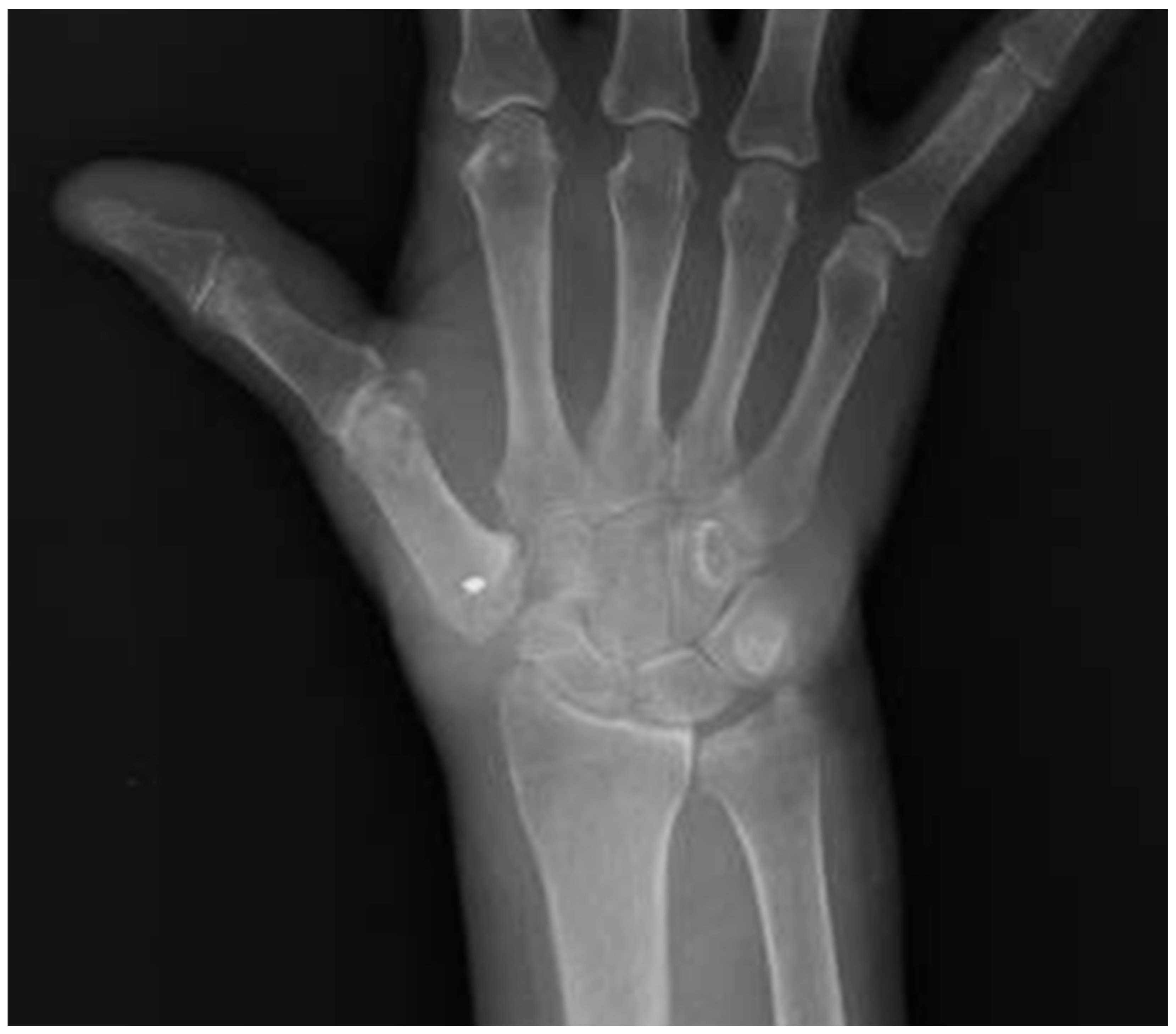
The same surgeon performed all the operations. Brachial plexus anesthesia was used for both surgical procedures. Just one surgical approach was required for these surgeries, which was to make a 4 cm longitudinal incision along the dorsal radial aspect of the TMJ. In the ulnar direction, the radial artery was retracted after being mobilized. The radial nerve’s cutaneous branch was located and separated. In order to generate a large distal triangular flap secured to the base of the first metacarpal bone, our team performed a Y-shaped capsulotomy to reveal the articular surfaces. GROUP A: AST by Altissimi (
Figure 2).
A trapeziectomy was performed. The flexor carpi radialis (FCR) tendon, located in the trapezial groove, was protected during this procedure. A segment of the FCR was dissected and released approximately 3–4 cm proximal to its insertion into the second metacarpal. The distal insertion remained intact. The free end of the FCR, passing through the triangular capsular space delineated during the capsulotomy around the base of the first metacarpal bone, was anchored using a suture anchor (Arthrex, Inc., Naples, FL, USA) on its radial edge to achieve suspension stability. The remaining portion of the FCR strip, distal to the anchor, was excised. GROUP B: Trapeziometacarpal Joint Arthroplasty (
Figure 3).
A stem was inserted into the diaphysis of the first metacarpal, followed by the placement of a cup implant in the trapezium. The cup was positioned in the trapezium with a slight anteversion to ensure optimal implant stability. The patients were fitted with the MAÏA™ dual mobility TMJ prosthesis (Group Lépin, Genay Cedex, France) [
16].
Finally, the capsule was sutured, and the skin was closed.
2.7. Postoperative Rehabilitation
Both groups followed an identical postoperative rehabilitation protocol, ensuring consistency in treatment and recovery. After surgery, the thumb and wrist were immobilized in a cast for three weeks to promote healing and prevent excessive movement. During this period, patients were advised to limit hand use and avoid weight-bearing activities. After cast removal, all patients underwent a structured rehabilitation program under the supervision of the same physiotherapist. Initially, passive and active range of motion exercises were introduced to restore mobility, focusing on thumb opposition to the fifth metacarpal and tip-to-tip pinch functionality. As mobility improved, progressive strengthening exercises targeting the thenar eminence muscles were implemented to enhance grip strength and thumb stability. Functional training exercises were gradually incorporated to facilitate the return to daily activities. Physiotherapy sessions were permitted starting one month after surgery, and patients were regularly monitored through follow-up visits and medical record reviews to assess recovery, identify complications, and ensure adherence to the rehabilitation protocol.
2.8. Statistical Analysis
We used IBM SPSS Statistics for Windows (Version 23.0, IBM Corp. 2015, Armonk, NY, USA) to analyze the data. Data are presented as means ± standard deviation. Differences between groups for continuous variables were assessed using either an unpaired student t test or Mann–Whitney U test, depending on the distribution of the data. Ninety-five percent (95%) confidence intervals were computed for significant differences in continuous variables. Statistical analysis included Bonferroni correction for multiple comparisons across the preoperative, 3-month, 6-month, 12-month, and 24-month assessments to minimize the risk of false-positive results.
3. Results
A total of 44 consecutive patients were treated. Eight patients were excluded from the study because they did not meet our inclusion criteria (six patients with rheumatoid arthritis, one patient with post-traumatic osteoarthritis, and one patient with osteomyelitis). The general characteristics of the study cohort are presented in
Table 1. Compared to the entire sample, eight patients underwent intra-articular injections at least six months prior to the surgery. The most common comorbidities were hypertension in fifteen patients, diabetes in nine patients, and hypercholesterolemia in thirteen patients. Two patients had both hypercholesterolemia and diabetes, while 4 patients had both diabetes and hypertension. All patients had grade III osteoarthritis, according to the Eaton–Littler classification.
Eighteen patients underwent AST by Altissimi (Group A), four males and fourteen females, while eighteen patients, six males and twelve females, were treated with TMJ arthroplasty (Group B). The mean age was 60 ± 7.6 for Group A and 61 ± 8.2 for Group B.
All results, including comprehensive details and relevant data, are presented in
Table 2 for better understanding. In Group A, the DASH score decreased from 71.2 ± 4.4 to 9.4 ± 6.2 (
p < 0.05). The VAS score decreased from 7.2 ± 3.3 preoperatively to 2.2 ± 1.3 (
p < 0.05) at three months, 2.0 ± 0.6 (
p < 0.05) at six months, 1.8 ± 0.5 (
p < 0.05) at twelve months, and 1.2 ± 0.7 (
p < 0.05) at twenty-four months post-surgery. The Range Of Motion (ROM) of the joint increased from 53.1° ± 2.1° to 75.8° ± 3.4° (
p < 0.05). Preoperatively, the Kapandji score was 4.0 ± 1.1 and 9.2 ± 1.2 twenty-four months after surgery (
p < 0.05). The strength (Kg), measured with a dynamometer for pulp pinch and hand grip, increased from 2.2 ± 0.4 to 3.2 ± 0.3 (
p = 0.13) and from 21.3 ± 2.3 to 23.3 ± 1.6 twenty-four months after surgery (
p = 0.23). The MHQ score was 67.2 ± 5.7 in a preoperative physical examination and 89.4 ± 7.3 in postoperative follow-up.
In Group B, the DASH score decreased from 74.8 ± 3.5 to 12.3 ± 4.2. The VAS score decreased from 7.4 ± 3.1 preoperatively to 1.2 ± 0.8 three months (p < 0.05), 1.1 ± 0.9 (p < 0.05) at six months, 0.3 ± 0.7 (p < 0.05) at twelve months, and 0.3 ± 0.7 (p < 0.05) at twenty-four months post-surgery. The Range Of Motion (ROM) of the joint increased from 55.3° ± 1.9° to 75.5° ± 2.8° (p < 0.05). The Kapandji score was 4.3 ± 0.8 preoperatively and 7.8 ± 1.4 twenty-four months after surgery (p < 0.05). The strength (Kg), measured with a dynamometer for pulp pinch and hand grip, increased from 2.9 ± 0.8 to 4.9 ± 0.5 (p = 0.11) and from 26.3 ± 1.1 to 31.6 ± 1.8 twenty-four months after surgery (p < 0.05). The MHQ score was 64.7 ± 6.2 in the preoperative physical examination and 91.2 ± 9.3 in the postoperative follow-up. Postoperative radiographs showed that the distance, or gap, between the base of the first TMJ and the scaphoid in Group A was 7.2 ±1.2 mm. At the final X-ray follow-up, this gap in Group A had decreased to 5.3 ±1.3 mm. By contrast, Group B’s immediate postoperative X-ray revealed a gap of 7.6 ±1.2 mm, which remained nearly unchanged at 7.3 ±1.4 mm at the last follow-up.
4. Discussion
The treatment of TMJ OA continues to be a subject of debate, with various surgical techniques detailed in the literature, including fusion, total trapeziectomy with or without ligament reconstruction, tendon interposition, arthroplasty, and joint replacement. However, the gold standard of care remains unclear [
17,
18,
19,
20].
The literature could not demonstrate any technique that conferred benefits over other techniques in terms of pain and physical function because the studies available were of poor quality [
21]. Joint arthroplasty demonstrated better results than trapeziectomy procedures in terms of strength and range of motion [
22]. De Jong et al. reported MHQ scores in their study and found no statistically significant differences between total joint arthroplasty and trapeziectomy [
23]. However, patients who underwent an arthroplasty returned to work sooner [
23]. The main findings of this study indicate that both the dual-mobility prosthesis for TMJ OA and AST were effective in improving postoperative clinical, functional, and radiological outcomes in patients with advanced trapeziometacarpal joint osteoarthritis (TMJ OA). Although some differences were observed in the outcomes, they were not statistically significant, supporting the secondary findings of this study. Therefore, TMJ prosthesis treatment is a safe treatment option for TMJ OA.
The comparative analysis revealed that both procedures significantly reduced DASH and VAS scores three months after surgery. These findings align with the established benefits of both techniques in providing pain relief and improving function [
23].
The ROM improvements were notable in both groups, with Group B showing a slightly higher increase than Group A. This finding suggests that while both techniques are effective, dual-mobility prostheses may offer a marginal advantage in restoring joint mobility. Guzzini et al. stated that joint replacement should be preferred to interposition arthroplasty as treatment for rhizarthrosis, opting for the latter in cases of prosthetic replacement complications or scaphoid-trapezium–trapezoid osteoarthritis [
24]. Ulrich–Vinther compares the outcomes of total basal joint replacement and tendon interposition for treating thumb basal joint osteoarthritis, highlighting that joint replacement leads to faster pain relief, stronger grip, improved range of motion, and quicker recovery, with no increased risk of complications compared to tendon interposition [
25]. Windhofer compares prosthesis and trapeziectomy with tendon interposition, finding that the prosthesis offers faster recovery and better pain relief in the first 3 months but carries a risk of loosening [
26]. The results of Tan et al. suggest, in the short term, that arthroplasty using the MAÏA™ prosthesis provides outcomes comparable to trapeziectomy with suspensionplasty [
27]. However, the difference in our sample was not statistically significant, highlighting that both methods are viable for maintaining or improving joint function.
The published evidence did not show that total arthroplasty was superior to trapeziectomy and its variants. On the other hand, using implants has a higher complication rate and a significant extra cost [
28]. The primary complications associated with these techniques include postoperative pain, De Quervain’s syndrome, radial nerve injuries, and, in the case of arthroplasty, prosthetic component mobilization. Literature reports indicate that prosthetic arthroplasty carries an 8% risk of implant loosening or mobilization over time, while suspension tenoplasty is often associated with persistent postoperative pain, occurring in approximately 15% of cases [
5,
24]. Despite these known risks, it is noteworthy that throughout the follow-up period in our study, none of the patients in each treatment group experienced these specific complications. Our findings suggest that both surgical approaches, when performed following a standardized protocol and postoperative care, may offer favorable outcomes with a low incidence of adverse events. Instead, revision surgery for trapeziometacarpal prostheses is typically just a standard trapeziectomy, with the same follow-up as a first-line trapeziectomy. By contrast, revision surgery for trapeziectomies is much more complex and yields uncertain results [
29]. In a systematic review, Hamasaki et al. found a high rate (12.3%) of postoperative complications in arthroplasty implants [
4]. Fones et al. found that suture suspensionplasty after trapeziectomy for thumb arthritis maintained 42% of the trapezial space at 2.1 years, achieving pain relief and functional outcomes comparable to other techniques [
30]. In our sample, we did not observe a similarly significant number of complications. The absence of significant complications in either group further underscores the safety of both procedures.
Group B had a slightly higher patient satisfaction and less pain at 3 months, possibly due to the perceived advantages of the dual-mobility prosthesis, such as better thumb length preservation and grip strength [
31]. However, these benefits did not translate into statistically superior outcomes, which aligns with previous literature suggesting no clear superiority among various surgical techniques for TMJ OA. It is important to note that the Eaton–Littler stage considered in our study was Stage III, where both techniques are particularly applicable. Additionally, transposition techniques are typically reserved for more advanced stages of the disease, further highlighting the need to tailor the surgical approach to the specific clinical scenario. Arthroplasty is associated with higher costs, which may influence decision-making in certain healthcare settings.
There are several limitations to consider in this study. The relatively small sample size and the midterm follow-up period limit the generalizability of the results. Long-term studies with larger cohorts are necessary to confirm these findings and assess the durability of the outcomes. Statistical differences across sex and age were not analyzed, despite these factors potentially influencing outcomes. Additionally, the single-surgeon setting, while ensuring consistency in surgical technique, may introduce bias and limit the applicability of the results to broader clinical practice. Failure to consider potential confounders, such as variations in rehabilitation adherence, hand dominance, or baseline differences in joint degeneration between groups, represented another limitation of the study.
All patients included in this study had Eaton–Littler stage III TMJ OA, which restricts the findings to this specific patient population and does not provide insight into the effectiveness of arthroplasty or AST in earlier or more advanced stages of the disease. Furthermore, while the study presents valuable clinical, functional, and radiological outcomes, it does not evaluate economic factors such as cost-effectiveness, which may influence surgical decision-making.


 ,
, 


