Article

Hematopoietic Stem Cells of Bone Marrow and Their Total RNA in Rat Liver Regeneration

by
Nina Onishchenko
1,†,
Murat Shagidulin
1,2,*,†,
Zalina Gonikova
1,
Alla Nikolskaya
1,
Ludmila Kirsanova
1,
Artem Venediktov
2,
Ksenia Pokidova
2,
Egor Kuzmin
2,
Natalia Kuznetsova
2,
Igor Kozlov
2,
Dmitry Telyshev
2,
Natalia Kartashkina
2,
Viacheslav Varentsov
2,
Maria Timofeeva
2,
Victor Sevastjanov
1,
Andrei Elchaninov
3,4,
Gennadii Piavchenko
2,*,‡ and
Sergey Gautier
1,2,‡
1
Shumakov National Medical Research Center of Transplantology and Artificial Organs, 123182 Moscow, Russia
2
Department of Human Anatomy and Histology, I. M. Sechenov First Moscow State Medical University (Sechenov University), 119991 Moscow, Russia
3
Kulakov National Medical Research Center for Obstetrics, Gynecology, and Perinatology, 117198 Moscow, Russia
4
Laboratory of Growth and Development, Avtsyn Research Institute of Human Morphology of FSBI “Petrovsky National Research Centre of Surgery”, 117418 Moscow, Russia
*
Authors to whom correspondence should be addressed.
These authors contributed equally to this work.
Appl. Sci. 2025, 15(7), 3782; https://doi.org/10.3390/app15073782
Submission received: 27 February 2025 / Revised: 25 March 2025 / Accepted: 27 March 2025 / Published: 30 March 2025
(This article belongs to the Special Issue Cell Biology: Latest Advances and Prospects)

Abstract

:
Hematopoietic stem cells derived from bone marrow are known to induce tissue repair. Their mechanisms to induce liver regeneration are not clear and may have numerous adverse effects. We compared the regenerative potential of intact hematopoietic stem cells (iHSCs), their total RNA, and apoptotic hematopoietic stem cells (aHSCs) in liver damage. Male Wistar rats (n = 40 per group) experienced a 75% liver resection and received single intraperitoneal injections of saline (control group), iHSCs, aHSCs, or total RNA of iHSCs. We recorded animal survival, liver mass, blood markers of liver cell lysis and function (albumin, aminotransferases, alkaline phosphatase), and liver histological features: mitotic index and expression of markers for proliferation and apoptosis (caspase-9, caspase-3, and Ki-67). We assessed the survival with the log-rank test and used Wilcoxon and t tests for the other parameters. Animals of all HSC- or RNA-treated groups survived until the end of the experiment with full blood marker recovery (p = 0.037). The liver mass enlarged mostly after apoptotic hematopoietic stem cells and total RNA. Mitotic index peaked in the group of total RNA with a lower but earlier increase for the aHSC group, and Ki-67 proliferation for the total RNA group was the highest (all the differences were significant with p values ˂ 0.05). We found the aHSCs and total RNA of iHSC group to be the most efficient for liver reparation. Total RNA from HSCs is preferred for further liver regeneration studies.

1. Introduction

Liver disease is a common medical problem that can be reversible due to the strong natural potential for liver regeneration [1]. Targeted signaling of regenerative stimuli in the body may be generally provided by cells of bone marrow origin, for example, by hematopoietic stem cells (HSCs) and mesenchymal stem cells (MSCs) [2,3,4]. In liver regeneration, HSCs and MSCs also exhibit a strong potential to launch regeneration [5].
For the last two decades, numerous studies have regarded a beneficial impact of HSCs in liver regeneration, e.g., in liver grafting [6], although no direct mechanism of HSC action has been revealed [7]. Animal experiments and clinical trials have shown the high regenerative potential of HSCs in the liver [8,9], with cholangiocytes starting to proliferate from hepatic triads and to replace damaged or removed parenchyma. However, regeneration may be more challenging than simply a proliferation of cholangiocytes. Thus, hepatocytes are known for their potential to experience hypertrophic changes in reaction to damage [10], and intracellular molecular chaperones can clear the debris and provide protein quality control in hepatocytes at the same time, provoking prooncogenic effects [11].
The challenging regulation and probable adverse effects of HSCs in liver regeneration prevent a rapid development of HSC-related approach for liver regeneration therapy [11]. Therefore, the potential of HSCs in liver regeneration requires profound and comprehensive research, and a solution with fewer risks than intact HSCs should be found.
HSC activity has already been shown to induce regenerative potential in various tissues, not only for intact HSCs [12,13,14] but also for apoptotic HSCs [15,16,17,18,19]. Many released elements of apoptotic HSCs, such as nanovesicles [19,20], lipids [21], exosomes, microRNAs, and proteins [16,17], may provide pro-regenerating changes.
Moreover, the total RNA of HSCs can also enhance reparation [3,22,23,24]. For instance, 23 miRNAs from bone marrow stem cells may participate in early liver regeneration with a reactivation of previously downregulated genes [24]. Not only do miRNAs exhibit regenerative potential [22,24], but so do long non-coding RNAs (Lnc-RNA), short interfering RNAs (siRNA), and short nuclear RNA [25,26,27,28,29]. Tishevskaya et al. reported total RNA from blood cells to launch regeneration [3]. In addition, total RNA may be preferable over protein-containing suspensions due to its lower immunogenicity.
Apoptotic HSCs and total RNA from HSCs cannot survive for a long period of time. We consider they exhibit fewer risks for adverse effects than intact HSCs have due to the aforementioned indirect mechanism of pro-regenerative HSC potential.
Our work aimed to compare the impact of intact HSCs (iHSCs), apoptotic HSCs (aHSCs), or total RNA derived from iHSCs on liver regeneration in rats after extended liver resection (ELR). We studied survival, biochemical, anatomical, and histological signs of liver damage and regeneration after the treatment.

2. Materials and Methods

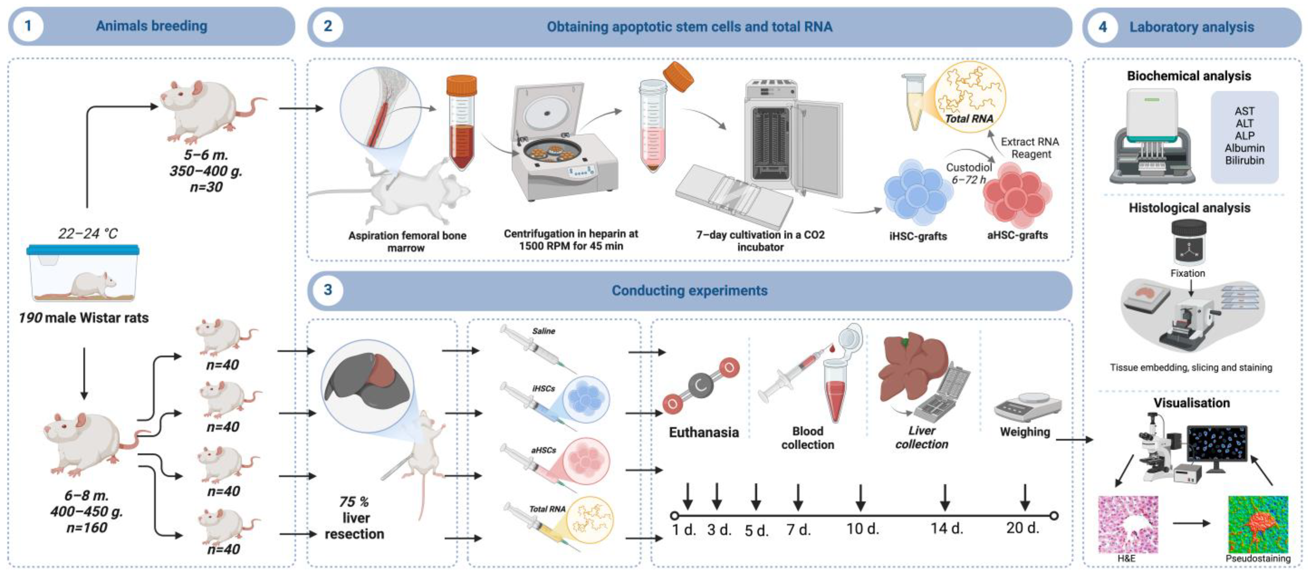
2.1. Animal Care and Extended Liver Resection

The study included male Wistar rats (n = 190). Among them, 160 six- to eight-month-old animals weighed 400–450 g, and 30 five- to six-month-old animals weighed 350–400 g. The rats stayed in our vivarium at 22–24 °C with 12 h light–dark cycle and a mixed diet (Laboratorkorm, Russia) ad libitum and free access to water (tap water was sterilized and distillated prior to the watering). We kept cages with animals in horizontal laminar flow cabinets for housing cages with laboratory rodents (CLJ–18; 3W, Moscow, Russia). The regulatory standards for the equipment accorded with GLP rules. Temperature and relative humidity were monitored twice a day using a thermometer and a psychrometer. The animal cage had a protruding upper bar, providing shading and allowing the animal to choose a dark area of the cage to stay in during the day with bright lighting.
Three rodents were kept per a cage, with respect to randomization, and sterile wood shavings delivered in vacuum bags served for the bedding (Laboratorkorm, Moscow, Russia). The experiments with animals took place from 9 am to 7 pm at 22–24 °C to avoid differences in mitotic activity of hepatocytes.
All the manipulations with animals fulfilled requirements of local Ethics Committee, National Guidelines on Animal Care, and European Convention for the Protection of Vertebrate Animals used for research and other scientific purposes. The animal study protocol was approved by the Ethics Committee of Federal State Budgetary Institution “Shumakov National Medical Research Center of Transplantology and Artificial Organs” of the Ministry of Health of the Russian Federation (protocol No. 050221-1/3e 05/02/21) for studies involving animals.
Younger rats (n = 30) served to obtain HSCs. Older rats (n = 160) experienced extended (75%) liver resection (ELR) according to common approaches [30,31]. Due to inability to accomplish all the experiments and surgeries on the same days, we performed eight consecutive rounds with 5 animals per group, considering the pilot experiment also, and then rejoined the numerical data for statistical processing.
The surgery was accomplished as follows. After an aseptic access to abdominal cavity by midline incision, we ligated and removed middle (35–40%), left lateral (25–30%), and right superior lobes (15–20%), then the peritoneum and skin were sutured. Surgical manipulations took place at 10–12 am with isoflurane anesthesia (Laboratories Karizoo, Barcelona, Spain). At the early post-surgery stage, the animals had signs of acute liver damage.
Animals were sacrificed by increasing CO2 concentration on Days 1, 3, 5, 7, 10, 14, and 20. Immediately after this, we collected their blood by notching the tail tip and removed the liver to weigh and then to put into 10% neutral buffered formalin Histosafe (cat. No. B06-001/L; ErgoProduction, Saint Petersburg, Russia).

2.2. Stem Cell Obtaining

We obtained HSCs according to Rumyantsev et al. [32]. After anesthesia, we aspirated femoral bone marrow from both sides in sterile conditions. We centrifuged portions of 25–30 mL in 100 U/mL heparin (Belmedpreparaty, Minsk, Belorussia) at 500 revolutions per min for 45 min, the supernatant was removed for one more centrifugation at 1500 revolutions per min for 10 min.
We transferred interphase from the surface of precipitated erythrocytes into a lysis solution (114 mM NH4Cl, 7.5 mM KHCO3, 100 μM EDTA; cat. No. R101p-2; Paneco, Moscow, Russia) at 1:4 for 5–8 min, then centrifuged the interphase at 1500 revolutions per min for 5 min and removed supernatant fractions. The lysis was repeated twice or thrice, and erythrocytic degradation occurred. We added the suspension (90–400 × 106 cells) purified from erythrocytes and platelets to plasma-derived suspension with Iscove’s medium (cat. No. 12-440-053; Gibco, Jenks, OK, USA), 0.58 g/L glutamine (cat. No. F032; Paneco, Moscow, Russia), 10% bovine serum (cat. No. SH3007001; HyClone, Logan, UT, USA), 0.4 μM insulin, 50 μg/mL gentamycin (Dalkhimpharm, Khabarovsk, Russia), 25 ng/mL granulocyte-monocyte colony-stimulating factor (cat. No. PSG030; Dia-M, Moscow, Russia), 10 ng/mL interleukin-3 (cat. No. I3769; SRP3243; Sigma-Aldrich, Saint Louis, MO, USA), 20 ng/mL insulin-like growth factor I (cat. No. I3769; Sigma-Aldrich, Saint Louis, MO, USA), and 25 mM 4-(2-hydroxyethyl)-1-piperazineethanesulfonic acid (cat. No. CAS 7365-45-9; Dia-M, Moscow, Russia).
These HSCs stayed in Petri dishes (d = 35 mm) at 1.5–2.0 mln cells/mL (Goryaev chamber; Minimed, Bryansk, Russia), 37 °C, 95% relative humidity inside incubator with 5% CO2 for 7 days with a single change of growth medium on Day 3. Non-attached HSCs served as iHSC grafts.

2.3. Obtaining Apoptotic Stem Cells and Total RNA

To obtain aHSCs, we incubated HSCs in Custodiol solution (histidine tryptophan-ketoglutarate solution; cat. No. P08-0735; Dr Franz Köhler Chemie, Bensheim, Germany) at 4–6 °C for 6, 18, 24, 48, or 72 h. Normally, Custodiol is common as a solution for conservation of grafted organs and cells, and that is also due to its potential to direct cells not to necrosis but to autophagy and reversible apoptosis, especially at low temperature [33]. We, therefore, employed Custodiol to provide a reversible damage to cells only. Moreover, we removed cells from incubating portions at different time points to obtain the maximal number of alive cells, as pro-apoptotic effects only stopped after 72 h of incubation in the experiment.
We validated apoptosis using Annexin V Apoptosis Detection Kit (Cat. No. 640922; BioLegend, San Diego, CA, USA). Cell suspension was added to 100 µL of Annexin V binding buffer (1 × 10⁷ cells per 1 mL), then treated with 5 µL of Annexin V (dye for apoptotic cells). After 5 min of incubation (no light, room temperature), we added 400 µL of Annexin V binding buffer. Stained cell analysis employed a Beckman Coulter Cytomics FC 500 flow cytometer (Beckman Coulter, Brea, CA, USA). Apoptotic cells served as aHSC-grafts.
We obtained total RNA from iHSCs according to manufacturer’s protocol [17] by Extract RNA Reagent (Cat. No. BC032; Eurogen, Moscow, Russia). The method allows obtaining RNA of high purity with almost no protein contaminants. Thus, approximately 148.5 ± 22.3 μg of total RNA came from each portion of 30–35 × 106 cells.

2.4. Experimental Design

Experimental design is given in Figure 1.
After ELR, we randomized rats into the following groups: (1) a control group (n = 40; Control) with single intraperitoneal dose of 1.0–1.5 mL saline; (2) group of iHSC received single intraperitoneal injections of intact hematopoietic stem cells at 30–35 × 106 cells per 1.0–1.5 mL saline (n = 40); (3) group of aHSC received single intraperitoneal injections of apoptotic hematopoietic stem cells at the same dose (n = 40); (4) group of total RNA received single intraperitoneal injections of total RNA from intact hematopoietic stem cells at a dose of 30 μg/100 g of body mass in 1.0–1.5 mL of saline (n = 40). The medications were given 3–5 h after ELR. The allocation to groups was based on column sorting with method of random numbers with respect to sex, age, and body mass as leading criteria.
We registered the time of animal survival after ELR. We also examined blood samples for markers of liver cell lysis: alanine aminotransferase (ALT), aspartate aminotransferase (AST), alkaline phosphatase (ALP), albumin, and total bilirubin levels by a biochemical analyzer AU 680 (Cat. No. AST-OSR 6109, ALT-OSR 6107, ALP-OSR 6104, Albumin-OSR 6102, Bilirubin-OSR 6112, respectively; Beckman Coulter, Brea, CA, USA) in animals before the surgery and dynamically after.

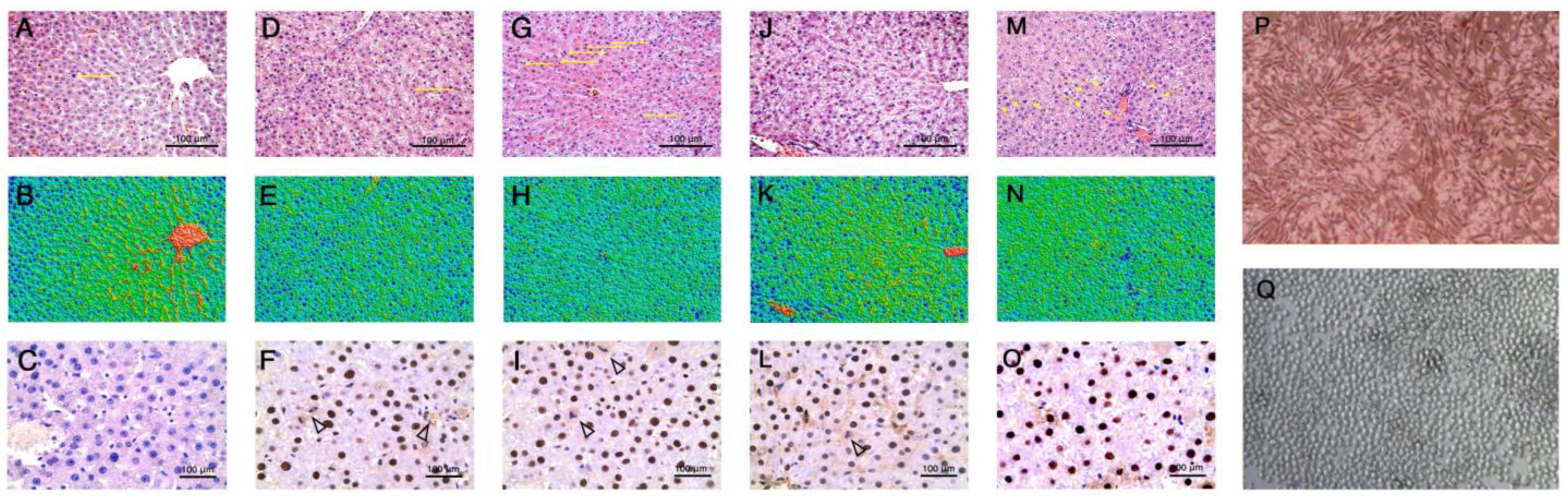
2.5. Histological Study

We weighed removed liver portions immediately after ELR (70% of total liver mass) and livers of lethalized rats with Adventurer Balances (OHAUS, Parsippany, NJ, USA) at room temperature. Then, liver samples of treated rats were fixed in 10% neutral buffered formalin, dehydrated in alcohols of increasing concentration, then embedded in paraffin with 5 µm sections obtained, and stained with hematoxylin and eosin (H&E) (hematoxylin solution cat. No. HK-G0-F250; ErgoProduction, Russia; eosin H, cat. No. CHM117739; AO Lenreaktiv, Saint Petersburg, Russia).
Immunohistochemical (IHC) study used anti-Ki-67 antibodies (cat. No. B68180; Beckman Coulter, Brea, CA, USA) to study proliferation, anti-caspase-3 (cat. No. AA626Ra82; Cloud-Clone, Katy, TX, USA) to study irreversible apoptosis, and anti-caspase-9 (cat. No. ab52298; Abcam, Cambridge, MA, USA) to study reversible apoptosis. The antibodies were diluted 1:100 in phosphate saline buffer solution (PBS) with 0.1% Tween 20 (cat. No. B-60261; Eco-Service, Moscow, Russia) and 5% bovine serum albumin (BSA; cat. No. PM-T1727; Biosera, Cholet, France). Washed samples were exposed to secondary antibodies against rabbit IgG conjugated to horseradish peroxidase (cat. No. HA710043; Huabio, Hangzhou, China) in PBS solution with 5% BSA, then employing detection by 3,3′-diaminobenzidine (DAB; cat. No. ab64238; Abcam, Cambridge, MA, USA). We counterstained nuclei with Mayer’s hematoxylin (cat. No. HK-G0-DL01; ErgoProduction, Saint Petersburg, Russia).
The calculation of mitotic index (MI) included the ratio between mitotic hepatocytes and their total number in 30 high-power fields at each time point by 24, 48, and 72 h, Days 5, 7, and 10 after ELR (‰) [34].
Morphometrics of H&E-stained sections employed light microscopy with camera by LeicaDM1000, Leica LTDCH9435, and DFC 295 (Leica Camera AG, Wetzlar, Germany), then software analysis with ImageScopeM, v12.3 (systems for microscopy and analysis, Moscow, Russia). Micrographs were obtained at ×400 and processed with ImageJ software, v.1.53h (NIH, Bethesda, MD, USA) [35,36]. We measured overall area of cells and their nuclei and then calculated areas of cytoplasm. We also counted the number of mono- and binuclear cells. An increase in cells with more than one nucleus was considered to be a feature of cell regeneration with no mitosis.
For transforming 2D into 3D images, we used a Leica DM 6000 B microscope (Wetzlar, Germany), Leica LTDCH9435 camera (Leica Camera AG, Wetzlar, Germany), and MATLAB software, v. R2021b, (MathWorks, Natick, MA, USA). The following types of structures are seen: oxyphilic cytoplasm of hepatocyte, basophilic nuclei of hepatocyte, connective tissue proper, intercellular substance, and empty spaces (vessels, adipocytes). Converting the images into grayscale, we constructed 3D surfaces, the models and colors specific for each image.

2.6. Statistics and Reproducibility
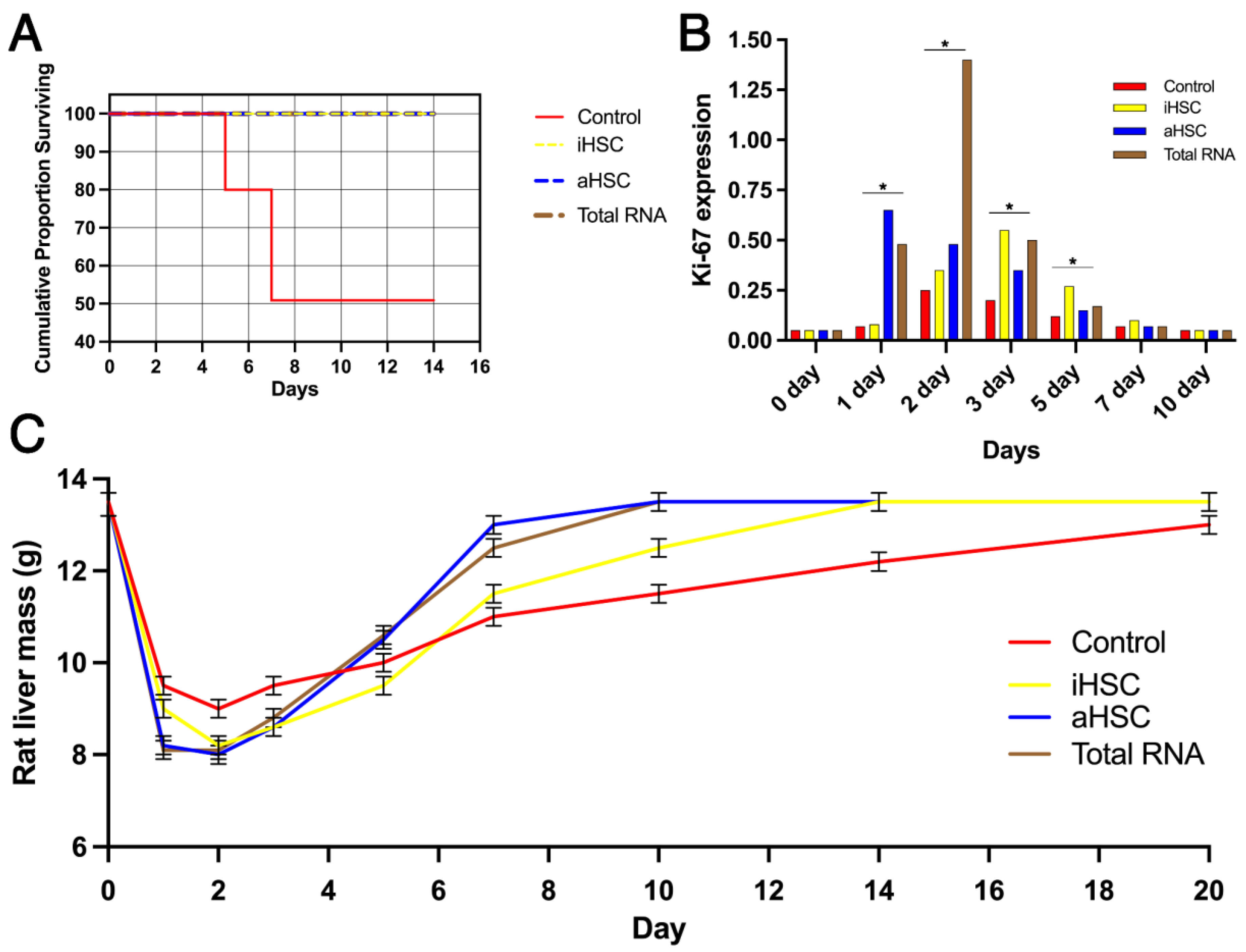
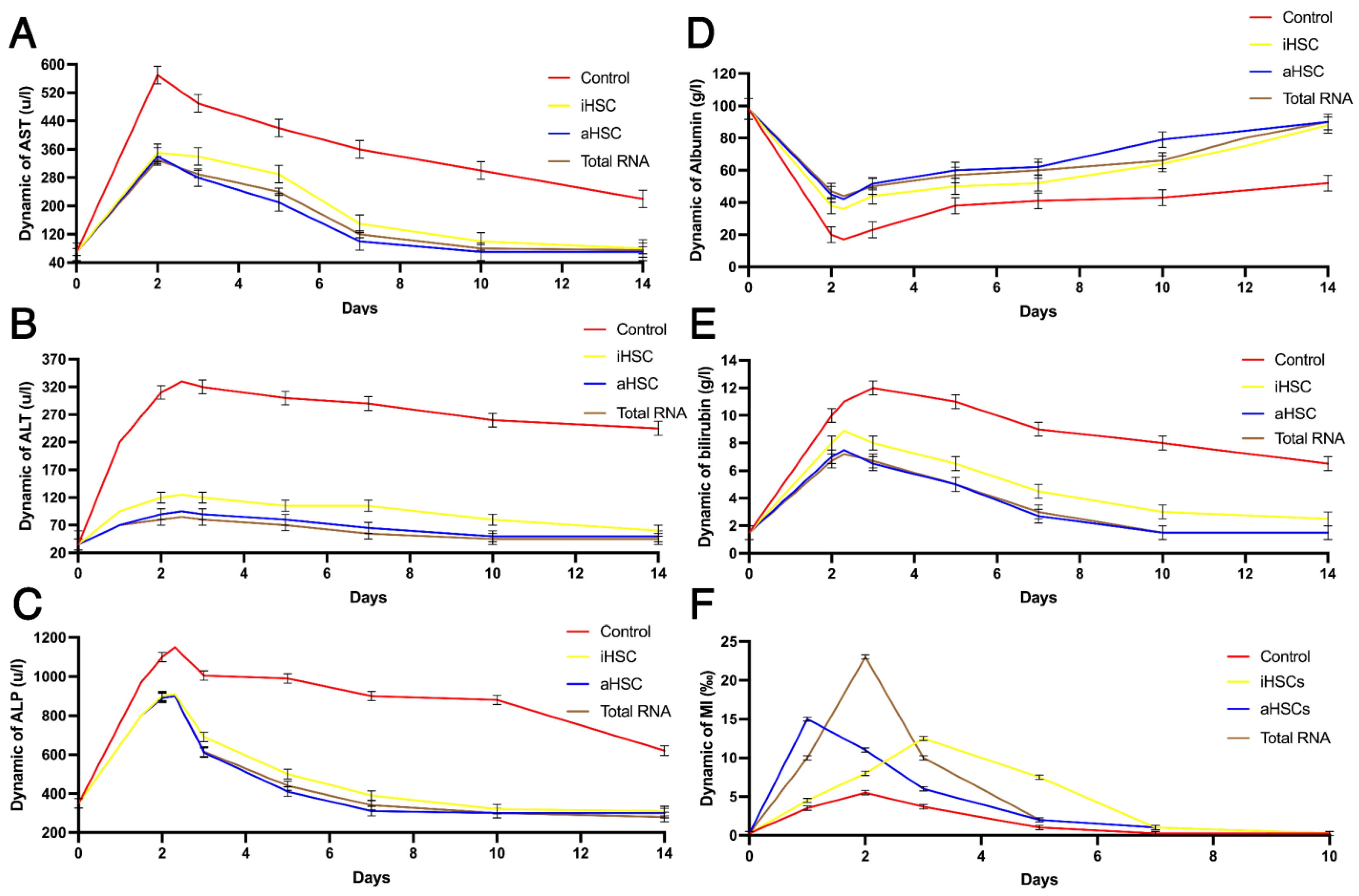
Sample size estimation was accomplished with respect to 3R principles with the least number of animals possible and was based on pilot part of the study that included 5 animals per group with targeted power as 0.8 and targeted statistical significance as 1.96.
The results were fixed in Microsoft Excel (Microsoft, Redmond, WA, USA) standard format. The analysis of the survival rate used log-rank test with Kaplan–Meier curves at Statistica software (TIBCO, Santa Clara, CA, USA) for Windows v.12. The differences were considered as significant at p < 0.05.
For blood sampling tests, we assessed the normality of distribution at any timepoint with Shapiro–Wilk test and then processed the obtained results with Biostat software (EpiInfo 5.0, CDC, Atlanta, GA, USA), t test with Bonferroni correction employed. The differences were considered significant at p < 0.05.
For histological study, the statistical processing recruited OriginPro software, Origin2024 (Origin, Farmington, ME, USA). Distribution models were defined with Shapiro–Wilk test and with paired Wilcoxon (for non-parametric values) and t test (for parametric values) with Holm–Bonferroni method employed. The statistical differences were regarded significant with p < 0.05.

3. Results

After 7 days of cultivation, the cell culture contained up to 40% stellate fibroblast-like cells attached to the polymer base (stromal progenitor cells) and up to 60% rounded and non-attached cells (hematopoietic cells, lymphocytes, and monocytes). The number of apoptotic cells reached 50–60%, and that of necrotic cells reached 7–10% (Figure 2P,Q).
For the first 7 days after ELR, the control group showed the survival rate almost equal to 50% as estimated using the log-rank test with p ˂ 0.05. No additional mortality occurred in the control group after Day 7. Groups of iHSC, aHSC, and total RNA had no fatal outcomes during the entire study period (Figure 3A).
The high mortality after surgery in the control group is consistent with acute liver failure confirmed by an increase on Day 2 compared to Day 0 in AST (572 ± 42.2 u/L to 71 ± 14.8 u/L), ALT (315 ± 3.7 u/L to 35 ± 2.3 u/L), ALP (1102 ± 37.2 u/L to 382 ± 29.8 u/L), and total bilirubin (2 ± 1.8 g/L to 10 ± 2.1 g/L with the increase continuing up to Day 12) levels (markers of cell lysis) in the blood of the rats (Figure 4A–E). The iHSC, aHSC, and total RNA groups exhibited a higher rate of biochemical recovery than the control group, as shown by repeated t test comparisons with p ˂ 0.05. In the iHSC, aHSC, and total RNA groups, cell lysis markers also increased to a lesser extent during the first two days but recovered completely by Day 12–14. Thus, the AST levels on Day 2 were the least for the total RNA group (330 ± 29.7 u/L), ALT levels were the least for the total RNA group (81 ± 6.4 g/L), ALP levels were the least for the aHSCs group (892 ± 32.4 u/L), and total bilirubin was the least for the total RNA (6 ± 1.2 g/L) group.
Albumin levels in the control group decreased with a peak by Day 2 (19.3 ± 1.73 g/L), then gradually increased almost up to the Day 0 values (98.0 ± 2.01 g/L) before the surgery on Day 14 (52.1 ± 1.54 g/L). The albumin levels in the iHSC (88.2 ± 1.94 g/L), aHSC (90.3 ± 1.23 g/L), and total RNA groups (90.5 ± 0.94 g/L) almost recovered by the end of the study.
Studying the histological specimens, we revealed no signs of bleeding, leukocytic infiltration, necrosis, or hemosiderin accumulation in the liver. A histological study also revealed cytoplasmic vacuolization of hepatocytes in all groups. The vacuolization developed by 24 h after ELR and increased by 48 h. However, the changes were not the same for all the groups. The aHSC and total RNA groups had a more pronounced and earlier vacuolization than the iHSC group and the control group. The histological slides of the liver were used to count the mitotic index (MI).
In the experiment, the MI was estimated with parametrical tests (t tests) and the p values were considered as significant if they were ˂0.05. The MI stayed increased for all groups (rats who had no ELR and served donors for HSCs had a liver MI equal to 0.2–0.3%). However, the groups of iHSC, aHSC, and total RNA had a more remarkable MI (Figure 4F) than the control group. The control group reached its peak of MI by 48 h (36 mitotic cells among 6653 counted cells). For the iHSC group, the peak occurred by 72 h with 93 mitoses per 8858 cells; for the aHSC group, the peak occurred by 24 h with 136 mitoses per 9762 cells; finally, for the total RNA group, the peak occurred by 48 h with 227 mitoses per 9678 cells. Counting the rate, the peak MI of the control group was equal to 5.4 ± 0.24 ‰; iHSC equal to 10.5 ± 0.47 ‰; aHSC equal to 14.9 ± 0.56 ‰; and total RNA was equal to 23.5 ± 0.21 ‰.
Regarding the timing, intraperitoneal administration of aHSCs and total RNA led to maximal MI values by 24 h or 48 h, respectively, whereas for iHSCs it was by 72 h. We calculated MI in the histological slides stained with H&E, as shown in Figure 3A,D,G,J,M. These slides revealed vacuolar dystrophy of hepatocytes by 48 h after ELR in all groups, while the effect was maximal in the aHSC group (Figure 2J,K).
We also studied the rate of proliferation and apoptosis by expression of Ki-67, caspase-3 and caspase-9 in hepatocytes 48 h after ELR. The IHC study with antibodies against Ki-67 revealed the maximal proliferation for groups aHSC and total RNA on Days 1, 2, and 3 after ELR as estimated by paired t tests with p ˂ 0.05 (M ± SD for logarithmic values of Ki-67 expression: 0.65 ± 0.07, 0.48 ± 0.05, 0.35 ± 0.06 for aHSC; 0.48 ± 0.04, 1.4 ± 0.09, 0.5 ± 0.06, respectively), and on Days 2 and 3 after ELR for the control group (0.25 ± 0.05, 0.2 ± 0.05, respectively) and iHSC (0.35 ± 0.03, 0.55 ± 0.05, respectively) (Figure 3B and Figure 5). Caspase-9 expression was remarkable in all groups by 48 h after ELR (Figure 2C,F,I,L,O), but no caspase-3 expression was found with null expression for all the groups, while the control slides from other organs were positive for the staining.
Measuring liver mass in rats (the parameter was normally distributed with the difference considered significant at p ˂ 0.05), we found that groups aHSC and total RNA had a higher rate of liver mass recovery (by Days 810; 13.5 ± 1.2 g and 13.6 ± 1.4 g, respectively) compared to the iHSC group (by Days 1214; 13.4 ± 2.1 g) and the control group (by Days 1820; 13 ± 0.9 g) after ELR (Figure 3C).
We also assessed the morphometrics of hepatocytes on the histological slides for the MI index count, considering that the changes in liver mass may be due not only to the mitotic activity of hepatocytes but also to their hypertrophic changes with no increase in cell number (Figure 6A–C). These parameters had a Gaussian data distribution and were estimated using the Wilcoxon test with p ˂ 0.05 being significant and data represented further as median with interquartile ranges.
In the iHSC, aHSC, and total RNA groups, we observed increased areas of both mononuclear and binuclear cells compared to the control group. Thus, the total RNA group had the largest cell area for binuclear cells: 430 μm2 (396; 475) to 353 μm2 (301; 397) in the controls, and in the aHSC group for mononuclear cells: 355 μm2 (292; 423) to 328 μm2 (290; 382) in the controls. The same results were observed for the areas of nuclei and cytoplasm. The total RNA group had the highest area of nuclei, and that is for binuclear cells (98 μm2 (86; 106) to 70 μm2 (57; 85) in controls), while the aHSC group exhibited the largest cytoplasm area, and for mononuclear cells (281 μm2 (230; 341) to 263 μm2 (223; 301) in controls).
At the same time, no significant changes were found in the number of binuclear cells at p ˂ 0.05. The control group showed a lesser number of binuclear cells with a larger rate of cell number per 50,000 μm2 (Figure 6D,E).

4. Discussion

Our study of ALF after ELR demonstrated that all the animals survived in the experimental groups (iHSC, aHSC, and total RNA), while the control group had a 50% mortality rate by Day 7. Therefore, the ELR model was adequate and appropriate for the assessment.
In the experimental groups, cell lysis markers (ALT, AST, ALP, and total bilirubin) first elevated and then decreased strongly compared to the control group, reaching normal ranges by Day 10–14. Moreover, the groups of aHSC and total RNA had a more rapid recovery rate than iHSC. The control group reached complete recovery biochemical markers and liver mass only up to Day 20. For albumin levels, the control group kept the same dynamics with no recovery achieved by Day 14 (Figure 4D). In the groups of iHSC, aHSC, and total RNA, the albumin levels decreased less than those in the control group. Generally, no fatal outcomes in animals at iHSC, aHSC, and total RNA (Figure 3A) together with a more efficient biochemical recovery (Figure 4A–E) reveal a successful liver regeneration.
Cytoplasm vacuolization on the histological slides of the liver can be a feature of cell autophagy [37], while a more pronounced and earlier vacuolization in the groups of aHSC and total RNA may be due to a more pronounced ability of apoptotic cells and their components to stimulate adaptive changes [38]. Interestingly, no histological signs for types of cellular death other than apoptosis and autophagy were observed, and no irreversible apoptosis was found at all.
The mitotic activity of hepatocytes showed differences between the groups. The experimental groups exhibited an increased mitotic activity compared to controls (Figure 4F). We observed the highest rate of MI for the aHSC group by 24 h, by 48 h in the total RNA group, and by 72 h for the iHSC group. The values of these MI peaks differed. For instance, the total RNA group provided the highest MI, while the aHSC group saw the most rapid increase. This finding may be explained by the immediate arrival of signal molecules from apoptotic cells to the liver [16].
Proliferation after ELR is, however, simultaneous with cell death, either reversible or irreversible. We found that most hepatocytes expressed elevated levels of caspase-9, a reversible apoptosis marker, in two days after ELR, i.e., at the same time with higher vacuolization. The data are consistent with reports about autophagy and reversible apoptosis involved in reparative regeneration [37,38,39]. Such an adaptation covers both metabolic and cellular mechanisms [40,41].
Extended liver resection changes the proliferative potential of hepatocytes, as we already demonstrated previously [42]. The current study develops this idea, demonstrating iHSCs, aHSCs, and total RNA to be adequate tools for autophagy and reversible apoptosis induction together with cell proliferation in the liver, and that is consistent with the common concepts of liver regeneration [43,44]. Interestingly, there was no increase in the absolute number of cells or binuclear cells per field of view in the experimental groups. Considering a higher rate of Ki-67 expression, we interpret this as, first of all, a hallmark for a strong apoptosis with mitigating cell number increase and, further, a strong role of hypertrophic regeneration and not only a proliferative one in response to HSC-derived stimuli.
The total RNA of HSCs, which is shown as the most appropriate method in the current study, has not been widely studied before in liver regeneration. Thus, such research with total RNA usually involves the employment of own total RNA from liver cells [45]. Nevertheless, the most common clinically or at least experimentally methods for liver regeneration do not comprise HSCs or their derivatives at all.
For instance, there exist such methods as hepatocyte grafting, gene editing, tissue engineering, liver organoid development, and siRNA treatment for certain genes, and all of them find their application [46]. This excellent review of Hora and Wuestefeld also outlines that stem cell therapy is appropriate for liver regeneration with bone marrow MSCs or induced pluripotent stem cells used as well as HSCs. However, it is more difficult to obtain MSCs or induced pluripotent stem cells than HSCs. Therefore, our finding proposes an option seldom considered earlier, and its machinery should be elucidated in further research.
In liver regeneration, there are plenty of pathways for metabolic and cellular crosstalk [47]. The most efficient way is regeneration due to the proliferation of cholangiocytes recruiting the Notch pathway [48]. The Notch pathway is suppressed in senescent cells, is dependent on macrophages, and is sensible to molecular signals from HSCs, including their protein and RNA signals [49]. Thereby, the Notch pathway may be an important target of HSC-derived total RNA in the liver.
Undoubtedly, the activity of total RNA and aHSCs in the liver may have an impact on oncogenic potential. The role of this impact remains unclear and requires further elucidation paying attention to a close interplay between proliferation in the liver and abdominal malignancies [50].

Limitations and Perspectives

  • The study aimed to compare different HSC-originating modes to enhance regeneration. Thus, the methodology applied is not relevant for revealing all the mechanisms of HSC communication, and the ratio between paracrine, direct, and immune-mediated machinery remains unclear. An analysis of the RNA sequence in new proliferating cells in the liver after HSC-derived total RNA would help to elucidate the problematics.
  • The study has not implied assessment of specific markers’ content by cellular and molecular methods, e.g., immunoblotting. These approaches are planned for future experiments in the field.
  • The tumorigenic potential of aHSCs and total RNA is challenging. The experiment is to be widened for rat follow-up for several consecutive months after the liver recovery to estimate the rate of oncological risks before extrapolation to humans.
  • Clinical implementation of the study results is limited due to legislative procedures. For instance, human stem cells are restricted for usage in certain countries’ jurisdictions, and the studies should absolutely respect the restrictions before planning any human extrapolation.

5. Conclusions

Our study showed that iHSC-, aHSC-, and iHSC-derived total RNA in a single-dosage regimen stimulated regeneration in the liver after ELR. However, the presence of aHSCs and total RNA is associated with a more prominent regeneration than that of iHSCs. The proliferation starts first with the usage of aHSCs and its rates are higher with the usage of total RNA. We conclude that aHSCs and total RNA of iHSCs take part in a powerful shift to repair the liver. Their application may be beneficial in liver disease, and further studies of the total RNA of iHSCs are required to elucidate its practical potential to become a targeted regenerative option.

6. Patents

Onishchenko N. A., Nikolskaya A. O., Gonikova Z. Z., Shagidulin M. Yu., Kirsanova L. A., Sevastyanov V. I. Method of treatment of acute liver failure. Patent No. 2744846, 16 March 2021.

Author Contributions

N.O.: conceptualization, methodology, validation, data curation, writing—original draft, writing—review and editing, supervision. M.S.: conceptualization, methodology, validation, data curation, writing—original draft, writing—review and editing, supervision. Z.G.: methodology, data curation. A.N.: methodology, data curation. L.K.: methodology, data curation. A.V.: data curation, writing—review and editing. K.P.: methodology, data curation. E.K.: methodology, data curation. N.K. (Natalia Kuznetsova): methodology, data curation. I.K.: formal analysis, Writing—review and editing. D.T.: conceptualization, supervision. N.K. (Natalia Kartashkina): methodology, writing—review and editing. V.V.: validation, data curation, writing—original draft. M.T.: validation, data curation, writing—original draft. V.S.: writing—original draft, writing—review and editing. A.E.: data curation, writing—review and editing. G.P.: conceptualization, methodology, writing—original draft, writing—review and editing. S.G.: methodology, writing—review and editing, supervision. All authors have read and agreed to the published version of the manuscript.

Funding

The study was carried out with the support of the state assignment of the Ministry of Health of the Russian Federation. (Topic No. 124031200061-0).

Institutional Review Board Statement

The animal study protocol was approved by the Ethics Committee of Federal State Budgetary Institution “Shumakov National Medical Research Center of Transplantology and Artificial Organs” of the Ministry of Health of the Russian Federation (protocol 050221-1/3e 05/02/21) for studies involving animals.

Informed Consent Statement

Not applicable.

Data Availability Statement

The data presented in this study are available upon request from the corresponding authors.

Acknowledgments

The authors are grateful to Fomenko E. and Shmerko N. for their research assistance.

Conflicts of Interest

The authors declare no conflicts of interest.

References

  1. Michalopoulos, G.K.; Bhushan, B. Liver regeneration: Biological and pathological mechanisms and implications. Nat. Rev. Gastroenterol. Hepatol. 2021, 18, 40–55. [Google Scholar] [CrossRef] [PubMed]
  2. Loffredo, L.F.; Savage, T.M.; Ringham, O.R.; Arpaia, N. Treg-tissue cell interactions in repair and regeneration. J. Exp. Med. 2024, 221, e20231244. [Google Scholar] [CrossRef] [PubMed]
  3. Babaeva, A.G.; Tishevskaya, N.V.; Gevorkyan, N.M. On the Morphogenetic Properties of RNA of Lymphoid and Stem Cells in Recovery Processes; RAMS, Scientific Research Institute of Human Morphology: Moscow, Russia, 2016. [Google Scholar]
  4. Tishevskaya, N.V.; Babaeva, A.G.; Gevorkyan, N.M. The role of lymphocytic RNA in intercellular information. Exchange and regulation of regenerative processes. Ross. Fiziol. Zhurnal Im. I.M. Sechenova 2016, 102, 1280–1301. (In Russian) [Google Scholar]
  5. Than, N.N.; Newsome, P.N. Stem cells for liver regeneration. QJM Mon. J. Assoc. Physicians 2014, 107, 417–421. [Google Scholar] [CrossRef]
  6. Liu, F.; Pan, X.; Chen, G.; Jiang, D.; Cong, X.; Fei, R.; Wei, L. Hematopoietic stem cells mobilized by granulocyte colony-stimulating factor partly contribute to liver graft regeneration after partial orthotopic liver transplantation. Liver Transplant. 2006, 12, 1129–1137. [Google Scholar] [CrossRef]
  7. Tsolaki, E.; Athanasiou, E.; Gounari, E.; Zogas, N.; Siotou, E.; Yiangou, M.; Anagnostopoulos, A.; Yannaki, E. Hematopoietic stem cells and liver regeneration: Differentially acting hematopoietic stem cell mobilization agents reverse induced chronic liver injury. Blood Cells Mol. Dis. 2014, 53, 124–132. [Google Scholar] [CrossRef] [PubMed]
  8. Venkitaraman, A.; De, A.; Verma, N.; Kumari, S.; Leishangthem, B.; Sharma, R.R.; Kalra, N.; Grover, S.; Singh, V. Multiple cycles of granulocyte colony-stimulating factor in decompensated cirrhosis: A double-blind RCT. Hepatol. Int. 2022, 16, 1127–1136. [Google Scholar] [CrossRef]
  9. Siapati, E.K.; Roubelakis, M.G.; Vassilopoulos, G. Liver regeneration by hematopoietic stem cells: Have we reached the end of the road? Cells 2022, 11, 2312. [Google Scholar] [CrossRef]
  10. Bird, T.G.; Müller, M.; Boulter, L.; Vincent, D.F.; Ridgway, R.A.; Lopez-Guadamillas, E.; Lu, W.Y.; Jamieson, T.; Govaere, O.; Campbell, A.D.; et al. TGFβ inhibition restores a regenerative response in acute liver injury by suppressing paracrine senescence. Sci. Transl. Med. 2018, 10, eaan1230. [Google Scholar] [CrossRef]
  11. Bi, J.; Zhang, J.; Ke, M.; Wang, T.; Wang, M.; Liu, W.; Du, Z.; Ren, Y.; Zhang, S.; Wu, Z.; et al. HSF2BP protects against acute liver injury by regulating HSF2/HSP70/MAPK signaling in mice. Cell Death Dis. 2022, 13, 830. [Google Scholar] [CrossRef]
  12. Fathi, E.; Valipour, B.; Jafari, S.; Kazemi, A.; Montazersaheb, S.; Farahzadi, R. The role of the hematopoietic stem/progenitor cells-derived extracellular vesicles in hematopoiesis. Heliyon 2024, 10, e35051. [Google Scholar] [CrossRef] [PubMed]
  13. Kitamura, T.; Terashima, T.; Katagi, M.; Ohashi, N.; Nozaki, K.; Tsuji, A. Bone marrow-derived mononuclear cells ameliorate neurological function in chronic cerebral infarction model mice via improvement of cerebral blood flow. Cytotherapy 2023, 25, 1186–1199. [Google Scholar] [CrossRef] [PubMed]
  14. Thum, T.; Bauersachs, J.; Poole-Wilson, P.A.; Volk, H.D.; Anker, S.D. The dying stem cell hypothesis: Immune modulation as a novel mechanism for progenitor cell therapy in cardiac muscle. J. Am. Coll. Cardiol. 2005, 46, 1799–1802. [Google Scholar] [CrossRef]
  15. Kerr, J.F.; Wyllie, A.H.; Currie, A.R. Apoptosis: A basic biological phenomenon with wide-ranging implications in tissue kinetics. Br. J. Cancer 1972, 26, 239–257. [Google Scholar] [CrossRef]
  16. Beer, L.; Zimmermann, M.; Mitterbauer, A.; Ellinger, A.; Gruber, F.; Narzt, M.S.; Zellner, M.; Gyöngyösi, M.; Madlener, S.; Simader, E.; et al. Analysis of the Secretome of Apoptotic Peripheral Blood Mononuclear Cells: Impact of Released Proteins and Exosomes for Tissue Regeneration. Sci. Rep. 2015, 5, 16662. [Google Scholar] [CrossRef]
  17. Ankersmit, H.J.; Hoetzenecker, K.; Dietl, W.; Soleiman, A.; Horvat, R.; Wolfsberger, M.; Gerner, C.; Hacker, S.; Mildner, M.; Moser, B.; et al. Irradiated cultured apoptotic peripheral blood mononuclear cells regenerate infarcted myocardium. Eur. J. Clin. Investig. 2009, 39, 445–456. [Google Scholar] [CrossRef]
  18. Beer, L.; Mildner, M.; Gyöngyösi, M.; Ankersmit, H.J. Peripheral blood mononuclear cell secretome for tissue repair. Apoptosis 2016, 21, 1336–1353. [Google Scholar] [CrossRef]
  19. Sirois, I.; Raymond, M.A.; Brassard, N.; Cailhier, J.F.; Fedjaev, M.; Hamelin, K.; Londono, I.; Bendayan, M.; Pshezhetsky, A.V.; Hébert, M.J. Caspase-3-dependent export of TCTP: A novel pathway for antiapoptotic intercellular communication. Cell Death Differ. 2011, 18, 549–562. [Google Scholar] [CrossRef]
  20. Huang, Q.; Li, F.; Liu, X.; Li, W.; Shi, W.; Liu, F.F.; O’Sullivan, B.; He, Z.; Peng, Y.; Tan, A.C.; et al. Caspase 3-mediated stimulation of tumor cell repopulation during cancer radiotherapy. Nat. Med. 2011, 17, 860–866. [Google Scholar] [CrossRef]
  21. Huleihel, L.; Scarritt, M.E.; Badylak, S.F. The Influence of Extracellular RNA on Cell Behavior in Health, Disease and Regeneration. Curr. Pathobiol. Rep. 2017, 5, 13–22. [Google Scholar] [CrossRef]
  22. Gonikova, Z.Z.; Nikolskaya, A.O.; Kirsanova, L.A.; Shagidulin, M.Y.; Onischenko, N.A.; Sevastjanov, V.I. Investigation of regenerative and tissue-specific activity of tot al RNA of bone marrow cells. Russ. J. Transplantology Artif. Organs 2018, 20, 64–69. (In Russian) [Google Scholar] [CrossRef]
  23. Gonikova, Z.Z.; Nikolskaya, A.O.; Kirsanova, L.A.; Shagidulin, M.Y.; Onischenko, N.A.; Sevastjanov, V.I. The comparative analysis of the effectiveness of stimulation of liver regeneration by bone marrow cells and total RNA of these cells. Russ. J. Transplantology Artif. Organs 2019, 21, 113–121. (In Russian) [Google Scholar] [CrossRef]
  24. Geng, X.; Chang, C.; Zang, X.; Sun, J.; Li, P.; Guo, J.; Xu, G. Integrative proteomic and microRNA analysis of the priming phase during rat liver regeneration. Gene 2016, 575, 224–232. [Google Scholar] [CrossRef] [PubMed]
  25. Li, L.; Guo, J.; Chen, Y.; Chang, C.; Xu, C. Comprehensive CircRNA expression profile and selection of key CircRNAs during priming phase of rat liver regeneration. BMC Genom. 2017, 18, 80. [Google Scholar] [CrossRef]
  26. Li, C.; Chang, L.; Chen, Z.; Liu, Z.; Wang, Y.; Ye, Q. The role of lncRNA MALAT1 in the regulation of hepatocyte proliferation during liver regeneration. Int J Mol Med. 2017, 39, 347–356. [Google Scholar] [CrossRef]
  27. Lauschke, V.M.; Vorrink, S.U.; Moro, S.M.; Rezayee, F.; Nordling, Å.; Hendriks, D.F.G.; Bell, C.C.; Sison-Young, R.; Park, B.K.; Goldring, C.E.; et al. Massive rearrangements of cellular MicroRNA signatures are key drivers of hepatocyte dedifferentiation. Hepatology 2016, 64, 1743–1756. [Google Scholar] [CrossRef]
  28. Qiao, J.; Yao, H.; Xia, Y.; Chu, P.; Li, M.; Wu, Y.; Li, W.; Ding, L.; Qi, K.; Li, D.; et al. Long non-coding RNAs expression profiles in hepatocytes of mice after hematopoietic stem cell transplantation. IUBMB Life 2016, 68, 232–241. [Google Scholar] [CrossRef]
  29. Mottaghitalab, F.; Rastegari, A.; Farokhi, M.; Dinarvand, R.; Hosseinkhani, H.; Ou, K.L.; Pack, D.W.; Mao, C.; Dinarvand, M.; Fatahi, Y.; et al. Prospects of siRNA applications in regenerative medicine. Int. J. Pharm. 2017, 524, 312–329. [Google Scholar] [CrossRef]
  30. Elchaninov, A.V.; Fatkhudinov, T.K. Mammalian Liver Regeneration: Intercellular Interactions; Nauka: Moscow, Russia, 2020. (In Russian) [Google Scholar]
  31. Saito, S.; Togo, S.; Morioka, D.; Matsuo, K.I.; Yoshimoto, N.; Nagano, Y.; Tanaka, K.; Kubota, T.; Nagashima, Y.; Shimada, H. A rat model of a repeat 70% major hepatectomy. J. Surg. Res. 2006, 134, 322–326. [Google Scholar] [CrossRef]
  32. Rumyantsev, A.G.; Maschan, A.A. Transplantation of Hematopoietic Stem Cells in Children; Medical information agency: Moscow, Russia, 2003. (In Russian) [Google Scholar]
  33. Baláz, P.; Matia, I.; Jackanin, S.; Rybárová, E.; Kron, I.; Pomfy, M.; Froněk, J.; Ryska, M. Preservation injury of jejunal grafts and its modulation by custodiol and University of Wisconsin perfusion solutions in Wistar rats. Eur. Surg. Res. 2004, 36, 192–197. [Google Scholar] [CrossRef]
  34. Baldo, G.; Giugliani, R.; Uribe, C.; Belardinelli, M.C.; Duarte, M.E.S.; Meurer, L.; da Silveira, T.R.; Matte, U. Bone marrow mononuclear cell transplantation improves survival and induces hepatocyte proliferation in rats after CCl4 acute liver damage. Dig. Dis. Sci. 2010, 55, 3384–3392. [Google Scholar] [CrossRef] [PubMed]
  35. Macenko, M.; Niethammer, M.; Marron, J.S.; Borland, D.; Woosley, J.T.; Guan, X.; Schmitt, C.; Thomas, N.E. A method for normalizing histology slides for quantitative analysis. In Proceedings of the 2009 IEEE International Symposium on Biomedical Imaging: From Nano to Macro, Boston, MA, USA, 28 June–1 July 2009; pp. 1107–1110. [Google Scholar] [CrossRef]
  36. Schneider, C.; Rasband, W.; Eliceiri, K. NIH image to ImageJ: 25 years of image analysis. Nat. Methods 2012, 9, 671–675. [Google Scholar] [CrossRef]
  37. Potapnev, M.P. Autophagy, apoptosis, cell necrosis and immune recognition of own and alien. Immunology 2014, 2, 95–102. (In Russian) [Google Scholar]
  38. Cheng, Y.; Wang, B.; Zhou, H.; Dang, S.; Jin, M.; Shi, Y.; Hao, L.; Yang, Z.; Zhang, Y. Autophagy is required for the maintenance of liver progenitor cell functionality. Cell Physiol. Biochem. 2015, 36, 1163–1174. [Google Scholar] [CrossRef] [PubMed]
  39. Lin, C.W.; Chen, Y.S.; Lin, C.C.; Chen, Y.J.; Lo, G.H.; Lee, P.H.; Kuo, P.L.; Dai, C.Y.; Huang, J.F.; Chung, W.L.; et al. Amiodarone as an autophagy promoter reduces liver injury and enhances liver regeneration and survival in mice after partial hepatectomy. Sci. Rep. 2015, 5, 15807. [Google Scholar] [CrossRef]
  40. Lv, H.; Fan, X.; Wang, L.; Feng, H.; Ci, X. Daphnetin alleviates lipopolysaccharide/d-galactosamine-induced acute liver failure via the inhibition of NLRP3, MAPK and NF-κB, and the induction of autophagy. Int. J. Biol. Macromol. 2018, 119, 240–248. [Google Scholar] [CrossRef]
  41. Onishchenko, N.A.; Nikolskaya, A.O.; Shagidulin, M.Y. Cytogenetic recapitulation, induced by medical preparations, as the universal stage of formation of urgent protection against damage at organ transplantation. Pathol. Physiol. Exp. Ther. 2016, 60, 148–153. (In Russian) [Google Scholar] [CrossRef]
  42. Elchaninov, A.V.; Fatkhudinov, T.K.; Kananykhina, E.Y.; Arutyunyan, I.V.; Makarov, A.V.; Charyeva, I.G.; Bolshakova, G.B.; Sukhikh, G.T. Cell proliferation and death in hepatocytes after subtotal rat liver resection (In Russ). Clin Exp Morph. 2016, 3, 22–29. [Google Scholar]
  43. Gazizov, I.M.; Gumerova, A.A.; Kiyasov, A.P. Apoptosis in regenerative histogenesis of the liver after partial hepatectomy in rats. Genes Cells 2015, 10, 22–26. [Google Scholar] [CrossRef]
  44. Budd, R.C. Death receptors couple to both cell proliferation and apoptosis. J. Clin. Investig. 2002, 109, 437–441. [Google Scholar] [CrossRef]
  45. Jun-Tang, L.; Cong-Rui, W.; Pramanik, J.; Hui-Yong, Z.; Hui-Gen, F.; Bao-Sheng, Y.; Yu-Chang, L.I.; Cun-Shuan, X. Study on construction of cDNA libraries from rat normal liver and regeneration liver with smart technique. Indian J. Clin. Biochem. 2004, 19, 177–180. [Google Scholar] [CrossRef] [PubMed]
  46. Hora, S.; Wuestefeld, T. Liver injury and regeneration: Current understanding, new approaches, and future perspectives. Cells 2023, 12, 2129. [Google Scholar] [CrossRef] [PubMed]
  47. Campana, L.; Esser, H.; Huch, M.; Forbes, S. Liver regeneration and inflammation: From fundamental science to clinical applications. Nat. Rev. Mol. Cell Biol. 2021, 22, 608–624. [Google Scholar] [CrossRef] [PubMed]
  48. Pu, W.; Zhu, H.; Zhang, M.; Pikiolek, M.; Ercan, C.; Li, J.; Huang, X.; Han, X.; Zhang, Z.; Lv, Z.; et al. Bipotent transitional liver progenitor cells contribute to liver regeneration. Nat. Genet. 2023, 55, 651–664. [Google Scholar] [CrossRef]
  49. Duan, J.L.; Ruan, B.; Song, P.; Zhi-Qiang, F.; Zhen-Sheng, Y.; Jing-Jing, L.; Guo-Rui, D.; Hua, H.; Lin, W. Shear stress-induced cellular senescence blunts liver regeneration through Notch-sirtuin 1-P21/P16 axis. Hepatology 2022, 75, 584–599. [Google Scholar] [CrossRef]
  50. Gatzidou, E.; Mantzourani, M.; Giaginis, C.; Giagini, A.; Patsouris, E.; Kouraklis, G.; Theocharis, S. Augmenter of liver regeneration gene expression in human colon cancer cell lines and clinical tissue samples. J. B.U.ON. 2015, 20, 84–91. [Google Scholar]
Figure 1. Experimental design.
Applsci 15 03782 g001
Figure 2. Histological slides of liver from rats before ELR and 48 h after. (A,B)—intact liver at Day 0; (D,E)—control group; (G,H)—group of iHSC; (J,K)—group of aHSC; (M,N)—group of total RNA. The second row shows pseudostaining of the corresponding images from the first row (hepatocyte nuclei are blue, cytoplasm is green, empty spaces are red). Yellow arrows indicate mitotic hepatocytes. H&E staining, ×200. Scale bar 100 μm. Caspase-9 expression on histological slides of liver from rats 48 h after ELR. (C)—intact liver at Day 0; (F)—control group; (I)—group of iHSC; (L)—group of aHSC; (O)—group of total RNA. Arrows indicate cells during mitosis. Although many of them are caspase-9-positive (i.e., in reversible apoptosis), the proliferation is evident. Anti-caspase-9 immunohistochemical staining with additional Mayer’s hematoxylin staining, ×200. Scale bar 100 μm. Cell cultures. (P)—culture of cells from the bone marrow of rats, 7 days of cultivation, ×100; (Q)—non-attached HSCs washed from the surface of cultivated cells, ×200, phase contrast.
Applsci 15 03782 g002
Figure 3. Dynamics of animal survival, Ki-67 expression, and rat liver mass. (A)—survival at percents after 75% ELR in rats (Kaplan–Meier). Groups of iHSC, aHSC, and total RNA increased the survival rate after ELR (p = 0.037, log-rank test); (B)—timepoints at dynamic logarithmic function of Ki-67 expression; *—the difference is significant compared with the control group; p < 0.05; (C)—recovery of rat liver mass in grams after ELR. Values are given as means ± SDs.
Applsci 15 03782 g003
Figure 4. Graphs for levels of blood markers and mitotic index. Daily dynamics of biochemical markers (units/Liter, u/L; or gram per liter, g/L) for all experimental groups: (A)—aspartate aminotransferase, AST, (B)—alanine aminotransferase, ALT, (C)—alkaline phosphatase, ALP, (D)—albumin, and (E)—total bilirubin in the blood. (F)—dynamics of mitotic index (MI, ‰) in hepatocytes of rats after ELR. Values are given as means ± SDs.
Applsci 15 03782 g004
Figure 5. Ki-67 expression at histological slides of liver from rats by 48 h after extended liver resection. (A)—Control group, Ki-67-negative; (C)—Group of iHSC with Ki-67-positive hepatocytes, widened sinusoids; (E)—Group of aHSC with a large number of Ki-67-positive hepatocytes; (G)—Group of total RNA with a large number of Ki-67-positive hepatocytes. Images (B,D,F,H) show pseudostaining of (A,C,E,G), respectively. Hepatocyte nuclei are blue, cytoplasm is green, empty spaces are red. Anti-Ki-67 immunohistochemical staining with additional Mayer’s hematoxylin staining, ×400. Scale bar 20 μm. (I)—the number of Ki67+ cells per field of view at the slides of the liver on Day 2. *—the difference is significant compared with the control group; p < 0.05; **—p < 0.01, ***—p < 0.001.
Applsci 15 03782 g005
Figure 6. Morphometrics for mono- and binuclear hepatocytes by 48 h after extended liver resection. Median values are provided with 25th and 75th percentiles: (A)—cell area, μm2; (B)—nucleus area, μm2; (C)—cytoplasm area, μm2; (D)—relative number of binuclear cells, %; (E)—total number of hepatocytes per 50,000 μm2. *—the difference is significant compared with the control group; p < 0.05.
Applsci 15 03782 g006
Disclaimer/Publisher’s Note: The statements, opinions and data contained in all publications are solely those of the individual author(s) and contributor(s) and not of MDPI and/or the editor(s). MDPI and/or the editor(s) disclaim responsibility for any injury to people or property resulting from any ideas, methods, instructions or products referred to in the content.

