Article

Morphofunctional Characteristics of the Foot and Ankle in Competitive Swimmers and Their Association with Muscle Pain

by
Cristina Jiménez-Braganza
1,
Antonia Sáez-Díaz
2 and
Pedro Vicente Munuera-Martínez
1,3,*
1
Faculty of Nursing, Physiotherapy and Podiatry. University of Seville, 41009 Seville, Spain
2
Department of Statistics, Axioma Comunicaciones, 41016 Seville, Spain
3
Department of Podiatry, University of Seville, 41092 Seville, Spain
*
Author to whom correspondence should be addressed.
Appl. Sci. 2025, 15(7), 3755; https://doi.org/10.3390/app15073755
Submission received: 19 February 2025 / Revised: 24 March 2025 / Accepted: 25 March 2025 / Published: 29 March 2025
(This article belongs to the Special Issue Advanced Research in Foot and Ankle Kinematics)

Abstract

:
The aim of this study was to analyze the joint characteristics of the foot and ankle in competitive swimmers aged 16–18 and 19–24 years and their relationship with the presence of muscle pain during swimming. A total of 74 swimmers were evaluated: 38 ‘junior’ (16–18 years) and 36 ‘senior’ (19–24 years). The following parameters were recorded: ankle dorsiflexion, rearfoot mobility, first metatarsophalangeal dorsiflexion, presence of hallux valgus, foot posture, first ray mobility, arch height, and plantar pressure. Additionally, the frequency and location of muscle pain in the triceps surae were analyzed. A cluster analysis was performed to identify variables that differentiated both groups. Ankle dorsiflexion was limited in both groups, with a greater restriction observed in adults (p < 0.001 with an extended knee; p < 0.014 with a flexed knee). The predominant foot type was the cavus foot. The most common pain was localized in the triceps surae, followed by the plantar musculature, with no significant differences between groups. Swimmers exhibited gastrocnemius shortening, which could limit ankle dorsiflexion and contribute to the onset of muscle pain in the leg and foot. These findings suggest the importance of incorporating lower limb flexibility strategies into the training of competitive swimmers.

1. Introduction

Swimming is a sport that is widely practiced throughout the world. The athlete’s goal is to swim a distance in the shortest possible time. The improvement of speed is conditioned by the perfection of swimming technique and mechanical efficiency, with elite swimmers standing out for their efficiency, power, and performance in propulsive forces [1]. Researchers have tried to find out which determinants influence performance in order to try to make swimmers faster with minimal risk of injury, so it is considered a very important topic of research. Although it is a non-impact sport with a low risk of injury, approximately 75% of swimmers have been injured at some time [2]. Competitive swimmers are affected by musculoskeletal injuries mainly due to cyclic and excessive joint movement, with most injuries occurring during training [3].
Although the shoulder is the joint most frequently affected by injuries in swimmers, with a prevalence rate of up to 91%, the function of the lower limb is also crucial for performance and can be a common site of musculoskeletal discomfort [4]. The joints and musculature of the lower limb enable leg movements that generate propulsion, body elevation, and balance. While lower limb function has been associated with improved swimming performance, studies describing the joint characteristics of the lower limb in swimmers, particularly the foot and ankle, remain limited to date [5]. Due to this lack of information, the aim of our study was to analyze the joint characteristics of the foot and ankle in competitive swimmers aged 16–18 and 19–24 years and their relationship with the presence of muscle pain in the posterior musculature and the plantar region during swimming.
Due to this lack of information, the objective of this study is to determine the morphofunctional characteristics of the foot and ankle in competitive swimmers of different age groups (junior and absolute) and analyze their potential relationship with muscular discomfort in the lower limb during swimming. By identifying joint mobility patterns, foot structure, and plantar pressures in swimmers, this study aims to contribute to the understanding of how these factors may predispose athletes to musculoskeletal pain or dysfunctions. The findings may provide useful insights for optimizing training strategies, preventing lower limb injuries, and improving biomechanical efficiency in competitive swimmers.

2. Methods

This is a cross-sectional study in which competitive swimmers from different swimming clubs in the province of Seville participated. The study was carried out between February 2022 and August 2023. All adult participants, as well as the parents of underage swimmers, gave their written consent to voluntarily participate in the study. The study was approved by the Research Ethics Committee of the University of Seville (internal code 250522). Seventy-four competitive swimmers voluntarily participated in the study, 38 ‘junior’ (age group 16–18 years, 20 men and 18 women) and 36 ‘absolute’ (age group 19–24 years, 19 men and 17 women). One hundred forty-eight lower limbs were analyzed. A total of 74 competitive swimmers were evaluated and divided into two groups according to their sports category. The “Junior” (JR) Group included 38 swimmers (20 men and 18 women) aged 16 to 18 years, who are still in the process of physical and technical development within their discipline but already compete at the national level. On the other hand, the “Absolute” (AB) Group consisted of 36 swimmers (19 men and 17 women) aged 19 to 24 years, considered athletes with greater maturity and competitive experience, who have surpassed the juvenile development phase and may have reached a high level of performance in national and international competitions.
The inclusion criteria were being registered in the junior (JR) and senior (AB) categories, having a minimum of five years of competitive swimming experience, and participating in national competitions in Spain. Swimmers who took part in triathlons, who had a bone fracture or surgery of the lower limb, or who had some degree of disability or functional diversity were excluded.
Firstly, demographic and anthropometric characteristics were analyzed, recording age in years and body measurements such as weight in kilograms and height in centimeters. These data provide context for the sample and allow for the analysis of potential differences between swimmer groups based on their physical development. Information was collected on the general characteristics of the swimmers, such as age, sex, and body mass index (BMI). Specific variables related to sporting practice were also recorded, such as weekly kilometers swam, main stroke, and years of swimming practice.
Next, several joint and biomechanical measurements of the foot and ankle were evaluated. Ankle dorsiflexion was measured in degrees using a goniometer (Fresco Podología, Barcelona, Spain), both with the knee extended and with the knee flexed [1]. Rearfoot mobility was assessed with an inclinometer, also expressed in degrees [2]. The range of inversion and eversion of the rearfoot was obtained by placing the athlete in the prone position with the knee in extension, according to the technique used by Taboadela [6]. The first metatarsophalangeal joint dorsiflexion (MPJ) [7] was measured using a goniometer [3], while first ray mobility was quantified in millimeters with a caliper [8,9]. The presence or absence of hallux valgus (HV) was quantified in degrees [10,11]. The presence and classification of HV were performed using the Manchester Scale described by Menz et al. [12]. Additionally, foot posture was evaluated using the Foot Posture Index (FPI) [13], a scoring system ranging from −12 to +12, which allows for the classification of different foot types [5].
Regarding morphological and baropodometric assessments, the arch height was measured in millimeters using specialized calipers [14]. Type of footprint (high-arched, normal, or low-arched) [15]. Arch height was calculated by measuring Clarke’s angle on footprints [12,16]. To record plantar pressure, the GAI + VIEW AFA-50 pressure plate (alFOOTs Co., Seoul, Republic of Korea) was used. The percentage load of the forefoot and rearfoot and the total load of each foot were obtained.
Furthermore, plantar pressure distribution was analyzed using a pressure platform, with values expressed in kilopascals (kPa), providing insights into the load distribution across different foot regions during standing posture [17]. An image of the footprint was digitized, and the angle was measured using AutoCAD 2022 software (Autodesk, San Francisco, CA, USA) [18].
Finally, muscle pain evaluation was conducted using the 11-point Numeric Pain Rating Scale (NPRS) [14], where 0 represents no pain, and 10 indicates the maximum possible pain [6]. The presence of pain in the posterior lower limb musculature and the plantar region was recorded, categorizing it based on location: triceps surae or plantar musculature [9]. To determine whether gastrocnemius tightness was present, the differences between dorsal flexion of the ankle with the knee flexed and extended were registered, according to the method described by Mil-Homens [19].
In Table 1, the data corresponding to the variables analyzed in both competition categories are presented, along with their respective units of measurement.

Data Analysis

Each lower limb was analyzed independently since the joint range of motion, arch height, plantar pressure distribution, and other morphofunctional parameters can differ between the right and left sides of the same individual. Considering each limb separately allows for a more detailed and accurate assessment of the characteristics under study.
For the descriptive analysis, the absolute frequency (N), relative frequency (%), mean values, standard deviation (SD), median and interquartile range (IQR) were calculated. For quantitative variables, normality was evaluated using the Shapiro–Wilk test. To determine whether there were differences between the JR and AB groups, the t-test for independent samples was applied if the variables were normal, or the Mann–Whitney U test if they were not. In the case of qualitative variables, the chi-square test was used. A two-stage cluster analysis was carried out using the silhouette measure of cohesion and separation coefficient, with the aim of finding out the values of the variables that differentiate the JR and AB groups at the global level. To determine whether foot type, classified according to the Foot Posture Index (FPI) and Clarke’s angle, influenced the 50 m swimming time, a one-way ANOVA test was conducted. A confidence level of 95% was taken into account so that the experimental p-value was compared with a significance level of 5%.
To assess intra-rater reliability, a subgroup of 12 randomly selected participants underwent repeated measurements 10 to 15 days after the initial assessment. The intraclass correlation coefficient (ICC) was calculated to evaluate the reliability of the measurements.
Statistical analysis was performed using the IBM SPSS Statistics 27 statistical package.
A post hoc power analysis was conducted by calculating the sample size, and the following results were obtained.
The variable ankle dorsiflexion with a flexed knee was taken as a reference, as it was the significant variable with the smallest effect size in this study. The sample size for the comparison of two means was calculated.
The formula applied is:
n = 2 s 2 ( z α 2 + z β ) 2 d 2
  • s. Standard deviation estimation based on this study. A mean value of 3.8 standard deviations was obtained.
  • α. Type I error. An a priori value of α = 0.05 was used.
  • β. Type II error. 1 − β is the power, which is the value to be calculated.
  • d. Minimum detectable difference. In this study, the mean difference was 2.7.
Therefore, the final equation is:
n = 2 s 2 ( z α 2 + z β ) 2 d 2 = 2 · 3.8 2 · ( 1.96 + z β ) 2 2.7 2 = 36
Solving for the equation, a value of z_β = 1.0545 is obtained, which results in β = 0.1469, meaning that the statistical power is 1 − β = 1 − 0.1469 = 0.8531.
It can be concluded that the statistical power of this study is above 85%.

3. Results

Seventy-four competitive swimmers voluntarily participated in the study: 38 in the junior category (16–18 years, 20 men and 18 women, mean age: 17.0 ± 0.8 years) and 36 in the senior category (19–24 years, 19 men and 17 women, mean age: 21.1 ± 1.5 years). At the time of examination, the JR swimmers had 8.0 ± 2.0 years of experience and averaged 35.3 ± 8.4 km per week in their training. The AB swimmers had 12.3 ± 3.4 years of experience and averaged 37.3 ± 9.8 km per week.
The intra-rater reliability analysis, assessed using the intraclass correlation coefficient (ICC) for all joint range of motion measurements, as well as the straight leg raise test and the seat and reach test, yielded optimal results. The lowest ICC obtained was 0.912 for ankle dorsiflexion with the knee flexed.
Specifically, absolute-category swimmers had a greater number of years of experience (12.3 ± 3.4 vs. 8.0 ± 2.0, p < 0.001), reflecting greater exposure to training load. However, no significant differences were found in weekly swimming distance (37.3 ± 9.8 km vs. 35.3 ± 8.4 km, p = 0.436), suggesting that training volume alone is not the only relevant factor in foot and ankle biomechanics.
The number of participants who presented muscular pain is shown in Table 2. There was no significant difference between the sexes (p = 0.587).
Significant differences were observed for the right FPI and the left HV (Table 3). As can be seen, in the AB category, we found a greater number of feet with a neutral category FPI, while the JR category has the highest stage A for HV. Regarding the arch height, we observed no differences between the two categories, with the JR category having the highest number of neutral feet, although both groups have a higher percentage of cavus feet among their swimmers.
The ranges of ankle dorsal flexion, rearfoot inversion, and eversion, first MPJ dorsal flexion, and first ray plantarflexion and dorsiflexion are shown in Table 4, as well as the results of the comparisons made between the two categories. As can be seen, dorsal flexion of the ankle with the knee extended was limited in both age groups.
In Table 5, we can observe the percentage of forefoot, rearfoot, and total load for each limb, with no statistically significant differences between both age groups.
To know which variables differed most between the JR and AB categories and their values, a cluster analysis was carried out (Figure 1), considering that the silhouette measure of cohesion and separation coefficient had to be greater than 0.5 and the variables within the clusters had to include the right and left sides. The silhouette measure of cohesion and separation coefficient was 0.533, so the model was classified correctly. The variables that differed most between the two groups were dorsal flexion of the ankle with the knee flexed (more limited in the AB category) and muscle pain in the triceps surae (more frequent in the AB category). Table 6 shows that the JR category is represented without triceps surae pain, and the measurement of ankle dorsal flexion with knee flexed is between 20 and 22 degrees. The AB category is represented by Triceps Surae pain, and the measurement of ankle dorsal flexion with knee flexion is between 13 and 18 degrees.

4. Discussion

The aim of this study was to analyze the joint characteristics of the foot and ankle in junior and young adult competitive swimmers and their relationship with muscular pain during training. Research on injuries in swimming has mainly focused on the upper limb [20], although from previous studies, it is known that leg kicking improves propulsion and speed by 31% [21], demonstrating that leg movement and muscle work is three to five times greater than arm movement [15]. To our knowledge, this is the first study to report joint characteristics of the feet and ankles of elite swimmers.
On the prevalence of muscle pain, several researchers sustain that women tend to injure themselves more often [2,4], possibly due to their shorter limb length, which implies a greater cadence [22]. On the other hand, Abgarov et al. [23] suggest that men are more likely to be injured. We agree with Kerr et al. [24] and Prieto-Andreu [25], as no significant differences were observed between men and women. Several studies have indicated that men exhibit a higher incidence of injuries compared to women in various sports, including swimming, due to differences in biomechanics, body composition, and levels of strength and flexibility [1]. Specifically, research has reported that male swimmers may be exposed to greater muscular loads in the lower limbs, which could influence the prevalence of muscle discomfort and injuries in this population [26]. The hamstrings are a frequent area of injury in athletes [27]. Lack of flexibility of the gastrocnemius muscles has an impact on pathologies related to the ankle and foot [28], hallux rigidus, plantar arch stiffness, and plantar fasciitis. Tightness in the posterior musculature limits the dorsal flexion of the ankle and has an impact on the appearance of muscular pain, maybe due to the maintained position in plantar flexion that swimmers adopt. McCullough et al. [27] state that maintaining a greater degree of plantar flexion and ankle inversion increases speed by 29.3% and makes the swimmer more effective. There are several studies that express the importance of maintaining ankle and foot posture in PF on swimming speed [29]. Miller [30] states that more experienced swimmers work with a smaller joint angle than more novice swimmers. The present study confirmed this finding, as the adult swimmers had a smaller joint angle than the junior swimmers. Both junior and adult swimmers showed a decrease in ankle dorsal flexion below values considered normal (15–20 degrees with the knee extended) [31].
In this study, participant classification was based on age category, as the main objective was to evaluate the differences between junior and adult swimmers in relation to plantar characteristics and the presence of muscle discomfort. Previous studies have indicated that, in competitive swimmers, biomechanical differences between men and women may be less pronounced due to sports adaptation and the similarity in training loads [4,21].
Regarding the distribution of muscle pain, our results showed no significant differences between sexes (p = 0.587), suggesting that gender was not a determining factor in this variable within our sample. This finding aligns with previous studies that have analyzed the prevalence of injuries in high-performance swimmers and have not found clear differences between men and women [17,18].
With regard to the characteristics of the first ray in swimmers, as well as the dorsal flexion of the first MPJ, previous research has shown that the etiology of the plantarflexed first ray may be due to muscular misbalance [32]. The results of this work show an increase in plantarflexion of the first ray. This could be due to a shortening of the posterior musculature and possibly of the plantar musculature because of the maintained plantarflexed position of the foot and ankle during the sports activity. In addition, the swimmers in our investigation presented a range of first MPJ dorsal flexion under 60 degrees, which is considered the normal range [33]. Although this is commonly associated with excessive foot pronation, the FPI of many of the participants did not show pronation. The mild limitation observed might also be a consequence of an imbalance in the calf muscles and plantar musculature affecting the first ray.
We observed that the most predominant type of plantar footprint corresponded to a high-arched foot, with no significant differences between the two age groups. López et al. [34] reported that overuse of the foot leads to biomechanical adaptations being different in pre- or post-competition. These modifications may become chronic with time, making the foot acquire morphological characteristics that depend on the sports modality. On the other hand, Martínez-Amat et al. [35] show that the cavus foot is the most predominant type of foot in athletes and swimmers, possibly due to the musculoskeletal adaptations that shape the plantar arch. Miguel-Andrés et al. [36] sustain that, in athletes, the prevalence of pes cavus is higher than in flat feet and that this finding is related to the demands of the foot towards plantar flexion. This could also affect the normal distribution of plantar pressure. In this work, the athletes presented a higher percentage of load in the rearfoot, both juniors and adults, which may be normal in unshod feet. Bernal et al. [37] mention that the distribution of plantar pressure in pes cavus is significantly greater in the rearfoot due to the fact that the arch elevation reduces the contact surface, increasing plantar pressure.
The findings of this study highlight a predominance of this specific foot type among competitive swimmers, which may be associated with the biomechanical demands imposed by the sport. The sustained plantar-flexed position of the foot and forefoot during stroke execution, particularly in propulsive phases, could contribute to morphological adaptations over time. This forced positioning may influence both musculoskeletal loading patterns and structural characteristics of the plantar region, potentially predisposing athletes to specific mechanical stresses.
The statistical findings obtained in the study have significant scientific and clinical importance, as ankle dorsiflexion is directly related to injury risk and sports performance in swimmers. Limitations in dorsiflexion can alter the biomechanics of the kick, increase tension in the posterior leg muscles (such as the triceps surae), and predispose athletes to overuse injuries, such as Achilles tendinopathy, plantar fasciitis, and tibial stress syndromes. This is particularly relevant in swimming, where repetitive movements and high joint loads make even small clinical variations in dorsiflexion significant.
Studies such as those by Baumbach et al. [28] and Willems et al. [29] support that limited dorsiflexion affects kick efficiency, reduces propulsion, and increases the risk of muscle fatigue and injuries. Additionally, the study found that swimmers with reduced dorsiflexion had a higher incidence of muscle pain in the triceps surae and the plantar region, suggesting a direct relationship between joint limitation and overload discomfort, as noted by McCullough et al. [27].
Different studies have reported varying values regarding the contribution of kicking to swimming speed. While some authors indicate that the lower limbs can increase relative swimming speed by approximately 10% [28], other studies have suggested that, under specific testing conditions, kicking can contribute up to 31% to the improvement of total propulsion [21]. These differences may be attributed to the methodology used in each study, including measurement conditions and the swimming styles analyzed.
We acknowledge that comparing with a group of non-swimmers could have helped differentiate which biomechanical characteristics are specific to swimmers compared to other athletes or the general population. However, our cross-sectional design aims primarily to describe and analyze characteristics within a specific population of competitive swimmers without attempting to establish causality in terms of adaptation to the sport. In fact, several biomechanical studies in athletes have used similar methodologies, focusing on describing adaptations within a single sports group [27,28].

5. Limitations of the Study

The results of this study should be understood with caution due to certain limitations. The variables measured in the lower extremities of swimmers have not been compared with those of the lower extremities of athletes in other disciplines, so we must not attribute the findings observed in this study exclusively to swimming. However, this is the first study to report foot characteristics in elite swimmers. Our research has been based exclusively on analyzing competitive swimmers and comparing our results with those obtained by other authors in previous research.
A longitudinal study would provide more detailed information on the progression of biomechanical adaptations in swimmers from their early stages to high-performance levels, allowing for causal relationships to be established between training load and changes in foot structure and function. Future research should consider long-term follow-up to examine the influence of continuous training on foot biomechanics and its possible relationship with the onset of muscular discomfort.
We have not been able to explain the differences found between the left and right feet of the swimmers with the methods used for the analysis without being able to guarantee whether the difference in length of the lower limbs represents a risk of injury or whether, on the contrary, they are a protection mechanism against injury.
Another limitation concerns the decision to analyze each lower limb independently. Although this approach allows for a more detailed assessment of biomechanical differences between limbs, it may be considered controversial, as both extremities belong to the same individual. However, in recent years, several studies have employed a similar methodology, including feet or lower extremities, instead of individuals in their samples [38,39,40,41]. This analytical strategy is based on the premise that functional and structural asymmetries between limbs may be clinically relevant and deserve to be evaluated separately.
While this study provides relevant information on the biomechanical characteristics of the foot and ankle in competitive swimmers, specific variables related to training intensity or specialization in swimming styles were not included, factors that could influence musculoskeletal adaptations. Future research could explore these aspects in greater depth through a more detailed analysis of training load type and its impact on swimmer biomechanics.
In future research, it would be interesting to take measurements before and after sports activity in order to observe anatomical changes.

6. Conclusions

The results of this study have shown that competitive swimmers have a shortening of the hamstring muscles and gastrocnemius muscles, which causes a limitation in ankle dorsal flexion and may be the main cause of pain in the posterior thigh and leg muscles, as well as in the plantar area. Furthermore, among the participants in this study, a decrease in the dorsal flexion of the first MPJ and ankle joint was observed, as well as an increase in plantar flexion of the first ray. The high-arched foot type was the most frequent among the swimmers. The variables that showed the most significant differences between the JR and AB categories were dorsal flexion of the ankle and triceps surae pain.

7. Perspective

The upper limb is the undisputed protagonist in the research on competitive swimmers. However, through the results of this study, it has become clear that the lower limb can be the source of muscular pain and that this type of discomfort could be related to the tightness of the posterior musculature of the lower limb. Although the muscle-joint work of the upper body is the most performed, according to the results of this research, it would be interesting to include stretching of the posterior musculature of the lower limb as a routine before and after training.

Author Contributions

Conceptualization, C.J.-B. and P.V.M.-M.; methodology, P.V.M.-M.; formal analysis, A.S.-D.; investigation, C.J.-B., A.S.-D. and P.V.M.-M.; resources, C.J.-B.; data curation, C.J.-B. and A.S.-D.; writing—original draft preparation, C.J.-B.; writing—review and editing, C.J.-B. and P.V.M.-M.; supervision, P.V.M.-M. All authors have read and agreed to the published version of the manuscript.

Funding

This research received no external funding.

Institutional Review Board Statement

The study was approved by the Research Ethics Committee of the University of Seville (internal code 250522).

Informed Consent Statement

Informed consent was obtained from all subjects involved in the study.

Data Availability Statement

The data that support the findings of this study are available on request from the corresponding author. The data are not publicly available due to privacy or ethical restrictions.

Conflicts of Interest

The authors declare no conflicts of interest.

References

  1. Nichols, A.W. Medical care of the aquatics athlete. Curr. Sports Med. Rep. 2015, 14, 389–396. [Google Scholar] [PubMed]
  2. Khodaee, M.; Edelman, G.T.; Spittler, J.; Wilber, R.; Krabak, B.J.; Solomon, D.; Riewald, S.; Kendig, A.; Borgelt, L.M.; Riederer, M.; et al. Medical care for swimmers. Sports Med.-Open 2016, 2, 27. [Google Scholar] [PubMed]
  3. De Jesus, K.; De Jesus, K.; Medeiros, A.I.A.; Gonçalves, P.; Figueiredo, P.; Fernandes, R.J.; Vilas-Boas, J.P. Neuromuscular activity of upper and lower limbs during two backstroke swimming start variants. J. Sports Sci. Med. 2015, 14, 591–601. [Google Scholar] [PubMed]
  4. Matsuura, Y.; Hangai, M.; Koizumi, K.; Ueno, K.; Hirai, N.; Akuzawa, H.; Kaneoka, K. Injury trend analysis in the Japan national swim team from 2002 to 2016: Effect of the lumbar injury prevention project. BMJ Open Sport Exerc. Med. 2019, 5, e000615. [Google Scholar]
  5. Martens, J.; Figueiredo, P.; Daly, D. Electromyography in the four competitive swimming strokes: A systematic review. J. Electromyogr. Kinesiol. 2015, 25, 273–291. [Google Scholar]
  6. Taboadela, C.H. Goniometria: Una herramienta para la evaluación de las incapacidades laborales. J. Chem. Inf. Model. 2013, 53, 1689–1699. Available online: https://aaot.org.ar/wp-content/uploads/2019/12/Taboadela-Claudio-H-Goniometria-Eval-Incap-Laborales-2007.pdf (accessed on 17 December 2023).
  7. Coetzee, J.C. Management of hallux rigidus in athletes. Oper. Tech. Sports Med. 2017, 25, 108–112. [Google Scholar]
  8. Munuera-Martínez, P.V.; Távara-Vidalón, P.; Monge-Vera, M.A.; Sáez-Díaz, A.; Lafuente-Sotillos, G. The validity and reliability of a new simple instrument for the measurement of first ray mobility. Sensors 2020, 20, 2207. [Google Scholar] [CrossRef]
  9. Távara Vidalón, P.; Lafuente Sotillos, G.; Manfredi Márquez, M.J.; Munuera-Martínez, P.V. Estudio radiográfico sobre la movilidad del primer radio en los planos sagital y frontal. Rev. Esp. Podol. 2021, 32, 27–35. [Google Scholar] [CrossRef]
  10. Iliou, K.; Paraskevas, G.; Kanavaros, P.; Barbouti, A.; Vrettakos, A.; Gekas, C.; Kitsoulis, P. Correlation between Manchester Grading Scale and American Orthopaedic Foot and Ankle Society Score in Patients with Hallux Valgus. Med. Princ. Pract. 2016, 25, 21–24. [Google Scholar] [CrossRef]
  11. Li, G.; Shen, J.; Smith, E.; Patel, C. Development of a Manual Measurement Device for Measuring Hallux Valgus Angle in Patients with Hallux Valgus. Int. J. Environ. Res. Public Health 2022, 19, 9108. [Google Scholar] [CrossRef] [PubMed]
  12. Menz, H.B.; Munteanu, S.E. Radiographic validation of the Manchester scale for the classification of hallux valgus deformity. Rheumatology 2005, 44, 1061–1066. [Google Scholar] [PubMed]
  13. Redmond, A.C.; Crosbie, J.; Ouvrier, R.A. Development and validation of a novel rating system for scoring standing foot posture: The Foot Posture Index. Clin. Biomech. 2006, 21, 89–98. [Google Scholar] [CrossRef]
  14. Breivik, H.; Borchgrevink, P.C.; Allen, S.M.; Rosseland, L.A.; Romundstad, L.; Breivik Hals, E.K.; Kvarstein, G.; Stubhaug, A. Assessment of pain. Br. J. Anaesth. 2008, 101, 17–24. [Google Scholar]
  15. Gonzalez-Martin, C.; Fernandez-Lopez, U.; Mosquera-Fernandez, A.; Balboa-Barreiro, V.; Garcia-Rodriguez, M.T.; Seijo-Bestilleiro, R.; Veiga-Seijo, R. Concordance between pressure platform and pedigraph. Diagnostics 2021, 11, 2322. [Google Scholar] [CrossRef]
  16. Pita-Fernández, S.; González-Martín, C.; Seoane-Pillado, T.; López-Calviño, B.; Pértega-Díaz, S.; Gil-Guillén, V. Validity of footprint analysis to determine flatfoot using clinical diagnosis as the gold standard in a random sample aged 40 years and older. J. Epidemiol. 2015, 25, 148–154. [Google Scholar]
  17. Requelo-Rodríguez, I.; Castro-Méndez, A.; Jiménez-Cebrián, A.M.; González-Elena, M.L.; Palomo-Toucedo, I.C.; Pabón-Carrasco, M. Assessment of selected spatio-temporal gait parameters on subjects with pronated foot posture on the basis of measurements using optogait. A case-control study. Sensors 2021, 21, 2805. [Google Scholar]
  18. Lara, S.; Lara, A.J.; Zagalaz, M.L.; Martínez-López, E.J. Análisis de los diferentes métodos de evaluación de la huella plantar (analysis of different methods to evaluate the footprint). Retos 2011, 19, 49–53. [Google Scholar]
  19. de Mil-Homens e Vinagre, L.G. Gastrocnemios Cortos: Estudio Baropodométrico del Efecto de la Fasciotomía del Gastrocnemio Medial en la Sobrecarga Metatarsal. Ph.D. Thesis, Universidad de Navarra, Pamplona, Spain, 2018. Available online: https://dadun.unav.edu/handle/10171/51505 (accessed on 3 February 2024).
  20. Matsuda, Y.; Hirano, M.; Yamada, Y.; Ikuta, Y.; Nomura, T.; Tanaka, H.; Oda, S. Lower muscle co-contraction in flutter kicking for competitive swimmers. Hum. Mov. Sci. 2016, 45, 40–52. [Google Scholar]
  21. Brukner, P. Hamstring injuries: Prevention and treatment—An update. Br. J. Sports Med. 2015, 49, 1241–1244. [Google Scholar]
  22. Chu, S.K.; Rho, M.E. Hamstring injuries in the athlete. Curr. Sports Med. Rep. 2016, 15, 184–190. [Google Scholar] [PubMed]
  23. Abgarov, A.; Fraser-Thomas, J.; Baker, J. Understanding trends and risk factors of swimming-related injuries in varsity swimmers. Clin. Kinesiol. 2012, 66, 24–28. [Google Scholar]
  24. Kerr, Z.; Baugh, C.M.; Hibberd, E.; Snook, E.M.; Hayden, R.; Dompier, T.P. Epidemiology of national collegiate athletic association men’s and women’s swimming and diving injuries from 2009/10 to 2013/14. Sport Med. 2015, 49, 465–471. [Google Scholar]
  25. Prieto Andreu, J. Asociación de variables deportivas y personales en la ocurrencia de lesiones deportivas. Ágora Para Educ. Física Deporte 2016, 18, 184–198. [Google Scholar]
  26. Wanivenhaus, F.; Fox, A.J.; Chaudhury, S.; Rodeo, S.A. Epidemiology of Injuries and Prevention Strategies in Competitive Swimmers. Sports Health 2012, 4, 246–251. [Google Scholar]
  27. McCullough, A.S.; Kraemer, W.J.; Volek, J.S.; Solomon-Hill, G.F., Jr.; Hatfield, D.L.; Vingren, J.L.; Ho, J.Y.; Fragala, M.S.; Thomas, G.A.; Häkkinen, K.; et al. Factors affecting flutter kicking speed in women who are competitive and recreational swimmers. J. Strength Cond. Res. 2013, 27, 69–75. [Google Scholar]
  28. Baumbach, S.F.; Brumann, M.; Binder, J.; Mutschler, W.; Regauer, M.; Polzer, H. The influence of knee position on ankle dorsiflexion—A biometric study. BMC Musculoskelet. Disord. 2014, 15, 246. [Google Scholar]
  29. Willems, T.M.; Cornelis, J.A.M.; De Deurwaerder, L.E.P.; Roelandt, F.; De Mits, S. The effect of ankle muscle strength and flexibility on dolphin kick performance in competitive swimmers. Hum. Mov. Sci. 2014, 36, 167–176. [Google Scholar]
  30. Miller, T.L. Endurance Sports Medicine—A Clinical Guide; Springer: Cham, Switzerland, 2016; p. 337. [Google Scholar]
  31. Norkin, C.C.; White, D.J. Manual de Goniometría: Evaluación de la Movilidad Articular; Paidotribo: Badalona, Spain, 2019; pp. 588–948. [Google Scholar]
  32. Van, C.; Greisberg, J. Mobility of the first ray: Review article. Foot Ankle Int. 2011, 32, 917–922. [Google Scholar]
  33. Munuera, P.V. Factores Morfológicos en la Etiología del Hallux Limitus y el Hallux Abductus Valgus. Ph.D. Thesis, Universidad de Sevilla, Sevilla, Spain, 2006. [Google Scholar]
  34. López, N.; Alburquerque, F.; Santos, M.; Sánchez, M.; Domínguez, R. Evaluation and analysis of the footprint of young individuals. A comparative study between football players and non-players. Eur. J. Anat. 2005, 9, 135–142. [Google Scholar]
  35. Martínez-Amat, A.; Hita-Contreras, F.; Ruiz-Ariza, A.; Muñoz-Jiménez, M.; Cruz-Díaz, D.; Martínez-López, E.J. Influencia de la práctica deportiva sobre la huella plantar en atletas españoles/influence of sport practice on the footprint in spanish athletes. Rev. Int. Med. Cienc. Act. Física Deporte 2016, 63, 423–438. [Google Scholar] [CrossRef]
  36. Miguel-Andrés, I.; Rivera-Cisneros, A.E.; Mayagoitia-Vázquez, J.J.; Orozco-Villaseñor, S.L.; Rosas-Flores, A. Flatfoot index and areas with the highest prevalence of musculoskeletal disorders in young athletes. Fisioterapia 2020, 42, 17–23. [Google Scholar] [CrossRef]
  37. Bernal, P. Plantar pressure of the most common feet pathologies. Eur. J. Podiatry 2016, 2, 57–68. [Google Scholar]
  38. Cairns, C.I.; Van Citters, D.W.; Chapman, R.M. The relationship between foot anthropometrics, lower-extremity kinematics, and ground reaction force in elite female basketball players: An exploratory study investigating arch height index and navicular drop. Biomechanics 2024, 4, 750–764. [Google Scholar] [CrossRef]
  39. Gordillo-Fernandez, L.M.; Ortiz-Romero, M.; Valero-Salas, J.; Benhamu-Benhamu, S.; Garcia-de-la-Peña, R.; Cervera-Marin, J.A. Effect by custom-made foot orthoses with added support under the first metatarso-phalangeal joint in hallux limitus patients: Improving on first metatarso-phalangeal joint extension. Prosthet. Orthot. Int. 2016, 6, 668–674. [Google Scholar] [CrossRef]
  40. Helme, M.; Tee, J.; Emmonds, S.; Low, C. Does lower-limb asymmetry increase injury risk in sport? A systematic review. Phys. Ther. Sport 2021, 49, 204–213. [Google Scholar] [CrossRef]
  41. Pappas, E.; Carpes, F.P. Lower extremity kinematic asymmetry in male and female athletes performing jump-landing tasks. J. Sci. Med. Sport 2012, 15, 87–92. [Google Scholar] [CrossRef]
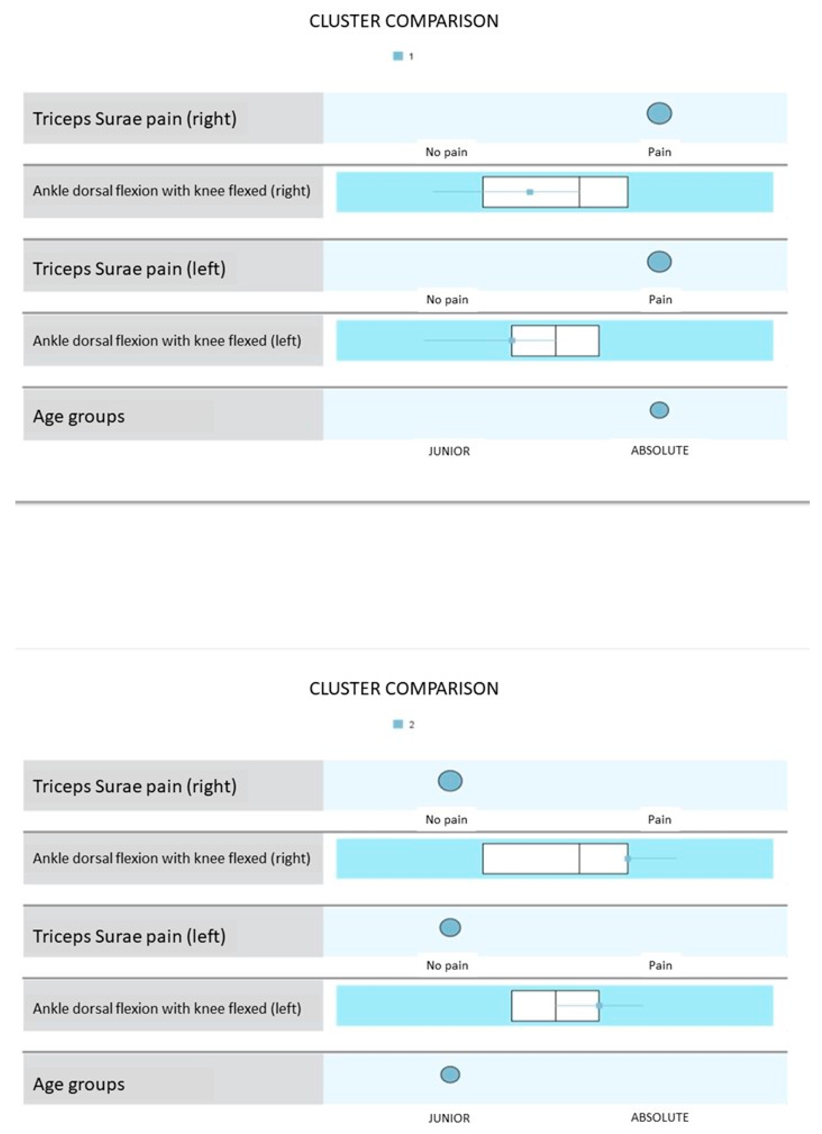
Figure 1. Graph showing the group comparison resulting from the two-stage cluster analysis.
Applsci 15 03755 g001
Table 1. Variables and Units of Measurement.
VariableMeasurement UnitGroup Junior (Media ± DE)Group Absolut (Media ± DE)
AgeYears17.0 ± 0.821.1 ± 1.5
Weightkg70.5 ± 5.473.8 ± 6.2
Heightcm175.2 ± 6.1178.4 ± 5.8
Ankle Dorsiflexion (Knee extended)Degrees (°)10.5 ± 2.18.7 ± 1.9
Ankle Dorsiflexion (Knee flexion)Degrees (°)12.3 ± 2.510.1 ± 2.3
Rearfoot MobilityDegrees (°)10.1 ± 2.37.5 ± 1.6
Dorsal Flexion of the 1st MTPDegrees (°)50.4 ± 5.247.2 ± 4.8
Firs Ray Mobilitymm6.5 ± 1.35.2 ± 1.5
Foot Posture (FPI)Scale (−12 to +12)4.0 ± 2.03.0 ± 2.0
Arch EightDegrees (Clarke’s angle)38.2 ± 3.536.8 ± 4.1
Plantar PressurekPa250.4 ± 30.2270.6 ± 28.9
Pain Presence (NPRS)Scale (0–10)3.8 ± 1.24.1 ± 1.3
Pain LocationCategoryTriceps surae, plantar musclesTriceps surae, plantar muscles
Table 2. Frequency (n and %) of muscular pain in triceps surae and plantar regions in both age groups, juniors, and adults.
Competition Category
Junior
N = 38 (51.4%)
Absolute
N = 36 (48.6%)
Chi-Square Tests
N%N%p 1Valuedf
Sex 0.5870.0001591
Man2052.61952.8
Woman1847.41747.2
Right Triceps Surae1847.42466.70.0752.8051
Left Triceps Surae2155.32775.00.0623.1601
Right Plantar Muscles1744.72363.90.0782.7301
Left Plantar Muscles1847.42158.30.2390.8921
1 Fisher’s Exact Test.
Table 3. Frequency (N and %) of neutral, pronated, and supinated feet; absent (A), mild (B), moderate (C), or severe (D) hallux valgus; and normal-arched, high-arched and low-arched feet.
Junior
N = 38 (51.4%)
Absolute
N = 36 (48.6%)
p
N%N%
Right FPI 0.030
Neutral1539.52466.7
Pronated1026.3822.2
Supinated1334.2411.1
Left FPI 0.110
Neutral1539.52363.9
Pronated1436.8822.2
Supinated923.7513.9
HV Right feet 0.201
A3297.02784.4
B13.0412.5
C0013.1
HV Left feet 0.037
A3193.92475.0
B26.1825.0
Arch height right Feet 0.676
Normal718.4411.1
Low37.938.3
High2873.72980.6
Arch height left Feet 0.327
Normal1026.3513.9
Low25.312.8
High2668.43083.3
FPI: Foot Posture Index; HV: hallux valgus.
Table 4. Range of ankle dorsal flexion with the knee extended and flexed, rearfoot inversion and eversion, first metatarsophalangeal joint dorsal flexion, and dorsiflexion and plantarflexion of the first ray. All ranges are expressed in degrees, except for the first ray dorsiflexion and plantarflexion, which are expressed in millimeters.
Competition Category
Junior
N = 38 (51.4%)
Absolute
N = 36 (48.6%)
MeanSDMedianIQRMeanSDMedianIQRpEffect Sizes
Ankle dorsal flexion with right knee extended10.32.2108–127.23.074.5–9.5<0.001 20.530 4
Ankle dorsal flexion with left knee extended10.32.41010–127.22.884.5–10.0<0.001 20.543 4
Ankle dorsal flexion with right knee flexion18.62.92018–2015.94.71612–200.014 20.287 4
Ankle dorsal flexion with left knee flexion18.73.11818–2015.94.91612–200.006 20.320 4
Right rearfoot inversion 27.15.82823.5–30.022.34.72220–26<0.001 10.891 3
Left rearfoot inversion 25.45.22422–3020.85.92118–24<0.001 10.830 3
Right rearfoot eversion9.72.8108–127.63.086–100.004 20.336 4
Left rearfoot eversion10.72.6119.5–12.08.72.886.5–10.00.002 20.360 4
Right first MPJ dorsal flexion58.17.45852–6453.211.35450–600.088 2
Left first MPJ dorsal flexion59.57.86054.0–62.553.210.05448–600.008 20.308 4
Right first ray dorsiflexion3.51.03.53–44.20.844–50.002 20.364 4
Left first ray dorsiflexion3.31.132–44.31.153.3–5.0<0.001 20.401 4
Right first ray plantarflexion5.50.965–65.41.05.55–60.550 2
Left first ray plantarflexion5.70.865–65.70.765–60.869 2
MPJ: Metatarsophalangeal joint. 1 t-test for independent samples. 2 Mann–Whitney U-test for independent samples. 3 d de Cohen. 4 r de Rosenthal.
Table 5. Plantar pressure distribution (%) under the forefoot, rearfoot, and whole foot.
Competition Category
Junior
N = 38 (51.4%)
Absolute
N = 36 (48.6%)
MeanSDMedianIQRMeanSDMedianIQRp
Right forefoot17.67.416.112.8–20.319.36.218.715.4–23.10.063 2
Left forefoot22.16.321.117.7–26.723.38.022.817.0–29.50.479 1
Right rearfoot30.07.531.526.9–34.227.58.127.125.4–32.90.086 2
Left rearfoot30.37.031.925.2–35.129.911.529.124.7–33.60.187 2
Whole right foot47.64.347.145.6–50.426.810.148.346.9–50.40.312 2
Whole left foot52.44.352.949.6–54.453.210.151.749.6–53.10.312 2
Both forefeet39.612.537.431.5–48.242.611.942.834.3–51.20.144 2
Both rearfeet60.412.562.751.8–68.557.411.957.348.8–65.70.144 2
1 t-test for independent samples. 2 Mann–Whitney U-test for independent samples.
Table 6. Values of the variables ‘triceps surae pain’ and ‘Ankle dorsal flexion with knee flexion’ in both age groups according to the cluster analysis.
Competition CategoryRight Triceps Surae PainLeft Triceps Surae PainRight Ankle Dorsal Flexion with Knee Flexion
(Degrees)
Left Ankle Dorsal Flexion with Knee Flexion
(Degrees)
JuniorNo painNo pain19.98–22.0118.03–22.02
AbsolutePainPain11.98–18.0012.04–18.03
Disclaimer/Publisher’s Note: The statements, opinions and data contained in all publications are solely those of the individual author(s) and contributor(s) and not of MDPI and/or the editor(s). MDPI and/or the editor(s) disclaim responsibility for any injury to people or property resulting from any ideas, methods, instructions or products referred to in the content.