Review

Is IBS a Food Allergy? Confocal Laser Endomicroscopy Findings in Patients with IBS: A Narrative Review

1
Department of Pathophysiology and Transplantation, University of Milan, 20122 Milan, Italy
2
Gastroenterology and Endoscopy Division, Foundation IRCCS Ca’ Granda Ospedale Maggiore Policlinico, 20122 Milan, Italy
3
Unit of Immunology, Rheumatology, Allergy and Rare Diseases (UniRAR), IRCCS San Raffaele Hospital, 20132 Milan, Italy
4
Allergy Unit, Department of Internal Medicine, Foundation IRCCS Ca’ Granda Ospedale Maggiore Policlinico, 20122 Milan, Italy
*
Author to whom correspondence should be addressed.
Appl. Sci. 2025, 15(7), 3717; https://doi.org/10.3390/app15073717
Submission received: 14 February 2025 / Revised: 21 March 2025 / Accepted: 24 March 2025 / Published: 28 March 2025
(This article belongs to the Special Issue New Diagnostic and Therapeutic Approaches in Food Allergy)

Abstract

:
Irritable bowel syndrome (IBS) is a gut–brain interaction disorder often associated with food-related triggers, yet the efficacy of common exclusion diets remains debated. Confocal laser endomicroscopy (CLE) offers real-time, high-resolution imaging of intestinal mucosal changes, allowing the visualization of food-induced barrier dysfunction. Early evidence indicates that a substantial subset of IBS patients exhibit acute mucosal reactions to specific foods, identified as fluorescein leakage and cell shedding on CLE, with over 70% showing symptom improvements after tailored exclusion diets. These findings suggest that localized immune responses and barrier defects may contribute to IBS symptoms beyond IgE-driven immunologic mechanisms. However, most CLE-based studies are small, unblinded, and heterogeneous, limiting definitive conclusions. Further research is needed to validate the diagnostic accuracy of CLE, refine protocols, and clarify how best to integrate CLE into personalized dietary management for difficult-to-treat IBS.

1. Introduction

Irritable bowel syndrome (IBS) remains one of the most common gastrointestinal disorders encountered by physicians [1]. IBS is a chronic disorder of gut–brain interaction (DGBI) and is still associated with areas of uncertainty, particularly regarding the optimal diagnostic workup and the most appropriate management approach, which may include exclusion diets [1,2,3,4,5]. In a recent review of 57 studies, covering 423,362 participants from 92 countries, the estimated global IBS prevalence using Rome III criteria was 9.2%, while Rome IV criteria showed a lower prevalence of 3.8%. IBS was more common in women than in men, highlighting significant gender differences [6]. Various mechanisms have been identified through which food can contribute to the onset of IBS symptoms. These include primary effects, such as osmotic, chemical, immunological, mechanical, and neuroendocrine responses, as well as secondary effects, including the production of fermentation byproducts, changes in intraluminal pH, and alterations in the gut microbiome [7]. More than 80% of individuals with IBS report food-related symptoms, particularly in response to fermentable carbohydrates and dietary fats, and many patients with IBS follow exclusion diets based on immunological test results or subjective perceptions of postprandial symptoms [8,9]. In this regard, several studies have demonstrated that the personalized management of adverse food reactions, supported by exclusion diets, can offer significant benefits for patients with IBS [9,10,11]. However, limited evidence supports the efficacy of exclusion diets based on IgG or IgG subclass testing [12]. These tests are supposed to be linked to increased intestinal permeability, broadening the understanding of interactions between diet and gut pathophysiology [11]. Although serum IgG antibodies against common food antigens are elevated in some IBS patients compared to healthy controls [13] no significant correlation has been found between symptom severity and elevated IgG [13,14]. This interest in exclusion diets highlights the need to understand the underlying mechanisms, particularly the role of epithelial barrier dysfunction in food-related immune responses. Numerous studies have shown a link between epithelial barrier dysfunction and food allergy. Increased intestinal permeability, by facilitating translocation of allergenic molecules across the intestinal epithelium, triggers the activation of an immune response involving epithelial cells and immune cells and causes an abnormal Th2-type response, leading to the production of allergen-specific immunoglobulin E (IgE) antibodies and subsequent activation of mast cells and basophils [15,16]. Gastrointestinal involvement includes nausea, abdominal pain, vomiting, and intermittent or persistent diarrhea. The onset of the gastrointestinal system symptoms is usually immediate, often within minutes up to 2 h [17]. The diagnosis of IgE-mediated food allergies is based on the combined use of a detailed medical history, in vivo/in vitro research of specific IgE, and in some cases, the double-blind placebo-controlled food challenge, which is the gold standard for the diagnosis of food allergies. The sole use of Specific IgE testing, although a quick and effective method for identifying allergic sensitization, is not recommended as it cannot confirm the presence of a clinical reaction to the allergen. Therefore, it is essential to distinguish between sensitization and true allergy, as some individuals with positive specific IgE may still tolerate the food, in order to prevent unnecessary dietary restrictions and the potential development of associated behavioral complications [18].
Larger population studies suggest correlations between atopy and DGBI, highlighting complex interactions between allergies and gut dysfunction [19,20]. For instance, Jones et al. found that patients with functional gastrointestinal disorders, including IBS and functional dyspepsia, had a significantly higher prevalence of atopic conditions than controls [19]. Similarly, in a Japanese study involving over 8000 patients, a significant association was identified between allergic diseases and functional gastrointestinal disorders, including IBS, suggesting shared immune mechanisms [20]. These studies, however, face some limitations, including non-standardized diagnostic criteria, lack of blinding, and uncertainty regarding the clinical relevance of observed associations.
Prolonged dietary restrictions can lead to nutritional deficiencies, necessitating careful medical and nutritional supervision. In this contest, an integrated and personalized approach with dietitians and nutritionists can help with and prevent IBS, also improving quality of life. Furthermore, IgG-based tests seem to be controversial as they do not necessarily indicate a pathological response but may reflect a normal immune response to food exposure [21,22,23]. This lack of specificity limits their diagnostic utility and may lead to overdiagnosis or misdiagnosis [24]. Diagnostic criteria lack standardization, with laboratories using variable methods and reference ranges, reducing result comparability across settings. Furthermore, many tests for food intolerance lack scientific validation, making it difficult to assess their true clinical efficacy [25].
Exclusion diets based on such tests also face some challenges. While some studies report symptom improvement with exclusion diets, methodological limitations such as short study duration and unrepresentative participant selection, cast doubts on the generalizability of findings. Perceived efficacy may partly result from placebo or nocebo effects rather than true clinical benefits [26]. However, recent findings suggest that maybe personalized IgG-guided diets may provide symptom relief, particularly in IBS-C and IBS-M patients [27]. Recently, the focus has shifted toward understanding local rather than systemic alterations, particularly within the intestinal mucosa, offering new perspectives on food reactions and immunological interactions in IBS.
There are currently no studies in the literature about the application of confocal laser endomicroscopy (CLE) for the most known non-IgE food allergy, Food Protein-Induced Enterocolitis Syndrome (FPEIS).
Confocal laser endomicroscopy (CLE) is an advanced and innovative diagnostic endoscopic imaging technology that provides real-time, high-resolution cellular-level visualization of the gastrointestinal epithelium [28,29,30,31]. Two main CLE systems have been marketed: endoscope-based CLE (eCLE), integrating the confocal microscope into the endoscope, and probe-based CLE (pCLE), using miniaturized confocal probes inserted through standard endoscopic accessory channels. pCLE is the only used technique currently (Figure 1).
This technique uses a low-power laser to illuminate tissue and collects reflected fluorescent light through a confocal optical system, ensuring high spatial resolution and real-time histological imaging [29,32]. Using intravenous contrast agents (such as fluorescein), visualization of cellular and subcellular structures is enhanced, enabling the detection of mucosal alterations with high sensitivity and specificity [28]. The high temporal resolution of CLE enables the recognition of dynamic changes in the mucosa [33]. CLE has been utilized in the evaluation and diagnosis of neoplasms, chronic inflammatory conditions (e.g., IBD), and both malignant and benign diseases of the pancreas and bile ducts [30,34,35]. It allows “optical biopsies”, reducing random sampling and enhancing diagnostic accuracy [34].

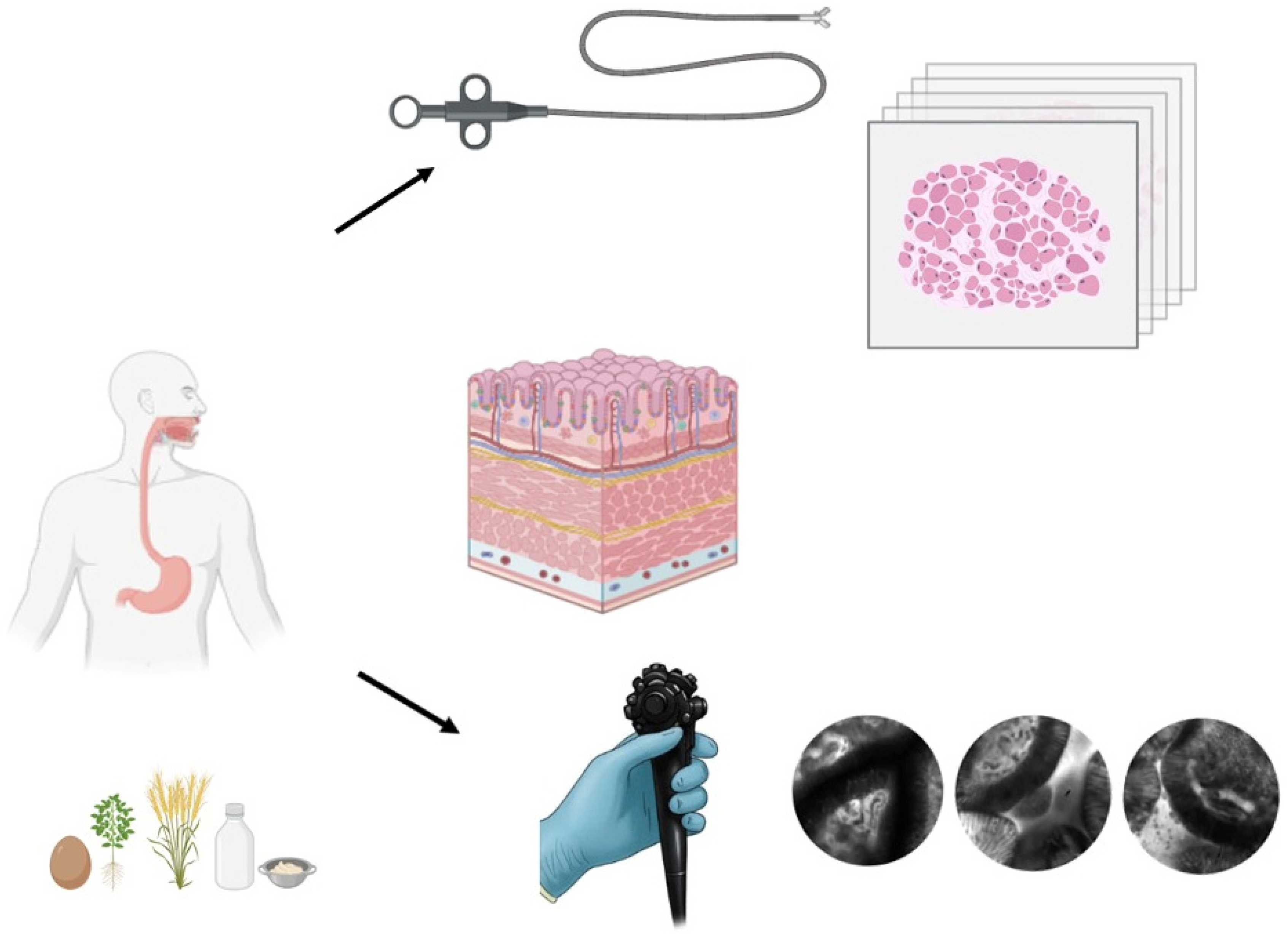
2. Materials and Methods

2.1. Literature Review

To identify relevant studies for this review, a literature search was conducted using the PubMed database. Articles exploring the connections between food allergies, atopy, IBS, and the use of advanced endoscopic techniques, such as microendoscopy, were included. The following MeSH terms were applied: “Food allergy” OR “atopy” OR “allergy” to include studies addressing food allergies, atopy, and general allergic reactions; “IBS” OR “irritable bowel syndrome” to limit the studies to those focused-on functional intestinal disorders; “Endoscopy” OR “microendoscopy, to focus on endoscopic techniques, particularly microendoscopy and CLE. Out of 128 studies identified, 74 were included in this review and all 74 have been selected for this review.

2.2. Inclusion Criteria

(1) Study Type: Original studies, abstracts, and articles describing or utilizing endoscopic techniques to investigate the intestinal barrier in IBS patients or those with allergic conditions. (2) Population: Patients diagnosed with IBS according to official criteria (e.g., Rome II-III-IV) and/or suspected food allergies or atopy. (3) Language: Articles published in English. (4) Publication Date: Studies published up to January 2025.

2.3. Exclusion Criteria

(1) Studies on pediatric populations or patients with organic gastrointestinal diseases (e.g., inflammatory bowel diseases or celiac disease). (2) Articles that do not use advanced endoscopic techniques to study intestinal permeability or the mucosal barrier.
Identified articles were screened for relevance to the themes of the review by evaluating abstracts and full texts. Selected studies were included in the discussion to provide a comprehensive overview of the available evidence.

3. Results

Results of the studies are reported in Table 1.

Mucosal Alterations and Food Allergy in IBS: Insights from Confocal Laser Endomicroscopy

The differences between the mucosal alteration diagnosed with CLE between patients with and without food allergy (FA) were investigated in one full-text journal article: both groups reported gastrointestinal symptoms, with the former associated with food intake and the latter not [36]. FA patients were diagnosed using a combination of clinical symptoms, food-specific IgE serological tests, and oral food challenges. These patients exhibited a higher frequency of intestinal barrier alterations detected through CLE performed in three sites: terminal ileum, cecum, and rectosigmoid junction. Fluorescein leakage, cell shedding, and micro erosions, interpreted as lamina propria exposure were compared to patients classified as non-FA. In the 96% of FA patients, a barrier dysfunction was observed in the terminal ileum, micro erosions were observed in 92.6% of FA patients. Barrier dysfunction in the terminal ileum was detected in 96% of patients with food allergy, with microerosions present in 92.6% of these cases, suggesting that CLE could serve as an effective screening test to rule out FA (Table 2).
Table 1. Summary of results on confocal laser endomicroscopy findings in patients with irritable bowel syndrome.
AutorPopulationMethodsCLE ResultsDietary
Intervention
Turcotte et al., 2013 [37]35 patients. 17 IBS and 18 HCCLE captured images post-fluorescein injection to examine epithelial gaps between villiIBS patients had significantly higher epithelial gap density compared to controlsno
Fritscher-Ravens et al., 2014 [38]36 IBS and 10 HCCLE for FCT in IBS and HS patients. Observed parameters: IELs, epithelial breaks, and intervillous space61.1% of IBS patients were CLE+. IELs increased from 18.7 ± 0.9 to 24.7 ± 1.4 (p = 0.0001). Not confirmed histologically. Epithelial breaks increased from 5.5 ± 0.6 to 11.9 ± 1.5 per 1000 epithelial cells (p < 0.001). Intervillous space widened from 8.1 ± 3.5 µm to 41.1 ± 2.5 µm (p = 0.0001)After an exclusion diet, 74% of CLE-positive patients experienced symptom reduction over 12 months
Fritscher-Ravens et al., 2019 [39]155 IBS patients and 10 healthy controlsCLE was used for FCT in IBS and HC by evaluating immune activation, epithelial integrity, and barrier function. Duodenal biopsies samples were analyzed for claudin-2, occludin, ECP, tryptase and intraepithelial lymphocytes.70% of IBS patients were CLE positive, showing increased intraepithelial lymphocytes (p = 0.001) and tight junction alterations, including elevated claudin-2 and reduced occludin levels (p < 0.05)After an exclusion diet, 73% of CLE-positive patients experienced symptom reduction over 6 months
Rath et al., 2021 [36]35 with FA and 27 non FA with GI symptomsPatients with and without FA, both experiencing gastrointestinal symptoms triggered by food intake. CLE observations included fluorescein leakage, cell shedding, and microerosionsBarrier dysfunction in the terminal ileum detected in 96% of FA patients vs. 33% of non-FA patients (p < 0.0001). Microerosions significantly more frequent in FA (92.6% vs. 24.2%, p < 0.0001). Crypt architecture showed no differences. CLE proved highly effective in ruling out FAno
Gjini et al., 2022 [40]34 patients with functional abdominal painCLE assessed spontaneous fluorescein leakage and DFC reactions. Duodenal biopsies analyzed inflammation and immune markers67.6% showed barrier dysfunction. Duodenal biopsies were normal, with no inflammation, atrophy, or structural abnormalities. Intraepithelial lymphocytes, mast cells, and dysfunction markers remained within normal limits, confirming no significant mucosal alterations69.5% reported pain relief after a four-week CLE-guided exclusion diet
Frieling et al., 2024 [41]71 patients with functional gastrointestinal symptoms. 52 patients with self-reported GARF and 19 patients without GARFCLE assessed spontaneous fluorescein leakage and DFC reactions. Duodenal biopsies analyzed inflammation and immune markers74% of GARF− and 70% of GARF+ showed fluorescein leakage and cell shedding. Duodenal biopsies showed no significant inflammationAfter four weeks of dietary changes, 79% of GARF+ patients experienced significant symptom reduction, compared to 14% of GARF−, (p < 0.05)
Grover et al., 2021 [42]26 IBS patients and 15 HCDuodenal lipid infusion. Intestinal permeability evaluated via a lactulose/mannitol excretion test; mucosal structure analyzed using CLE. Transcriptomic analysis to assess TRPV channel expressionNo change in intestinal permeability between groups. IBS patients experienced more severe symptoms. Increased TRPV1 and TRPV3 expression in the duodenum and jejunum correlated with symptoms. TRPV1 linked to pain and lower rectal sensitivity thresholds (r = −0.48, p < 0.05), while TRPV3 associated with bloating and urgency, especially in femalesno
Heßler et al., 2023 [43]172 IBS patientsSelective diet based on CLE results compared to a restrictive diet. Symptom improvement assessed via FAQLQ-AF and IBS-SSS, with support from a mobile appThe 12-week protocol included microbiome monitoring, though results are still pending.yes
Kiesslich et al., 2021 (Abstract) [44]256 IBS patientsA positive reaction was defined by increased fluorescein leakage and cell shedding after FCT and subsequent exclusion diet60% showed mucosal alteration.85% improved symptoms after 6 weeks. 74,6% after 6 months.
Kiesslich et al., 2020 (Abstract) [45]56 IBS patientsA positive reaction was defined by increased fluorescein leakage and cell shedding after FCT and subsequent exclusion diet58.9% showed mucosal alteration.84% improved symptoms after exclusion diet after six weeks
Blomsten et al., 2024
(Abstract) [46]
43 IBS patientsA positive reaction was defined by increased fluorescein leakage and cell shedding after FCT, subsequent exclusion diet and reintroduction.Half of the
patients were CLE+
Significant
improvement after food elimination, and worsening after reintroduction,
IBS: Irritable bowel syndrome; CLE: Confocal Laser Endomicroscopy; FCT: food challenge test; ECP: eosinophilic cationic protein; FA: food allergy; GI: Gastrointestinal; GARF: gastrointestinal adverse reactions to food; TRPV: Transient Receptor Potential Vanilloid; FAQLQ-AF: Food Allergy Quality of Life Questionnaire—Adult Form; IBS-SSS: Irritable Bowel Syndrome—Symptom Severity Score.
In recent years, the field of microendoscopy has provided new insights into microscopic alterations in the intestinal mucosa, even in IBS patients [37]. Real-time imaging with CLE has been employed to evaluate duodenal mucosal changes following acute food exposure in a pioneering study [38]. Baseline imaging of the mucosa is performed, and then food components are introduced via the endoscope’s working channel to observe changes linked to altered mucosal permeability. Normally, fluorescein is absent in the lumen due to an intact mucosa, but acute food reactions cause fluorescein extravasation and luminal particles (“cell shedding”), appearing as dark spots against the illuminated lumen. Fritscher-Ravens and colleagues analyzed a pattern of mucosal changes via CLE, suggesting that these may represent non-classical forms of food allergy due to localized immune reactions in patients with negative IgE serological tests [38]. In their study, 22 out of 36 IBS patients (61.1%) showed positive responses to food challenges administered in liquid or standardized suspension forms (CLE+), while no reactions were observed in 14 CLE− IBS patients and 10 healthy controls. Specifically, there was an increase in the number of intraepithelial lymphocytes (IELs) from an average of 18.7 ± 0.9 to 24.7 ± 1.4 IELs/field (p = 0.0001), increase in epithelial breaks from 5.5 ± 0.6 to 11.9 ± 1.5 per 1000 epithelial cells (p < 0.001), and significant widening of intervillous spaces from 8.1 ± 3.5 µm to 41.1 ± 2.5 µm (p = 0.0001). Subsequently, CLE+ patients followed an exclusion diet based on the food antigen provocation test results. After four weeks, 19 out of 22 CLE-positive patients reported a greater than 50% reduction in symptoms (abdominal pain, bloating, diarrhea, or constipation). At 12 months, the average symptom reduction reached 74%, with some patients achieving complete remission. The same group further explored mucosal reactions to food challenges in 155 IBS patients and 10 healthy controls [39]. Duodenal biopsies, as well as molecular and histological analyses, were also conducted to investigate changes in tight junctions and inflammatory responses. CLE+ patients underwent an exclusion diet based on the food identified during challenges. Among IBS participants, 70% of IBS participants exhibited immediate reactions to food components (CLE+), with 60.5% reacting to wheat. Patients who excluded reactive foods showed significant long-term symptom improvements (up to 6 months of follow-up). Additionally, the authors reported increased claudin-2 expression and reduced occludin levels in the duodenal mucosa of CLE+ patients. Tryptase levels in duodenal fluid showed no differences between CLE+ and CLE− patients or HCs and remained unchanged after antigen exposure. Duodenal fluid eosinophilic cationic protein (ECP) levels were significantly higher in CLE+ patients compared to healthy controls, with a post-stimulation average of 29.4 ± 51.2 mg/L in CLE+ versus 6.5 ± 7.9 mg/L in controls (p = 0.03). Compared to CLE− patients, ECP levels were higher but did not reach statistical significance (CLE−: 15.5 ± 19 mg/L) [39]. Heßler and colleagues conducted a study comparing a selective diet based on CLE results with a more restrictive diet excluding five major allergens (milk, eggs, wheat, soy, and yeast) [43]. The 172 IBS participants, who had non-IgE-mediated food intolerances and positive CLE results, received support via a digital app providing personalized guidelines, recipes, and tools to monitor adherence and symptoms. Primary endpoints included improvements in quality of life (FAQLQ-AF) and reductions in gastrointestinal symptom severity (IBS-SSS). The 12-week protocol also monitored microbiome changes, but results are pending [43]. Abstracts reported symptomatic improvement in more than 80% of IBS patients with negative IgE serological tests following an unblinded dietary intervention based on CLE results excluding trigger nutrients [44,45]. The elimination of trigger foods improved symptoms, which worsened upon reintroduction after 4 weeks [46].
A German research group recently published two studies evaluating the effectiveness of CLE in detecting food-induced mucosal alterations. The studies focused on two heterogenous patient populations: one with “functional abdominal pain”, selected using the Digestive Symptom Frequency Questionnaire (DSFQ) [40], and the other with “gastrointestinal adverse reactions to food” (GARF) [41]. The latter study placed greater emphasis on the relationship between symptoms and food, while still utilizing the same standardized symptom assessment tool. Gjini and colleagues found that most patients with functional abdominal pain exhibited food-induced mucosal barrier alterations, primarily triggered by soy and wheat [40]. Of these, 67.6% (23/34) showed a positive fluorescein test, indicating barrier dysfunction and 69.5% (16/23) experienced significant pain reduction after an exclusion diet guided by CLE results. Duodenal biopsies following the food challenge test revealed a normal mucosa, with no signs of inflammation, atrophy, or structural abnormalities. Intraepithelial lymphocytes (IELs) and mast cells were within normal ranges. Mast cells showed no anomalies in distribution or morphology, and markers for mast cell dysfunction, such as tryptase and diamine oxidase, were within normal limits. In another study conducted by Frieling et al. [41]. CLE was used to study mucosal reactions to foods in 71 patients, all of whom had functional gastrointestinal symptoms, including 52 patients with self-reported gastrointestinal adverse reactions to food (GARF+) and 19 without food-related symptoms (GARF−). The main symptoms were bloating, abdominal pain, and diarrhea. Patients with positive stimulation tests underwent personalized exclusion diets. Among GARF + patients, 70% showed food-related reactions, primarily to soy, wheat, and milk, with fluorescein leakage into the duodenal lumen. Even 74% of GARF− patients displayed a local mucosal response with duodenal food challenge. After four weeks of dietary changes, 79% of GARF+ patients experienced significant symptom reduction, compared to 14% of GARF−, with a statistically significant difference between groups (p < 0.05) [41].
Lastly, Grover and colleagues analyzed symptom changes (abdominal pain, bloating, urgency) before and after intraduodenal lipid infusion to stimulate intestinal sensitivity, comparing healthy individuals and IBS patients [42]. A total of 41 individuals were included: 26 IBS patients without organic disease and 15 healthy volunteers. Intestinal permeability was evaluated using CLE and a lactulose/mannitol test. Transcriptomic analyses were performed to assess gene expression in Transient Receptor Potential Vanilloid (TRPV) channels. After lipid infusion, no significant differences in intestinal permeability were observed between groups. However, IBS patients reported more severe symptoms, correlated with higher TRPV1 and TRPV3 expression in the duodenum and jejunum. TRPV1 was positively associated with pain and inversely correlated with rectal sensitivity thresholds (r = −0.48, p < 0.05), while TRPV3 was linked to bloating and urgency, with a significant association in female patients [42].

4. Discussion

CLE is a rapidly evolving field in gastroenterology that bridges endoscopy and histology, enhancing the ability to visualize living tissues in real-time and provide therapies within the same setting [34]. Moreover, CLE has the privilege of being able to evaluate in real-time some functional dynamics related to barrier integrity (e.g., cell shedding, fluorescein leakage) that do not have any histological counterpart. Several studies demonstrate how CLE can serve as an innovative diagnostic tool capable of providing an immediate correlation between mucosal alterations and clinical response, leading to a personalized approach to intestinal symptoms triggered by food, regardless of the etiological hypothesis. The evidence in this field is still limited. The study conducted by Rath et al. [36] appears to be unique in applying CLE to directly investigate intestinal barrier dysfunction in patients with suspected FA, including patient stratification based on barrier integrity parameters for further diagnostic workup. Alterations were also reported in 33% of non-FA patients who experienced gastrointestinal symptoms not directly linked to specific food consumption (p < 0.0001). Crypt architecture parameters (crypt diameter, inter-crypt distance, and lumen diameter) did not differ significantly between FA and non-FA groups [36].
Other studies [38,39,42,43,44,45,46] have explored similar concepts in the context of IBS patients who tested negative for IgE. In these studies, CLE was used to assess intestinal barrier integrity, with a focus on direct stimulation with food antigens and subsequent dietary interventions based on the observed results [38,39,40,41,43,44,45,46]. Alterations in the distribution of key proteins, such as occludin and claudin-1, have already been associated with increased intestinal permeability in IBS patients [47]. This finding has been further supported by hypotheses formulated in the study by Fritscher-Ravens, which linked intestinal barrier defects observed through CLE with alterations in these proteins [39]. The authors suggested that the observed reactions could be mediated by local immune responses of the duodenal mucosa in response to commonly used food antigens such as wheat [38,39,41,43] which was the most frequent cause of positive food challenge tests in the reported studies. This finding is further supported by other studies [48,49,50] which report the benefits of a gluten-free diet in patients with functional gastrointestinal symptoms and negative for celiac disease through the concept of non-celiac gluten sensitivity [51]. However, some authors discourage using CLE for diagnosing this condition due to its low specificity, which does not justify its use as an invasive initial test in IBS patients suspected of wheat sensitivity [52]. Nevertheless, CLE may help elucidate the pathomechanisms underlying mucosal and submucosal changes during food-induced gastrointestinal alterations [52]. The mucosal barrier alterations observed in GARF− patients in Frieling’s study are particularly noteworthy [41]. The study analyzed a heterogeneous group of patients with functional gastrointestinal symptoms. This broad categorization encompassed a diverse population, potentially including patients with IBS. Intestinal barrier disruptions, such as fluorescein leakage, were identified in both GARF+ and GARF− patients, indicating that mucosal damage is not exclusive to those with a clear food-symptom correlation. However, 74% of GARF− patients exhibited mucosal alterations but only 14% improved with dietary intervention underscoring CLE’s low specificity (68%) and therefore its inadequacy as a standalone diagnostic tool. The different sizes of both groups might cause some bias, therefore, to prove the results by a larger group of patients GARF—is needed. This finding supports the concept that intestinal epithelial barrier dysfunction is a common pathophysiological mechanism in functional gastrointestinal disorders, including patients with gastrointestinal symptoms not directly linked to specific foods but responsive to antigenic stimulation.
Not only were mucosal alterations observed; for example, the group led by Fritscher-Ravens reported an increase in eosinophil activity in the duodenum and the secretion of eosinophilic cationic protein (ECP) in duodenal fluids following exposure to specific foods [39]. These findings suggest that eosinophils may play a role in mucosal reactions triggered by food antigens, unfortunately, the lack of data on ECP levels pre- and post-reaction limits conclusions about whether this activity was triggered acutely. An increase in eosinophil activity has been observed in functional dyspepsia [53,54] and even in eosinophilic esophagitis [55,56]. This reaction also occurs in the absence of IgE or positive skin tests for allergens, suggesting a different mode of immunologic response, likely in the context of mild intestinal mucosal inflammation. The authors observed an increase in intraepithelial lymphocytes (IEL) on confocal laser endomicroscopy (CLE) in patients experiencing acute reactions, both initially and after the reaction. However, this finding was not corroborated by CD3 immunohistochemistry. Moreover, the reported lack of correlation between IEL identified through CLE and immunohistochemistry raises doubts about the reliability of using CLE to quantify lymphocytes [38]. In the larger follow-up study, patients with reactions showed increased IEL at baseline, but no changes were observed following acute reactions [39]. Recent research revealed normal lymphocyte levels and no variations in mast cell counts in biopsies taken after acute alterations [40,41]. However, no baseline comparison analyses were conducted. Low-grade inflammation is well-known to contribute to the disruption of gastrointestinal reflexes and the activation of the visceral sensory system, leading to symptoms associated with IBS, such as visceral hypersensitivity [57]. Some studies have shown that ion channels and their receptors are critical for gastrointestinal sensitivity [58,59,60]. Specifically, TRPV channels have been linked to heightened intestinal sensitivity in IBS patients [61,62,63,64]. These channels, part of the broader TRP family (Transient Receptor Potential), play essential physiological roles, including pain perception, thermoregulation, chemosensitivity, and epithelial barrier maintenance [65]. Grover and colleagues observed mucosal structural changes via CLE following lipid infusion rather than direct antigenic stimulation. This study represents an important step in correlating intestinal mucosal stimulation by food mixtures with increased TRPV channel expression, heightened intestinal symptoms, and CLE-detected permeability changes. These findings provide insights into the relationship between mucosal alterations and sensory perception in IBS patients [42]. Although this technique shows promise, its application in clinical practice remains unproven, and prescribing a diet based on these findings requires further thorough evaluation.
A literature search in the PubMed database shows no connection between Food Protein Induced Enterocolitis Syndrome (FPIES), and the use of advanced endoscopic techniques. FPIES is the most well-known type of non-IgE-mediated food allergy [66], is a delayed inflammatory reaction to specific food proteins that primarily affects the gastrointestinal tract. It is characterized by inflammatory lymphocyte and neutrophil infiltrates, edema and vascular congestion, villous atrophy, crypt structural changes, and, in a more advanced stage, tissue necrosis. Additionally, there is an increase in intestinal permeability characterized by the production of inflammatory cytokines like IL-4, IL-5, and IL-13, activation of the complement system, (C3a and C5a), and an increase in the activity of CD4+ and CD8+ T cells, with a preponderance of CD4+ T cells in the intestinal mucosa [67]. The confocal laser endomicroscopy method would be crucial for exploring FPIES.

5. Conclusions

CLE is an emerging technology with significant potential for evaluating intestinal barrier alterations, both in patients with classic food allergies and in those with functional gastrointestinal disorders such as IBS. Current data suggest that intestinal barrier dysfunction may represent a shared pathophysiological mechanism between FA and IBS, highlighting the need for further research to elucidate the connections between these two conditions. Specifically, it would be valuable to determine whether the alterations detected by CLE are specific markers for allergies or a more general phenomenon associated with intestinal disorders. CLE is not limited to the identification of classic food allergies but offers a unique opportunity to investigate the role of local intestinal barrier dysfunctions in non-IgE-mediated conditions, expanding its utility beyond traditional diagnostic applications. IELs, in a process partly mediated by the secretion of interferon-gamma [68], likely enhance epithelial permeability by activating myosin light chain kinase, leading to phosphorylation of myosin light chains within the contractile actomyosin ring of the tight junctions. This results in a redistribution of tight junction proteins such as occludin and claudins [69]. For instance, CLE has shown preliminary findings suggest the occurrence of acute reactions in other conditions such as functional dyspepsia, eosinophilic esophagitis, and inflammatory bowel disease [70,71,72,73] The mucosa of the ileum and colon have been analyzed in some studies investigating intestinal permeability in patients with inflammatory bowel disease (IBD), but without stimulation tests using food antigens. Furthermore, an important aspect of these studies lies in the ability to correlate previously described IBS characteristics, such as increased TRPV channel expression or altered distribution of junctional proteins that regulate intestinal barrier permeability, with direct stimulation through food mixtures [39,42]. Future developments in this technique could include real-time correlation of observed mucosal changes with findings from transcriptomic studies, thereby deepening the understanding of molecular mechanisms involved in functional gastrointestinal disorders. One of the main findings was correlated with transcriptomic analyses, which revealed a significant reduction in claudin-1 protein expression [70]. This approach offers an innovative perspective, enabling not only the identification of mucosal alterations in response to food antigens but also their direct correlation with clinical symptoms reported by patients. However, it is important to acknowledge the limitations of these studies. The available literature linking clinical outcomes to acute reactions identified via CLE is limited, mostly uncontrolled and unblinded, and often conducted on heterogeneous patient populations [74]. Many studies have involved small sample sizes, often in single-center settings, with limited observation periods, particularly during post-elimination diet phases. Additionally, despite being a highly useful tool, CLE has significant challenges: it is expensive, technically demanding, and requires prolonged sedation. CLE is also currently available only in a few specialized centers. These factors limit the generalizability and applicability of such studies in routine clinical practice. Despite these limitations, these studies provide a starting point for better understanding IBS pathophysiology. CLE could potentially form the basis for developing standardized therapeutic strategies aimed at reducing symptoms through targeted and personalized interventions. Moreover, an improved understanding of the underlying mechanisms could help reduce the economic burden on healthcare systems by decreasing the need for frequent specialist visits and unnecessary diagnostic tests. Addressing these issues through larger, well-designed studies in the future could represent a significant step forward in managing IBS and gastrointestinal symptoms, ultimately improving patients’ quality of life and optimizing healthcare resource allocation.

Author Contributions

F.P. and A.C. contributed to conceptualization, writing, review, and editing; G.E.T., L.E., N.S., G.L., M.D. and A.S. contributed to the review and editing; M.V. supervised. All authors have read and agreed to the published version of the manuscript.

Funding

This study was partially funded by the Italian Ministry of Health—current research, IRCCS year 2024.

Data Availability Statement

The original contributions presented in the study are included in the article, further inquiries can be directed to the corresponding author.

Conflicts of Interest

The authors declare no conflicts of interest.

References

  1. Vasant, D.H.; Paine, P.A.; Black, C.J.; Houghton, L.A.; Everitt, H.A.; Corsetti, M.; Agrawal, A.; Aziz, I.; Farmer, A.D.; Eugenicos, M.P.; et al. British Society of Gastroenterology guidelines on the management of irritable bowel syndrome. Gut 2021, 70, 1214–1240. [Google Scholar] [CrossRef] [PubMed]
  2. Enck, P.; Aziz, Q.; Barbara, G.; Farmer, A.D.; Fukudo, S.; Mayer, E.A.; Niesler, B.; Quigley, E.M.M.; Rajilić-Stojanović, M.; Schemann, M.; et al. Irritable bowel syndrome. Nat. Rev. Dis. Prim. 2016, 2, 16014. [Google Scholar] [CrossRef] [PubMed]
  3. Savarino, E.; Zingone, F.; Barberio, B.; Marasco, G.; Akyuz, F.; Akpinar, H.; Barboi, O.; Bodini, G.; Bor, S.; Chiarioni, G.; et al. Functional bowel disorders with diarrhoea: Clinical guidelines of the United European Gastroenterology and European Society for Neurogastroenterology and Motility. United Eur. Gastroenterol. J. 2022, 10, 556–584. [Google Scholar] [CrossRef]
  4. Holtmann, G.J.; Ford, A.C.; Talley, N.J. Pathophysiology of irritable bowel syndrome. Lancet Gastroenterol. Hepatol. 2016, 1, 133–146. [Google Scholar] [CrossRef]
  5. Pessarelli, T.; Sorge, A.; Elli, L.; Costantino, A. The low-FODMAP diet and the gluten-free diet in the management of functional abdominal bloating and distension. Front. Nutr. 2022, 9, 1007716. [Google Scholar] [CrossRef]
  6. Oka, P.; Parr, H.; Barberio, B.; Black, C.J.; Savarino, E.V.; Ford, A.C. Global prevalence of irritable bowel syndrome according to Rome III or IV criteria: A systematic review and meta-analysis. Lancet Gastroenterol. Hepatol. 2020, 5, 908–917. [Google Scholar] [CrossRef]
  7. Spencer, M.; Chey, W.D.; Eswaran, S. Dietary Renaissance in IBS: Has Food Replaced Medications as a Primary Treatment Strategy? Curr. Treat. Options Gastroenterol. 2014, 12, 424–440. [Google Scholar] [CrossRef]
  8. Simrén, M.; Månsson, A.; Langkilde, A.M.; Svedlund, J.; Abrahamsson, H.; Bengtsson, U.; Björnsson, E.S. Food-related gastrointestinal symptoms in the irritable bowel syndrome. Digestion 2001, 63, 108–115. [Google Scholar] [CrossRef] [PubMed]
  9. Atkinson, W.; Sheldon, T.A.; Shaath, N.; Whorwell, P.J. Food elimination based on IgG antibodies in irritable bowel syndrome: A randomised controlled trial. Gut 2004, 53, 1459–1464. [Google Scholar] [CrossRef]
  10. Ostrowska, L.; Wasiluk, D.; Lieners, C.F.J.; Gałęcka, M.; Bartnicka, A.; Tveiten, D. Igg food antibody guided elimination-rotation diet was more effective than FODMAP diet and control diet in the treatment of women with mixed IBS—Results from an open label study. J. Clin. Med. 2021, 10, 4317. [Google Scholar] [CrossRef]
  11. Vita, A.A.; Zwickey, H.; Bradley, R. Associations between food-specific IgG antibodies and intestinal permeability biomarkers. Front. Nutr. 2022, 9, 962093. [Google Scholar] [CrossRef]
  12. Zar, S.; Mincher, L.; Benson, M.J.; Kumar, D. Food-specific IgG4 antibody-guided exclusion diet improves symptoms and rectal compliance in irritable bowel syndrome. Scand. J. Gastroenterol. 2005, 40, 800–807. [Google Scholar] [CrossRef] [PubMed]
  13. Zuo, X.L.; Li, Y.Q.; Li, W.J.; Guo, Y.T.; Lu, X.F.; Li, J.M.; Desmond, P.V. Alterations of food antigen-specific serum immunoglobulins G and E antibodies in patients with irritable bowel syndrome and functional dyspepsia. Clin. Exp. Allergy 2007, 37, 823–830. [Google Scholar] [CrossRef] [PubMed]
  14. Vara, E.J.; Valeur, J.; Hausken, T.; Lied, G.A. Extra-intestinal symptoms in patients with irritable bowel syndrome: Related to high total IgE levels and atopic sensitization? Scand. J. Gastroenterol. 2016, 51, 908–913. [Google Scholar] [CrossRef] [PubMed]
  15. Poto, R.; Fusco, W.; Rinninella, E.; Cintoni, M.; Kaitsas, F.; Raoul, P.; Caruso, C.; Mele, M.C.; Varricchi, G.; Gasbarrini, A.; et al. The Role of Gut Microbiota and Leaky Gut in the Pathogenesis of Food Allergy. Nutrients 2024, 16, 92. [Google Scholar] [CrossRef]
  16. Abril, A.G.; Villa, T.G.; Sánchez-Pérez, Á.; Notario, V.; Carrera, M. The Role of the Gallbladder, the Intestinal Barrier and the Gut Microbiota in the Development of Food Allergies and Other Disorders. Int. J. Mol. Sci. 2022, 23, 14333. [Google Scholar] [CrossRef]
  17. Anvari, S.; Miller, J.; Yeh, C.-Y.; Davis, C.M. IgE-Mediated Food Allergy. Clin. Rev. Allergy Immunol. 2019, 57, 244–260. [Google Scholar] [CrossRef]
  18. Bartha, I.; Almulhem, N.; Santos, A.F. Feast for thought: A comprehensive review of food allergy 2021–2023. J. Allergy Clin. Immunol. 2024, 153, 576–594. [Google Scholar] [CrossRef]
  19. Jones, M.P.; Walker, M.M.; Ford, A.C.; Talley, N.J. The overlap of atopy and functional gastrointestinal disorders among 23 471 patients in primary care. Aliment. Pharmacol. Ther. 2014, 40, 382–391. [Google Scholar] [CrossRef]
  20. Yamamoto, Y.; Furukawa, S.; Miyake, T.; Watanabe, J.; Nakamura, Y.; Taguchi, Y.; Yamamoto, T.; Kato, A.; Kusumoto, K.; Yoshida, O.; et al. Multimorbidity of Allergic Diseases Is Associated with Functional Gastrointestinal Disorders in a Young Japanese Population. J. Neurogastroenterol. Motil. 2024, 30, 229–235. [Google Scholar] [CrossRef]
  21. Stapel, S.O.; Asero, R.; Ballmer-Weber, B.K.; Knol, E.F.; Strobel, S.; Vieths, S.; Kleine-Tebbe, J. Testing for IgG4 against foods is not recommended as a diagnostic tool: EAACI Task Force Report. Allergy Eur. J. Allergy Clin. Immunol. 2008, 63, 793–796. [Google Scholar] [CrossRef] [PubMed]
  22. Ahmed, T.; Sumazaki, R.; Nagai, Y.; Shibasaki, M.; Takita, H. Immune response to food antigens: Kinetics of food-specific antibodies in the normal population. Pediatr. Int. 1997, 39, 322–328. [Google Scholar] [CrossRef]
  23. Whelan, K.; Ford, A.C.; Burton-Murray, H.; Staudacher, H.M. Dietary management of irritable bowel syndrome: Considerations, challenges, and solutions. Lancet Gastroenterol. Hepatol. 2024, 9, 1147–1161. [Google Scholar] [CrossRef] [PubMed]
  24. Bischoff, S.; Crowe, S.E. Gastrointestinal food allergy: New insights into pathophysiology and clinical perspectives. Gastroenterology 2005, 128, 1089–1113. [Google Scholar] [CrossRef]
  25. Zar, S.; Kumar, D.; Benson, M.J. Food hypersensitivity and irritable bowel syndrome. Aliment. Pharmacol. Ther. 2001, 15, 439–449. [Google Scholar] [CrossRef]
  26. Stierstorfer, M.; Toro, B. Patch Test–Directed Dietary Avoidance in the Management of Irritable Bowel Syndrome. Cutis 2021, 108, 91–95. [Google Scholar] [CrossRef]
  27. Singh, P.; Chey, W.D.; Takakura, W.; Cash, B.D.; Lacy, B.E.; Quigley, E.M.; Randall, C.W.; Lembo, A. A novel, IBS-specific IgG ELISA-based elimination diet in irritable bowel syndrome: A randomized, sham-controlled trial. Gastroenterology 2025. Epub ahead of print. [Google Scholar] [CrossRef]
  28. Dunbar, K.; Canto, M. Confocal endomicroscopy. Curr. Opin. Gastroenterol. 2008, 24, 631–637. [Google Scholar] [CrossRef] [PubMed]
  29. Wang, T.D. Confocal microscopy from the bench to the bedside. Gastrointest. Endosc. 2005, 62, 696–697. [Google Scholar] [CrossRef]
  30. Fugazza, A.; Gaiani, F.; Carra, M.C.; Brunetti, F.; Lévy, M.; Sobhani, I.; Azoulay, D.; Catena, F.; De’angelis, G.L.; De’angelis, N. Confocal Laser Endomicroscopy in Gastrointestinal and Pancreatobiliary Diseases: A Systematic Review and Meta-Analysis. BioMed Res. Int. 2016, 2016, 4638683. [Google Scholar] [CrossRef]
  31. Neumann, H.; Kiesslich, R.; Wallace, M.B.; Neurath, M.F. Confocal laser endomicroscopy: Technical advances and clinical applications. Gastroenterology 2010, 139, 388–392.e2. [Google Scholar] [CrossRef] [PubMed]
  32. Gheonea, D.I.; Saftoiu, A.; Ciurea, T.; Popescu, C.; Georgescu, C.V.; Malos, A. Confocal laser endomicroscopy of the colon. J. Gastrointest. Liver Dis. 2010, 19, 207–211. [Google Scholar] [PubMed]
  33. Kiesslich, R.; Goetz, M.; Angus, E.M.; Hu, Q.; Guan, Y.; Potten, C.; Allen, T.; Neurath, M.F.; Shroyer, N.F.; Montrose, M.H.; et al. Identification of Epithelial Gaps in Human Small and Large Intestine by Confocal Endomicroscopy. Gastroenterology 2007, 133, 1769–1778. [Google Scholar] [CrossRef]
  34. De Palma, G.D. Confocal laser endomicroscopy in the “in vivo” histologicaldiagnosis of the gastrointestinal tract. World J. Gastroenterol. 2009, 15, 5770–5775. [Google Scholar] [CrossRef]
  35. Tontini, G.E.; Mudter, J.; Vieth, M.; Günther, C.; Milani, V.; Atreya, R.; Rath, T.; Nägel, A.; Hatem, G.; Sturniolo, G.C.; et al. Prediction of clinical outcomes in Crohn’s disease by using confocal laser endomicroscopy: Results from a prospective multicenter study. Gastrointest. Endosc. 2018, 87, 1505–1514.e3. [Google Scholar] [CrossRef]
  36. Rath, T.; Dieterich, W.; Kätscher-Murad, C.; Neurath, M.F.; Zopf, Y. Cross-sectional imaging of intestinal barrier dysfunction by confocal laser endomicroscopy can identify patients with food allergy in vivo with high sensitivity. Sci. Rep. 2021, 11, 12777. [Google Scholar] [CrossRef]
  37. Turcotte, J.-F.; Kao, D.; Mah, S.J.; Claggett, B.; Saltzman, J.R.; Fedorak, R.N.; Liu, J.J. Breaks in the wall: Increased gaps in the intestinal epithelium of irritable bowel syndrome patients identified by confocal laser endomicroscopy (with videos). Gastrointest. Endosc. 2013, 77, 624–630. [Google Scholar] [CrossRef]
  38. Fritscher-Ravens, A.; Schuppan, D.; Ellrichmann, M.; Schoch, S.; Röcken, C.; Brasch, J.; Bethge, J.; Böttner, M.; Klose, J.; Milla, P.J. Confocal endomicroscopy shows food-associated changes in the intestinal mucosa of patients with irritable bowel syndrome. Gastroenterology 2014, 147, 1012–1020.e4. [Google Scholar] [CrossRef]
  39. Fritscher-Ravens, A.; Pflaum, T.; Mösinger, M.; Ruchay, Z.; Röcken, C.; Milla, P.J.; Das, M.; Böttner, M.; Wedel, T.; Schuppan, D. Many Patients with Irritable Bowel Syndrome Have Atypical Food Allergies Not Associated with Immunoglobulin E. Gastroenterology 2019, 157, 109–118.e5. [Google Scholar] [CrossRef]
  40. Gjini, B.; Melchior, I.; Euler, P.; Kreysel, C.; Kalde, S.; Krummen, B.; Kiesslich, R.; Hemmerlein, B.; Frieling, T. Food intolerance in patients with functional abdominal pain: Evaluation through endoscopic confocal laser endomicroscopy. Endosc. Int. Open 2023, 11, E67–E71. [Google Scholar] [CrossRef]
  41. Frieling, T.; Gjini, B.; Melchior, I.; Euler, P.; Kreysel, C.; Kalde, S.; Krummen, B.; Kiesslich, R.; Hemmerlein, B. Gastrointestinal adverse reaction to food (GARF) and endoscopic confocal laser endomicroscopy (eCLE). Z. Gastroenterol. 2024, 62, 1201–1206. [Google Scholar] [CrossRef] [PubMed]
  42. Grover, M.; Berumen, A.; Peters, S.; Wei, T.; Breen-Lyles, M.; Harmsen, W.S.; Busciglio, I.; Burton, D.; Roque, M.V.; DeVault, K.R.; et al. Intestinal chemosensitivity in irritable bowel syndrome associates with small intestinal TRPV channel expression. Aliment. Pharmacol. Ther. 2021, 54, 1179–1192. [Google Scholar] [CrossRef] [PubMed]
  43. Heßler, N.; Kordowski, A.; Sasse, J.; Ahlemann, G.; Schulz, F.; Schröder, T.; Exner, A.; Jablonski, L.; Jappe, U.; Bischoff, S.C.; et al. Study protocol to investigate the efficacy of confocal laser endomicroscopy-based selective single-elimination diet over standard fivefold elimination diet in patients with endomicroscopically proven food intolerance: App-assisted, monocentric, double-blind, randomised and controlled trial in Germany. BMJ Open 2023, 13, e072024. [Google Scholar] [CrossRef]
  44. Kiesslich, R.; Adib-Tezer, H.; Teubner, D.; Frieling, T.; Bayerl, C.; Wenda, N.K.; Gosepath, J. ID: 3526039 Food Allergy Sensitivity Test (Fast) with Endomicroscopy of the Duodenum Enables Tailored Exclusion Diet in Patients with Irritable Bowel Syndrome. Gastrointest. Endosc. 2021, 93, AB207. [Google Scholar] [CrossRef]
  45. Kiesslich, R.; Adib-Tezer, H.; Teubner, D.; Frieling, T.; Mudter, J.; Bayerl, C.; Wenda, N.K.; Gosepath, J. Su1344 Endomicroscopic Detection of Atypical Food Allergy in Patients with Irritable Bowel Syndrome—A New Diagnostic Era? Gastroenterology 2020, 158, S-558–S-559. [Google Scholar] [CrossRef]
  46. Blomsten, A.; van Gils, T.; Algera, J.P.; Josefsson, A.; Hreinsson, J.P.; Hedenström, P.; Störsrud, S.; Törnblom, H.; Simren, M. Tu1682 food-induced intestinal mucosal reactions in irritable bowel syndrome detected with confocal laser endomicroscopy. Gastroenterology 2024, 166, S-1377–S-1378. [Google Scholar] [CrossRef]
  47. Piche, T.; Barbara, G.; Aubert, P.; Varannes, S.B.D.; Dainese, R.; Nano, J.L.; Cremon, C.; Stanghellini, V.; De Giorgio, R.; Galmiche, J.P.; et al. Impaired intestinal barrier integrity in the colon of patients with irritable bowel syndrome: Involvement of soluble mediators. Gut 2009, 58, 196–201. [Google Scholar] [CrossRef]
  48. Elli, L.; Tomba, C.; Branchi, F.; Roncoroni, L.; Lombardo, V.; Bardella, M.T.; Ferretti, F.; Conte, D.; Valiante, F.; Fini, L.; et al. Evidence for the presence of non-celiac gluten sensitivity in patients with functional gastrointestinal symptoms: Results from a multicenter randomized double-blind placebo-controlled gluten challenge. Nutrients 2016, 8, 84. [Google Scholar] [CrossRef]
  49. Barone, M.; Gemello, E.; Viggiani, M.T.; Cristofori, F.; Renna, C.; Iannone, A.; Di Leo, A.; Francavilla, R. Evaluation of non-celiac gluten sensitivity in patients with previous diagnosis of irritable bowel syndrome: A randomized double-blind placebo-controlled crossover trial. Nutrients 2020, 12, 705. [Google Scholar] [CrossRef]
  50. Barbaro, M.R.; Cremon, C.; Wrona, D.; Fuschi, D.; Marasco, G.; Stanghellini, V.; Barbara, G. Non-celiac gluten sensitivity in the context of functional gastrointestinal disorders. Nutrients 2020, 12, 3735. [Google Scholar] [CrossRef]
  51. Costantino, A.; Aversano, G.M.; Lasagni, G.; Smania, V.; Doneda, L.; Vecchi, M.; Roncoroni, L.; Pastorello, E.A.; Elli, L. Diagnostic management of patients reporting symptoms after wheat ingestion. Front. Nutr. 2022, 9, 1007007. [Google Scholar] [CrossRef]
  52. Bojarski, C.; Tangermann, P.; Barmeyer, C.; Buchkremer, J.; Kiesslich, R.; Ellrichmann, M.; Schreiber, S.; Schmidt, C.; Stallmach, A.; Roehle, R.; et al. Prospective, double-blind diagnostic multicentre study of confocal laser endomicroscopy for wheat sensitivity in patients with irritable bowel syndrome. Gut 2022, 71, 1567–1576. [Google Scholar] [CrossRef] [PubMed]
  53. Walker, M.M.; Aggarwal, K.R.; Shim, L.S.; Bassan, M.; Kalantar, J.S.; Weltman, M.D.; Jones, M.; Powell, N.; Talley, N.J. Duodenal eosinophilia and early satiety in functional dyspepsia: Confirmation of a positive association in an Australian cohort. J. Gastroenterol. Hepatol. 2014, 29, 474–479. [Google Scholar] [CrossRef] [PubMed]
  54. Du, L.; Shen, J.; Kim, J.J.; Yu, Y.; Ma, L.; Dai, N. Increased Duodenal Eosinophil Degranulation in Patients with Functional Dyspepsia: A Prospective Study. Sci. Rep. 2016, 6, 34305. [Google Scholar] [CrossRef]
  55. Kalach, N.; Kapel, N.; Waligora-Dupriet, A.-J.; Castelain, M.-C.; Cousin, M.O.; Sauvage, C.; Ba, F.; Nicolis, I.; Campeotto, F.; Butel, M.J.; et al. Intestinal permeability and fecal eosinophil-derived neurotoxin are the best diagnosis tools for digestive non-IgE-mediated cow’s milk allergy in toddlers. Clin. Chem. Lab. Med. 2013, 51, 351–361. [Google Scholar] [CrossRef]
  56. Saitoh, O.; Kojima, K.; Sugi, K.; Matsuse, R.; Uchida, K.; Tabata, K.; Nakagawa, K.; Kayazawa, M.; Hirata, I.; Katsu, K.-I. Fecal eosinophil granule-derived proteins reflect disease activity in inflammatory bowel disease. Am. J. Gastroenterol. 1999, 94, 3513–3520. [Google Scholar] [CrossRef] [PubMed]
  57. Barbara, G.; De Giorgio, R.; Stanghellini, V.; Cremon, C.; Corinaldesi, R. A role for inflammation in irritable bowel syndrome? Gut 2002, 51 (Suppl. S1), i41–i44. [Google Scholar] [CrossRef] [PubMed] [PubMed Central]
  58. Fuentes, I.M.; Christianson, J.A. Ion channels, ion channel receptors, and visceral hypersensitivity in irritable bowel syndrome. Neurogastroenterol. Motil. 2016, 28, 1613–1618. [Google Scholar] [CrossRef]
  59. Azpiroz, F.; Bouin, M.; Camilleri, M.; Mayer, E.A.; Poitras, P.; Serra, J.; Spiller, R.C. Mechanisms of hypersensitivity in IBS and functional disorders. Neurogastroenterol. Motil. 2007, 19, 62–88. [Google Scholar] [CrossRef]
  60. Christianson, J.A.; Davis, B.M. The Role of Visceral Afferents in Disease. In Translational Pain Research: From Mouse to Man; Kruger, L., Light, A.R., Eds.; CRC Press/Taylor & Francis: Boca Raton, FL, USA, 2010; Chapter 3. [Google Scholar] [PubMed]
  61. Brierley, S.M.; Page, A.J.; Hughes, P.A.; Adam, B.; Liebregts, T.; Cooper, N.J.; Holtmann, G.; Liedtke, W.; Blackshaw, L.A. Selective Role for TRPV4 Ion Channels in Visceral Sensory Pathways. Gastroenterology 2008, 134, 2059–2069. [Google Scholar] [CrossRef]
  62. Cenac, N.; Bautzova, T.; Le Faouder, P.; Veldhuis, N.A.; Poole, D.P.; Rolland, C.; Bertrand, J.; Liedtke, W.; Dubourdeau, M.; Bertrand-Michel, J.; et al. Quantification and potential functions of endogenous agonists of transient receptor potential channels in patients with irritable bowel syndrome. Gastroenterology 2015, 149, 433–444.e7. [Google Scholar] [CrossRef] [PubMed]
  63. Perna, E.; Aguilera-Lizarraga, J.; Florens, M.V.; Jain, P.; Theofanous, S.A.; Hanning, N.; De Man, J.G.; Berg, M.; De Winter, B.; Alpizar, Y.A.; et al. Effect of resolvins on sensitisation of TRPV1 and visceral hypersensitivity in IBS. Gut 2021, 70, 1275–1286. [Google Scholar] [CrossRef] [PubMed]
  64. Wouters, M.M.; Balemans, D.; Van Wanrooy, S.; Dooley, J.; Cibert-Goton, V.; Alpizar, Y.A.; Valdez-Morales, E.E.; Nasser, Y.; Van Veldhoven, P.P.; Vanbrabant, W.; et al. Histamine Receptor H1–Mediated Sensitization of TRPV1 Mediates Visceral Hypersensitivity and Symptoms in Patients with Irritable Bowel Syndrome. Gastroenterology 2016, 150, 875–887.e9. [Google Scholar] [CrossRef] [PubMed]
  65. Liedtke, W. Role of TRPV ion channels in sensory transduction of osmotic stimuli in mammals. Exp. Physiol. 2007, 92, 507–512. [Google Scholar] [CrossRef]
  66. Baker, M.G.; Sampson, H.A. Recent trends in food protein–induced enterocolitis syndrome (FPIES). J. Allergy Clin. Immunol. 2023, 151, 43–46. [Google Scholar] [CrossRef]
  67. Berin, M.C. Immunopathophysiology of food protein–induced enterocolitis syndrome. J. Allergy Clin. Immunol. 2015, 135, 1108–1113. [Google Scholar] [CrossRef]
  68. Wang, F.; Graham, W.V.; Wang, Y.; Witkowski, E.D.; Schwarz, B.T.; Turner, J.R. Interferon-gamma and tumor necrosis factor-alpha synergize to induce intestinal epithelial barrier dysfunction by up-regulating myosin light chain kinase expression. Am. J. Pathol. 2005, 166, 409–419. [Google Scholar] [CrossRef]
  69. Martinez, C.; Gonzalez-Castro, A.M.; Vicario, M.; Santos, J. Cellular and molecular basis of intestinal barrier dysfunction in the irritable bowel syndrome. Gut Liver 2012, 6, 305–315. [Google Scholar] [CrossRef]
  70. Nojkov, B.; Zhou, S.-Y.; Dolan, R.D.; Davis, E.M.; Appelman, H.D.; Guo, X.; Jackson, K.; Sturm, M.B.; Wang, T.D.; Owyang, C.; et al. Evidence of Duodenal Epithelial Barrier Impairment and Increased Pyroptosis in Patients with Functional Dyspepsia on Confocal Laser Endomicroscopy and “Ex Vivo” Mucosa Analysis. Am. J. Gastroenterol. 2020, 115, 1891–1901. [Google Scholar] [CrossRef]
  71. Schol, J.; Balsiger, L.M.; Toth, J.; Wauters, L.; Houte, K.V.D.; Huang, I.; Vanuytsel, T.; Carbone, F.; Tack, J.F. Tu1346: Role Of Atypical Food Allergies in Functional Dyspepsia: Evaluation by Six-Food Elimination Diet and Confocal Laser Endomicroscopy Food Allergy Testing. Gastroenterology 2022, 162, S926. [Google Scholar] [CrossRef]
  72. Langhorst, J.; Bittel, M.; Öznur, Ö.; Schnitker, J.; Buslei, R.; Förster, S.; Tannapfel, A. 1139: Primary and food-induced secondary barrier dysfunction in patients with crohns disease. Gastroenterology 2022, 162, S-267. [Google Scholar] [CrossRef]
  73. Frieling, T.; Gjini, B.; Melchior, I.; Hemmerlein, B.; Kiesslich, R.; Kuhlbusch-Zicklam, R. Eosinophilic esophagitis and duodenal food challenge—Evaluation through endoscopic confocal laser endomicroscopy. Z. Gastroenterol. 2023, 62, 399–403. [Google Scholar] [CrossRef] [PubMed]
  74. Balsiger, L.M.; Rusticeanu, M.; Langhorst, J.; Sina, C.; Benamouzig, R.; Huang, C.; Tack, J.; Kiesslich, R. Review: Food-induced mucosal alterations visualized using endomicroscopy. Neurogastroenterol. Motil. 2024, 37, e14930. [Google Scholar] [CrossRef] [PubMed]
Figure 1. Characteristics of intestinal tissue findings: arrows indicate histological aspect (above) and confocal laser endomicroscopy (below) with the possible dynamic changes (normal, fluorescence leakage, and cell shedding) after the food antigen provocation test.
Applsci 15 03717 g001
Table 2. Diagnostic accuracy of confocal laser endomicroscopy findings (barrier dysfunction in the terminal ileum and in the colon).
Diagnostic ParameterSensitivity (%)Specificity (%)PPV (%)NPV (%)
Barrier dysfunction in the terminal ileum96.366.670.395.7
Barrier dysfunction in the colon37.081.862.561.4
PPV: predictive positive value; NPV: negative predictive value.
Disclaimer/Publisher’s Note: The statements, opinions and data contained in all publications are solely those of the individual author(s) and contributor(s) and not of MDPI and/or the editor(s). MDPI and/or the editor(s) disclaim responsibility for any injury to people or property resulting from any ideas, methods, instructions or products referred to in the content.

