Article

Examination of the Bond Strength of Retrograde Filling in Teeth with Failed Apical Resection After Retreatment

1
Salt Dental Clinic, 34944 Istanbul, Turkey
2
Department of Endodontics, Faculty of Dentistry, Ordu University, 52200 Ordu, Turkey
*
Author to whom correspondence should be addressed.
Appl. Sci. 2025, 15(7), 3441; https://doi.org/10.3390/app15073441
Submission received: 4 December 2024 / Revised: 10 February 2025 / Accepted: 12 March 2025 / Published: 21 March 2025

Abstract

:
Background/Objectives: The primary purpose of the study is to investigate the bond strength of apical fillings following retreatment in teeth with failed apical resection. Methods: After the preparation and obturation of the 120 human upper central and canine teeth, apical 3 mm was resected and separated into two main groups to prepare retrograde cavities using tungsten carbide burs or ultrasonic retro-tips. Each main group was separated into three subgroups according to retrograde filling material (Glass ionomer cement, MTA and Biodentine), and each subgroup was divided according to placement technique: manual condensation and indirect ultrasonic vibration. After the retrograde filling, retreatment procedures were performed, and 2 mm sections were removed from the apical filling and analyzed for push-out test. A Kolmogorov–Smirnov test was used to check the normal distribution of the data while Levene’s test was used to check the homogenity of group variances. The data were analyzed using three-way ANOVA. Results: The analysis of variance demonstrated a significant difference between cavities prepared with tungsten carbide and ultrasonic retro tips in push-out bond strength. Conclusions: There was no effect on the bond strength of the retrograde filling material and the placement technique of the material.

1. Introduction

The aim of endodontic treatment is to remove bacteria from the root canal system and prevent the migration of microorganisms or their products into the periapical tissues, thus creating an effective barrier. Retreatment or periapical surgery can be required in case of failure of endodontic treatment. Periapical surgery consists of curettage of the infected or inflammatory tissue, excision of the root apex of the infected or damaged root (apicoectomy), root-end cavity preparation (retropreparation), and retrograde placement of a filling material to avoid contact of the root canal system with the periapical tissues [1].
It has been demonstrated that it is sufficient to resect 3 mm of the root tip as 93% of the lateral canals and 98% of the apical branches are removed [2]. The minimal bevel angle resection technique has resulted in less exposure of the dentinal tubules, thus preventing bacterial leakage and contamination [3]. The optimal root apex cavity has parallel walls and is 3–4 mm deep in accordance with the anatomical contours of the root canal space [2]. The preparation of the root-end is traditionally carried out with the use of tungsten carbide burs or, nowadays, with the use of ultrasonic and laser tips [4]. However, the conventional method may result in some complications including non-parallel cavity walls, access difficulties to the root-end and perforation of the lingual dentin [5]. Ultrasonic tips have significantly reduced the risk of perforation by enabling the preparation of smaller, more centralized and more parallel-walled root end cavities [6].
The placement of retrograde filling material into the apical cavity is to provide apical sealing that prevents residual irritants’ movement from the root canal into the periradicular tissues [7]. Because of its high sealing and biocompatibility properties, mineral trioxide aggregate (MTA) is commonly used as a retrograde filling material. Nevertheless, MTA has some negative features like discoloration, application difficulties and long curing time [8]. On the other hand, Biodentine is a tricalcium silicate (Ca3SiO5) that has been presented as a biomaterial that can be used as a substitute for dentin. Biodentine offers advantages such as a shorter hardening time than MTA, no discoloration, easy manipulation, and similar compressive strength to dentin [9]. Additionally, glass ionomer cement is a type of retrograde filling material that has been used for many years. Their most important advantage is that they can chemically bind to tooth structures. The conventional glass ionomer cements possess sufficient biological compatibility [10].
Due to leaking retrograde fillings, inadequate coronal restorations and incompletely prepared isthmuses, surgical attempts to keep the remaining micro-organisms in the root canal system and just to prevent communication with the periapical tissues may fail [11]. With the aid of an operating microscope, complete preparation of the entire root canal system, use of effective disinfection and irrigation protocols, and hermetic obturation of all canals, accordingly high success rates can also be expected in orthograde retreatment after failed or unsuccessful apical surgery [12,13].
Orthograde retreatment should always be the first option after failed root canal treatment instead of apical surgery. If a patient experiences post-treatment disease in a tooth that underwent apical surgery instead of orthograde retreatment, the endodontist should still formulate a treatment plan [14]. However, available data on the results of non-surgical retreatment after failed apical resection are limited. There are few case reports describing a non-surgical retreatment approach following failed apical resection [15,16].
The purpose of this study is to examine retrograde filling materials’ bonding ability following retreatment in cases of the recurrence of apical periodontitis after apical resection using different techniques and methods. Moreover, this study also aims to clarify whether a new apical surgery will be required after the retreatment of teeth with retrograde fillings. The first hypothesis is that there will be a difference between the retrograde cavity preparation methods, and the second hypothesis is that there will be difference between the cement types.

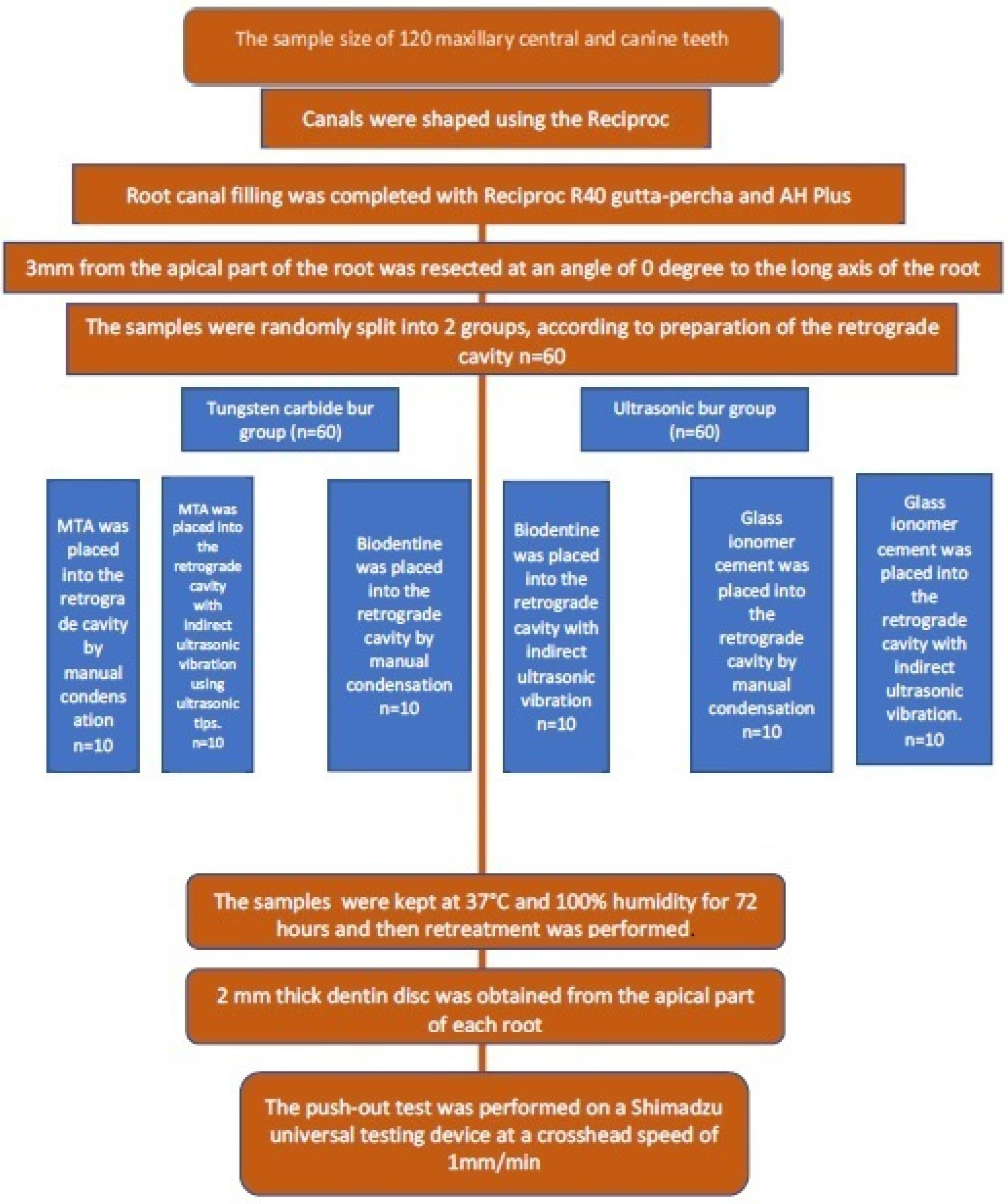
2. Materials and Methods

2.1. Ethics Committee Approval

This study was carried out to investigate the bond strength of retrograde filling after retreatment in teeth with failed apical resection. In order to carry out this study, an ethics committee’s approval was obtained from T.C. Ordu University Clinical Research Ethics Committee with the decision dated 18 October 2021 and numbered 2021/238. The experimental stages of the study were carried out at Ordu University Faculty of Dentistry, Department of Endodontics.

2.2. Preparation of Experimental Teeth

In order to determine the sample size, an independent t test was performed with G*Power, version 3.1 program based on the reference article [17]. Considering the bond strength parameter, it was determined that at least 10 samples should be taken in each group with 95% confidence, 80% test power, and 1.17 effect size. A total of 120 human upper central and upper canine teeth with one root and one canal were extracted for periodontal and orthodontic reasons. The inclusion criteria of the teeth were as follows:
-
Root tip development must be completed,
-
There should be no defects or signs of resorption at the root,
-
The root should not be too long, too short, too wide or too narrow,
-
Should not have more than 10 degrees of curvature,
-
Must be single-rooted and single-canal,
-
Root canal anatomy should not show variation,
-
Root canal treatment must not have been applied before,
-
No visible cracks or fractures on the root.
The dental crowns of the teeth chosen for the study were cut away from the cementum–enamel boundary using a diamond separe bur (Dedeco International Inc., New York, NY, USA) under water cooling to a root length of 15 mm. Working lengths were defined with a 10 K type file (Dentsply Mailfer, Ballaigues, Switzerland).

2.3. Preparation and Obturation of Root Canals

Following defining the working lengths, the canals were instrumented using the Reciproc Blue (VDW, Munich, Germany) system according to the manufacturer’s instructions using R25 and R40 files, respectively. The root canals were irrigated with 2 mL of 5% sodium hypoclorite (NaOCl) (Wizard, Rehber Kimya San. Tic. Aş., Istanbul, Turkey) between each file change, totaling a volume of 10 mL. The final irrigation was performed with solutions of 5 mL 17% ethylenediamine tetraacetic acid (EDTA) (Werax; Spot Dis Deposu AŞ, Izmir, Turkey), 2 mL NaOCl, and 5 mL distilled water. After the completion of the irrigation procedures, the root canals dried by paper points were filled using a cold lateral condensation technique with R 40 gutta-percha (VDW, Munich, Germany) and #25 accessory cones with AH Plus (Dentsply Maillefer, Ballaigues, Switzerland) sealer, and kept at 37 °C 100% humidity for 1 month.

2.4. Apical Resection Procedure

In order to perform an apical resection, a 3 mm section of the apical part of the root was resected at an angle of 90 degrees in the bucco-lingual direction, perpendicularly to the long axis of the tooth, with a speed-controlled, torque-monitored, and saline-irrigated surgical motor (NSK Surgic Pro, NSK Nakanishi Inc., Tochigi, Japan) using a fissure tungsten carbide bur (Meisinger, Neuss, Germany) (clockwise at 1200 rpm and 50 Ncm torque setting) [18].

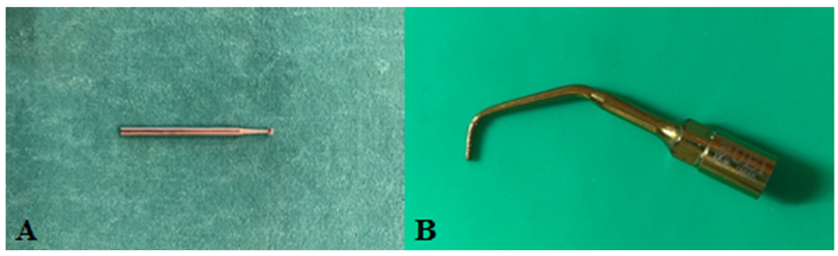
2.5. Retrograde Cavity Preparation

Resected teeth were randomly divided into two main groups by tossing coins according to the retrograde cavity preparation methods. The experimental study design of each group was shown in Figure 1.
Group 1 (TC): In this group, 3 mm deep, 1.1 ± 0.5 mm diameter retrograde cavity was prepared using a speed-controlled, torque-monitored, and saline irrigated surgical motor (NSK Surgic Pro, NSK Nakanishi Inc., Tochigi, Japan) with a 1.1 mm round tungsten carbide bur (Meisinger, Germany) (Figure 2) [18].
Group 2 (U): In this group, an ultrasonic piezo (NSK Vario Surg 3, NSK Nakanishi Inc., Tochigi, Japan) and an ultrasonic retro-tip (Woodpecker DTE, UE1, Foshan, Guangdong, China) (Figure 2) at 40% power in ‘E’ ENDO mode were used for the preparation of the same retrograde cavity [18]. The prepared teeth were kept in saline after these procedures. The 2 main groups were separated into 6 subgroups consisting of (n: 10) teeth in each group, making a total of 12 groups. The subgroups were created according to the retrograde filling materials placed in the prepared cavities and the method of placement of these materials. Before the retreatment process, the teeth were embedded in acryl blocks.
Subgroup 1a: According to the manufacturer’s instructions, MTA (Dentsply, Dental Tulsa, Switzerland) was hand-mixed in a powder to liquid ratio of 3:1 on a clean glass surface. The prepared cement was inserted into the cavities with an MTA gun (Dentsply Maillefer, Ballaigues, Switzerland), was and manually compacted with an endodontic plugger (Dentsply, Maillefer, Switzerland) [19].
Subgroup 1b: In the indirect ultrasonic vibration group, MTA was placed with the help of a hand carrier. The material was compacted with indirect ultrasonic vibration through the amalgam fulvar for 3 s with VDW Ultra Ultrasound (Munich, Germany) using Start-X number 4 tip (Dentsply, Sirona, Switzerland) [20].
Subgroup 2a: A Biodentine (Septodont, Saint Maur des Fossés, France) capsule included a single dose container. A total of 5 drops of liquid were dropped into the capsule and placed in an amalgamator (ADM 9002, Medident GbR, Treffurt, Germany) and mixed for 30 s. Biodentine was gradually placed into the retrograde cavities of the specimens with the help of plastic spatulas taken from the product box and compacted with the help of a hand carrier. Excess material was removed from the surface of the specimens with a plastic spatula.
Subgroup 2b: The specimens in the indirect ultrasonic vibration group were performed as in subgroup 1b.
Subgroup 3a: The glass ionomer cement (Nova Glass F, Imicryl, Turkey) (GIS) was mixed on a clean glass surface in a 3:2 powder/liquid ratio according to the manufacturer’s instructions. The prepared cement was carried into the retrograde cavity with a spatula, gradually placed, and compacted with a hand tool. Excess material was removed with a spatula.
Subgroup 3b: In the indirect ultrasonic vibration group, the procedures were performed as in subgroup 1b.

2.6. Retreatment Procedures

The obtained samples were wrapped with wet gauze in Petri dishes and kept at 37 °C and 100% humidity for 72 h. Then, retreatment was performed using Reciproc Blue (VDW, Munich, Germany) R25 and R40 instruments with the Dentsply X-smart IQ motor (Dentsply, Sirona, Switzerland). Root canals were irrigated with 2 mL 5% NaOCl after all three pecking movements. A 10 K-type hand file was used to obtain apical clearance. For the final shaping, a Reciproc Blue R40 file was used. During the retreatment of each tooth, the use of the file was limited to a total of 6 pecking movements. As this file system is a single-use system, only a single instrument was used in each specimen. For the final irrigation, 5 mL of 17% EDTA was administered for 3 min. Afterwards, the teeth were irrigated with 5 mL of 5% NaOCl. The irrigation process was completed after the teeth were irrigated with 5 mL of distilled water. Preparation was performed up to the last point reached by the Reciproc Blue R25 instrument for cases without apical exposure, and these teeth were included in the study as well.

2.7. Sample Preparation and Push-Out Test

From the apical side of each retreated tooth, one 2 mm thick dentin disk was taken perpendicularly to the long axis of the root, with a diamond disk of 0.5 mm thickness on a water-cooled cutting device (Microtome, Mecatom T180; Presi SA, Angonnes, France) rotating at a low speed (250–300 rpm). The section’s thickness was measured with a digital caliper. Within the limitations of the precision cutting device, it was adjusted so that the desired dimensions could be achieved with minimal loss. Every single dentin disk was assigned a number to avoid confusion among the groups. The specimens were tested for bond strength using the Shimadzu universal testing machine (Autograph AGS X; Shimadzu Co., Kyoto, Japan) [21]. In the experimental groups, each specimen was placed in contact with a 0.8 mm diameter stainless steel cylindrical tip through the cement, which was prepared to perform the push-out test only. The pushing force was performed at a rate of 1 mm/min, coronally to apically, until bond failure occurred. The forces were registered in Newtons (N). Coupling strength values in MPa were calculated with the formula below.
B o n d   s t r e n g t h M P a =       F o r c e   N S u r f a c e   A r e a = 2 π r h ,
(bonding surface area of the cement mm2), (π = 3.14, r = radius of the inner channel cavity, h = height in mm).

2.8. Analysis of Fracture Types

Using a stereo motorized research microscope (Nikon SMZ25, Nikon Ltd., Tokyo, Japan), the fracture surfaces of all specimens were examined under 1.00×, 3.00×, and 7.00× magnification after fracture. Following the examination, types of fracture of the samples were recorded in three categories, similar to Kadić et al. [22]. The obtained data were analyzed as percentage (%).
Adhesive type fracture: A fracture that occurs in the adhesive layer between the cement and dentin.
Cohesive type fracture: A fracture that occurs completely in the cement.
Mixed fracture type: The type of fracture in which adhesive and cohesive fracture are both seen in a specimen.

2.9. Statistical Analysis
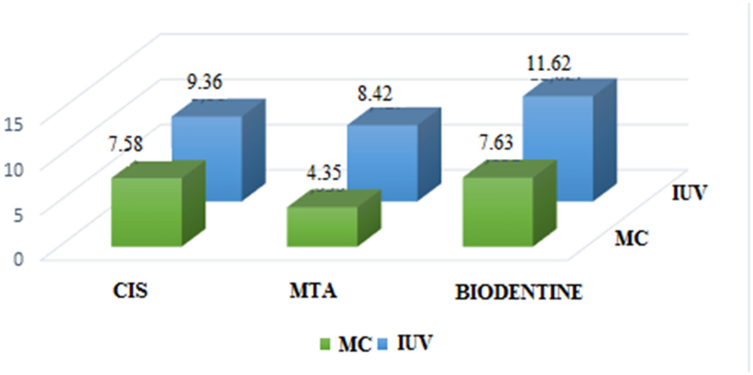
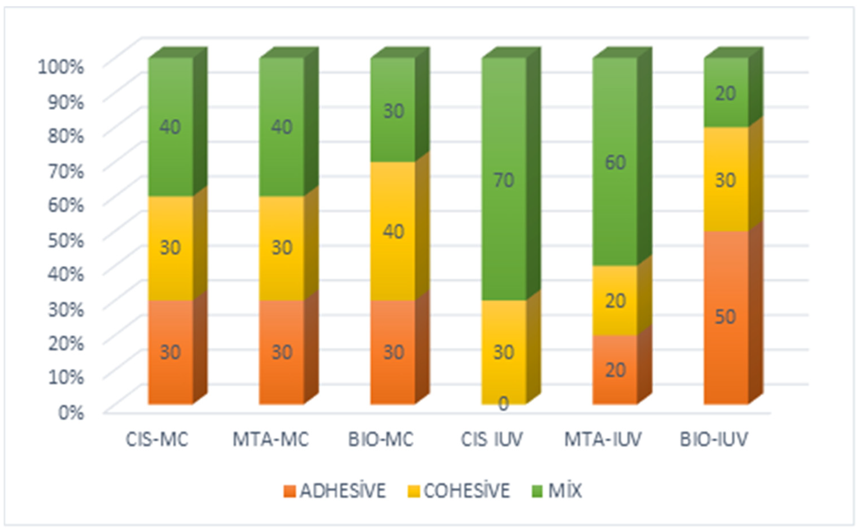
A Kolmogorov–Smirnov test was used to check the normal distribution of the data, while Levene’s test was used to check the homogenity of group variances. The data were analyzed using three-way ANOVA. A 5% significance level (α) was applied in the analysis and interpretation of the results. The SPSS v27 (IBM Inc., Chicago, IL, USA) statistical package program was used for all calculations.
This manuscript was produced with Microsoft 2013 (15.0.5589.1001) (Microsoft Corporation by impressa systems, Santa Rosa, CA, USA).

3. Results

3.1. Bond Strength

The MTA retrograde filling groups had a mean bond strength of 15.403 MPa, while those placed with Biodentine had a mean bond strength of 17.957 MPa. Also, those placed with CIS had a mean bond strength of 14.12 MPa as a result of three-way analysis of variance. Although the bond strength value of retrograde fillings with manual condensation was 15.323 MPa, it was calculated as 16.268 MPa for those placed with indirect ultrasonic vibration. Retrograde fillings placed in retrograde cavities prepared with tungsten carbide burs had a mean bond strength of 8.244 MPa, whereas retrograde fillings placed in cavities prepared with ultrasonic retro-tips had a mean bond strength of 22.981 MPa (Table 1, Figure 3 and Figure 4).
The cavity*material*placement triple interaction between cavity*material*placement was not statistically significant (p = 0.939), as seen in Table 2, which shows the results of three-way analysis of variance for MPa. Cavity*material, cavity*placement, and material*placement binary interactions were also not statistically significant (p = 0.191, p = 0.104, and p = 0.543, respectively). Placement factors were not statistically significant (p = 0.128 and p = 0.508), whereas the difference between the levels of the cavity method factor was statistically significant (p = 0.000). The mean bonding value of retrograde fillings placed in retrograde cavities prepared with an ultrasonic retro tip was significantly higher than the mean bonding value of retrograde fillings placed in retrograde cavities prepared with a tungsten carbide bur (Table 2).

3.2. Apical Reach

The apical access evaluation showed that there was no statistically significant difference in the placement of CIS using manual condensation or indirect ultrasonic vibration in cavities prepared with tungsten carbide or ultrasonic tips (p = 0.350), as can be seen in Table 3. There was no statistically significant difference between the cavity preparation methods in the manual condensation of the MTA group’s apical access (p = 0.087), but there was a statistically significant difference in the indirect ultrasonic vibration group (p = 0.011). A statistically significant difference was found between the cavity preparation methods in the groups placed by manual condensation and indirect ultrasonic vibration in the apical access of the Biodentine group (p = 0.000; p = 0.003, respectively) (Table 3).
When apical access was evaluated according to the type of retrograde filling placement, as seen in Table 4, it was found that the presence or absence of apical access in all materials, and in both cavity preparation methods showed no statistically significant difference (p > 0.05) (Table 4).
When evaluated in terms of retrograde cavity material, there is a statistically significant difference in apical access in all materials according to retrograde cavity and filling placement technique (p < 0.05), as seen in Table 5. MTA and Biodentine placed through manual condensation in tungsten carbide cavities provided 100% apical access. For cements placed by indirect ultrasonic vibration in tungsten carbide cavities, apical access was reported as 100% in the MTA group, 70% in the Biodentine group, and 50% in the CIS group. Manual condensation in ultrasonic cavities resulted in 60% apical access in the MTA group, 20% in the CIS group, and no apical access in the Biodentine group. Indirect ultrasonic vibration in ultrasonic cavities resulted in 40% apical access in the MTA group, 20% in the CIS group, and no apical access in the Biodentine group. For all groups, the MTA group had the highest rate of apical access, while the CIS group had the lowest rate of apical access (Table 5).
In both cavity preparation methods, bond strength values were significantly higher where apical access could not be achieved (Table 6).

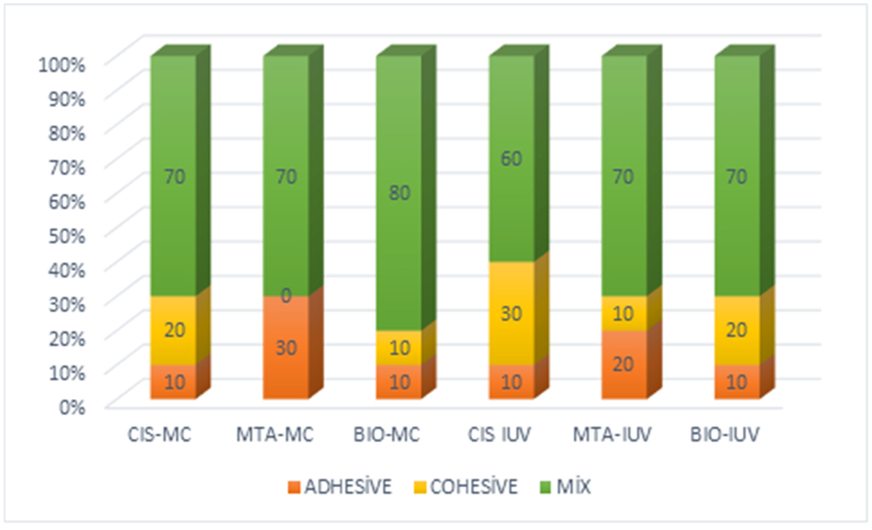
3.3. Stereomicroscope Analysis

After the push-out tests, 120 specimens were examined under stereo motorized microscope, and fracture types were also analyzed. The adhesive failure type had the highest percentage in all cements placed by manual condensation in retrograde cavities that opened with tungsten carbide burs, and in the MTA group placed by manual condensation in retrograde cavities that opened with ultrasonic retro tips. Cohesive failure was highest in the Biodentine group placed by manual condensation in retrograde cavities that opened with a tungsten carbide bur (Figure 5 and Figure 6).

4. Discussion

In our study, the fillings in retrograde cavities prepared with ultrasonic retro tips were found to have significantly higher bond strength after retreatment compared to prepared with tungsten carbide bur, thus confirming the first hypothesis; however, no difference could be found between cements after retreatment and the second hypothesis was not confirmed.
The cross-sectional plane should be considered during root resection. Ideally, the inclination as perpendicular as possible to the long axis of the tooth (0°) preserves the root length and exposes fewer dentinal tubules, thus reducing the number of tubules exposed to the periphery, resulting in less microleakage. In our study, a (0)° bevel was given for the same reasons [23].
The marginal adaptation has been identified as the proximity degree and the clamping of a filling material to the cavity wall [24]. It is possible that the failure of marginal adaptation of the material and the presence of cracks or gaps at the interface between the material and the dentin wall may lead to apical leakage, resulting in treatment failure [25]. O’Connor et al. [26] demonstrated that there is no significant difference between preparation with ultrasonic tips or micro-handpiece preparations in the sealing of retrograde fillings.
The ideal root-end filling should prevent the exit of any bacteria, bacterial products, or toxic materials into the surrounding periradicular tissues while sealing the contents of the root canal system within the canal [2]. Taschieri et al. [27] investigated persistent cases of periapical lesions after endodontic surgery. Due to defective sealing at the interface between the root-end filling and the cavity, the failure of apical surgery occurred. Such a gap would support a continuous leakage of bacteria from the infected root canal system into the periapical tissue, thus sustaining inflammation. This results in the reoccurrence of periapical disease after endodontic surgery. Nevertheless, there are limited studies for retreatment after endodontic surgery [15,16]. Retrograde filling is not available in these studies.
The fact that the bond strength values of the ultrasonic cavity group were significantly (p < 0.000) higher than the tungsten carbide cavity group regardless of retrograde filling material is important. This study was inspired by the ability to prepare smaller, centralized and desired depth cavities as shown in the study of Bernardes et al. [28].
The popularity of bioceramic-based retrograde filling materials has recently increased [4,29]. It was demonstrated that MTA has superior properties in terms of sealing ability, biocompatibility, and periradicular tissue regeneration when compared to conventional retrograde filling materials [30].
Among some studies in which the average bonding value of MTA in retrograde cavities prepared by traditional methods were examined, Formosa et al. [31] reported 5.08 ± 2.41 MPa, Aggarwal et al. [32] calculated 5.2 ± 0.4 MPa after a curing time of 24 h, 9.0 ± 0.9 MPa after curing for 7 days, and Ertaş et al. [33] calculated 4.5 ± 1.5 MPa. Despite the removal force during the retreatment process, the bonding value of MTA placed in the traditional retrograde cavity prepared with a tungsten carbide bur was calculated as 4.35 ± 3.68 MPa in our study, similar to other studies in the literature.
Several studies have investigated filling bonding in the retrograde cavity using diamond-coated ultrasonic retro tips. Vivan et al. [34] calculated the bond strength of MTA as 19.18 ± 4.7 MPa, Marques et al. [35] calculated the bond strength of Angelus MTA as 1.82 ± 0.47 MPa, and Shokouhinejad et al. [36] calculated the bond strength of MTA as 7.77 ± 1.34 MPa. In the present study, we found that the bonding value of Angelus MTA was higher than other studies in the literature with a value of 23.86 ± 8.04 MPa, although it was similar to the study of Vivan et al. [34] despite the extraction force during the retreatment process.
According to Akbulut et al. [37], the average bond strength value of Biodentine was calculated as 17.55 ± 5.67 MPa, and there was no significant difference between Angelus MTA. Marques et al. [35] investigated the bonding of different tricalcium silicate-based materials, they stated that there was no significant difference between MTA and Biodentine bonding ability. But, a difference was found between the failure types of the materials. While MTA Angelus mostly showed mixed failure, Biodentine demonstrated mixed and cohesive failure at similar rates. The material properties of Biodentine, enhanced by the higher tricalcium silicate ratio than MTA and the high calcium ion release of Biodentine, led to mixed and cohesive failure [38]. However, in our study, cohesive failure was observed only in the Biodentine group, which was placed into the cavity by manual condensation and prepared with a tungsten carbide bur.
Kadic et al. [22] reported that, although the average bonding value of Biodentine was higher, there was no significant difference with MTA. Similarly, in the present study, there was no significant difference between MTA and Biodentine when placed in cavities that prepared with diamond-coated ultrasonic retro-tips.
During the retreatment procedures, it is necessary to reach the apical area of the root canal. Once apical access has been achieved, all principles of endodontic treatment had to be performed. The coronal access must be completed, all previous root filling material must be removed, canal obstructions must be managed, and they must be achieved to their full working length [39]. Mente et al. [15] suggested that orthograde retreatment combined with orthograde placement of an apical mineral trioxide aggregate plug is a promising long-term treatment option for teeth with postsurgical failure.
In our study, 100% of apical access was achieved in MTA and Biodentine, which were placed into the cavity by manual condensation and prepared with a tungsten carbide bur. On the other hand, apical access was achieved at a rate of 100% and 70% for MTA and Biodentine placed by indirect ultrasonic vibration in the cavities prepared with tungsten carbide burs, respectively.
When looking at the cavities prepared with ultrasonic retro-tips, apical access was achieved in 60% of MTA and 20% of CIS placed with manual condensation, whereas no apical access was achieved in the Biodentine group. The MTA group that placed the indirect ultrasound vibration achieved 40% apical access and 20% apical access in CIS, whereas no apical access was achieved in the Biodentine group. Biodentine showed higher bond strength than all other groups in cavities prepared with ultrasonic retro-tips, regardless of whether they were placed by manual condensation or indirect ultrasonic vibration, but no significant difference was detected. Additionally, it should be considered that other factors could influence bond strength, such as the presence of debris or substrate contamination. Different cleaning protocols were evaluated for removing sealer residues on the dentin surface [40]. Blood contamination during the bonding procedure of conventional and hydrophilic primers significantly lowers their bond strength values [41]. These protocols would be applied in future studies. Appel et al. [42] evaluated the clinical outcome of orthograde endodontic retreatment after failed apicectomy. They stated that, even teeth with pre-existing retrograde fillings can be treated successfully by an orthograde retreatment, as the mean success rate for these cases did not differ significantly from the mean success rate obtained for cases without any preexisting retrograde filling.
Although apical access in cavities prepared with tungsten carbide burs is desirable, bond strength values were statistically lower compared to ultrasonically prepared cavities, which affects the success of treatment. However, Appel et al. [42] reported that, in one case, the periapical lesion showed through radiographic healing that a pre-existing retrograde filling was dislodged into the apical tissue. Therefore, further in vivo and in vitro studies were needed.
In this in vitro research study, in vivo clinical situations could not be reflected. Blood contamination could have altered the results of this study. This is a limitation of the present study. It is difficult to determine the resection method performed in cases of failed apical resection in clinical conditions. In our study, possible scenarios were constructed with the awareness of the technique used. This may be another limitation of our study. The acidic apical infection environment in clinical situations may affect the physical and chemical properties of the retrograde filling material. Therefore, the bond strength values would have been decreased. This is another limitation of the present study.

5. Conclusions

Within the limitations of the present study, in teeth with failed apical resection, if retrograde cavity was prepared with tungsten carbide bur, apical plug or re-apical surgery can be planned because retrograde filling bonding is lower in cases where apical access is provided. In cases where apical access is not achieved, re-apical surgical treatment should be planned due to incomplete cleaning of the root canal system.
In teeth that were prepared with ultrasonic retro tips, routine root canal treatment can be performed because the bonding of the retrograde filling is significantly better in cases where apical access is achieved. However, the method used may often be unknown. In such cases, a second resection may be necessary.
The acidic apical infection environment may affect the physical and chemical properties of the retrograde filling material. Our study was performed under in vitro conditions, and even if it is not possible to reflect the intraoral environment completely in the study, we tried to obtain the closest results by following the information in the literature. Therefore, in vitro studies must be supported by clinical studies to investigate the actual bonding performance of the materials.

Author Contributions

Idea, hypothesis, experimental design: L.B.A., Performed the experiments: S.T., Wrote the manuscript: S.T. and L.B.A., Proofread the manuscript: L.B.A. All authors have read and agreed to the published version of the manuscript.

Funding

This study was supported by the project numbered B-2103 of Ordu University Scientific Research Projects Coordination Office.

Institutional Review Board Statement

In order to conduct this study, the approval of the ethics committee was obtained from Ordu University Clinical Research Ethics Committee with decision number 2021/238 (18 October 2021).

Informed Consent Statement

Informed consent was obtained from all subjects involved in the study.

Data Availability Statement

The original contributions presented in the study are included in the article, further inquiries can be directed to the corresponding author.

Conflicts of Interest

The authors declare no conflict of interest.

References

  1. Kruse, C.; Spin-Neto, R.; Reibel, J.; Wenzel, A.; Kirkevang, L.L. Diagnostic validity of periapical radiography and CBCT for assessing periapical lesions that persist after endodontic surgery. Dentomaxillofac. Radiol. 2017, 46, 20170210. [Google Scholar] [CrossRef] [PubMed]
  2. Kim, S.; Kratchman, S. Modern endodontic surgery concepts and practice: A review. J. Endod. 2006, 32, 601–623. [Google Scholar] [CrossRef]
  3. Chong, B.S.; Ford, T.R.; Kariyawasam, S.P. Tissue response to potential root-end filling materials in infected root canals. Int. Endod. J. 1997, 30, 102–114. [Google Scholar] [CrossRef] [PubMed]
  4. Premjith; Shetty, D.; Kailar, A.; Pare, S.; Kumar, P.; Ragher, M. The Effect of Root End Cavity Preparation Using Er,Cr:YSGG Laser, Ultrasonic Retrotip, and Bur on the Apical Microleakage of Retrograde Cavity Filled with MTA Plus. J. Pharm. Bioallied Sci. 2020, 12, S299–S303. [Google Scholar] [CrossRef] [PubMed]
  5. Karlovic, Z.; Pezelj-Ribaric, S.; Miletic, I.; Jukic, S.; Grgurevic, J.; Anic, I. Erbium:YAG laser versus ultrasonic in preparation of root-end cavities. J. Endod. 2005, 31, 821–823. [Google Scholar] [CrossRef]
  6. Rubinstein, R.A.; Kim, S. Long-term follow-up of cases considered healed one year after apical microsurgery. J. Endod. 2002, 28, 378–383. [Google Scholar] [CrossRef]
  7. Torabinejad, M.; Watson, T.F.; Pitt Ford, T.R. Sealing ability of a mineral trioxide aggregate when used as a root end filling material. J. Endod. 1993, 19, 591–595. [Google Scholar] [CrossRef]
  8. Alsubait, S.A.; Hashem, Q.; AlHargan, N.; AlMohimeed, K.; Alkahtani, A. Comparative evaluation of push-out bond strength of ProRoot MTA, bioaggregate and biodentine. J. Contemp. Dent. Pract. 2014, 15, 336–340. [Google Scholar] [CrossRef]
  9. Prati, C.; Gandolfi, M.G. Calcium silicate bioactive cements: Biological perspectives and clinical applications. Dent. Mater. 2015, 31, 351–370. [Google Scholar] [CrossRef]
  10. Chong, B.S.; Pitt Ford, T.R.; Watson, T.F. Light-cured glass ionomer cement as a retrograde root seal. Int. Endod. J. 1993, 26, 218–224. [Google Scholar] [CrossRef]
  11. Friedman, S. The prognosis and expected outcome of apical surgery. Endod. Top. 2005, 11, 219–262. [Google Scholar] [CrossRef]
  12. de Chevigny, C.; Dao, T.T.; Basrani, B.R.; Marquis, V.; Farzaneh, M.; Abitbol, S.; Friedman, S. Treatment outcome in endodontics: The Toronto study—phases 3 and 4: Orthograde retreatment. J. Endod. 2008, 34, 131–137. [Google Scholar] [CrossRef] [PubMed]
  13. Ng, Y.L.; Mann, V.; Gulabivala, K. Outcome of secondary root canal treatment: A systematic review of the literature. Int. Endod. J. 2008, 41, 1026–1046. [Google Scholar] [CrossRef]
  14. Dioguardi, M.; Stellacci, C.; La Femina, L.; Spirito, F.; Sovereto, D.; Laneve, E.; Manfredonia, M.F.; D’Alessandro, A.; Ballini, A.; Cantore, S.; et al. Comparison of Endodontic Failures between Nonsurgical Retreatment and Endodontic Surgery: Systematic Review and Meta-Analysis with Trial Sequential Analysis. Medicina 2022, 58, 894. [Google Scholar] [CrossRef]
  15. Mente, J.; Leo, M.; Michel, A.; Gehrig, H.; Saure, D.; Pfefferle, T. Outcome of orthograde retreatment after failed apicoectomy: Use of a mineral trioxide aggregate apical plug. J. Endod. 2015, 41, 613–620. [Google Scholar] [CrossRef]
  16. Stefopoulos, S.; Tzanetakis, G.N.; Kontakiotis, E.G. Non-surgical retreatment of a failed apicoectomy without retrofilling using white mineral trioxide aggregate as an apical barrier. Braz. Dent. J. 2012, 23, 167–171. [Google Scholar] [CrossRef]
  17. Vivan, R.R.; Guerreiro-Tanomaru, J.M.; Bosso-Martelo, R.; Costa, B.C.; Duarte, M.A.H.; Tanomaru-Filho, M. Push-out Bond Strength of Root-end Filling Materials. Braz. Dent. J. 2016, 27, 332–335. [Google Scholar] [CrossRef]
  18. Angerame, D.; De Biasi, M.; Lenhardt, M.; Porrelli, D.; Bevilacqua, L.; Generali, L.; La Rosa, G.R.M.; Pedulla, E. Root-end resection with or without retrograde obturation after orthograde filling with two techniques: A micro-CT study. Aust. Endod. J. 2022, 48, 423–430. [Google Scholar] [CrossRef]
  19. Chaudhari, P.S.; Chandak, M.G.; Jaiswal, A.A.; Ikhar, A. Comparative evaluation of push-out bond strength of three retrograde filling materials in teeth with root apices resected at 90 degrees: Mineral trioxide aggregate Angelus, Zirconomer, and Bioactive bone cement. J. Conserv. Dent. 2022, 25, 605–609. [Google Scholar] [CrossRef]
  20. Parashos, P.; Phoon, A.; Sathorn, C. Effect of ultrasonication on physical properties of mineral trioxide aggregate. Biomed. Res. Int. 2014, 2014, 191984. [Google Scholar] [CrossRef]
  21. Skidmore, L.J.; Berzins, D.W.; Bahcall, J.K. An in vitro comparison of the intraradicular dentin bond strength of Resilon and gutta-percha. J. Endod. 2006, 32, 963–966. [Google Scholar] [CrossRef] [PubMed]
  22. Kadic, S.; Baraba, A.; Miletic, I.; Ionescu, A.; Brambilla, E.; Ivanisevic Malcic, A.; Gabric, D. Push-out bond strength of three different calcium silicate-based root-end filling materials after ultrasonic retrograde cavity preparation. Clin. Oral. Investig. 2018, 22, 1559–1565. [Google Scholar] [CrossRef] [PubMed]
  23. Gilheany, P.A.; Figdor, D.; Tyas, M.J. Apical dentin permeability and microleakage associated with root end resection and retrograde filling. J. Endod. 1994, 20, 22–26. [Google Scholar] [CrossRef] [PubMed]
  24. Taylor, M.J.; Lynch, E. Marginal adaptation. J. Dent. 1993, 21, 265–273. [Google Scholar] [CrossRef]
  25. Ishikawa, H.; Sawada, N.; Kobayashi, C.; Suda, H. Evaluation of root-end cavity preparation using ultrasonic retrotips. Int. Endod. J. 2003, 36, 586–590. [Google Scholar] [CrossRef]
  26. O’Connor, R.P.; Hutter, J.W.; Roahen, J.O. Leakage of amalgam and Super-EBA root-end fillings using two preparation techniques and surgical microscopy. J. Endod. 1995, 21, 74–78. [Google Scholar] [CrossRef]
  27. Taschieri, S.; Bettach, R.; Lolato, A.; Moneghini, L.; Fabbro, M.D. Endodontic surgery failure: SEM analysis of root-end filling. J. Oral. Sci. 2011, 53, 393–396. [Google Scholar] [CrossRef]
  28. Bernardes, R.A.; de Moraes, I.G.; Garcia, R.B.; Bernardineli, N.; Baldi, J.V.; Victorino, F.R.; Vasconcelos, B.C.; Duarte, M.A.; Bramante, C.M. Evaluation of apical cavity preparation with a new type of ultrasonic diamond tip. J. Endod. 2007, 33, 484–487. [Google Scholar] [CrossRef]
  29. Jung, J.; Kim, S.; Kim, E.; Shin, S.J. Volume of Voids in Retrograde Filling: Comparison between Calcium Silicate Cement Alone and Combined with a Calcium Silicate-based Sealer. J. Endod. 2020, 46, 97–102. [Google Scholar] [CrossRef]
  30. Bueno, C.R.E.; Vasques, A.M.V.; Cury, M.T.S.; Sivieri-Araujo, G.; Jacinto, R.C.; Gomes-Filho, J.E.; Cintra, L.T.A.; Dezan-Junior, E. Biocompatibility and biomineralization assessment of mineral trioxide aggregate flow. Clin. Oral. Investig. 2019, 23, 169–177. [Google Scholar] [CrossRef]
  31. Formosa, L.M.; Mallia, B.; Camilleri, J. Push-out bond strength of MTA with antiwashout gel or resins. Int. Endod. J. 2014, 47, 454–462. [Google Scholar] [CrossRef] [PubMed]
  32. Aggarwal, V.; Singla, M.; Miglani, S.; Kohli, S. Comparative evaluation of push-out bond strength of ProRoot MTA, Biodentine, and MTA Plus in furcation perforation repair. J. Conserv. Dent. 2013, 16, 462–465. [Google Scholar] [CrossRef] [PubMed]
  33. Ertas, H.; Kucukyilmaz, E.; Ok, E.; Uysal, B. Push-out bond strength of different mineral trioxide aggregates. Eur. J. Dent. 2014, 8, 348–352. [Google Scholar] [CrossRef] [PubMed]
  34. Vivan, R.R.; Guerreiro-Tanomaru, J.M.; Bernardes, R.A.; Reis, J.M.; Hungaro Duarte, M.A.; Tanomaru-Filho, M. Effect of ultrasonic tip and root-end filling material on bond strength. Clin. Oral. Investig. 2016, 20, 2007–2011. [Google Scholar] [CrossRef] [PubMed]
  35. Stefaneli Marques, J.H.; Silva-Sousa, Y.T.C.; Rached-Junior, F.J.A.; Macedo, L.M.D.; Mazzi-Chaves, J.F.; Camilleri, J.; Sousa-Neto, M.D. Push-out bond strength of different tricalcium silicate-based filling materials to root dentin. Braz. Oral. Res. 2018, 32, e18. [Google Scholar] [CrossRef]
  36. Shokouhinejad, N.; Razmi, H.; Fekrazad, R.; Asgary, S.; Neshati, A.; Assadian, H.; Kheirieh, S. Push-out bond strength of two root-end filling materials in root-end cavities prepared by Er,Cr:YSGG laser or ultrasonic technique. Aust. Endod. J. 2012, 38, 113–117. [Google Scholar] [CrossRef]
  37. Akbulut, M.B.; Bozkurt, D.A.; Terlemez, A.; Akman, M. The push-out bond strength of BIOfactor mineral trioxide aggregate, a novel root repair material. Restor. Dent. Endod. 2019, 44, e5. [Google Scholar] [CrossRef]
  38. Camilleri, J.; Sorrentino, F.; Damidot, D. Investigation of the hydration and bioactivity of radiopacified tricalcium silicate cement, Biodentine and MTA Angelus. Dent. Mater. 2013, 29, 580–593. [Google Scholar] [CrossRef]
  39. Roda, R.S.; Gettleman, B.H.; Jhonson, S.C. Nonsurgical retreatment. In Cohen’s Pathways of the Pulp, 12th ed.; Berman, L.H., Ed.; Elsevier: Philadelphia, PA, USA, 2021; pp. 343–410. [Google Scholar]
  40. Goncalves Galoza, M.O.; Fagundes Jordao-Basso, K.C.; Escalante-Otarola, W.G.; Victorino, K.R.; Rached Dantas, A.A.; Kuga, M.C. Effect of cleaning protocols on bond strength of etch-and-rinse adhesive system to dentin. J. Conserv. Dent. 2018, 21, 602–606. [Google Scholar] [CrossRef]
  41. Cacciafesta, V.; Sfondrini, M.F.; Scribante, A.; De Angelis, M.; Klersy, C. Effects of blood contamination on the shear bond strengths of conventional and hydrophilic primers. Am. J. Orthod. Dentofacial Orthop. 2004, 126, 207–212. [Google Scholar] [CrossRef]
  42. Appel, D.; Schafer, E.; Appel, C. Success rate of orthograde endodontic retreatment after failed apicectomy—A retrospective study. Int. Endod. J. 2023, 56, 819–826. [Google Scholar] [CrossRef] [PubMed]
Figure 1. Flowchart of the methodology.
Applsci 15 03441 g001
Figure 2. (A) 1.1 mm round tungsten carbide bur, (B) ultrasonic retro-tip.
Applsci 15 03441 g002
Figure 3. Retrograde filling materials placed in the cavity prepared with tungsten carbide drill and mean MPa values after retreatment, according to the placement factors of the materials. CIS: Cam ionomer cement, MTA: Mineral trioxide aggregate, MC: Manual condensation, IUV: Indirect ultrasonic vibration.
Applsci 15 03441 g003
Figure 4. Retrograde filling materials placed in the cavity prepared with ultrasonic retro tips and mean MPa values after retreatment according to the placement factors of the materials. CIS: Cam ionomer cement, MTA: Mineral trioxide aggregate, MC: Manual condensation, IUV: Indirect ultrasonic vibration.
Applsci 15 03441 g004
Figure 5. Fracture types in cavities prepared with tungsten carbide bur. CIS: Glass ionomer cement, MC: Manual compaction, BIO: Biodentin, IUV: Indirect ultrasonic vibration.
Applsci 15 03441 g005
Figure 6. Fracture types in cavities prepared with ultrasonic retro tips. CIS: Glass ionomer cement, MC: Manual compaction, BIO: Biodentin, IUV: Indirect ultrasonic vibration.
Applsci 15 03441 g006
Table 1. Introductory statistical values of MPa.
MaterialMethodCavities
TCUTotal
nMeanSDnMeanSDnMeanSD
CISMC107.584.641021.905.474014.129.23
IUV109.365.991017.6311.34
MTAMC84.353.68823.868.043515.4011.08
IUV98.427.061023.757.79
BIOMC97.634.771025.3411.683817.9511.34
IUV911.624.811025.568.96
TotalMC 5515.3211.00
IUV5816.2610.26
Total558.245.485822.989.18
CIS: Cam ionomer cement, MTA: Mineral trioxide aggregate, BIO: Biodentin, MC: Manual condensation, IUV: Indirect ultrasonic vibration, SD: Standard deviation, TC: Tungsten carbide, U: Ultrasonic.
Table 2. Three-way analysis of variance results of MPa.
VariancesSum of SquaresSDMean SquareF-Valuep-Value
Cavity6183.87416183.874108.4740.000 *
Material238.8852119.4422.0950.128
Placement25.130125.1300.4410.508
Cavity × Material191.669295.8341.6810.191
Cavity × Placement153.1071153.1072.6860.104
Material × Placement70.094235.0470.6150.543
Cavity × Material × Placement7.18223.5910.0630.939
SD: Standard Deviation. ×: interactions between the groups, * statistically significant according to three-way analysis.
Table 3. Apical access chi-square test according to cavity methods.
MaterialPlacementCavityThe Apical Accessp
YesNo
n%n%
CISMCTC550.0550.00.350
U220.0880.0
IUVTC550.0550.00.350
U220.0880.0
MTAMCTC10100.000.00.087
U660.0440.0
IUVTC10100.000.00.011 *
U440.0660.0
BIOMCTC10100.000.00.000 *
U00.010100.0
IUVTC770.0330.00.003 *
U00.010100.0
CIS: Cam ionomer cement, MTA: Mineral trioxide aggregate, BIO: Biodentin, MC: Manual condensation, IUV: Indirect ultrasonic vibration, TC: Tungsten carbide cavity, U: Ultrasonic cavity, * statistically significant according to chi-square analysis.
Table 4. Apical access chi-square test according to placement method.
MaterialCavityPlacementThe Apical Accessp
YesNo
n%n%
CISTCMC550.0550.01.000
IUV550.0550.0
UMC220.0880.01.000
IUV220.0880.0
MTATCMC10100.000.0-
IUV10100.000.0
UMC660.0440.00.656
IUV440.0660.0
BIOTCMC10100.000.00.211
IUV770.0330.0
UMC00.010100.0-
IUV00.010100.0
CIS: Cam ionomer cement, MTA: Mineral trioxide aggregate, BIO: Biodentin, MC: Manual condensation, IUV: Indirect ultrasonic vibration, TC: Tungsten carbide cavity, U: Ultrasonic cavity.
Table 5. Apical access chi-square test according to materials.
Placement Method Material Apical Accessp
YesNo
n%n%
MCTCCIS550.0550.00.001 *
MTA10100.000.0
BIO10100.000.0
UCIS220.0880.00.003 *
MTA660.0440.0
BIO00.010100.0
IUVTCCIS550.0550.00.013 *
MTA10100.000.0
BIO770.0330.0
UCIS220.0880.00.038 *
MTA440.0660.0
BIO00.010100.0
CIS: Cam ionomer cement, MTA: Mineral trioxide aggregate, BIO: Biodentin, MC: Manual condensation, IUV: Indirect ultrasonic vibration, TC: Tungsten carbide cavity, U: Ultrasonic cavity, * statistically significant according to chi-square analysis.
Table 6. Bond strength according to apical access in cavity methods using Student’s t-test.
Cavity Methods Apical AccessnMeanSDp
TCMPaYes476.375.610.002 *
No1311.823.92
UMPaYes1416.5211.070.013 *
No4623.949.00
TC: Tungsten carbide cavity, U: Ultrasonic cavity, * statistically significant according to Student’s t-test.
Disclaimer/Publisher’s Note: The statements, opinions and data contained in all publications are solely those of the individual author(s) and contributor(s) and not of MDPI and/or the editor(s). MDPI and/or the editor(s) disclaim responsibility for any injury to people or property resulting from any ideas, methods, instructions or products referred to in the content.

