Article

The Osteoinductive Effect of Water-Soluble Matrix from Nano-Nacre Particles of Haliotis diversicolor (H. diversicolor) Abalone on MC3T3-E1 Osteoblasts

by
Chanyatip Suwannasing
1,*,
Ausanai Prapan
1,
Piyaporn Surinlert
2,3,
Chanyarak Sombutkayasith
4 and
Wattana Weerachatyanukul
4
1
Department of Radiological Technology, Faculty of Allied Health Science, Naresuan University, Phitsanulok 65000, Thailand
2
Thammasat University Research Unit in Synthesis and Applications of Graphene, Thammasat University, Pathum Thani 12120, Thailand
3
Chulabhorn International College of Medicine, Thammasat University, Pathum Thani 12120, Thailand
4
Department of Anatomy, Faculty of Science, Mahidol University, Bangkok 10400, Thailand
*
Author to whom correspondence should be addressed.
Appl. Sci. 2025, 15(6), 2907; https://doi.org/10.3390/app15062907
Submission received: 16 January 2025 / Revised: 3 March 2025 / Accepted: 6 March 2025 / Published: 7 March 2025

Abstract

:
Osteoporosis is characterized by an imbalance between osteoblastic bone formation and osteoclastic bone resorption, leading to an increased risk of fractures. The water-soluble matrix (WSM) of nacre exhibits osteoinductive properties in osteoblastic cells, both in vitro and in vivo. However, its release from natural nacre remains challenging due to its solid and compact surface. This study aimed to prepare nano-nacre particles with smaller diameters than intact aragonite crystals to enhance WSM release and to investigate its effects on osteoblast differentiation. Size analysis and SEM imaging showed that the nano-nacre particles had an average size of about 600 nm. Furthermore, their effects on osteoblast differentiation and mineralization were evaluated through qPCR and ARS assay. The results showed that WSM significantly upregulated key osteogenic genes, including RUNX2, ALP, and OCN, in a dose- and time-dependent manner over 14 days, with fold-changes ranging from 1.6 to 3.6. Additionally, the mineralization effects showed calcium deposition levels comparable to those of the positive group. These findings suggest that WSM may be a promising soluble factor for osteoblast differentiation and mineralization. Therefore, understanding the effects of the WSM from H. diversicolor nano-nacre particles on osteoblasts in vitro may provide evidence suggesting that it could be a promising anti-osteoporosis agent.

1. Introduction

Osteoporosis, a common condition in aging societies, is particularly severe among postmenopausal women due to estrogen deficiency. This deficiency accelerates bone turnover, disrupting the balance between bone formation and resorption, leading to a decrease in bone density and an increased risk of fractures, especially in the spine and hips. These fractures are a leading cause of morbidity and mortality among the elderly population [1,2]. Current pharmaceutical treatments, such as bisphosphonates, selective estrogen receptor modulators (SERMs), and newer agents like denosumab and romosozumab, are effective in reducing fracture risks but can have significant side effects, including infection risks, venous thromboembolism, and bone loss after discontinuation [3,4]. Estrogen therapy, while beneficial in reducing bone loss and fracture risk, is also associated with cardiovascular events and certain types of cancer [5]. Consequently, there is a pressing need for new treatment options that can effectively stimulate bone density recovery and restore bone structure. Nacre, or mother of pearl, is a composite material primarily composed of calcium carbonate crystals in aragonite form, along with an organic WSM consisting of polysaccharides, proteins, and glycoproteins [6]. This composition is similar to bone, as both structures utilize organic matrices to guide controlled mineral deposition in biomineralization processes [7]. Moreover, studies suggest that nacre has strong osteoinductive properties. For example, implants of fragmented nacre from Pinctada maxima (P. maxima) have been shown to stimulate bone formation in animal models, suggesting its potential for promoting bone healing [8,9,10,11,12]. In vitro studies also show that WSM extracted from nacre can induce human bone marrow stromal cells (hBMSCs) and pre-osteoblasts to differentiate into osteoblasts. These cells exhibit osteogenic markers, such as alkaline phosphatase and osteocalcin, indicative of active bone tissue formation [13,14]. Nacre’s organic matrix contains bioactive proteins like nacrein, MSI60, and perlucin, which are critical for its osteoinductive effects. These proteins are non-immunogenic, making nacre biocompatible and suitable for use in bone repair without causing adverse immune reactions [15,16,17,18,19,20,21,22,23,24]. Studies have shown that fragmented nacre releases bioactive molecules into culture media, further promoting osteoblast differentiation and mineralization. However, achieving the release of WSM from natural nacre has been challenging due to its solid and compact surface [25]. Recent advances, such as the development of nano-nacre particles, have helped to enhance the release of WSM and calciumions by increasing the surface area [14]. However, it is well established that the osteoinductive effect of nacre is attributed to the organic components within the WSM. Therefore, the molecules responsible for promoting bone formation need to be identified and elucidated. In this study, nano-nacre particles were developed with a smaller diameter than the intact aragonite crystal of nacre to enhance the release of the organic matrix and identify potential osteogenic molecules within the WSM. This approach improves the bioavailability of nacre’s active components, making it more effective in promoting osteogenesis. This innovation is particularly significant for the development of therapeutic strategies for osteoporosis and bone regeneration.

2. Materials and Methods

2.1. Nano-Nacre Particles Preparation

The shells of adult H. diversicolor abalone (50–55 mm in length) were provided by Phuket Abalone Farm (Phuket, Thailand). To prepare the nano-nacre particles, the outer surface of the shells was cleaned using a metal brush, and then the prismatic layer was removed with sandpaper. The shells were then fragmented into small pieces (2–10 mm) and ground into a powder. The powder was suspended in a nano-grinding facility for further grinding, resulting in particles ranging in size from 10 to 1000 nm, which were then freeze-dried at −50 °C for 48 h (FreeZone, Labconco, Kansas City, MO, USA). For structural analysis, the generated nano-nacre particles were placed on a sample plate for scanning electron microscopy (SEM). The samples were diluted in distilled water, and a drop of the suspension was applied to copper tape and allowed to dry at room temperature overnight. The dried samples were then sputter-coated with gold, and SEM images were obtained under high vacuum conditions with an operation voltage of 15 kV using a JSM-5910 SEM (JEOL USA, Peabody, MA, USA). Similar methods have been used in other studies to prepare and analyze nano-particles from natural sources for biomedical applications [26].

2.2. Extraction of WSM from Nano-Nacre Particles

The WSM from nano-nacre particles was extracted by dissolving 100 mg of nano-nacre particles in 100 mL of phosphate-buffered saline (PBS). The solution was stirred overnight at 4 °C, and then subjected to centrifugation at 30,000× g for 20 min. The supernatant was then lyophilized to obtain the WSM in a powdered form [27].

2.3. Western Blotting for WSM Protein Profiling

To analyze the protein profile of the WSM from nano-nacre particles, Western blotting was performed as follows: Approximately 10 μg of WSM proteins was loaded onto a 12.5% SDS-PAGE gel for separation. Following electrophoresis, proteins were transferred to a polyvinylidene difluoride (PVDF) membrane (Millipore, Bedford, MA, USA) for analysis. The PVDF membrane was immersed in a blocking solution containing 10% skim milk in TBS-T (Tris-buffered saline with 0.05% Tween-20) at 4 °C overnight to prevent non-specific binding. The membrane was incubated with goat anti-BMP-2/4 antibody (Abcam, Cambridge, UK) at a dilution of 1:1000 for 2 h at room temperature (RT). After extensive washing with PBST (PBS with Tween-20), the membrane was incubated with a corresponding secondary antibody conjugated to streptavidin–horseradish peroxidase (HRP) (Jackson ImmunoResearch, West Grove, PA, USA) at a dilution of 1:3000 for 2 h at RT. Immunoreactive bands were visualized using an enhanced chemiluminescence (ECL) detection kit (Amersham Pharmacia, Buckinghamshire, UK) [28].

2.4. Cell Culture and WSM Treatment

Mouse calvarial MC3T3-E1 pre-osteoblasts (American Type Culture Collection, Rockville, MD, USA) were seeded at a density of 25 × 103 cells per well in 24-well plates. The cells were cultured in α-Minimum Essential Medium (α-MEM) (Gibco, Waltham, MA, USA) supplemented with 10% fetal bovine serum (FBS) (Gibco, USA), 1% penicillin (100 U/mL) and streptomycin (100 μg/mL) (Gibco, USA) and maintained at 37 °C in a 5% CO2 humidified incubator. To induce osteoblastic differentiation, MC3T3-E1 cells were treated with water-soluble matrix (WSM) at concentrations of 0.025%, 0.05%, and 0.1% w/v. Culture media were changed every 3 days, and osteogenic supplements (ascorbic acid at 50 μg/mL and β-glycerophosphate at 10 mM) were freshly added during each medium change. Positive and negative controls were set with α-MEM supplemented with ascorbic acid (50 μg/mL) and β-glycerophosphate (10 mM), respectively, as described in previous studies [27,28,29]. Each experiment was repeated three times to ensure the reproducibility and reliability of the results.

2.5. Cell Viability

To determine whether WSM is non-immunogenic, making nacre biocompatible and suitable for using without causing adverse immune reactions, a 3-(4,5-dimethylthiazol-2-yl)-2,5-diphenyl tetrazolium bromide (MTT) assay was conducted to evaluate the impact of WSM on the viability of MC3T3-E1 osteoblast cells. This assay involved seeding approximately 5 × 10³ cells per well in a 96-well microplate. WSM at varying concentrations was introduced, and the cells were incubated for 24, 48, and 72 h so acute and late immune reaction effects could be observed. At each time point, an MTT solution (0.5 mg/mL in PBS) was added to each well, and the cells were further incubated for 4 h to allow the formation of formazan crystals by metabolically active cells. The formazan was dissolved by adding 200 μL of dimethyl sulfoxide (DMSO). The absorbance was measured at 570 nm using a microplate reader. This method enables the quantitative analysis of cell metabolic activity, providing insights into the cytotoxic or proliferative effects of WSM on osteoblasts. A high absorbance value at 570 nm indicates higher cell viability, suggesting a non-cytotoxic effect of WSM at the tested concentrations and time points [28].

2.6. Detection of Osteogenic Gene Markers

The alteration of osteoblastic mRNA marker levels was analyzed using quantitative PCR (qPCR). Total RNA was extracted from WSM-treated groups using the Direct-Zol RNA MiniPrep Kit (Zymo Research, Tustin, CA, USA) according to the manufacturer’s protocol. Approximately 500 ng of total RNA was reverse transcribed into complementary DNA (cDNA) using the Superscript III RT Kit (Invitrogen, Carlsbad, CA, USA). The qPCR was performed using SYBR Green PCR Mastermix (Applied Biosystems, Warrington, UK) with the following primer sequences: GAPDH (internal control), 5′-CATGTTCCAGTATGACTCCACTC-3′ and 5′-GGCCTCACC CCATTTGATGT-3′; Runx2 (Runt-related transcription factor 2), 5′-CGCCCCTCCCTGAA CTCT-3′ and 5′-TGCCTGCCTGGGATCTGTA-3′; alkaline phosphatase (ALP), 5′CGGGT CATAAGCTTCGTT-3′ and 5′-CCGCAGGTTCACCTACGG-3′; Osteocalcin (OCN), 5′-CAGCGGCCCTGAGTCTGA-3′ and 5′-GCCGGAGT CTGTTCACT ACCTTA-3′. Amplifications were performed in a program of 45 PCR cycles as follows: The first cycle consisted of 10 min at 95 °C, and the next 44 cycles consisted of 15 s at 95 °C, 30 s at 57 °C for RUNX2 and ALP, and 64 °C for OCN. The 2−△△CT method was applied for relative quantification of mRNA levels, allowing comparison between WSM-treated samples and the negative control group. Data were collected from triplicate experiments and presented as fold-changes. This approach ensures robust and reproducible quantification of gene expression changes in osteoblastic differentiation markers [24].

2.7. Quantitative Calcium Deposition

Matrix mineralization in MC3T3-E1 cells treated with WSM for 7 and 14 days was assessed using an Alizarin Red Staining (ARS)-based assay to evaluate calcium deposition. Briefly, the procedure was as follows: WSM-treated cells were washed twice with PBS, fixed using 4% paraformaldehyde for 30 min at room temperature, and rinsed twice with deionized water. Fixed cells were stained with a 40 mM ARS solution (pH 4.2) for 20 min under continuous agitation. After incubation, excess dye was removed by washing with deionized water followed by PBS. For quantitation, 10 mM sodium phosphate (pH 7.0) containing 10% Cetylpyridinium Chloride (CPC, Sigma-Aldrich, St. Louis, MO, USA) was added to ARS-stained wells. The plates were agitated at 37 °C for 1 h to dissolve the calcium-bound dye. The dissolved dye solution was transferred to a 96-well plate, and the absorbance was measured at 564 nm using a microplate reader. This absorbance reflects the extent of calcium deposition, providing quantitative insights into matrix mineralization [24].

2.8. Statistics Analyses

All experiments were performed in triplicate to ensure the reproducibility and reliability of the results. Data are presented as the mean ± standard deviation (SD). Statistical analyses were conducted using one-way analysis of variance (ANOVA) for comparisons across multiple groups. Post hoc analyses were performed where necessary to identify significant differences between groups. The GraphPad Prism software (version 10.0.2) was utilized for statistical computations, and a value of p < 0.05 was established to determine statistical significance.

3. Results

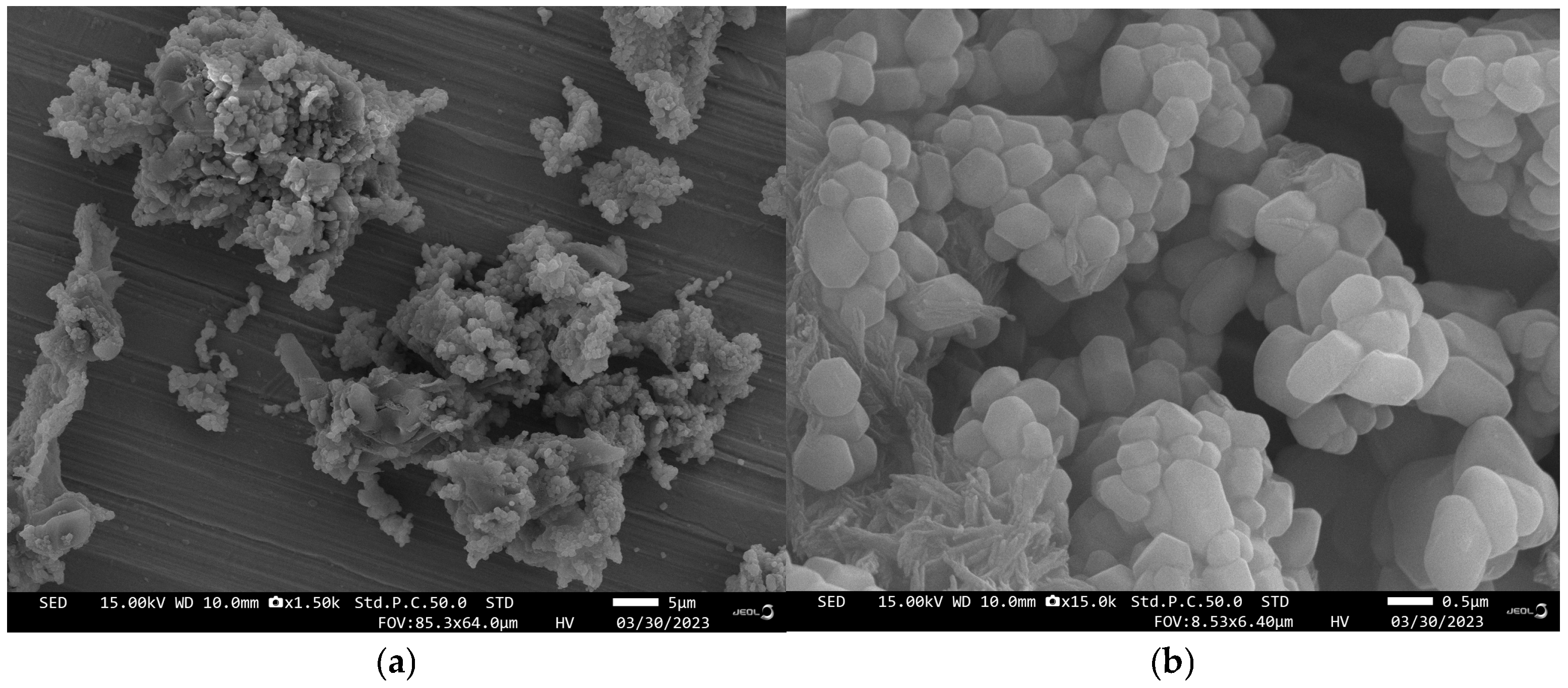
3.1. Characterization of Nano-Nacre Particles

After the mechanical crushing of the nacre, scanning electron microscopy (SEM) analysis confirmed that the nano-nacre particles had experienced a substantial reduction in size to approximately 600 nm (Figure 1), which is considerably smaller than the natural intact aragonite crystals of nacre, which typically exceed 50 µm in diameter. This significant reduction in particle size highlights the efficiency of the mechanical grinding process and suggests an increased surface area, which is crucial for enhancing bioactive compound release.

3.2. Western Blotting

The Western blot analysis revealed distinct protein profiles in all lysate samples derived from nano-nacre particle WSM, foot tissue, and mantle tissue (MT). Strong protein signals were detected at molecular weights equal to or larger than 75 kDa, with lighter bands observed near 25 kDa (Figure 2a). Notably, there was no detection of the pre-protein (~47 kDa) or the secreted (mature) protein (~11 kDa) forms of BMP-2/4 in any lysate sample.
To confirm the specificity of antibody staining, a competition assay was performed using a tenfold higher concentration of HdBMP-2/4 peptide. This competitive binding completely inhibited the staining of the BMP-2/4 antibody (Figure 2b). These results suggest that the HdBMP-2/4 peptide is a constituent of the WSM derived from nano-nacre particles of H. diversicolor. This specificity underscores the potential role of HdBMP-2/4 in the bioactivity of WSM.

3.3. The WSM from Nano-Nacre Particles Is Non-Cytotoxic to MC3T3-E1 Cells

The MTT assay was used to assess the cytotoxicity of WSM on MC3T3-E1 pre-osteoblastic cells, revealing no adverse effects on cell viability across a broad concentration range (0.005% w/v, 0.025% w/v, 0.05% w/v, and 0.1% w/v). At all time points studied (24, 48, and 72 h), cell viability remained above 90%, comparable to the untreated control group (Figure 3). These results demonstrate that WSM does not induce cytotoxicity, highlighting its biocompatibility and potential for osteogenic applications.

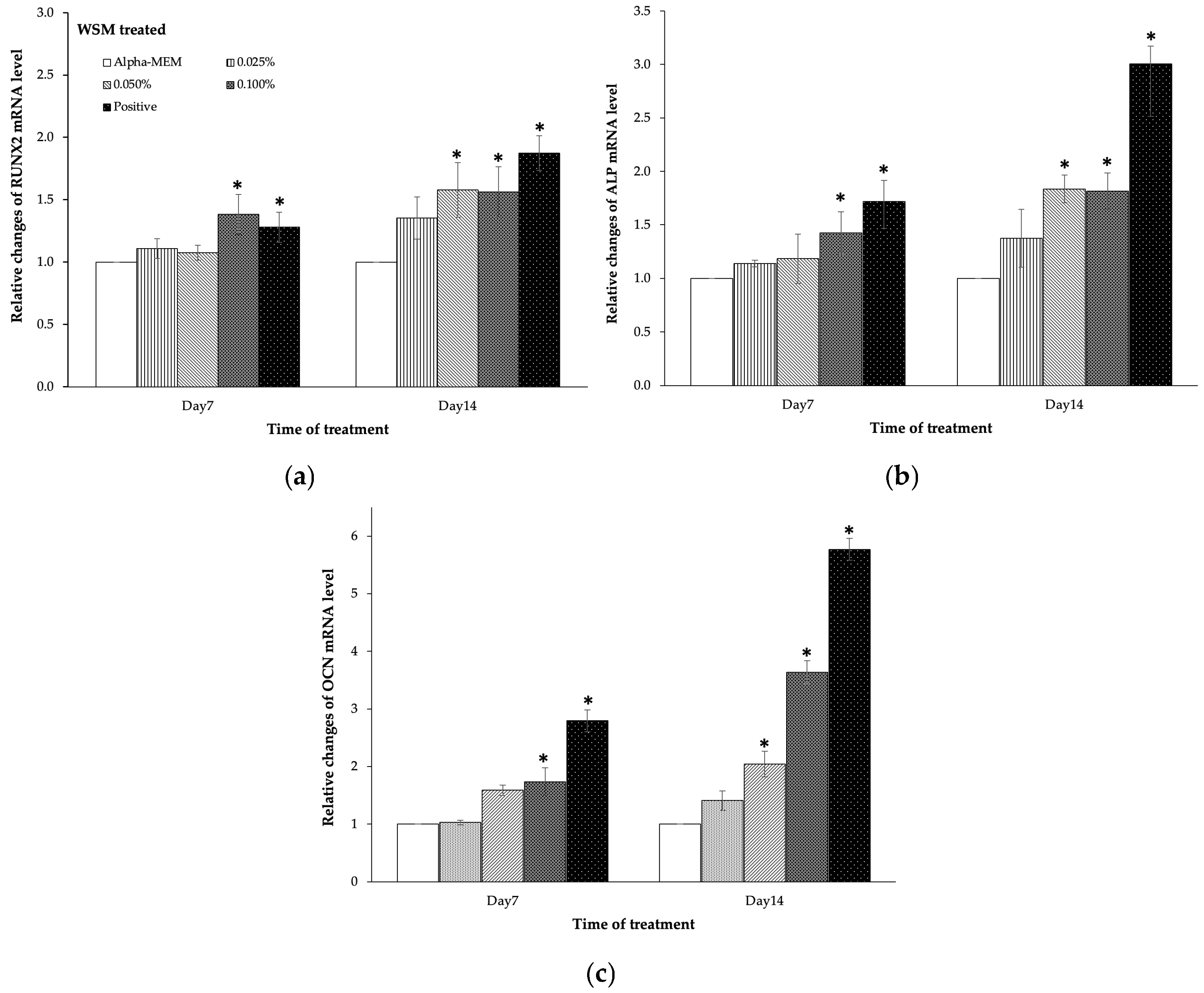
3.4. The WSM from Nano-Nacre Particles Promoted MC3T3-E1 Cells Differentiation

The WSM extracted from nano-nacre particles significantly promoted the differentiation of MC3T3-E1 pre-osteoblastic cells by upregulating both early and late osteogenic markers (Figure 4a–c). Early markers, including RUNX2 and ALP [30], exhibited increased mRNA levels following WSM treatments, with ALP showing a higher fold-change than RUNX2. The highest fold-change for RUNX2 (~1.6) was observed on day 14 at WSM concentrations of 0.05% and 0.1% w/v, both significantly different from the control (p = 0.005, p = 0.0062). For ALP, the effective WSM concentration was as low as 0.025% w/v, yielding a fold-change of ~1.4 by day 14, with the maximum (~1.8) observed in the 0.05% and 0.1% w/v treatments (p < 0.01, p < 0.0121). Late osteogenic marker OCN [31] exhibited a more pronounced response, with its mRNA levels showing a maximum fold-change of ~3.6 on day 14 at 0.1% w/v (p < 0.0001). Across all durations, OCN mRNA fold-changes consistently exceeded 1.5 at 0.05% w/v (p < 0.009), demonstrating WSM’s potent osteoinductive capacity.

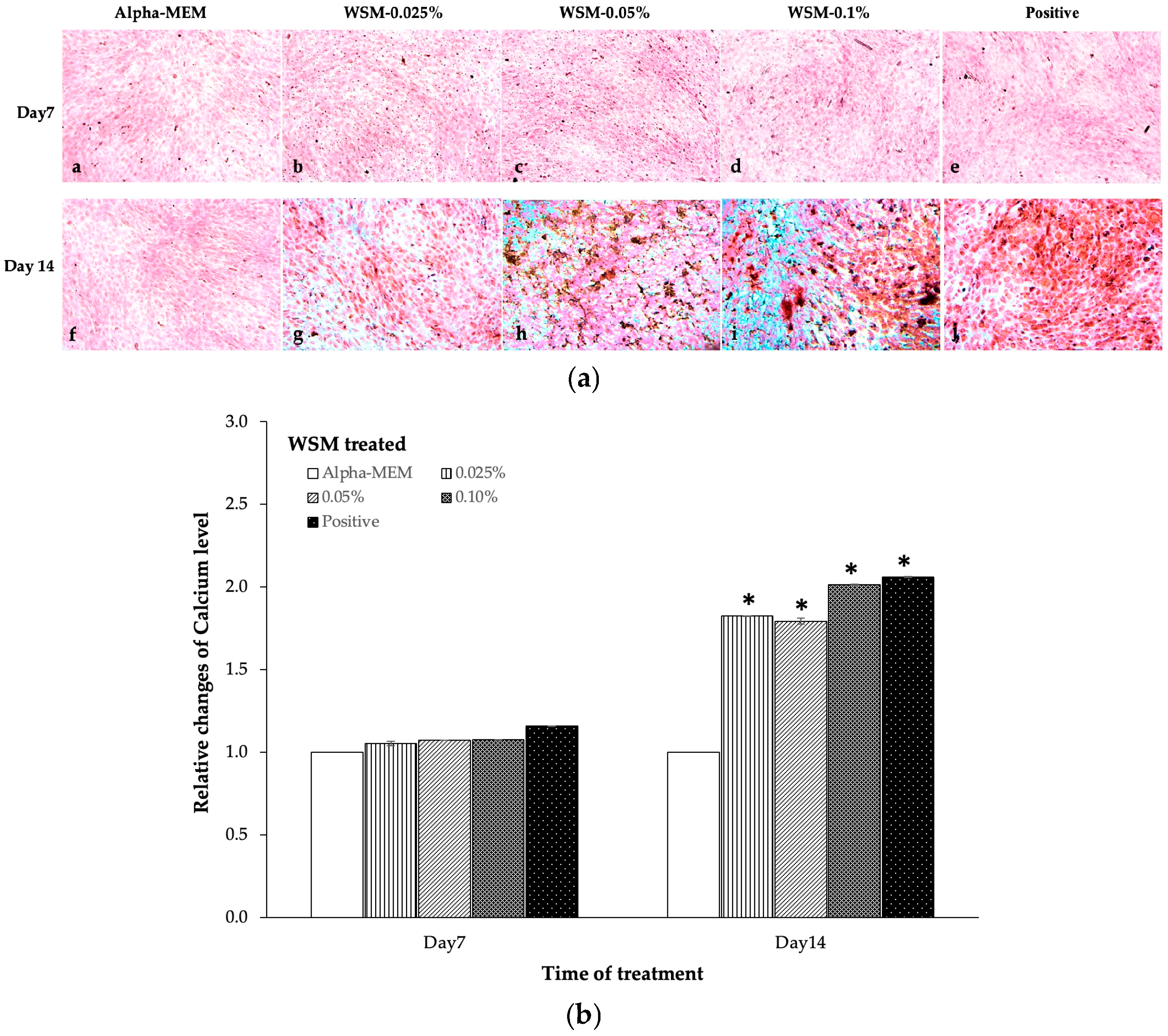
3.5. The WSM from Nano-Nacre Particles Accelerated Mineralization of MC3T3-E1 Cells

The WSM derived from nano-nacre particles significantly accelerated the mineralization of MC3T3-E1 pre-osteoblastic cells in vitro. ARS demonstrated enhanced calcium deposition within the extracellular matrix, with an increasingly intense red coloration observed upon WSM treatment. This effect was both concentration and duration dependent, highlighting the osteogenic potential of WSM (Figure 5a). Quantitative analysis of ARS via CPC solubilization and absorbance measurement (A564) confirmed a significant increase in calcium deposition. Fold-changes ranged from 1.8 to 2.0 when cells were treated with WSM concentrations of 0.025–0.1% w/v for 14 days, compared to the control group (Figure 5b). These findings underscore that WSM not only promotes differentiation of MC3T3-E1 cells but also effectively accelerates matrix mineralization, a critical step in bone formation.

4. Discussion

In this study, we explored the osteoinductive potential of WSM from nacre, particularly focusing on its ability to enhance the differentiation of pre-osteoblasts into mature osteoblasts. Previous studies have demonstrated that nacre and its extracts stimulate bone formation and differentiation of human bone marrow stem cells (hBMSC) without affecting the physical size of the nacre [8,9,10]. The osteoinductive properties of nacre have been attributed to an organic WSM consisting of polysaccharides, water-soluble protein, and glycosaccharides, which can be slowly released from the organic matrix of nacre and subsequently mediate osteoblastic differentiation [13,14]. However, the challenge lies in the limited release of osteoinductive factors from natural nacre due to its dense and compact surface [25]. To address this issue, we synthesized nano-nacre particles with a size of approximately 600 nm, smaller than the intact aragonite crystals of nacre (greater than 50 µm). This reduction in particle size facilitates the enhancement of the release of WSM, resulting in faster pre-osteoblast differentiation. Notably, while osteoblasts typically require 21 days to differentiate and form mineralized nodules in standard mineralizing media [32], our results showed that 0.1% w/v WSM significantly increased RUNX2, ALP mRNA, and OCN mRNA expression by 1.5- and 1.8-fold, respectively, compared to the control within 7 days, while 0.05% w/v WSM achieved the same within 14 days (Figure 4). Additionally, ARS also confirmed significant calcium deposits in MC3T3-E1 treated with 0.1% w/v WSM within 7 days, indicating hydroxyapatite mineralization (Figure 5). Meanwhile, the MC3T3-E1 pre-osteoblasts also entered the mature stage of differentiation earlier. Therefore, WSM may accelerate bone formation, completing the process in just 7 days in a dose-dependent manner. In our study, WSM showed a prominent protein band at approximately 75 and 25 kDa (as shown in Figure 2). This finding is consistent with the results reported by Tanasawet et al., who indicated the presence of a protein band at 25, 52, 60, and 72 kDa in acid-extracted nacreous protein (AEP) from H. diversicolor shell [33]. The protein band at 25 kDa was described as sometsuke, an abalone mantle protein, which has biomineralization and calcium-binding properties [34,35]. According to the composition of nacre, which has similarities to bones in both its organic and inorganic components, it plays a crucial role in mineralization, which is regulated by the mantle tissue in mollusks [16,26,36]. Mineral deposition occurs in specific regions of the mantle, where epithelial cells influence the formation of the nacreous layer [37,38]. For nacreous proteins to be deposited into the nacreous layer, they must exhibit characteristics typical of secretory proteins, such as a signal peptide, glycosylation sites, and cysteine residues forming disulfide bridges for proper conformation. The presence of such features was confirmed in molecular analyses of the HdBMP2/4 transcript from the univalve H. diversicolor and the PfBMP2 transcript from the bivalve P. fucata [18,24]. As shown in Figure 2, we performed WB by using a polyclonal antibody against BMP-2/4, but there was no detection of the pre-protein (~47 kDa) or the secreted (mature) protein (~11 kDa) forms of BMP-2/4 in the WSM. However, we analyzed its competition with the HdBMP-2/4 peptide and the result showed that the protein band at 25 kDa was absent. One explanation for the lack of pre-BMP-2/4 (~47 kDa) protein may be that it is in a dimeric or multimeric form, bound together by disulfide bridges or by electrostatic interaction, as also reported for other nacreous proteins [39,40], which are subsequently disrupted by reducing or high salt conditions. Therefore, our competition assay further supported that HdBMP2/4 peptides are some of the integral components of WSM. As shown in Figure 3, the osteoinductive mechanism of WSM appears to involve the canonical BMP signaling pathway. Specifically, WSM likely promotes pre-osteoblast differentiation by engaging the dimerized HdBMP2/4 with BMP receptors on the cell surface. This interaction triggers phosphorylation of Smad1/5/8, which then forms a complex with Smad4. The complex translocates to the nucleus to regulate the expression of osteoblastic transcription factors, including RUNX2, which subsequently stimulates osteoblast-related genes such as ALP, collagen type I (Col IA), and OCN [41,42]. This cascade of events is essential for the induction of osteoblastic differentiation and bone matrix deposition. Our findings corroborate this mechanism by showing upregulated expression of RUNX2, ALP, and OCN in response to WSM treatment, which also led to calcium deposition in the extracellular matrix, as confirmed by ARS (Figure 5). While the canonical pathway is the primary mechanism identified, the potential involvement of non-canonical pathways, such as a RUNX2-related mechanism [43], cannot be excluded, and further research is needed to fully elucidate the diverse signaling pathways activated by WSM in osteoinduction.

5. Conclusions

The function of WSM from nano-nacre particles of H. diversicolor in osteoinduction is signified by the triggering of pre-osteoblastic MC3T3-E1 cell differentiation. As mollusk nacreous materials that have high homology with bone proteins (including HdBMP2/4, reported herein) continuously exert their potency in inducing osteogenesis in higher mammals, the potential clinical applications of these marine biomaterials (particularly those that are non-allergic to mammalian systems) should thus be successfully achieved in the future.

Author Contributions

Conceptualization, C.S. (Chanyatip Suwannasing); methodology, C.S. (Chanyatip Suwannasing) and A.P.; validation, C.S. (Chanyatip Suwannasing), P.S. and C.S. (Chanyarak Sombutkayasith); formal analysis, C.S. (Chanyatip Suwannasing) and A.P.; investigation, C.S. (Chanyatip Suwannasing); resources, C.S. (Chanyarak Sombutkayasith), P.S. and W.W.; data curation, C.S. (Chanyatip Suwannasing) and W.W.; writing—original draft preparation, C.S. (Chanyatip Suwannasing); writing—review and editing, C.S. (Chanyatip Suwannasing); visualization, C.S. (Chanyatip Suwannasing); supervision, W.W.; project administration, C.S. (Chanyatip Suwannasing); funding acquisition, C.S. (Chanyatip Suwannasing) All authors have read and agreed to the published version of the manuscript.

Funding

This research was funded by Naresuan University, Thailand Science Research and Innovation (TSRI), and the National Science, Research and Innovation Fund (NSRF). (Fundamental Fund: grant no. R2565B093).

Institutional Review Board Statement

Not applicable.

Informed Consent Statement

Not applicable.

Data Availability Statement

Data is contained within the article.

Acknowledgments

The authors would like to thank Phuket Abalone Farm, Phuket province, Thailand, for providing the abalone shells and the facilities at the Centre Laboratory, Faculty of Allied Health Science, Naresuan University, and the Faculty of Science, Mahidol University, for providing instrumental support throughout this work.

Conflicts of Interest

The authors declare no conflicts of interest.

References

  1. Kanis, J.A.; McCloskey, E.V.; Johansson, H.; Cooper, C.; Rizzoli, R.; Reginster, J.-Y.; on behalf of the Scientific Advisory Board of the European Society for Clinical and Economic Aspects of Osteoporosis and Osteoarthritis (ESCEO) and the Committee of Scientific Advisors of the International Osteoporosis Foundation (IOF). European guidance for the diagnosis and management of osteoporosis in postmenopausal women. Osteoporos. Int. 2013, 24, 23–57. [Google Scholar] [CrossRef] [PubMed]
  2. Popp, A.W.; Varathan, N.; Buffat, H.; Senn, C.; Perrelet, R.; Lippuner, K. Bone Mineral Density Changes After 1 Year of Denosumab Discontinuation in Postmenopausal Women with Long-Term Denosumab Treatment for Osteoporosis. Calcif. Tissue Int. 2018, 103, 50–54. [Google Scholar] [CrossRef] [PubMed]
  3. Guise, T.A.; Yin, J.J.; Mohammad, K.S. Role of Endothelin-1 in Osteoblastic Bone Metastases. Cancer 2003, 97, 779–784. [Google Scholar] [CrossRef]
  4. McClung, M.R. Romosozumab for the treatment of osteoporosis. Osteoporos. Sarcopenia 2018, 4, 11–15. [Google Scholar] [CrossRef]
  5. Farkas, S.; Szabó, A.; Hegyi, A.E.; Török, B.; Fazekas, C.L.; Ernszt, D.; Kovács, T.; Zelena, D. Estradiol and Estrogen-like Alternative Therapies in Use: The Importance of the Selective and Non-Classical Actions. Biomedicines 2022, 10, 861. [Google Scholar] [CrossRef]
  6. Vecchio, K.S.; Zhang, X.; Massie, J.B.; Wang, M.; Kim, C.W. Conversion of bulk seashells to biocompatible hydroxyapatite for bone implants. Acta Biomater. 2007, 3, 910–918. [Google Scholar] [CrossRef]
  7. Rey, C.; Combes, C.; Drouet, C.; Glimcher, M.J. Bone mineral: Update on chemical composition and structure. Osteoporos. Int. 2009, 20, 1013–1021. [Google Scholar] [CrossRef]
  8. Camprasse, S.; Camprasse, G.; Pouzol, M.; Lopez, E. Artificial dental root made of natural calcium carbonate (Bioracine). Clin. Mater. 1990, 5, 235–250. [Google Scholar] [CrossRef]
  9. Camprasse, G.; Camprasse, S.; Gill, G.A. Substitution of the dental root by aquatic invertebrate skeletons in animals and man. C. R. Acad.Sci. 1988, 307, 485–491. [Google Scholar]
  10. Berland, S.; Delattre, O.; Borzeix, S.; Catonne, Y.; Lopez, E. Nacre/bone interface changes in durable nacre endosseous implants in sheep. Biomaterials 2005, 26, 2767–2773. [Google Scholar] [CrossRef]
  11. Zhang, G.; Brion, A.; Willemin, A.-S.; Piet, M.-H.; Moby, V.; Bianchi, A.; Mainard, D.; Galois, L.; Gillet, P.; Rousseau, M. Nacre, a natural, multi-use, and timely biomaterial for bone graft substitution. J. Biomed. Mater. Res. Part A 2017, 105, 662–672. [Google Scholar] [CrossRef] [PubMed]
  12. Iandolo, D.; Laroche, N.; Nguyen, D.K.; Normand, M.; Met, C.; Zhang, G.; Vico, L.; Mainard, D.; Rousseau, M. Preclinical safety study of nacre powder in an intraosseous sheep model. BMJ Open Sci. 2022, 6, e100231. [Google Scholar] [CrossRef] [PubMed]
  13. Green, D.W.; Kwon, H.-J.; Jung, H.-S. Osteogenic Potency of Nacre on Human Mesenchymal Stem Cells. Mol. Cells 2015, 38, 267–272. [Google Scholar] [CrossRef]
  14. Xu, J.; Rao, Y.; Wu, X.; Jiang, J.; Yu, M.; Chen, X.; Wang, H. The osteoinductive effect of nano-nacre particles on MC-3T3 E1 preosteoblast through controlled release of water-soluble matrix and calciumions. Dent. Mater. J. 2019, 38, 981–986. [Google Scholar] [CrossRef]
  15. Miyamoto, H.; Miyashita, T.; Okushima, M.; Nakano, S.; Morita, T.; Matsushiro, A. A carbonic anhydrase from the nacreous layer in oyster pearls. Proc. Natl. Acad. Sci. USA 1996, 93, 9657–9660. [Google Scholar] [CrossRef] [PubMed]
  16. Sudo, S.; Fujikawa, T.; Nagakura, T.; Ohkubo, T.; Sakaguchi, K.; Tanaka, M.; Nakashima, K.; Takahashi, T. Structures of mollusc shell framework proteins. Nature 1997, 387, 563–564. [Google Scholar] [CrossRef]
  17. Samata, T.; Hayashi, N.; Kono, M.; Hasegawa, K.; Horita, C.; Akera, S. A new matrix protein family related to the nacreous layer formation of Pinctada fucata. FEBS Lett. 1999, 462, 225–229. [Google Scholar] [CrossRef]
  18. Miyashita, T.; Takagi, R.; Okushima, M.; Nakano, S.; Miyamoto, H.; Nishikawa, E.; Matsushiro, A. Complementary DNA Cloning and Characterization of Pearlin, a New Class of Matrix Protein in the Nacreous Layer of Oyster Pearls. Mar. Biotechnol. 2000, 2, 409–418. [Google Scholar] [CrossRef]
  19. Weiss, I.; Kaufmann, S.; Mann, K.; Fritz, M. Purification and Characterization of Perlucin and Perlustrin, Two New Proteins from the Shell of the Mollusc Haliotis laevigata. BBRC Biochem. Biophys. Res. Commun. 2000, 267, 17–21. [Google Scholar] [CrossRef]
  20. Zhang, C.; Li, S.; Ma, Z.; Xie, L.; Zhang, R. A Novel Matrix Protein p10 from the Nacre of Pearl Oyster (Pinctada fucata) and Its Effects on Both CaCO3 Crystal Formation and Mineralogenic Cells. Mar. Biotechnol. 2006, 8, 624–633. [Google Scholar] [CrossRef]
  21. Yano, M.; Nagai, K.; Morimoto, K.; Miyamoto, H. A novel nacre protein N19 in the pearl oyster Pinctada fucata. BBRC Biochem. Biophys. Res. Commun. 2007, 362, 158–163. [Google Scholar] [CrossRef] [PubMed]
  22. Ma, J.Y.; Wong, K.L.; Xu, Z.Y.; Au, K.Y.; Lee, N.L.; Su, C.; Su, W.-W.; Li, P.-B.; Shaw, P.-C. N16, a Nacreous Protein, Inhibits Osteoclast Differentiation and Enhances Osteogenesis. J. Nat. Prod. 2016, 79, 204–212. [Google Scholar] [CrossRef]
  23. Mann, K.; Cerveau, N.; Gummich, M.; Fritz, M.; Mann, M.; Jackson, D.J. In-depth proteomic analyses of Haliotis laevigata (greenlip abalone) nacre and prismatic organic shell matrix. Proteome Sci. 2018, 16, 11. [Google Scholar] [CrossRef]
  24. Suwannasing, C.; Buddawong, A.; Khumpune, S.; Habuddha, V.; Weerachatyanukul, W.; Asuvapongpatana, S. Bone Morphogenetic Protein 2/4 in Mollusk, Haliotis diversicolor: Its Expression and Osteoinductive Function In Vitro. Mar. Biotechnol. 2021, 23, 836–846. [Google Scholar] [CrossRef]
  25. Pereira-Mouriès, L.; Almeida, M.J.; Ribeiro, C.; Peduzzi, J.; Barthélemy, M.; Milet, C.; Lopez, E. Soluble silk-like organic matrix in the nacreous layer of the bivalve Pinctada maxima. Eur. J. Biochem. 2002, 269, 4994–5003. [Google Scholar] [CrossRef]
  26. Shi, Y.; Zheng, X.; Zhan, X.; Wang, A.; Gu, Z. cDNA microarray analysis revealing candidate biomineralization genes of the pearl oyster, Pinctada fucata martensii. Mar. Biotechnol. 2016, 18, 336–348. [Google Scholar] [CrossRef]
  27. Chaturvedi, R.; Singha, P.K.; Dey, S. Water Soluble Bioactives of Nacre Mediate Antioxidant Activity and Osteoblast Differentiation. PLoS ONE 2013, 8, e84584. [Google Scholar] [CrossRef]
  28. Cheng, Y.; Zhang, W.; Fan, H.; Xu, P. Water-soluble nano-pearl powder promotes MC3T3-E1 cell differentiation by enhancing autophagy via the MEK/ERK signaling pathway. Mol. Med. Rep. 2018, 18, 993–1000. [Google Scholar] [CrossRef]
  29. Wang, X.; Harimoto, K.; Fuji, R.; Liu, J.; Li, L.; Wang, P.; Akaike, T.; Wang, Z. Pinctada fucata mantle gene 4 (PFMG4) from pearl oyster mantle enhances osteoblast differentiation. Biosci. Biotechnol. Biochem. 2015, 79, 558–565. [Google Scholar] [CrossRef]
  30. Peverali, F.A.; Basdra, E.K.; Papavassiliou, A.G. Stretch-mediated activation of selective MAPK subtypes and potentiation of AP-1 binding in human osteoblastic cells. J. Mol. Med. 2001, 7, 68–78. [Google Scholar] [CrossRef]
  31. Lian, J.B.; Stein, G.S.; Stein, J.L.; van Wijnen, A.J. Osteocalcin gene promoter: Unlocking the secrets for the regulation of osteoblast growth and differentiation. J. Cell. Biochem. 1998, 72, 62–72. [Google Scholar] [CrossRef]
  32. Rousseau, M.; Pereira-Mouriès, L.; Almeida, M.J.; Milet, C.; Lopez, E. The water-soluble matrix fraction from the nacre of Pinctada maxima produces earlier mineralization of MC3T3-E1 mouse pre-osteoblasts. Comp. Biochem. Physiol. B Biochem. Mol. Biol. 2003, 135, 1–7. [Google Scholar] [CrossRef]
  33. Tanasawet, S.; Withyachumnarnkul, B.; Changsangfar, C.; Cummins, S.F.; Sroyraya, M.; Sangsuwan, P.; Kitiyanant, Y.; Asuvapongpatana, S.; Weerachatyanukul, W. Isolation of Organic Matrix Nacreous Proteins from Haliotis diversicolor and Their Effect On In Vitro Osteoinduction. Malacologia 2013, 56, 107–119. [Google Scholar] [CrossRef]
  34. Jackson, D.J.; McDougall, C.; Green, K.; Simpson, F.; Wörheide, G.; Degnan, B.M. A rapidly evolving secretome builds and patterns a sea shell. BMC Biol. 2006, 4, 40. [Google Scholar] [CrossRef]
  35. Marie, B.; Marie, A.; Jackson, D.J.; Dubost, L.; Degnan, B.M.; Milet, C.; Marin, F. Proteomic analysis of the organic matrix of the abalone Haliotis asinina calcified shell. Proteome Sci. 2010, 8, 54. [Google Scholar] [CrossRef]
  36. Zhang, Y.; Xie, L.; Meng, Q.; Jing, T.; Pu, R.; Chen, L.; Zhang, R. A novel matrix protein participating in the nacre framework formation of pearl oyster, Pinctada fucata. Comp. Biochem. Physiol. Part B Biochem. Mol. Biol. 2003, 135, 565–573. [Google Scholar] [CrossRef]
  37. Checa, A. A new model for periostracum and shell formation in Unionidae (Bivalvia, Mollusca). Tissue Cell 2000, 32, 405–416. [Google Scholar] [CrossRef]
  38. Marin, F.; Roy, N.L.; Marie, B. The formation and mineralization of the mollusk shell. Front. Biosci. Schol. Ed. 2012, 4, 1099–1125. [Google Scholar] [CrossRef]
  39. Marin, F.; Amons, R.; Guichard, N.; Stigter, M.; Hecker, A.; Luquet, G.; Layrolle, P.; Alcaraz, G.; Riondet, C.; Westbroek, P. Caspartin and calprismin, two proteins of the shell calcitic prisms of the Mediterranean fan mussel Pinna nobilis. J. Biol. Chem. 2005, 280, 33895–33908. [Google Scholar] [CrossRef]
  40. Kong, Y.; Jing, G.; Yan, Z.; Li, C.; Gong, N.; Zhu, F.; Li, D.; Zhang, Y.; Zheng, G.; Wang, H.; et al. Cloning and Characterization of Prisilkin-39, a Novel Matrix Protein Serving a Dual Role in the Prismatic Layer Formation from the Oyster Pinctada fucata. J. Biol. Chem. 2009, 284, 10841–10854. [Google Scholar] [CrossRef]
  41. Hayashi, M.; Maeda, S.; Aburatani, H.; Kitamura, K.; Miyoshi, H.; Miyazono, K.; Imamura, T. Pitx2 prevents osteoblastic transdifferentiation of myoblasts by bone morphogenetic proteins. J. Biol. Chem. 2008, 283, 565–571. [Google Scholar] [CrossRef] [PubMed]
  42. Chen, G.; Deng, C.; Li, Y.P. TGF-β and BMP signaling in osteoblast differentiation and bone formation. Int. J. Biol. Sci. 2012, 8, 272–288. [Google Scholar] [CrossRef]
  43. Ge, C.; Xiao, G.; Jiang, D.; Yang, Q.; Hatch, N.E.; Roca, H.; Franceschi, R.T. Identification and functional characterization of ERK/MAPK phosphorylation sites in the Runx2 transcription factor. J. Biol. Chem. 2009, 284, 32533–32543. [Google Scholar] [CrossRef]
Figure 1. Nano-nacre particles observed by scanning electron microscopy. Scale bars represent 5 μm (a) and 0.5 μm (b).
Applsci 15 02907 g001
Figure 2. Protein expression of BMP-2/4 in the WSM from nano-nacre particles of H. diversicolor. Western blotting of protein lysates from three tissues: WSM from nano-nacre particles (WSM) (lane 1), foot tissue (lane 2), and mantle tissue (MT) (lane 3) using a polyclonal antibody against BMP-2/4 (a) and its competition with HdBMP-2/4 peptide (b). Note the reactive band of BMP-2/4 antibody at 47 kDa.
Applsci 15 02907 g002
Figure 3. Effect of WSM from nano-nacre particles of H. diversicolor on cell viability percentage as measured by MTT assay, indicating the non-toxic nature of WSM for MC3T3-E1 cells. Each value is the mean ± SD of three independent experiments when compared with the DMEM group. * p < 0.05 indicates significant differences compared with the control group.
Applsci 15 02907 g003
Figure 4. Effect of WSM from nano-nacre particles of H. diversicolor on osteogenic gene expressions (ac) using the MC3T3-E1 pre-osteoblastic cell line as a model. The cells were incubated with WSM for 2 consecutive days and collected for mRNA analysis at days 7 and 14 post-treatment. Osteogenic markers assessed include RUNX2 (a), ALP (b), and OCN (c). Each value is the mean ± SE of three independent experiments when compared with the DMEM group. * p < 0.05 indicates significant differences compared with the control group.
Applsci 15 02907 g004
Figure 5. Enhanced biomineralization of MC3T3-E1 pre-osteoblasts with WSM from nano-nacre particles of H. diversicolor. The cells were treated with various concentrations (0–0.1% w/v WSM) and positive control at day 7 (ae) and day 14 (fj), and calcium nodule formation was detected by Alizarin Red Staining (a) and further quantified by PCP solution and spectrophotometric absorbance at 564 nm (A564) (b). Each value is the mean ± SD of three independent experiments when compared with the DMEM group. * p < 0.05 indicates significant differences compared with the control group.
Applsci 15 02907 g005
Disclaimer/Publisher’s Note: The statements, opinions and data contained in all publications are solely those of the individual author(s) and contributor(s) and not of MDPI and/or the editor(s). MDPI and/or the editor(s) disclaim responsibility for any injury to people or property resulting from any ideas, methods, instructions or products referred to in the content.

