Article

Evaluation of Condylar and Airway Volume in Skeletal Class I Patients with Different Vertical Growth Patterns

1
Department of Orthodontics, Faculty of Dentistry, İnönü University, Malatya 44280, Türkiye
2
Department of Oral and Maxillofacial Radiology, Faculty of Dentistry, İnönü University, Malatya 44280, Türkiye
*
Author to whom correspondence should be addressed.
Appl. Sci. 2025, 15(5), 2794; https://doi.org/10.3390/app15052794
Submission received: 3 February 2025 / Revised: 27 February 2025 / Accepted: 3 March 2025 / Published: 5 March 2025

Abstract

:
Objective: This study aimed to investigate the correlation between condylar volume and airway dimensions in skeletal Class I malocclusion patients with different vertical growth patterns. Cone-beam computed tomography (CBCT) files were analyzed using AI-performed segmentation to ensure accurate measurements. Materials and Methods: A total of 93 individuals with skeletal Class I malocclusion (55 females and 38 males; average age 21.3 ± 3.0 years) were classified into three groups (normodivergent, hyperdivergent, and hypodivergent) according to their vertical growth patterns. Upper airway and condylar volumes were calculated following AI-assisted segmentation, and their correlation was evaluated. Results: In the hyperdivergent group, both airway volume (11.2 ± 5.0 cm3) and condylar volume (1.2 ± 0.2 cm3) were significantly lower compared to the normodivergent (airway: 14.4 ± 4.9 cm3; condyle: 1.5 ± 0.3 cm3) and hypodivergent groups (airway: 14.1 ± 6.3 cm3; condyle: 1.5 ± 0.3 cm3) (p < 0.05). Although no statistically significant correlation was detected between airway volume and right condylar volume across the three groups (normodivergent: r = −0.204, p = 0.280; hypodivergent: r = 0.015, p = 0.936; hyperdivergent: r = −0.007, p = 0.971), a strong positive correlation was identified between the right and left condylar volumes in all groups (r > 0.8, p < 0.01). Conclusions: No significant statistical correlation was detected between condylar volume and airway volume across the evaluated groups. However, hyperdivergent individuals were found to have smaller condylar volumes and narrower airway volumes, which may contribute to increased airway resistance and a higher risk of respiratory dysfunctions. These findings highlight the importance of considering vertical growth patterns in orthodontic and orthopedic treatment planning, especially when evaluating airway dimensions. Additionally, a strong and statistically notable positive correlation was detected between the right and left condylar volumes across all groups.

1. Introduction

The connection between craniofacial anatomy and the upper respiratory tract continues to be a relevant subject in dentofacial anomaly research. The Functional Matrix Theory, proposed by Melvin L. Moss in the 1960s, has been recognized as one of the fundamental mechanisms of craniofacial growth. According to this theory, skeletal structures are passive elements, and their growth and development are directed by the surrounding functional matrices. Unlike traditional perspectives, Moss suggested that the growth of the craniofacial skeleton is influenced not primarily by genetic factors but rather by the demands of functional matrices, such as respiration, mastication, swallowing, and speech [1]. It has been concluded that obstructions in the airway or dysfunctions within the functional matrix, such as issues related to mastication and swallowing, may significantly impact craniofacial growth [1]. Studies have also reported that the volume and morphology of soft tissues may change due to craniofacial skeletal anomalies [2,3].
Vertical malocclusions are linked to the pharyngeal airway, with narrowed dimensions observed in hyperdivergent individuals [4,5,6]. This condition has been explained as the narrowing of the pharyngeal airway due to the downward and backward rotation of the mandible and the posterior displacement of the tongue [7]. These malocclusions may arise during adolescence due to factors like maxillary/mandibular development, tooth eruption, and tongue function [8]. Schudy [9] and Isaacson et al. [10], noted that when condylar growth lags behind maxillary sutures, the mandible rotates backward, causing an open bite. This results in a high-angle or hyperdivergent growth pattern. Conversely, forward mandibular growth results in a hypodivergent pattern. This suggests that condylar volume may influence mandibular rotation and growth direction, thereby altering airway volume. Posteriorly positioned or small condyles may cause the mandible to rotate backward and downward. This condition, as seen in hyperdivergent individuals, may lead to a reduction in airway volume [11].
Research suggests that airway volumes vary with vertical and sagittal growth patterns. Paul et al. [12] found that horizontal growth patterns correlate with larger airway volumes. Saccucci et al. [13] reported that the condylar volumes of skeletal Class III patients are greater compared to Class II and Class I patients. Other studies have also shown that different skeletal and vertical patterns are associated with variations in the shape, size, and positions of the condyles [14,15,16]. Xu et al. [11], through CBCT analysis, detected notable variations in the pharyngeal airway space among adult skeletal Class II patients based on different condylar positions, including anterior, posterior and central placements.
Accurately identifying and segmenting teeth from CBCT images plays a significant role in assisting clinicians with diagnosis and treatment planning. However, achieving precise segmentation is a challenging process due to various anatomical and technical difficulties. The close proximity and occasional overlapping of anatomical structures to be segmented further complicate the segmentation process [17]. Additionally, the similar densities of these structures can make it difficult to distinguish different tissues in radiographic images [18]. Among the technical challenges, artifacts and low-contrast regions in CBCT images are significant factors that can affect segmentation accuracy. In particular, artifacts caused by metal restorations and orthodontic appliances can degrade image quality, making the segmentation process even more complex [17]. Current tooth segmentation approaches encompass techniques such as edge-based, region-based, and threshold-based segmentation methods [16]. These methods often face difficulties in accurately determining segmentation results [19,20]. With the advancement of artificial intelligence, machine learning and deep learning methods are increasingly becoming popular for segmenting medical images. Unlike traditional manual segmentation approaches, which often require the creation of complex rules, data-driven artificial intelligence models demonstrate superior accuracy and generalization capabilities [21,22]. These models reduce user dependency, providing faster, more repeatable, and consistent results. It has been reported that AI-based segmentation can be performed successfully in pediatric patients in the period of deciduous and mixed dentition as well as individuals in the permanent dentition [23]. AI-assisted CBCT file (DICOM) segmentation reduces user dependency, enabling faster, more repeatable, and consistent results. These systems can process large datasets in a short time, offering a significant advantage in analyzing anatomical structures such as airway volume and condylar morphology with greater precision than manual methods. Additionally, they minimize potential human errors, thereby enhancing measurement accuracy. Studies have demonstrated that AI-assisted segmentation methods achieve successful results in evaluating airway volume in CBCT images and provide more objective analyses compared to traditional techniques [18,22]. AI-performed CBCT DICOM segmentations enable the analysis of relationships between craniofacial structures, the measurement of these structures, and the creation of patient-specific appliances for treatment processes. Therefore, using an AI-assisted segmentation method provides significant advantages for our study. While previous studies [7,12,24] have examined airway dimensions and vertical growth patterns, few have investigated their impact on condylar volume. Traditional segmentation methods introduce operator-dependent variability, which can compromise measurement accuracy. To address these limitations, AI-assisted segmentation ensures precise and reproducible volume assessments. The key novelty of this study lies in utilizing AI-assisted segmentation to evaluate relationships between craniofacial structure volumes, reducing human-related segmentation errors for a more objective analysis.
H0: 
There is no statistically significant correlation between airway volume and condylar volume segmented using AI-assisted methods in skeletal Class I malocclusion patients with different vertical growth patterns.

2. Materials and Methods

The Ethics Committee of İnönü University approved this retrospective study prior to its implementation. All patients provided consent forms permitting the use of their data for scientific research purposes. The study used the CBCT images of 93 individuals who met the inclusion criteria from among the records of 2750 patients who had applied to İnönü University.
Inclusion criteria were defined as follows:
  • Age range: Individuals between 18 and 30 years old,
  • Skeletal Class I structure,
  • No history of orthodontic or orthognathic surgical treatment,
  • CBCT images of sufficient quality and resolution,
  • No respiratory tract pathology or craniofacial syndromes.
Exclusion criteria included low-quality or incomplete CBCT images, a history of previous orthodontic treatment, or the presence of relevant craniofacial anomalies.
Skeletal class I patients were included according to ANB and Wits angles and divided into three groups based on their vertical growth patterns. The GoGn-SN angle was used to determine these groups, classifying individuals with angles of 26–38° as normodivergent, angles >38° as hyperdivergent, and angles <26° as hypodivergent (Table 1) [7].
CBCT imaging was conducted with all patients in a standard supine posture using the same device, with scanning parameters including a scanning time of 14–18 s, a collimation height of 13 cm, an exposure time of 3.6 s, and a voxel size of 0.25 mm3. During the scan, patients were instructed to bite in maximum intercuspation, refrain from swallowing, and remain motionless.
The acquired 3D images were converted to Digital Imaging and Communications in Medicine (DICOM). The DICOM images were loaded into NemoStudio (NemoFAB, v2020, NemoTec, Madrid, Spain). Orientations were adjusted according to the Frankfurt horizontal plane, and the orientation was saved using the “apply changes” feature (Figure 1).

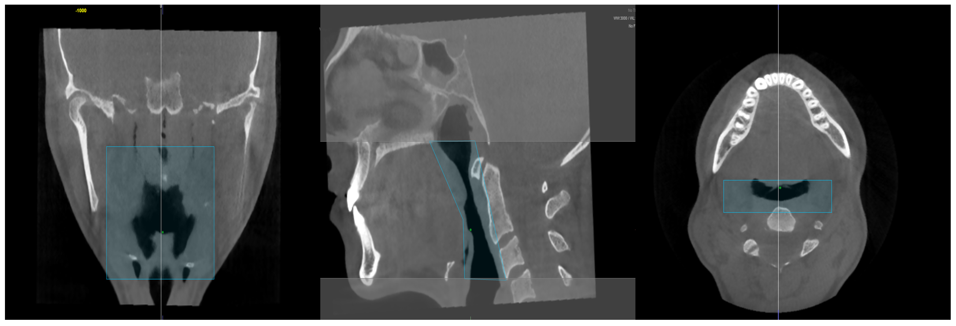
2.1. Airway Volume Calculation

To calculate airway volume, the “define region” feature in the “Airway” section was applied on the midsagittal slice to delineate the boundaries of the airway. The upper boundary was defined as the line extending from the posterior nasal spine (PNS) to the pterygomaxillary junction, while the lower boundary was the parallel line at the level of the third cervical vertebra [24]. The posterior boundary was determined by the pharyngeal wall, and the anterior boundary was defined between the base of the tongue and the soft palate. These airway volume boundaries were selected as they allow for a comprehensive assessment of pharyngeal airway dimensions. We believe that this measurement is crucial for evaluating respiratory efficiency and potential airway obstructions.
Once the boundaries were determined, points were placed on the radiolucent area forming the airway using the “place points” feature. The airway boundaries were then reviewed in all reconstructed planes (coronal, axial, and sagittal) to confirm that the designated points were included within the airway limits. After these checks, the “volume detection” feature was utilized to calculate the total airway volume (cm3) (Figure 2).
While calculating the volume of the created three-dimensional airway model, the minimum axial airway area (mm2) in the pharyngeal region was recorded (Figure 3). After these measurements, the airway volume calculation was completed.

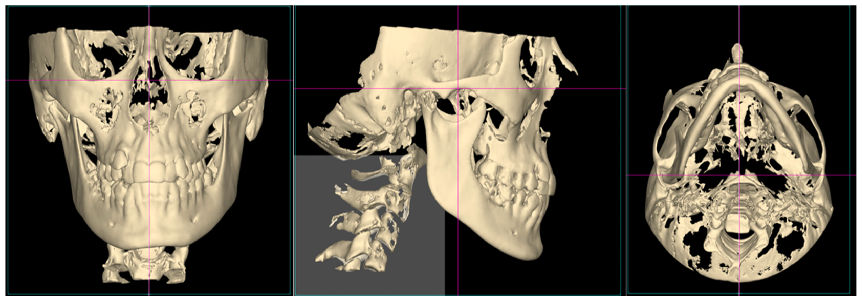
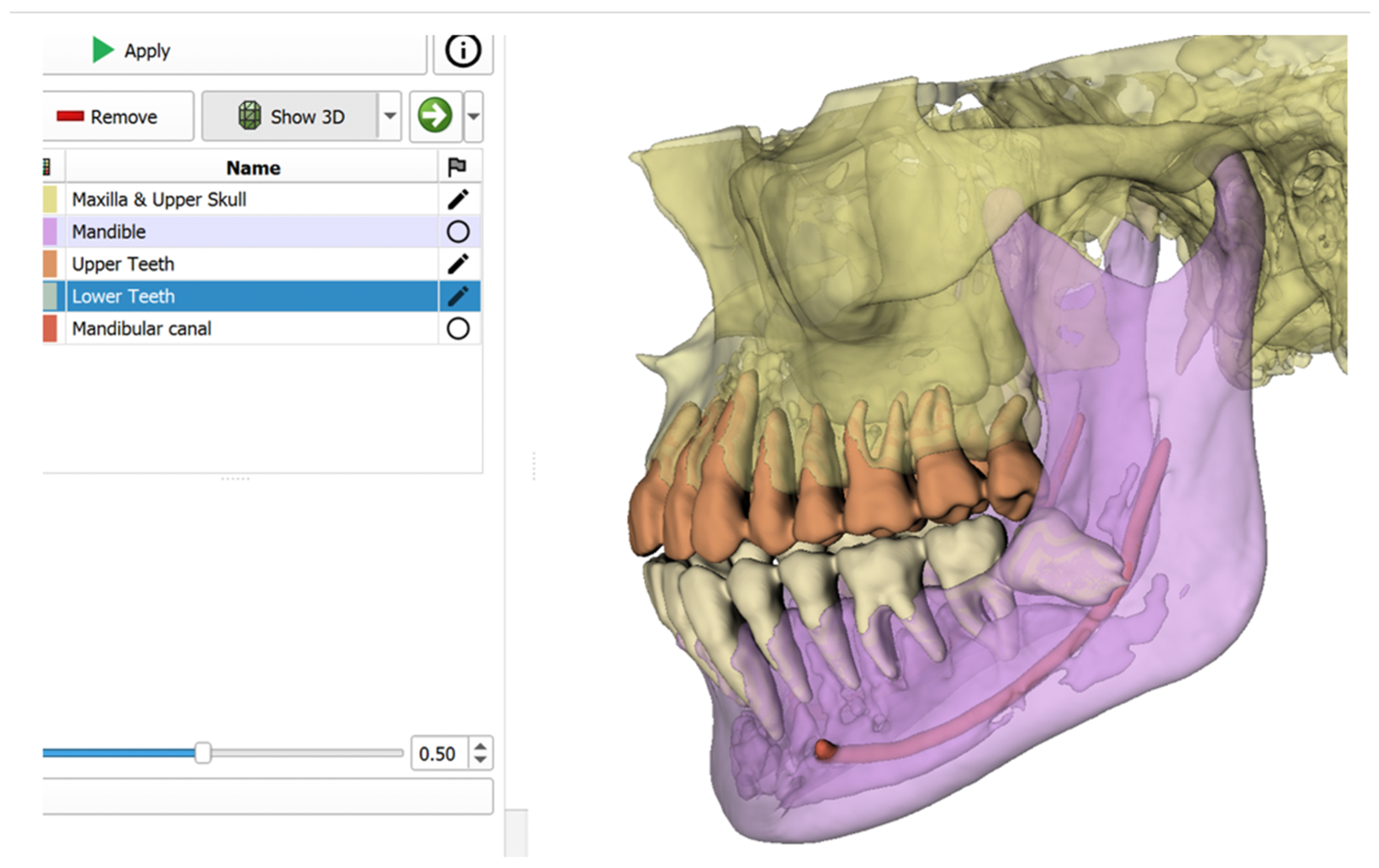
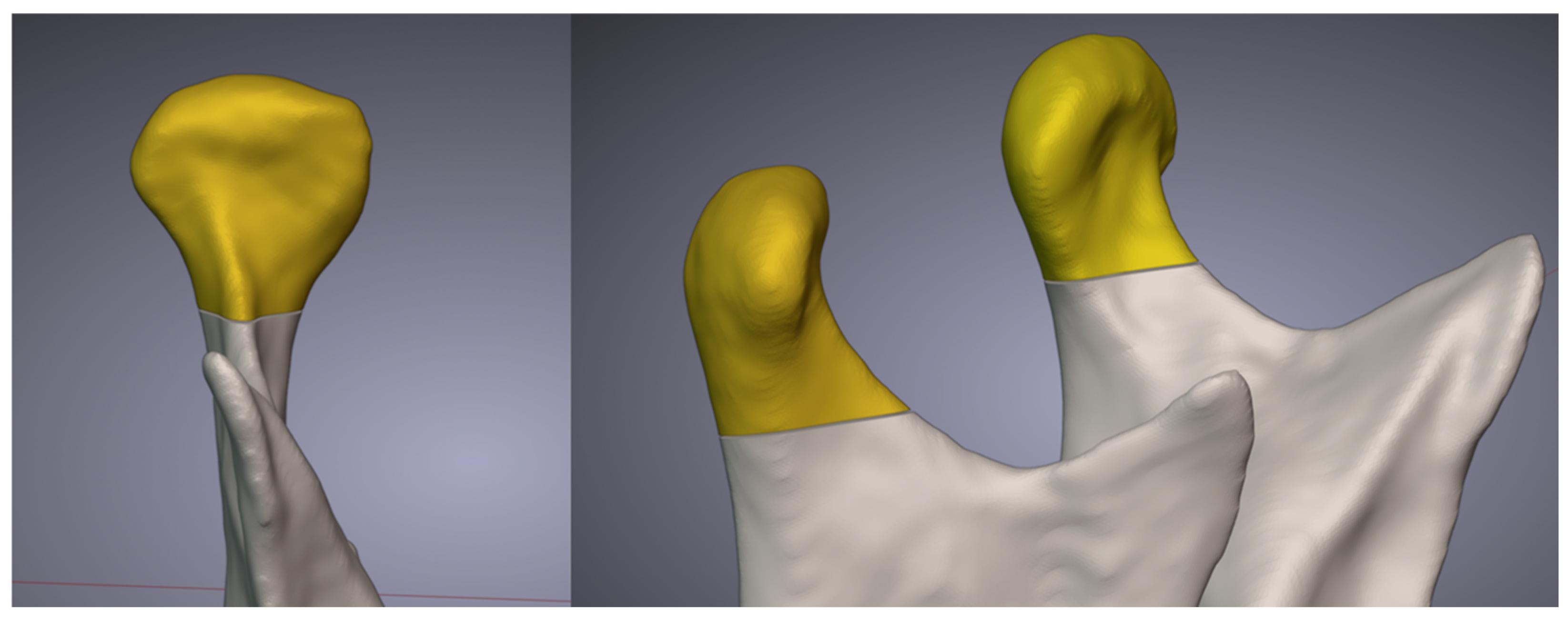
2.2. Condyle Volume Calculation

CBCT DICOM files of the patients were imported into the 3D Slicer (version 5.7). The mandible, maxilla and upper skull, upper teeth, lower teeth, and mandibular canal were segmented using the DentalSegmentator tool, an artificial intelligence extension de-veloped by Gauter et al. [25] (Figure 4) (https://www.youtube.com/watch?v=BEG-XhjjiaY, (accessed on 4 March 2025)).
Through the Blender Slicer Bridge Tool, which facilitates the transfer between 3D Slicer and Blenderfordental (Blenderfordental 2024, Dubai, United Arab Emirates) the generated STL files were transferred to Blenderfordental (B4D) software. Condylar volume was measured instead of linear measurements to provide a more accurate and three-dimensional assessment. Taking previous studies as a reference, the CUT tool in the Guide Module available in B4D (Figure 5) [13,26] was used solely to separate the inferior border of the condyle at the point where the sigmoid notch disappears. Since segmentation was performed by AI, this approach ensured that the entire condylar region up to the sigmoid notch was incorporated into the study and analyzed. Volume measurement was performed in mm3 using the 3D Print module in B4D. These values were then converted to cm3. The measured volumes of the right and left condyles were documented in an Excel table.
These measurements were repeated by the same operator (F.O) after one month for 10 individuals randomly selected from each group using the www.calculatorsoupplatform.com (accessed on 4 March 2025). Another researcher (S.B) also performed measurements on 10 randomly selected individuals from each group using the same methodology and recorded the results in an Excel table. (Microsoft Office, 365, Redmond, Washington, DC, USA).
From these data, the DICOM files of 20 randomly selected individuals were segmented by another artificial intelligence platform (DentalMesh AI), and the consistency of AI-assisted segmentation was evaluated by comparing the total mandibular volume obtained from both platforms. According to the total mandibular volume, a comparison between the two artificial intelligence methods was conducted. The Intraclass Correlation Coefficient (ICC) was used to assess the consistency between the two AI programs. Moreover, Gautier et al.’s [25] study was considered as a validation study. The agreement between the two AI platforms was regarded as an additional strength.

2.3. Statistical Analysis

Data analysis was performed using SPSS software (version 28.0; IBM Corp., Armonk, NY, USA), with descriptive statistics computed, including mean and standard deviation (SD). Group differences were assessed using ANOVA for data with a normal distribution, while the Kruskal–Wallis test was employed for data that did not meet normality assumptions. In cases where a significant difference was found in the ANOVA test, a post hoc Tamhane’s test was performed, and for the Kruskal–Wallis test, Dunn’s test was performed. As the data were not normally distributed, the Spearman correlation coefficient was applied to examine the association between airway and condylar volumes. Significance level was set as p < 0.05.

3. Results

To assess the reliability of segmentation semi-automatic (airway calculation by NemoFab), both intra-operator and inter-operator reliability tests were conducted. Two independent observers segmented the same dataset, and intra-class correlation coefficients (ICC) were calculated to evaluate reproducibility. In all groups, intra-operator ICC values exceeded 0.986, inter-operator ICC values surpassed 0.9, and inter-AI ICC values exceeded 0.95, collectively indicated excellent agreement.
Notable variations in condylar and airway volumes were identified across different facial divergence groups. In the normodivergent group, the average volume of the right condylar volume was 1.5 cm3, and the mean airway volume was 14.40 cm3. In the hyperdivergent group, the average volume of the right condylar volume was 1.2 cm3, and the mean airway volume was 11.23 cm3. In the hypodivergent group, the average volume of the right condylar volume was 1.5 cm3, and the mean airway volume was 14.33 cm3 (Table 2). The hyperdivergent group exhibited notably lower airway volumes compared to the normodivergent and hypodivergent groups. A similar trend was observed for condylar volumes, with hyperdivergent individuals having significantly smaller condylar volumes than the other groups.
The ANOVA test revealed a significant difference in condylar volume among the groups (p = 0.006) (Table 3).
Post hoc analyses using the Tukey HSD test indicated a significant difference in right condylar volume between the normodivergent and hyperdivergent groups (p = 0.038), with an even more pronounced difference observed between the hyperdivergent and hypodivergent groups (p = 0.007) (Table 4).
For airway volume data that did not meet normality assumptions, the Kruskal–Wallis test was performed, revealing significant differences among the groups (p = 0.028) (Table 3). Post hoc evaluation with Dunn’s test revealed significant difference in airway volume between the hyperdivergent and normodivergent groups (p = 0.027), whereas no notable variation was detected between the hyperdivergent and hypodivergent groups (p > 0.05).
As the data did not follow a normal distribution, Spearman correlation analysis was conducted, showing no significant relationship between condylar volume and airway volume (p > 0.05).
  • Normodivergent Group: A weak and nonsignificant negative relationship was found between airway volume and right condylar volume (r = −0.204, p = 0.280).
  • Hyperdivergent Group: No significant relationship was observed between airway volume and right condylar volume (r = −0.007, p = 0.971).
  • Hypodivergent Group: A nonsignificant positive relationship was detected between airway volume and right condylar volume (r = 0.015, p = 0.936).
A strong positive correlation was observed between right and left condylar volumes in all groups (p < 0.01).

4. Discussion

Two-dimensional radiographic volume measurements are considered insufficient due to the limited visualization of anatomical structures [1]. Consequently, the use of three-dimensional imaging methods, which provide more comprehensive anatomical information, has been increasingly evaluated. CBCT offers significant advantages over conventional methods by providing higher accuracy and precision in airway and condylar volume measurements, particularly in the detailed assessment of bony structures. The rapid acquisition of CBCT scans enhances patient comfort while minimizing motion artifacts. Additionally, compared to conventional computed tomography (CT), CBCT is more cost-effective and exposes patients to lower radiation doses, making it increasingly preferred in clinical applications [27,28].
However, CBCT has certain limitations. Unlike magnetic resonance imaging (MRI), it does not provide soft tissue contrast, which restricts the detailed analysis of the airway and surrounding soft tissues [17]. Furthermore, CBCT is a static imaging method and lacks the capability to assess airway function in real-time. Therefore, in cases requiring dynamic respiratory analysis, MRI or dynamic CT should be used as complementary imaging modalities [11].
In the present study, airway volume measurements were performed semi-automatically, with the mandible fully segmented using AI-assisted methods from DICOM files. Condyles were separated in accordance with the literature, and their volumes were measured using B4D [13,26]. These AI-assisted volume measurements were found to be highly consistent. Compared to manual segmentation, AI-assisted segmentation of anatomical structures not only allows segmentation to be completed in a shorter time but also eliminates potential human errors [21].
The accuracy of AI-assisted segmentation was visually assessed using a DICOM viewer and compared with results from another AI platform, DentalMesh AI. A strong level of agreement was observed between the two. The use of AI in evaluating anatomical structures and understanding their relationships has the potential to enhance our knowledge of human craniofacial development.
In individuals with skeletal Class I, different vertical growth patterns can develop. In this study, skeletal Class I individuals were categorized into three groups based on their vertical growth patterns: normodivergent, hyperdivergent, and hypodivergent. Existing research explores the association between vertical growth patterns and pharyngeal airway dimensions [6,29,30]. Grauer et al. [29] examined pharyngeal airway volume across various vertical growth patterns using CBCT and concluded that total airway volumes did not differ significantly. Differences in sample selection criteria, such as anatomical variation and age distribution, may have influenced these results. Additionally, variations in segmentation algorithms and measurement techniques could explain these discrepancies.
Unlike these studies, the present study found that the hyperdivergent group had significantly lower airway volumes compared to the normodivergent and hypodivergent groups (p < 0.05). Hyperdivergent individuals often exhibit mandibular backward rotation, which can narrow the pharyngeal airway by displacing the tongue and soft palate, increasing airway resistance. Additionally, increased facial height and weak masticatory muscle activity may further contribute to reduced airway width. These factors may lead to increased airway resistance and a higher risk of obstructive sleep apnea (OSA) in hyperdivergent individuals [31].
This finding indicates that hyperdivergent growth patterns should be carefully considered in orthodontic treatment planning. Since hyperdivergent individuals exhibit smaller condylar volumes and narrower airway dimensions, evaluating airway patency and mandibular growth potential before initiating orthodontic treatment is crucial. Orthopedic or functional appliances designed to improve mandibular position and enhance airway volume may be particularly beneficial for these patients.
However, this relationship may vary in normodivergent and hypodivergent individuals. In this regard, the present study yielded results like those of Freitas et al. [6], Flores et al. [30] and Celikoglu et al. [24]. In their study, Celikoğlu et al. [24] evaluated airway dimensions in patients with varying vertical growth patterns and found that the hyperdivergent group exhibited the lowest oropharyngeal, nasopharyngeal and total airway volumes. Similarly, Paul et al. [12] reported that the average airway volume in hypodivergent individuals was significantly higher than in hyperdivergent individuals. Conversely, Salehi et al. [32] found no significant association between airway volume and vertical growth patterns. These discrepancies may stem from methodological differences, including variations in software, sample size, and imaging modalities used across studies.
This study aimed to assess mandibular condyle volume in different vertical growth patterns and its correlation with airway volume. Burke et al. [33], found that hyperdivergent individuals had reduced superior joint spaces and posteriorly inclined condyles, whereas normodivergent and hypodivergent individuals had larger joint spaces and anteriorly positioned condyles. Similarly, Park et al. [34] reported smaller, more superiorly positioned condyles in hyperdivergent individuals. Consistent with these findings, this study observed significantly lower condylar volumes in the hyperdivergent group. Since the mandible in hyperdivergent tends to rotate downward and backward, condylar development may be affected. Additionally, their masticatory muscles are typically less developed with lower muscle tone [35], leading to reduced mechanical loading on the condyle, potentially limiting bone growth and contributing to smaller condylar volume.
Xu et al. [11] investigated the relationship between condylar position (anterior, posterior, central) and airway volume using CBCT, finding significant differences in pharyngeal airway volume based on condylar position. Unlike their study, this research focused on condylar volume and its correlation with airway volume but found no significant association, suggesting that condylar volume may not directly influence airway volume or that its effect was not evident in the analyzed sample. A larger sample or different measurement parameters may provide further insight. However, a strong correlation was observed between right and left condylar volumes across all groups, aligning with Orhan et al. [26], while Al-Koshab et al. [36] reported a significantly larger right condyle, highlighting potential anatomical variations.
This study has several limitations. The sample, limited to individuals with skeletal Class I malocclusion, may affect the generalizability of the findings. Variations in patient positioning and motion artifacts could also impact segmentation accuracy. While AI-assisted segmentation improves accuracy, CBCT imaging has inherent limitations, including motion artifacts, the need for semi-automatic airway adjustments, and its inability to assess dynamic airway function, unlike 4D techniques.
Future studies should include larger, more diverse samples with varying skeletal classifications, demographics, and physiological factors to enhance validation and investigate condylar and airway morphology variations. Advancements in AI-assisted segmentation should also be explored to improve accuracy and reduce operator dependency.

5. Conclusions

The null hypothesis was accepted. No significant correlation was found between the condyle volume and airway volume in all groups. This may indicate that condyle volume does not directly affect pharyngeal airway width, or its effect is not significant in the analyzed sample. Vertical growth patterns significantly affected both pharyngeal airway width and condylar volume, with hyperdivergent individuals exhibiting smaller condylar volumes and narrower airways. Additionally, the strong positive correlation between right and left condylar volumes indicates a tendency for symmetrical growth of mandibular condyles.

Author Contributions

F.O.: conceptualization, methodology, validation, investigation, writing—original draft preparation. S.B.: methodology, validation, writing—review and editing. A.K.: measurement analysis, data curation, M.K.: data curation, measurement analysis. All authors have read and agreed to the published version of the manuscript.

Funding

This research received no external funding.

Institutional Review Board Statement

The study was conducted in accordance with the Declaration of Helsinki, and approved by the Ethics Committee of İnönü University (protocol code 2024/6368 and date of approval 11.09.2024).

Informed Consent Statement

Informed consent was obtained from all subjects involved in the study.

Data Availability Statement

The original contributions presented in the study are included in the article, further inquiries can be directed to the corresponding author.

Conflicts of Interest

The authors declare no conflicts of interest.

References

  1. Moss, M.L.; Salentijn, L. The primary role of functional matrices in facial growth. Am. J. Orthod. 1969, 55, 566–577. [Google Scholar] [CrossRef]
  2. El, H.; Palomo, J.M. Three-dimensional evaluation of upper airway following rapid maxillary expansion: A CBCT study. Angle Orthod. 2014, 84, 265–273. [Google Scholar] [CrossRef]
  3. Li, L.; Liu, H.; Cheng, H.; Han, Y.; Wang, C.; Chen, Y.; Song, J.; Liu, D. CBCT evaluation of the upper airway morphological changes in growing patients of class II division 1 malocclusion with mandibular retrusion using twin block appliance: A comparative research. PLoS ONE 2014, 9, e94378. [Google Scholar] [CrossRef]
  4. Alhammadi, M.S.; Almashraqi, A.A.; Halboub, E.; Almahdi, S.; Jali, T.; Atafi, A.; Alomar, F. Pharyngeal airway spaces in different skeletal malocclusions: A CBCT 3D assessment. Cranio 2021, 39, 97–106. [Google Scholar] [CrossRef]
  5. Brito, F.C.; Brunetto, D.P.; Nojima, M.C. Three-dimensional study of the upper airway in different skeletal Class II malocclusion patterns. Angle Orthod. 2019, 89, 93–101. [Google Scholar] [CrossRef]
  6. de Freitas, M.R.; Alcazar, N.M.P.V.; Janson, G.; de Freitas, K.M.S.; Henriques, J.F.C. Upper and lower pharyngeal airways in subjects with Class I and Class II malocclusions and different growth patterns. Am. J. Orthod. Dentofac. Orthop. 2006, 130, 742–745. [Google Scholar] [CrossRef]
  7. Ucar, F.I.; Uysal, T. Orofacial airway dimensions in subjects with Class I malocclusion and different growth patterns. Angle Orthod. 2011, 81, 460–468. [Google Scholar] [CrossRef]
  8. Nielsen, I.L. Vertical malocclusions: Etiology, development, diagnosis and some aspects of treatment. Angle Orthod. 1991, 61, 247–260. [Google Scholar]
  9. Schudy, F.F. The rotation of the mandible resulting from growth: Its implications in orthodontic treatment. Angle Orthod. 1965, 35, 36–50. [Google Scholar]
  10. Isaacson, J.R.; Isaacson, R.J.; Speidel, T.M.; Worms, F.W. Extreme variation in vertical facial growth and associated variation in skeletal and dental relations. Angle Orthod 1971, 41, 219–229. [Google Scholar]
  11. Xu, J.; Sun, R.; Wang, L.; Hu, X. Cone-beam evaluation of pharyngeal airway space in adult skeletal Class II patients with different condylar positions. Angle Orthod. 2019, 89, 312–316. [Google Scholar] [CrossRef]
  12. Paul, P.; Thakur, A.; Mathur, A.; Chitra, P. Airway dimensions and mandibular position in adults with different growth patterns: A cone-beam computed tomography study. J. Stomatol. 2022, 75, 87–92. [Google Scholar] [CrossRef]
  13. Saccucci, M.; D’Attilio, M.; Rodolfino, D.; Festa, F.; Polimeni, A.; Tecco, S. Condylar volume and condylar area in Class I, Class II and Class III young adult subjects. Head Face Med. 2012, 8, 34. [Google Scholar] [CrossRef]
  14. Arieta-Miranda, J.M.; Silva-Valencia, M.; Flores-Mir, C.; Paredes-Sampen, N.A.; Arriola-Guillen, L.E. Spatial analysis of condyle position according to sagittal skeletal relationship, assessed by cone beam computed tomography. Prog. Orthod. 2013, 14, 36. [Google Scholar] [CrossRef]
  15. Paknahad, M.; Shahidi, S.; Iranpour, S.; Mirhadi, S.; Paknahad, M. Cone-beam computed tomographic assessment of mandibular condylar position in patients with temporomandibular joint dysfunction and in healthy subjects. Int. J. Dent. 2015, 2015, 301796. [Google Scholar] [CrossRef]
  16. Barandiaran, I.; Macía, I.; Berckmann, E.; Wald, D.; Dupillier, M.P.; Paloc, C.; Grana, M. An automatic segmentation and reconstruction of mandibular structures from CT-data. In Proceedings of the Intelligent Data Engineering and Automated Learning-IDEAL 2009: 10th International Conference, Burgos, Spain, 23–26 September 2009; pp. 649–655. [Google Scholar]
  17. Caruso, S.; Storti, E.; Nota, A.; Ehsani, S.; Gatto, R. Temporomandibular joint anatomy assessed by CBCT images. BioMed Res. Int. 2017, 2017, 2916953. [Google Scholar] [CrossRef]
  18. Sin, Ç.; Akkaya, N.; Aksoy, S.; Orhan, K.; Öz, U. A deep learning algorithm proposal to automatic pharyngeal airway detection and segmentation on CBCT images. Orthod. Craniofacial Res. 2021, 24, 117–123. [Google Scholar] [CrossRef]
  19. Evain, T.; Ripoche, X.; Atif, J.; Bloch, I. Semi-automatic teeth segmentation in cone-beam computed tomography by graph-cut with statistical shape priors. In Proceedings of the 2017 IEEE 14th International Symposium on Biomedical Imaging (ISBI 2017), Melbourne, Australia, 18–21 April 2017; pp. 1197–1200. [Google Scholar]
  20. Indraswari, R.; Kurita, T.; Arifin, A.Z.; Suciati, N.; Astuti, E.R.; Navastara, D.A. 3D region merging for segmentation of teeth on cone-beam computed tomography images. In Proceedings of the 2018 Joint 10th International Conference on Soft Computing and Intelligent Systems (SCIS) and 19th International Symposium on Advanced Intelligent Systems (ISIS), Toyama, Japan, 5–8 December 2018; pp. 341–345. [Google Scholar]
  21. Tarce, M.; Zhou, Y.; Antonelli, A.; Becker, K. The Application of Artificial Intelligence for Tooth Segmentation in CBCT Images: A Systematic Review. Appl. Sci. 2024, 14, 6298. [Google Scholar] [CrossRef]
  22. Orhan, K.; Shamshiev, M.; Ezhov, M.; Plaksin, A.; Kurbanova, A.; Ünsal, G.; Gusarev, M.; Golitsyna, M.; Aksoy, S.; Mısırlı, M. AI-based automatic segmentation of craniomaxillofacial anatomy from CBCT scans for automatic detection of pharyngeal airway evaluations in OSA patients. Sci. Rep. 2022, 12, 11863. [Google Scholar] [CrossRef]
  23. Elsonbaty, S.; Elgarba, B.M.; Fontenele, R.C.; Swaity, A.; Jacobs, R. Novel AI-based tool for primary tooth segmentation on CBCT using convolutional neural networks: A validation study. Int. J. Paediatr. Dent. 2025, 35, 97–107. [Google Scholar] [CrossRef]
  24. Celikoglu, M.; Bayram, M.; Sekerci, A.E.; Buyuk, S.K.; Toy, E. Comparison of pharyngeal airway volume among different vertical skeletal patterns: A cone-beam computed tomography study. Angle Orthod. 2014, 84, 782–787. [Google Scholar] [CrossRef] [PubMed]
  25. Dot, G.; Chaurasia, A.; Dubois, G.; Savoldelli, C.; Haghighat, S.; Azimian, S.; Taramsari, A.R.; Sivaramakrishnan, G.; Issa, J.; Dubey, A. DentalSegmentator: Robust open source deep learning-based CT and CBCT image segmentation. J. Dent. 2024, 147, 105130. [Google Scholar] [CrossRef] [PubMed]
  26. Orhan, K.; Karslioğlu, H.; Özemre, M.Ö.; Köseoğlu Seçgin, C.; Kamburoğlu, K. Kondil hacmi ve şeklinin konik ışınlı bilgisayarlı tomografi kullanılarak değerlendirilmesi. Turk. Klin. J. Dent. Sci. 2021, 27, 199–204. [Google Scholar] [CrossRef]
  27. Castro-Silva, L.; Monnazzi, M.S.; Spin-Neto, R.; Moraes, M.; Miranda, S.; Gabrielli, M.F.R.; Pereira-Filho, V.A. Cone-beam evaluation of pharyngeal airway space in Class I, II, and III patients. Oral Surg. Oral Med. Oral Pathol. Oral Radiol. 2015, 120, 679–683. [Google Scholar] [CrossRef]
  28. Muto, T.; Yamazaki, A.; Takeda, S. A cephalometric evaluation of the pharyngeal airway space in patients with mandibular retrognathia and prognathia, and normal subjects. Int. J. Oral Maxillofac. Surg. 2008, 37, 228–231. [Google Scholar] [CrossRef]
  29. Grauer, D.; Cevidanes, L.S.; Styner, M.A.; Ackerman, J.L.; Proffit, W.R. Pharyngeal airway volume and shape from cone-beam computed tomography: Relationship to facial morphology. Am. J. Orthod. Dentofac. Orthop. 2009, 136, 805–814. [Google Scholar] [CrossRef]
  30. Flores-Blancas, A.P.; Carruitero, M.J.; Flores-Mir, C. Comparison of airway dimensions in skeletal Class I malocclusion subjects with different vertical facial patterns. Dent. Press J. Orthod. 2017, 22, 35–42. [Google Scholar] [CrossRef]
  31. Enache, A.M.; Nimigean, V.R.; Mihaltan, F.; Didilescu, A.C.; Munteanu, I.; Nimigean, V. Assessment of sagittal and vertical skeletal patterns in Romanian patients with obstructive sleep apnea. Rom. J. Morphol. Embryol. 2010, 51, 505–508. [Google Scholar]
  32. Salehi, P.; Shahidi, S.; Majdi, B. Evaluation of the relationship between airway volumes and vertical facial growth patterns in adult patients. J. Dentomaxillofacial Radiol. Pathol. Surg. 2015, 4, 20–30. [Google Scholar] [CrossRef]
  33. Burke, G.; Major, P.; Glover, K.; Prasad, N. Correlations between condylar characteristics and facial morphology in Class II preadolescent patients. Am. J. Orthod. Dentofac. Orthop. 1998, 114, 328–336. [Google Scholar] [CrossRef]
  34. Park, I.-Y.; Kim, J.-H.; Park, Y.-H. Three-dimensional cone-beam computed tomography based comparison of condylar position and morphology according to the vertical skeletal pattern. Korean J. Orthod. 2015, 45, 66–73. [Google Scholar] [CrossRef] [PubMed]
  35. Proffit, W.; Fields, H. Occlusal forces in normal-and long-face children. J. Dent. Res. 1983, 62, 571–574. [Google Scholar] [CrossRef] [PubMed]
  36. Al-koshab, M.; Nambiar, P.; John, J. Assessment of condyle and glenoid fossa morphology using CBCT in South-East Asians. PLoS ONE 2015, 10, e0121682. [Google Scholar]
Figure 1. Adjusting the Frankfurt horizontal plane to be parallel to the ground and orientation control.
Applsci 15 02794 g001
Figure 2. Defining the boundaries of the upper airway.
Applsci 15 02794 g002
Figure 3. Three-dimensional rendering of the airway and measurement of the minimum axial cross-section (mm2).
Applsci 15 02794 g003
Figure 4. Segmentation of craniofacial structures from DICOM files using the DentalSegmentator AI module in 3D Slicer software (version 5.7).
Applsci 15 02794 g004
Figure 5. Separation of the condyle using the CUT tool in Blenderfordental.
Applsci 15 02794 g005
Table 1. Descriptive statistics for the groups.
GroupNumber of PatientsFemalesMalesMean Age (Years ± SD)
Normodivergent30171321.3 ± 2.8
Hyperdivergent33201320.7 ± 3.1
Hypodivergent30181221.9 ± 3.2
SD: Standard Deviation.
Table 2. Descriptive Statistics and comparisons of airway volumes and condylar volumes.
ParameterNormodivergent (n = 30)Hyperdivergent (n = 33)Hypodivergent (n = 30)Test Statisticp-Value
Airway Volume (cm3)14.4 ± 4.911.2 ± 5.014.1 ± 6.3χ2 = 7.4440.024 *
Condylar Volume (Right) (cm3)1.5 ± 0.31.2 ± 0.21.5 ± 0.3F = 5.5010.006 **
Airway Volume: Kruskal–Wallis Test was used due to the absence of normal distribution (p < 0.05 indicates statistical significance). Condylar Volume (Right): ANOVA was used for parametric data. Statistical significant: * p < 0.05: ** p < 0.01.
Table 3. Correlation between right and left condylar volumes.
GroupPearson Correlation (r)p-Value
Normodivergent0.909 ***<0.001
Hyperdivergent0.840 ***<0.001
Hypodivergent0.927 ***<0.001
r: Pearson correlation coefficient; p < 0.05: Statistically significant: *** p < 0.001.
Table 4. Pairwise comparison of condyle volume in different vertical groups.
Mean Difference (1–3)Std. Errorp-Value 95% CI Lower Bound95% CI Upper Bound
1 vs. 2211.41784.6210.038 *9.758413.076
1 vs. 3−51.60286.6120.823−258.007154.803
2 vs. 3−263.01984.6210.007 **−464.678−61.359
Significance levels are marked as * p < 0.05 and ** p < 0.01, Post hoc Tukey HSD test; 1: Normodivergent, 2: Hyperdivergent, 3: Hypodivergent.
Disclaimer/Publisher’s Note: The statements, opinions and data contained in all publications are solely those of the individual author(s) and contributor(s) and not of MDPI and/or the editor(s). MDPI and/or the editor(s) disclaim responsibility for any injury to people or property resulting from any ideas, methods, instructions or products referred to in the content.

