Communication

Does Mental Imagery Influence Muscles Activity? A Proof of Concept Study on Franklin Method® Effectiveness in Dance Training

Department of Biomechanics, Faculty of Sport Sciences, Poznan University of Physical Education, 61-871 Poznań, Poland
*
Author to whom correspondence should be addressed.
Appl. Sci. 2025, 15(4), 1902; https://doi.org/10.3390/app15041902
Submission received: 13 December 2024 / Revised: 10 February 2025 / Accepted: 10 February 2025 / Published: 12 February 2025
(This article belongs to the Special Issue Advances in the Biomechanics of Sports)

Abstract

:

Featured Application

Biomechanical tools such as electromyography, dynamometry, or motion capture systems are widely used for training effectiveness or performance correctness verification. Our work indicates that electromyography may be used to evaluate the impact of the specific training (i.e., using mental imagery in dance performance).

Abstract

Mental imagery influences the body, movement, and technical skills of the dancer. The aim of this study was to identify the influence of dance imagery on the electromyographic parameters of selected muscles in a professional ballet dancer during three ballet tasks: parallel position, demi pointe relevé, and demi plié. Five mental imageries according to the Franklin Method® were used: foot dome, the wheelbarrow, pushing the toes, space behind the kneecap, and the kneecap float. Electromyographic signals were recorded bilaterally for lumbar erector spinae, rectus abdominis, vastus medialis, long head of biceps femoris, lateral head of gastrocnemius, tibialis anterior, and fibularis longus. All of the mental imageries resulted in increased activity (above 20% compared with no-imagery performance) of the selected muscles in the studied classical dance positions and tasks. Overall, the ankle muscles were influenced the most. This study indicates that mental images effectively influence a physiological parameter, as indicated by an electromyographic signal.

1. Introduction

Dance imagery influences the dancer’s body and movement [1] as well as their technical skills [2]. These voluntary mental creations are used in training sessions and performances [3,4].
Mental imagery in dance is mostly presented as a teaching tool used to learn specific dance movements. Dance teachers mainly use metaphors to help students properly execute different choreographic elements. Dance students are encouraged to imagine a particular movement in order to perform it correctly and perfect their technique [5,6]. Mental images are used in postures and movements of varying difficulty and are gradually incorporated in dance training. Primary dance students first learn how to use mental imagery in a static parallel position (i.e., standing straight with feet joined together). Next, they learn dynamic movements of medium difficulty like demi pointe relevé in parallel position (i.e., from parallel position lifting the heels above the floor). Next, they move into more complex movements like demi plié in first classical ballet position (i.e., bending the hips and knees with heels joined together and feet in opposite direction).
The goals of mental imagery include mastering technical skills, enhancing self-confidence, or taking inspiration for choreography [1]. According to Eric Franklin, mastering mental imagery is a long-lasting task and resembles learning a new language [7]. Regular exercises and patience are essential to achieve the intended goal [7]. The Franklin Method® is a system based on using mental imagery techniques for improvements in musculoskeletal system function [1,8]. According to Eric Franklin, his method is effective and evidence-based, recommended on every dance education stage. The Franklin Method® is focused on the body and its functions and improves the quality of movement [9]. However, according to Heiland and Rovetti (2012), dancers, especially those younger and less experienced, are often not aware if the imagery is actually effective [8]. As a matter of fact, mental imagery is used by dancers of all ages and ability levels: from primary-aged children through amateurs to professional dancers. It turns out that the more experienced the dancer, the more complex images they use [1]. Professional, high-level dancers mostly employ mental techniques to improve their posture, visualize emotions, or increase jump height [4,10].
According to Pavlik et al. (2016), despite the variety of research focused on mental imagery methods, the conclusions are not consistent [1]. This indicates that the use of mental images in the field of dance or sport training or performance is still to be discovered and verified with objective measurement methods.
Biomechanical tools such as electromyography, dynamometry, or motion capture systems provide an objective assessment of the training effectiveness or performance correctness [11,12,13]. However only a few studies have been found on using electromyography on dancers for verification of the mental imagery effectiveness using the Franklin Method® on jump height [14] or other mental techniques on demi plié performance [15].
Therefore the aim of this study was to identify the influence of dance imagery based on the Franklin Method® on the electromyographic parameters of selected muscles in a professional ballet dancer during three ballet tasks of varying difficulty: parallel position, demi pointe relevé, and demi plié.

2. Materials and Methods

2.1. Participant

A male professional dancer and dance teacher with 13 years of experience in using dance imagery based on the Franklin Method® in his work with dance students participated in the study. The study subject was healthy and had not had any serious injuries for the past 6 months. The study obtained the approval of the Bioethical Committee at the Poznan University of Medical Sciences (Poland) (decision no. 796/09). The study was carried out in accordance with the latest version of the Declaration of Helsinki.

2.2. Study Design and Procedures

Surface electromyography (EMG) was used to investigate the electrical activity of selected muscles in three different classical ballet tasks: (1) standing in parallel position; (2) demi-pointe relevé in the parallel position; and (3) demi plié in first classical ballet position. Each task was conducted in the classical technique (no imagery) and with dance imagery according to the Franklin Method® [7]. The rationale for choosing these ballet tasks for analysis was as follows: parallel position, demi pointe relevé, demi plié in first classical ballet position are dance elements of gradually increasing difficulty used by the study subject in everyday training in the ballet school. The mental images described below were chosen according to the Franklin Method®. Franklin described these mental images as specific for the positions examined in our study [7]. The study subject performed three tasks barefoot with and without dance imagery, each repeated three times:
  • Standing in a parallel position for 10 s. A parallel position involved an upright standing position with the feet located parallel and medial borders of the feet adhered to each other (Figure 1). The task was carried out in classical technique (no imagery) and with mental imagery called foot dome, according to the Franklin Method® [7] (p. 247). In this imagery, the study subject visualized a waterspout spraying upward from between the ankles [7] (p. 247).
  • Demi pointe relevé in the parallel position (Figure 1). The task was divided into three phases: (1) lifting the heel from parallel position into demi pointe relevé (3 s); (2) standing in demi pointe relevé (5 s); (3) lowering the heel from demi pointe relevé into parallel position (3 s). The task was conducted in classical technique (no imagery) and with mental imagery called the wheelbarrow, according to the Franklin Method® [7] (p. 115). In this imagery, the study subject visualized that: (1) his foot turned into a wheelbarrow as he lifted his heel off the ground; (2) the toes push against the floor as the weight of the body is lifted upward [7] (p. 115).
  • Demi plié in first classical ballet position (Figure 2). The task was divided into two phases: (1) bending the knees from first position into demi plié (4 s), and (2) straightening the knees from the demi plié into first position (4 s). The task was carried out in the classical technique (no imagery) and with two mental imageries called (1) the kneecap float, and (2) space behind the kneecap [7] (p. 119). In this imagery, the study subject respectively visualized: (1) the kneecap floating perpendicularly away from the femur as he extended the knee; and (2) the space behind the kneecap increasing, creating a little cushion of space for the kneecap to glide on [7] (p. 119).

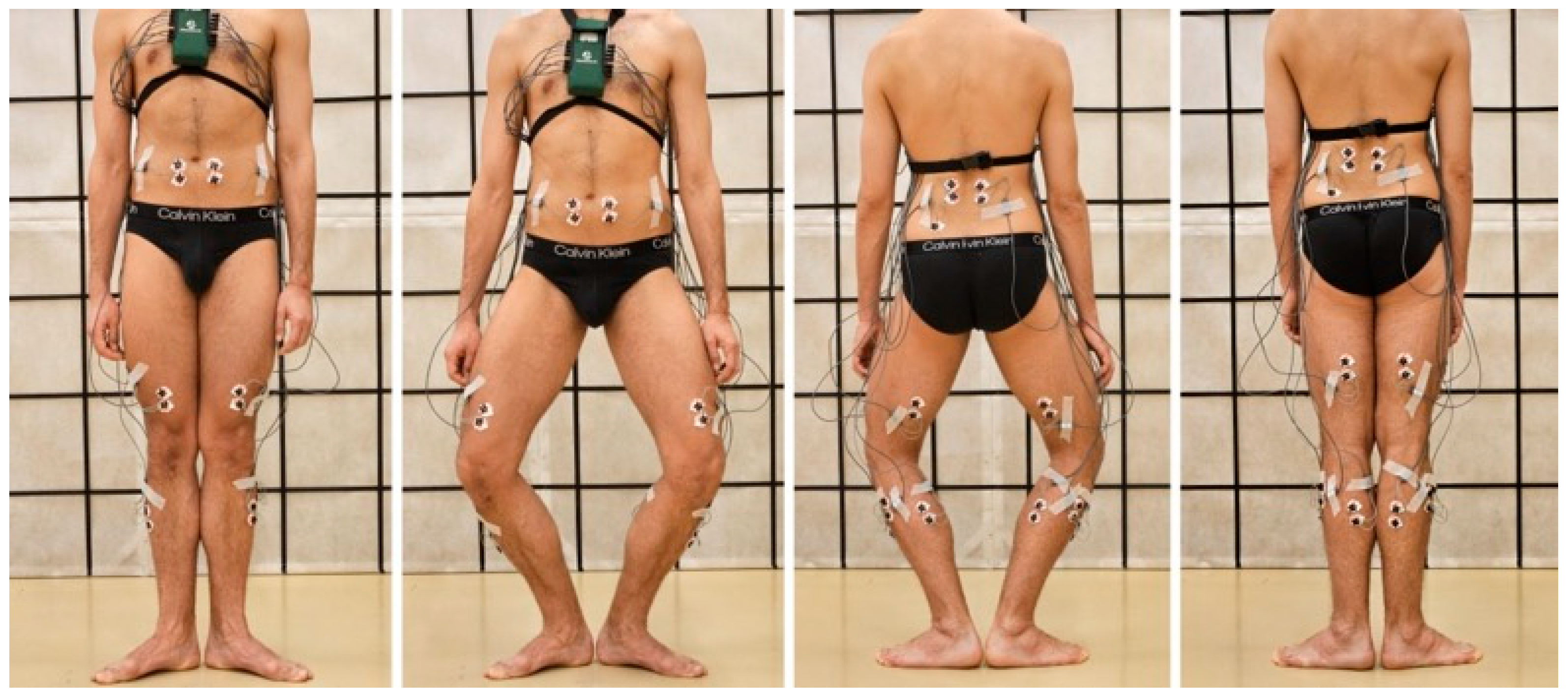
2.3. Electromyography

A Telemyo 2400T G2 device (Noraxon, Scottsdale, AZ, USA) compatible with the MyoResearch XP 1.07 Master Edition software was used to record the electrical activity of seven muscles bilaterally: lumbar erector spinae (ES), rectus abdominis (RA), vastus medialis (VM), long head of biceps femoris (BF), lateral head of gastrocnemius (LGAS), tibialis anterior (TA), and fibularis longus (FIB) with the ground electrode placed over the right posterior superior iliac spine. The rationale for choosing these muscles for analysis was as follows: ES and RA muscles are trunk stabilizers, helping dancers to keep their pelvis in neutral position. VM and BF stabilize the knee during demi plié. VM is a medial stabilizer of the patella, preventing its lateral dislocation. BF, besides flexing the knee, also laterally rotates the lower leg in first classical ballet position. The LGAS, TA, and FIB muscles mainly act on the ankle joints. LGAS is a strong ankle plantar flexor during demi pointe relevé and also externally rotates the lower leg in first classical ballet position. TA and FIB act antagonistically to stabilize the feet. TA is a foot supinator while FIB is a foot pronator. Figure 1 illustrates the electrode placement. The study subject was prepared for the test following the SENIAM recommendations [16]. First, the skin prepared for the electrode placement was shaved and cleaned with an abrasive and disinfecting agent. Next, a pair of Ag/AgCl surface electrodes (SORIMEX, Toruń, Poland, 1 cm in diameter) was attached in the bipolar configuration along the longitudinal axis of the examined muscles. The distance between the electrodes was 2 cm. The correctness of the attachments was verified through the observation of a raw EMG signal during the testing of the muscles. The EMG signal was sampled at a frequency of 1000 Hz and filtered with a hardware bandpass filter (bandwidth: 10–500 Hz).

2.4. Data Analysis

The EMG signal was processed using the MyoResearch XP Master Edition software program (Noraxon, USA). Related artifacts and noise were inspected visually. The EMG signal was full-wave rectified and smoothed using the root mean square algorithm with a 50-millisecond window.
In the parallel position, the mean value of the EMG signal recorded for 10 s was taken for further analysis. In the demi pointe relevé and demi plié, the maximum value of the EMG signal recorded in each phase of the movement was taken for further analysis.
The values obtained from the three repetitions were averaged for each task. Next, the EMG signal obtained in the tasks carried out using dance imagery was normalized to the signal from tasks carried out classically (no imagery), according to the formula:
EMGnormalized = (EMGimagery/EMGclassical) × 100%
Values above 100% indicate a higher activity of a given muscle in the task carried out using mental imagery in comparison with the same task carried out using the classical technique (no imagery). The muscle activity was marked as increased if the EMG signal obtained in a task carried out with imagery exceeded the signal obtained with no imagery by at least 20%.

3. Results

A summary of the EMG assessment is presented in Table 1. In the parallel position, the activity of all but one muscles increased with foot dome imagery. In the demi pointe relevé, during the wheelbarrow imagery, the activity of BF and the trunk muscles did not increase, but the VM and ankle muscles were more active. In the pushing the toes imagery, only VM was more active on both sides and in every phase of the task. Other muscles were more active on one side or in selected phases with the exception of RA, whose activity did not increase at all. In the second phase of demi plié with the space behind the kneecap imagery, the ES and TA were more active on both sides, while RA and LGAS were more active only on the right side. The activity of other muscles did not increase. In the kneecap float, no muscle was more active on both sides. Only ES, LGAS, and FIB were more active on one side, and the activity of the other muscles did not increase.

4. Discussion

The most important finding from this study is that mental imagery influenced the muscle activity in parallel position, demi pointe relevé, and demi plié. Five mental imageries according to the Franklin Method® were used: foot dome, the wheelbarrow, pushing the toes, space behind the kneecap, and the kneecap float. All of these resulted in increased activity of the selected muscles in the studied ballet tasks. These findings show how mental creations impact body functioning.
The dancer who was tested in this study had 13 years of experience in using dance imagery in his work with dance students. His expertise in the field of mental imagery based on the Franklin Method® ensured that the basic dance posture and movements used in this study were performed correctly. Therefore, the differences between performances carried out with and without imagery resulted from the mental techniques used, and not from random variables connected to lack of expertise.
Dance imagery influenced the ankle muscles the most. Activity of the LGAS muscle increased in all trials. In the case of FIB and TA, only one imagery in demi plié did not result in an increase in muscle activity. ES muscle activity also increased in all but one trials in demi pointe relevé. The knee extensor was more active in parallel position and demi pointe relevé with dance imagery, but no influence of imagery in demi plié was noticed. RA muscle activity increased bilaterally only in the parallel position with imagery and unilaterally in one visualization in demi plié. The BF muscle responded to dance imagery only partly and unilaterally in one case in demi pointe relevé.
In parallel position, all but one muscles were more active bilaterally with foot dome visualization. In demi plié, only half of the studied muscles were influenced by dance imagery, and most of them responded to visualization only unilaterally. In the demi pointe relevé activity, most of the studied muscles increased with dance imagery. However, in most cases, the increase occurred unilaterally or in selected phases of the task.
Based on the description of the studied visualizations [7], an increase in the activity of the selected muscles was expected with every dance imagery. The foot dome imagery aims to increase one’s awareness of foot arches by visualizing a waterspout spraying upward from between the ankles [7] (p. 247). Therefore, with this imagery, the dancer focuses on the ankle muscles to supinate the feet. In our study, not only were the ankle muscles (TA, FIB, LGAS) activated, but also postural muscles (RA, ES, VM) that stabilize the upright standing position.
In the wheelbarrow imageries, the dancer visualizes that (1) the foot turns into a wheelbarrow as the heels are lifted off the ground or (2) the toes push against the floor as the weight of the body is lifted upward [7] (p. 115). These visualizations are aimed at activation of the ankle muscles with LGAS lifting the body upward and TA with FIB stiffening the feet. In our study, the activity of the ankle muscles increased with the wheelbarrow imageries during demi pointe relevé; however, in most cases, it was a partial and unilateral increase. On the other hand, the knee extensor’s activity increased bilaterally with the wheelbarrow imageries, which may be co-activation in response to increased knee flexor (LGAS) activity.
In the kneecap float and space behind the kneecap visualizations, the dancer imagines the lengthening of the knee extension moment arm [7] (p. 119), therefore, an increase in knee extensor activity was not expected. As the main burst of the EMG activity of the studied muscles in demi plié occurred when the dancer was straightening their knees from demi plié into first position, only the second phase of the movement was analyzed. As expected, the imagery did not influence the VMO activity. However, an increase in the bilateral muscle activity occurred only in the space behind the kneecap visualization for ES and TA. Lumbar extensor and foot dorsiflexor may additionally stabilize the trunk and lower limb during the second phase of the demi plié.
Biomechanical tools such as EMG, dynamometry, or motion capture systems are widely used for training effectiveness or performance correctness verification [11,12,13,14,15]. EMG was used by Heiland and Rovetti (2012), who analyzed the influence of the visualizations based on the Franklin Method® on jump height [14]. By using cameras and a motion capture system, they confirmed the effectiveness of the rocket and spring imageries in their study. According to Heiland and Rovetti [14], Franklin created the most diverse mental imagery system and provided guidance as to how to use mental imagery in dance [7,10,17,18]. A biomechanical objective tool such as EMG may be used to evaluate the impact of specific training (i.e., using mental imagery on dance performance). In classical ballet, dancers struggle with static loads that impact their bodies [19]. Dance imagery may serve to decrease these loads. No study in the field of using dance imagery in reducing loads in the everyday training of artists has been found. Most studies have highlighted the importance of feedback for movement improvement and to assess the efficacy of the mental technique. Therefore, EMG is a suitable objective method that can provide feedback for dancers, sportsmen, teachers, or coaches. Furthermore, this methodology would also be valuable in primary ballet schools in the education of young dance students.

5. Conclusions

The main contribution of this work was the identification of the impact of mental imagery based on the Franklin Method® on a physiological parameter, which was the EMG signal. The described experiment showed that using the EMG measurement for verification of the mental techniques’ effectiveness is feasible. It is known that dance training benefits from mental imagery. Dance analysis could take place at a biomechanics laboratory where complex biomechanical measurements on a specific target group can be conducted. There are many mental techniques available, and therefore, there are plenty of possible research studies that could evaluate the effectiveness of mental imagery in dance training.

Author Contributions

Conceptualization, J.G.; Data curation, A.F.; Formal analysis, J.G. and A.F.; Investigation, J.G. and A.F.; Methodology, J.G. and A.F.; Project administration, J.G.; Resources, J.G. and A.F.; Software, J.G. and A.F.; Supervision, J.G.; Validation, J.G. and A.F.; Visualization, A.F.; Writing—original draft, J.G. and A.F.; Writing—review and editing, J.G. and A.F. All authors have read and agreed to the published version of the manuscript.

Funding

This research received no external funding.

Institutional Review Board Statement

This study was reviewed by the Bioethical Committee at the Poznan University of Medical Sciences (Poland) (decision no. 796/09, dated 3 September 2009). The study was conducted in accordance with the Declaration of Helsinki.

Informed Consent Statement

Informed consent was obtained from all subjects involved in the study.

Data Availability Statement

The data that support the findings of this study are available on request from the corresponding author, A.F.

Conflicts of Interest

The authors declare no conflicts of interest.

References

  1. Pavlik, K.; Nordin-Bates, S. Imagery in dance. A literature review. J. Danc. Med. Sci. 2016, 20, 51–63. [Google Scholar] [CrossRef] [PubMed]
  2. Afremow, J.; Overby, L.; Vadocz, E. Using mental imagery to enhance sport and dance skills of children. J. Int. Counc. Health Phys. Educ. Recreat. Sport Danc. 1997, 33, 44–48. [Google Scholar]
  3. Krasnow, D.H.; Chatfield, S.J.; Barr, S.; Jensen, J.L.; Dufek, J.S. Imagery and conditioning practices for dancers. Danc. Res. J. 1997, 29, 43–64. [Google Scholar] [CrossRef]
  4. Nordin, S.M.; Cumming, J. Measuring the content of dancers’ images: Development of the Dance Imagery Questionnaire (DIQ). J. Danc. Med. Sci. 2006, 10, 85–98. [Google Scholar] [CrossRef]
  5. Bolles, G.; Chatfield, S.J. The intersection of imagery ability, imagery use, and learning style. J. Danc. Educ. 2009, 9, 6–16. [Google Scholar] [CrossRef]
  6. Sacha, T.J.; Russ, S.W. Effects of pretend imagery on learning dance in preschool children. Early Child. Educ. J. 2006, 33, 341–345. [Google Scholar] [CrossRef]
  7. Franklin, E. Dynamic Alignment Through Imagery, 2nd ed.; Human Kinetics: Champaign, IL, USA, 2012. [Google Scholar]
  8. Franklin, E. Dance Imagery for Technique and Performance; Human Kinetics: Champaign, IL, USA, 1996. [Google Scholar]
  9. Available online: https://franklinmethod.com/ (accessed on 29 January 2025).
  10. Hanrahan, C.; Vergeer, I. Multiple uses of mental imagery by professional modern dancers. Imagin. Cogn. Personal. 2001, 20, 231–255. [Google Scholar] [CrossRef]
  11. Krasnow, D.; Wilmerding, M.V.; Stecyk, S.; Wyon, M.; Koutedakis, Y. Biomechanical research in dance: A literature review. Med. Probl. Perform. Artist. 2011, 26, 3–23. [Google Scholar] [CrossRef]
  12. Abraham, A.; Dunsky, A.; Dickstein, R. The effect of motor imagery practice on Elevé performance in adolescent female dance students: A randomized controlled trial. J. Imag. Res. Sport Phys. Act. 2017, 12, 20160006. [Google Scholar] [CrossRef]
  13. Gildea, J.E.; VAN DEN Hoorn, W.; Hides, J.A.; Hodges, P.W. Trunk dynamics are impaired in ballet dancers with back pain but improve with imagery. Med. Sci. Sports Exerc. 2015, 47, 1665–1671. [Google Scholar] [CrossRef] [PubMed]
  14. Heiland, T.; Rovetti, R. Examining effects of Franklin Method metaphorical and anatomical mental images on college dancers’ jumping height. Res. Danc. Educ. 2012, 14, 141–161. [Google Scholar] [CrossRef]
  15. Couillandre, A.; Lewton-Brain, P.; Portero, P. Exploring the effects of kinesiological awareness and mental imagery on movement intention in the performance of demi-plié. J. Danc. Med. Sci. 2008, 12, s91–s98. [Google Scholar] [CrossRef]
  16. Hermens, H.J.; Freriks, B.; Disselhorst-Klug, C.; Rau, G. Development of recommendations for SEMG sensors and sensor placement procedures. J. Electromyogr. Kinesiol. 2000, 10, 361–374. [Google Scholar] [CrossRef] [PubMed]
  17. Franklin, E. Dynamic Alignment Through Imagery; Human Kinetics: Champaign, IL, USA, 1996. [Google Scholar]
  18. Franklin, E. Conditioning for Dance; Human Kinetics: Champaign, IL, USA, 2004. [Google Scholar]
  19. Gorwa, J.; Kabaciński, J.; Murawa, M.; Fryzowicz, A. On the track of the ideal turnout: Electromyographic and kinematic analysis of the five classical ballet positions. PLoS ONE 2020, 15, e0230654. [Google Scholar] [CrossRef] [PubMed]
Figure 1. Bilateral placement of the electrodes on the tested muscles: lumbar erector spinae, rectus abdominis, vastus medialis, long head of biceps femoris, lateral head of gastrocnemius, tibialis anterior, and fibularis longus with the ground electrode placed over the right posterior superior iliac spine. The first and last pictures present the parallel position. The second to fourth pictures present standing in demi pointe relevé.
Applsci 15 01902 g001
Figure 2. The first and last pictures present the first classical ballet position. The second and third pictures present bending the knees from first position into demi plié.
Applsci 15 01902 g002
Table 1. Summary of the electromyographic assessment.
ParallelRelevé (1st, 2nd, and 3rd Phases)Plié (2nd Phase)
Examined muscleFoot domePushing the toesThe wheelbarrowThe kneecap floatSpace behind the kneecap
Rectus abdominisIncreasedNoNoNoIncreased (right)
Erector spinaeIncreasedIncreased (left)NoIncreased (left)Increased
Vastus medialisIncreasedIncreasedIncreasedNoNo
Biceps femorisNoIncreased (left in 2nd, 3rd phases)NoNoNo
GastrocnemiusIncreasedIncreased (except for left, 1st phase)IncreasedIncreased (right)Increased (right)
Tibialis anteriorIncreasedIncreased (right in 2nd, 3rd phases)Increased (except for right, 1st phase)NoIncreased
Fibularis longusIncreasedIncreased (both sides in 3rd phase; right in 1st phase)Increased (except for left, 3rd phaseIncreased (left)No
“Increased” means that the EMG signal obtained in a task carried out with imagery exceeded the signal obtained with no imagery by at least 20% (colour green, if the increase was on both sides in majority of phases; colour yellow, if the increase was on one side or both sides but in minority of phases); “no” means that the threshold of 20% increase in EMG signal was not achieved in a task carried out with imagery compared with no imagery (colour red); comments in the brackets describe in which phase of the movement or on which side of the body the increase occurred.
Disclaimer/Publisher’s Note: The statements, opinions and data contained in all publications are solely those of the individual author(s) and contributor(s) and not of MDPI and/or the editor(s). MDPI and/or the editor(s) disclaim responsibility for any injury to people or property resulting from any ideas, methods, instructions or products referred to in the content.

