Article

The Prevalence and Clinical Implications of Atlantal Posterior Arch Defects: A CBCT Analysis of 500 Italian Patients

by
Monica Macrì
1,*,
Abdulaziz Alhotan
2,
Mario Festa
3,
Fabiola Rendina
1 and
Felice Festa
1
1
Department of Innovative Technologies in Medicine & Dentistry, University “G. D’Annunzio” of Chieti-Pescara, 66100 Chieti, Italy
2
Department of Dental Health, College of Applied Medical Sciences, King Saud University, Riyadh 12372, Saudi Arabia
3
Department of Anatomic, Histologic, Medical-Legal Sciences and of Locomotor System, Rome University Sapienza, 00185 Rome, Italy
*
Author to whom correspondence should be addressed.
Appl. Sci. 2025, 15(4), 1877; https://doi.org/10.3390/app15041877
Submission received: 5 August 2024 / Revised: 2 December 2024 / Accepted: 6 February 2025 / Published: 12 February 2025
(This article belongs to the Section Applied Dentistry and Oral Sciences)

Abstract

:
Atlas Posterior Arch Deficiency (APAD) is a rare anatomical variation of the atlas vertebra detected using cone-beam computed tomography (CBCT). This study assessed the prevalence, classification, and clinical implications of APAD in an Italian cohort. Methods: We analyzed CBCT images from 500 Italian patients (17–60 years) from the University “G. D’Annunzio” in Chieti. The sample was evenly split between genders. The sample was evenly split between genders. APAD classification was performed, identifying five morphological types. Imaging used a low-dose protocol with a Pax-Zenith 3D CBCT machine and Dolphin imaging software (12.0.9.58). Results: APAD was identified in thirteen patients—twelve with partial APAD and one with complete APAD. Two cases also had Ponticulus Posticus. Type A anomalies were most common; no cases of types B, C, D, or E were observed. None of the cases were symptomatic. Conclusions: APAD is rare in this cohort, with partial forms being the most frequent. CBCT is effective for diagnosing APAD, highlighting the importance of routine imaging in orthodontic assessments. Despite the absence of symptoms, awareness and monitoring for potential symptoms are important. Further research is needed to understand APAD’s clinical significance across different populations.

1. Introduction

This study aims to investigate the prevalence and anatomical variations of Atlas Posterior Arch Deficiency (APAD) using CBCT imaging. The objective is to enhance the understanding of this rare anatomical variant and to provide insights into its implications for both clinical practice and future research. Atlas Posterior Arch Deficiency (APAD) is a congenital anomaly characterized by the partial or complete absence of the posterior arch of the atlas vertebra (C1), the first cervical vertebra of the spine [1]. This condition results from a failure of the posterior ossification centers to fuse during embryonic development, leading to a structural defect in the atlas. Consequently, the posterior arch, which normally forms a protective ring around the spinal cord and vertebral arteries, may be deficient, predisposing the individual to cervical spine instability and potential neurological compromise [1,2,3].
The cervical region contains several vital structures, including the cervical vertebrae, muscles, blood vessels, nerves, and various glands. Each cervical vertebra is numbered C1 through C7, with C1 being the first cervical vertebra, also known as the atlas [1,3]. The term “atlas” is derived from Greek mythology, reflecting its function in supporting the weight of the head, akin to the mythical role of Atlas.
The ring-like structure of the atlas (C1) is unique among the cervical vertebrae as it lacks a vertebral body and spinous process. Instead, it consists of anterior and posterior arches and two lateral masses. In contrast, the other cervical vertebrae (C2–C7) have a more typical vertebral structure with a vertebral body, spinous process, and transverse processes. C1 supports the weight of the skull and allows for the nodding or “yes” motion of the head, articulating with the occipital condyles of the skull to form the atlanto-occipital joint [1,3,4].
The articulation of the atlas is unique and plays a crucial role in supporting the weight of the head and facilitating its movement. Dysfunction or injury to these articulations can lead to a range of clinical conditions and symptoms, emphasizing their importance in maintaining overall spinal health and function [2,3].
During embryonic development, the vertebrae form from segments of mesodermal tissue called somites. The atlas specifically arises from the first occipital sclerotome, a specialized region of the mesoderm located at the base of the skull. The mesenchymal cells within the first occipital sclerotome undergo chondrification, differentiating into cartilage. Ossification, the process of bone formation, begins around the seventh week of gestation. The atlas ossifies from multiple centers, including separate centers for the anterior and posterior arches, as well as for the lateral masses [3,4,5]. These ossification centers gradually fuse during childhood and adolescence to form the mature atlas. All synchondroses of the C1 fuse slightly earlier in females, as reported by Wu et al. (2022) [5].
The etiology of APAD involves disturbances in embryonic development during the formation of the atlas. Various hypotheses have been proposed, including that disruptions in chondrogenesis, vascular insufficiency, or genetic factors influence skeletal development. The exact pathogenesis remains incompletely understood, and further research is needed to elucidate the underlying mechanisms contributing to APAD [3,4,5].
APAD is frequently asymptomatic and may remain undiagnosed unless discovered incidentally on imaging studies. Symptomatic cases may present with a range of clinical manifestations, including neck pain, stiffness, headaches, or neurologic symptoms such as sensory disturbances, motor weakness, or gait abnormalities. Symptomatic presentation may occur secondary to associated conditions such as spinal cord compression, instability, or traumatic injuries [1,2,4,6].
The precise incidence of APAD is not well established, as it is often asymptomatic and underreported. Studies have estimated the prevalence to range from 0.5% to 4% in the general population [6,7,8,9]. APAD may be more commonly encountered in certain populations, such as individuals with underlying congenital syndromes or those with a history of cervical spine trauma. The detection of APAD in orthodontic patients is typically incidental during routine imaging for orthodontic treatment planning.
Studies investigating the sex distribution of APAD have yielded conflicting results, with some suggesting a slight female predominance and others finding no significant difference between genders. The prevalence of female incidence might be attributable to embryological causes, as all synchondroses of the C1 fuse slightly earlier in females, as reported by Wu et al. (2022). In the posterior arch, the posterior synchondrosis fused at 5.4 years in males and at 4.4 years in females [5].
While traditional X-rays and MRI offer valuable diagnostic insights, cone-beam computed tomography (CBCT) provides superior imaging for the assessment of posterior atlas arch deficits. The high-resolution, three-dimensional capabilities of CBCT enable the precise evaluation of complex bony structures and spatial relationships within the atlas, significantly enhancing the diagnostic accuracy and clinical management of such anomalies [3,4,7,10,11].
APAD is typically incidentally identified on imaging studies performed for unrelated reasons, and its prevalence may vary across different age groups. While congenital APAD may manifest from birth, it is often discovered incidentally in older individuals during imaging studies for degenerative spinal conditions or traumatic injuries. The incidence of non-congenital APAD may escalate with age due to the cumulative effects of degenerative changes in the cervical spine [2,6,8].

2. Materials and Methods

Study Design and Participants: This cross-sectional study was conducted using cone-beam computed tomography (CBCT) images from the archives of the Department of Medical, Oral, and Biotechnological Sciences at the University “G. D’Annunzio” in Chieti. A total of 500 Italian patients aged between 17 and 60 years of age were included, with a balanced distribution of 250 males and 250 females. The sample was selected from patients who presented for orthodontic diagnosis and treatment planning.
The CBCT scans included in the study were distributed across the age groups as follows:
17–29 years: 121 scans (24.2%)
30–39 years: 127 scans (25.4%)
40–49 years: 123 scans (24.6%)
50–60 years: 129 scans (25.8%)
Ethical Considerations: The study received ethical approval (number 23) from the Independent Ethics Committee of the Hospital of Chieti. All procedures were conducted in accordance with the European Union Good Practice Rules and the Helsinki Declaration. Written informed consent was obtained from all participants for the use of their images and data.
Inclusion and Exclusion Criteria: Patients were included if they were aged 17 years or older (under 60 years old) and had no history of congenital craniofacial syndromes, cervical spine trauma, or surgery. Exclusion criteria comprised the following:
  • Insufficient image quality (e.g., due to patient movement);
  • Region of interest cut-off;
  • Presence of congenital lip and palate anomalies or other craniofacial syndromes;
  • History of trauma or surgery in the cervical spine;
  • Non-Italian nationality.
Imaging Protocol: All CBCT scans were acquired using a Pax-Zenith 3D CBCT machine (Vatech Corporation Ltd., Hwaseong, Republic of Korea) following a low-dose acquisition protocol. The parameters included the following:
  • Field-of-view (FOV): 240 × 190 mm;
  • Normal resolution quality;
  • Tube voltage: 80 kVp;
  • Tube current: 5 mA;
  • Acquisition time: 15 s.
Patients were positioned with the Frankfurt plane parallel to the floor, and a 360° rotation scan was performed with a duration of 15–20 s. The raw data were then processed into multiplanar and three-dimensional images using Dolphin imaging software (12.0.9.58, Dolphin Imaging & Management Solutions, Chatsworth, CA, USA).
Image Analysis: To determine the presence and type of APAD, each CBCT image was individually assessed by the authors. Discrepancies in evaluation were resolved through consensus or by consulting a third observer. Only images where all observers agreed on the presence or absence of APAD were included in the final analysis. To prevent evaluation fatigue, no more than 50 images were analyzed in a single session.
Classification and Measurement: APAD was classified according to the system proposed by Currarino et al. (1994) [6] and is illustrated in Figure 1, which categorizes anomalies into five types based on morphological characteristics:
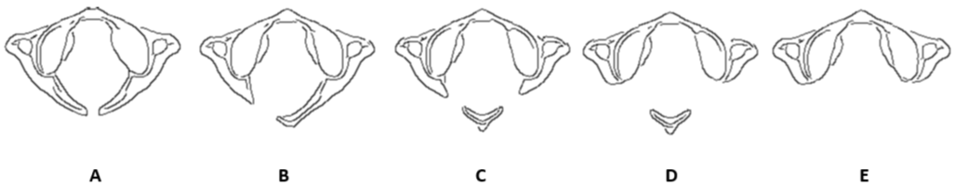
  • Type A: Failure of posterior midline fusion of the two hemiarches;
  • Type B: Unilateral defect;
  • Type C: Bilateral defects;
  • Type D: Absence of the posterior arch with a persistent posterior tubercle;
  • Type E: Absence of the entire posterior arch, including the tubercle.
Figure 1. Classification of C1 Posterior Arch Hypoplasia According to Currarino [6]. Currarino’s classification of C1 posterior arch hypoplasia includes midline fusion failure (A), unilateral arch defect (B), bilateral clefts with dorsal preservation (C), asymmetrical arch absence with an unattached tubercle (D), and complete arch and tubercle absence (E).
Applsci 15 01877 g001
For cephalometric analysis, the NHP (Natural Head Position) orientation was set using Dolphin software (12.0.9.58), which included three reference planes:
  • The transverse plane coinciding with the Frankfurt plane (FH), passing through Orbital (Or) and porion (Po);
  • The sagittal plane coinciding with the mid-sagittal plane (MSP), perpendicular to the FH and passing through the Crista galli (Cg) and Basion (Ba);
  • The coronal plane coinciding with the anteroposterior (PO) plane, perpendicular to the FH and MSP, and passing through the right and left porion.
Data Recording and Analysis: All findings and measurements were recorded using Microsoft Excel version 16.0 for Windows 10. The prevalence and distribution of APAD types were analyzed, with particular attention paid to the occurrence of any associated conditions such as Ponticulus Posticus.

3. Results

3.1. Prevalence and Classification of APAD

In our study cohort of 500 Italian patients, we identified a total of 13 cases of Atlas Posterior Arch Deficiency (APAD). The demographic distribution was balanced, with 50% males and 50% females. The ages of the participants ranged from 17 to 60 years.
According to the Currarino classification system, 13 cases of APAD were identified as type A. No type B, C, D, or E defects were observed. An ulterior classification was performed, in which the following results were obtained:
  • Partial APAD was the most common form, occurring in twelve cases;
  • Complete APAD was observed in one case.
Additionally, two cases of APAD were associated with the presence of Ponticulus Posticus.
Table 1 summarizes the distribution and classification of APAD in our study cohort.

3.2. Age Distribution

The age distribution of participants with APAD is detailed in Table 2. The results indicate the following distribution of APAD cases:
  • 17–29 years: two cases (15.4% of APAD cases; 1.7% within this age group)
  • 30–39 years: four cases (30.8% of APAD cases; 3.1% within this age group)
  • 40–49 years: three cases (23.1% of APAD cases; 2.4% within this age group)
  • 50–60 years: four cases (30.8% of APAD cases; 3.1% within this age group)
Table 2 provides a detailed breakdown of the age distribution among participants with APAD.
Table 2. Age distribution of participants with APAD.
Age RangeNumber of CasesPercentage (%)
17–29215.4
30–39430.8
40–49323.1
50–60430.8

3.2.1. Case Report 1: Bilateral Complete Ponticulus Posticus and Complete Atlantal Posterior Arch Defect Type A in a 17-Year-Old Female: Clinical and Imaging Findings

Diagnosis: Bilateral Complete Ponticulus Posticus with Complete APAD Type A.
Clinical Presentation: The patient, a 17-year-old female, was referred for a routine orthodontic assessment. Imaging with cone-beam computed tomography (CBCT) revealed a bilateral complete Ponticulus Posticus, accompanied by a complete absence of the posterior arch of the atlas (APAD Type A), as in Figure 2.
Symptoms: The patient reported occasional mild neck stiffness and localized discomfort, but no significant neurological symptoms or deficits were noted. The findings were incidental and did not significantly impact her daily activities.
Management: Given the asymptomatic nature of the condition, the patient was advised to continue regular monitoring. No immediate intervention was required. The patient was educated about the potential for future symptoms and the importance of reporting any changes.
Outcome: The patient remained symptom-free during the follow-up period, and no interventions were necessary. Routine imaging will continue to monitor for any future developments.

3.2.2. Case Report 2: Unilateral Partial Ponticulus Posticus and Partial Atlantal Posterior Arch Defect Type A in a 54-Year-Old Female: Clinical Presentation and Imaging Characteristics

Diagnosis: Unilateral Partial Ponticulus Posticus with Partial APAD Type A.
Clinical Presentation: A 54-year-old female presented for evaluation due to persistent neck pain and occasional sensory disturbances in the upper limb. CBCT imaging revealed a unilateral partial Ponticulus Posticus on the right side, associated with a partial absence of the posterior arch of the atlas (APAD Type A). The defect was characterized by the incomplete fusion of the posterior arch on the affected side, with a partially preserved posterior tubercle, as in Figure 3.
Symptoms: The patient experienced mild to moderate neck pain and intermittent tingling sensations in the right upper limb. These symptoms were exacerbated by prolonged periods of neck strain and certain head movements.
Management: The patient was initially managed with conservative measures, including physical therapy and nonsteroidal anti-inflammatory drugs (NSAIDs) for pain relief. Follow-up imaging was recommended to assess the stability of the defect and any progression of symptoms.
Outcome: The patient’s symptoms improved with conservative treatment, and no significant progression of the defect was observed on follow-up imaging. She continues to be monitored for any potential changes in her condition.

4. Discussion

This study sheds light on the prevalence and clinical significance of Atlas Posterior Arch Deficiency (APAD) among a cohort of 500 Italian patients. The detection of APAD through CBCT imaging has proven to be a valuable diagnostic tool, revealing that this anatomical variation, though relatively rare, holds notable clinical importance.
Congenital anomalies of the posterior atlas arch, such as atlanto-occipital assimilation and posterior arch aplasia (APAD), often present diagnostic challenges due to their typically asymptomatic nature and incidental discovery during radiographic examination. Effective identification and assessment of these anomalies are crucial for accurate diagnosis and appropriate management [10,11,12].
Cone-beam computed tomography (CBCT) has proven invaluable in the precise visualization and classification of APAD. CBCT provides high-resolution, three-dimensional imaging of the cervical spine, enabling the detailed examination of anatomical structures and abnormalities. This advanced imaging modality offers superior spatial resolution compared to conventional radiography, facilitating the detection of subtle variations in the atlas arch morphology associated with APAD.
In this study, the 13 identified cases of Type A APAD are described in Table 3 and reflect findings consistent with prior research. For instance, Choi et al. (2011) [7] described the congenital cleft of the anterior arch and the partial aplasia of the posterior arch, highlighting the diagnostic importance of such anomalies. Similarly, Klimo Jr. et al. (2003) reported a case where the partial aplasia of the posterior arch led to myelopathy, emphasizing the potential neurological consequences [8].
Hyun et al. (2018) [9] and Elmalky et al. (2013) [10] have contributed valuable insights regarding the prevalence and clinical presentations of C1 arch anomalies, reinforcing the need for thorough evaluation in patients with cervical spine symptoms. The classification systems by Izaki et al. (2009) [13] and Junior et al. (2021) [14] aid in categorizing these anomalies, improving communication among clinicians. Additionally, Jin et al. (2014) [15] and Butt et al. (2021) [16] discuss the associations between anterior arch hypertrophy and the congenital nonunion of the posterior arch, illustrating the complex interplay of structural variations within the C1 vertebra.
Clinical presentations such as cervical myelopathy, as detailed by Ogata et al. (2012) [17] and Chau et al. (2009) [18], highlight the importance of early recognition and management to prevent neurological sequelae. Despite the generally benign nature of certain anomalies, as noted by Tan et al. (2007) [19], individualized treatment approaches are essential based on the specific characteristics and clinical context of each case.
While Type A APAD is often asymptomatic, conservative management, including physical therapy and pain relief, is effective when symptoms arise. Surgical intervention, such as excision of the defective posterior arch, may be considered for persistent or significant symptoms. This approach can provide relief and prevent further complications [20,21,22,23,24,25,26].
Dental professionals play a critical role in the initial detection of APAD, particularly through the use of CBCT. Dental radiographic examinations, which are routine in orthodontic and other dental assessments, may reveal incidental findings of APAD. Awareness of these anomalies among dental practitioners is crucial for timely referral and management [21,25,27].
CBCT offers detailed imaging of the atlas arch morphology, allowing for the accurate identification of APAD variants and their associated anomalies. This capability enhances diagnosis and treatment planning, especially in cases where traditional radiographic techniques may be inconclusive.
Compared to traditional CT, CBCT uses lower radiation doses while providing high-quality imaging. This reduction in radiation exposure is advantageous for pediatric patients and those requiring repeated imaging.
In orthodontic practice, CBCT is commonly used for treatment planning and the assessment of craniofacial structures. Detection of APAD on CBCT can influence orthodontic treatment decisions, ensuring the safety and efficacy of interventions.
The effective management of APAD requires collaboration between dental and medical professionals. Dental practitioners may be the first to identify cervical spine abnormalities and should facilitate referrals to specialists for further evaluation and treatment. This interdisciplinary approach ensures comprehensive care and optimal patient outcomes.
Our findings indicate a predominance of partial APAD, with only a single case of complete APAD observed. This distribution aligns with the existing literature, which suggests that partial APAD is more common than complete APAD. Additionally, the presence of Ponticulus Posticus in two cases highlights the potential for additional anatomical variants that may impact clinical management.
The Currarino classification system (1994) [6] applied in this study exclusively identified Type A anomalies, reflecting a higher prevalence of this defect type in our cohort. This finding is consistent with other studies [20,21,22,23,24,25,26] and reinforces the utility of CBCT in the accurate detection and classification of APAD. CBCT’s detailed imaging capabilities make it an essential tool for orthodontists and other clinicians in detecting such variations and planning appropriate treatments.
In this paper, two representative cases were selected for detailed reporting to illustrate the diverse clinical presentations of APAD and its implications for orthodontic treatment. These cases were chosen for their ability to highlight the key aspects and potential complications associated with APAD [27]. The remaining cases, while not detailed individually, were consistent with these findings and support the overall conclusions of the study.
Although no symptomatic cases of APAD were found in our study, it is important for patients diagnosed with this condition to be informed about the potential for future symptoms and monitored accordingly. This proactive approach ensures that any emerging issues can be addressed promptly.
This study also highlights the need for further research to explore the clinical implications of APAD, especially in symptomatic cases and among diverse demographic groups with varying risk factors. Future studies should investigate the impact of APAD on clinical outcomes and patient management, aiming to refine diagnostic and treatment strategies.
Overall, this research provides a foundational understanding of APAD’s prevalence and classification, setting the stage for future investigations into its broader clinical significance.
The limitations of this study warrant further research to determine the true prevalence of APAD. Not all patients undergo CBCT imaging, and when performed, the primary indication is often unrelated to cervical spine evaluation. As a result, the prevalence of APAD may be underestimated. To address these limitations, future studies should include larger and more diverse populations from various institutions (not limited to orthodontic departments), along with longitudinal designs, to provide a more comprehensive understanding of the condition and its long-term outcomes.

5. Conclusions

This study provides a comprehensive evaluation of Atlas Posterior Arch Deficiency (APAD) in a cohort of 500 Italian patients using cone-beam computed tomography (CBCT). Our findings indicate that while APAD is a relatively rare anatomical variation, it has significant clinical relevance. We identified thirteen cases of APAD among the participants, which were predominantly classified as partial APAD, with only one instance of complete APAD observed. Additionally, two cases were associated with Ponticulus Posticus.
The application of the Currarino classification system (1994) [6] in our study revealed that only Type A anomalies were present, consistent with the existing literature which also reports a higher prevalence of Type A defects. This underscores the critical role of CBCT in accurately detecting and categorizing APAD, reinforcing its importance in orthodontic treatment planning and routine imaging for incidental findings.
Importantly, no symptomatic cases of APAD were detected in this cohort, and no specific interventions were required. Nevertheless, patients diagnosed with APAD should be informed about their condition and advised to monitor for any potential future symptoms.
Our study provides valuable insights into the prevalence and classification of APAD within this population and underscores the need for ongoing research to better understand its clinical implications. Future research should focus on exploring the impact of APAD on clinical outcomes and patient management to enhance our understanding and treatment strategies.

Author Contributions

Conceptualization: M.M., F.F., M.F. and F.R.; Methodology: M.M.; Software: M.M.; Validation: M.M., A.A., M.F. and F.R; Formal Analysis: M.M.; Investigation: M.M. and F.F.; Resources: M.M.; Data Curation: M.M.; Writing—Original Draft Preparation: M.M.; Writing—Review and Editing: M.M. and A.A.; Visualization: M.M.; Supervision: M.M.; Project Administration: M.M.; Funding Acquisition: M.M. All authors have read and agreed to the published version of the manuscript.

Funding

This research did not receive external funding.

Institutional Review Board Statement

Ethical approval (number 23, 8 November 2018) was obtained by the Independent Ethics Committee of Chieti hospital. The study protocol was drawn following the European Union Good Practice Rules and the Helsinki Declaration.

Informed Consent Statement

Written informed consent was obtained from all patients. Sensitive data were anonymized, retaining only age, gender, and exam date to ensure patient privacy.

Data Availability Statement

The data presented in this study are available on request from the corresponding author. The data are not publicly available due to privacy.

Conflicts of Interest

The authors declare no conflicts of interest.

References

  1. Pasini, M.; Giuca, M.R.; Ligori, S.; Mummolo, S.; Fiasca, F.; Marzo, G.; Quinzi, V. Association between Anatomical Variations and maxillary canine impaction: A retrospective study in orthodontics. Appl. Sci. 2020, 10, 5638. [Google Scholar] [CrossRef]
  2. Visocchi, M.; Signorelli, F.; Parrilla, C.; Paludetti, G.; Rigante, M. Multidisciplinary approach to the craniovertebral junction. Historical insights, current and future perspectives in the neurosurgical and otorhinolaryngological alliance. ACTA Otorhinolaryngol. Ital. 2021, 41 (Suppl. S1), S51. [Google Scholar] [CrossRef] [PubMed]
  3. Izzo, R.; Popolizio, T.; Balzano, R.F.; Simeone, A.; Gasparotti, R.; Scarabino, T.; Muto, M. Imaging of cranio-cervical junction traumas. Eur. J. Radiol. 2020, 127, 108960. [Google Scholar] [CrossRef] [PubMed]
  4. Khanna, R.; Smith, Z.A.; Dlouhy, B.J.; Dahdaleh, N.S. Complete absence of the posterior arch of C1: Case report. J. Craniovertebral Junction Spine 2014, 5, 176–178. [Google Scholar] [CrossRef]
  5. Wu, W.L.; Shao, X.B.; Shen, Y.G.; Chen, J.S.; Shu, Q. Sex-specific differences in ossification patterns of the atlas and axis: A computed tomography study. World J. Pediatr. WJP 2022, 18, 263–270. [Google Scholar] [CrossRef]
  6. Currarino GRollins, N.; Diehl, J.T. Congenital defects of the posterior arch of the atlas: A report of seven cases including an affected mother and son. Am. J. Neuroradiol. 1994, 15, 249–254. [Google Scholar]
  7. Choi, J.W.; Jeong, J.H.; Moon, S.M.; Hwang, H.S. Congenital cleft of anterior arch and partial aplasia of the posterior arch of the c1. J. Korean Neurosurg. Soc. 2011, 49, 178. [Google Scholar] [CrossRef]
  8. Klimo, P., Jr.; Blumenthal, D.T.; Couldwell, W.T. Congenital partial aplasia of the posterior arch of the atlas causing myelopathy: Case report and review of the literature. Spine 2003, 28, E224–E228. [Google Scholar] [CrossRef]
  9. Hyun, G.; Allam, E.; Sander, P.; Hasiak, C.; Zhou, Y. The prevalence of congenital C1 arch anomalies. Eur. Spine J. 2018, 27, 1266–1271. [Google Scholar] [CrossRef]
  10. Elmalky, M.M.; Elsayed, S.; Arealis, G.; Mehdian, H. Congenital C1 arch deficiency: Grand Round presentation. Eur. Spine J. 2013, 22, 1223–1226. [Google Scholar] [CrossRef]
  11. Macrì, M.; Perrella, G.; Varvara, G.; Murmura, G.; Traini, T.; Rendina, F.; Festa, F. Assessments of Prevalence of Ponticulus Posticus, Atlas Posterior Arch Deficiency, Sella Turcica Bridging, Maxillary Canine Impaction, and Associations Among Them in 500 CBCTs of Italian Orthodontic Patients. Front. Dent. Med. 2021, 2, 708169. [Google Scholar] [CrossRef]
  12. Torriani, M.; Lourenço, J.L.G. Agenesis of the posterior arch of the atlas. Rev. Do Hosp. Das Clínicas 2002, 57, 73–76. [Google Scholar] [CrossRef] [PubMed]
  13. Izaki, T.; Shiota, E.; Zhang, J. Aplasia of the posterior arch of the atlas: A proposal for a new clinical classification. Med. Bull. Fukuoka Univ. 2009, 36, 11–16. [Google Scholar]
  14. Junior, M.G.P.; Ribeiro, R.T.; Landeiro, J.A.; Pessoa, B.L. Hypoplasia of C1’s posterior arch: Is there an ideal anatomical classification? Surg. Neurol. Int. 2021, 12, 623. [Google Scholar] [CrossRef]
  15. Jin, M.; Asadoorian, M.; Hiller, L.P.; Hughes, T.H. Hypertrophy of the anterior arch of the atlas associated with congenital nonunion of the posterior arch: A retrospective case-control study. Spine J. 2014, 14, 1155–1158. [Google Scholar] [CrossRef]
  16. Butt, B.B.; Gagnet, P.; Patel, R.; Aleem, I. Congenital defect of the posterior arch of C1: A case report. J. Spine Surg. 2021, 7, 214. [Google Scholar] [CrossRef]
  17. Ogata, T.; Morino, T.; Hino, M.; Miura, H. Cervical myelopathy caused by atlantoaxial instability in a patient with an os odontoideum and total aplasia of the posterior arch of the atlas: A case report. J. Med. Case Rep. 2012, 6, 171. [Google Scholar] [CrossRef]
  18. Chau, A.M.T.; Wong, J.H.Y.; Mobbs, R.J. Cervical myelopathy associated with congenital C2/3 canal stenosis and deficiencies of the posterior arch of the atlas and laminae of the axis: Case report and review of the literature. Spine 2009, 34, E886–E891. [Google Scholar] [CrossRef]
  19. Tan, E.C.; Soon, H.C.; Kevin, M.; To, S. Congenital absence of the posterior arch of the atlas—A strange but benign anomaly. Malays. Orthop. J. 2007, 1, 30–32. [Google Scholar] [CrossRef]
  20. Sabuncuoğlu, H.; Özdoğan, S.; Karadağ, D.; Timurkaynak, E. Congenital hypoplasia of the posterior arch of the atlas: Case report and extensive review of the literature. Turk. Neurosurg. 2011, 21, 97–103. [Google Scholar] [CrossRef]
  21. Monsarrat, P.; Galibourg, A.; Nasr, K.; Telmon, N.; Maret, D. Incidental findings in dental radiology are concerning for family doctors. Open Med. 2019, 14, 467–478. [Google Scholar] [CrossRef] [PubMed]
  22. Senoglu, M.; Safavi-Abbasi, S.; Theodore, N.; Bambakidis, N.C.; Crawford, N.R.; Sonntag, V.K. The frequency and clinical significance of congenital defects of the posterior and anterior arch of the atlas. J. Neurosurg. Spine 2007, 7, 399–402. [Google Scholar] [CrossRef] [PubMed]
  23. Chitroda, P.K.; Katti, G.; Baba, I.A.; Najmudin, M.; Ghali, S.R.; Kalmath, B.; Vijay, G. Ponticulus posticus on the posterior arch of atlas, prevalence analysis in symptomatic and asymptomatic patients of Gulbarga population. J. Clin. Diagn. Res. JCDR 2013, 7, 3044. [Google Scholar] [PubMed]
  24. He, Q.; Xu, J. Congenital anterior midline cleft of the atlas and posterior atlanto-occipital fusion associated with symptomatic anterior atlantoaxial subluxation. Eur. J. Orthop. Surg. Traumatol. 2012, 22, 35–39. [Google Scholar] [CrossRef] [PubMed]
  25. Chu, C.Y.; Chan, R.T.M.; Fung, V.K.P.; Cheng, C.S. Congenital posterior arch defect of the atlas: Report of a rare anomaly. Hong Kong J. Radiol. 2011, 14, 102–105. [Google Scholar]
  26. Kim, M.S. Anatomical variant of atlas: Arcuate foramen, occpitalization of atlas, and defect of posterior arch of atlas. J. Korean Neurosurg. Soc. 2015, 58, 528–533. [Google Scholar] [CrossRef]
  27. Macrì, M.; Rendina, F.; Feragalli, B.; Pegreffi, F.; Festa, F. Prevalence of ponticulus posticus and migraine in 220 orthodontic patients: A cross-sectional study. Biology 2023, 12, 471. [Google Scholar] [CrossRef]
Figure 2. Imaging findings and clinical presentation of Case Report 1.
Applsci 15 01877 g002
Figure 3. Imaging findings and clinical presentation of Case Report 2.
Applsci 15 01877 g003
Table 1. Distribution and classification of APAD.
ClassificationNumber of CasesPercentage (%)
Partial APAD1292.3
Complete APAD17.7
APAD with Ponticulus Posticus215.4
Table 3. Summary of symptoms, management, and outcomes of 13 cases of Atlantal Posterior Arch Defect (APAD).
CaseAgeType of APADPresence of Ponticulus PosticusSymptomsManagementOutcome
117 yearsComplete APADBilateral CompleteOccasional mild neck stiffness; no significant neurological symptomsRoutine monitoring; no immediate intervention requiredRemained symptom-free; routine imaging for follow-up
254 yearsPartial APADUnilateral PartialMild to moderate neck pain; intermittent tingling in right upper limbPhysical therapy and NSAIDs; follow-up imaging recommendedSymptoms improved; no significant defect progression
335 yearsPartial APADNoneOccasional neck pain; no significant symptoms reportedConservative treatment; monitoring for any symptoms or changesStable condition; no significant progression noted
442 yearsPartial APADNoneMild neck discomfort; no significant symptomsConservative management; regular follow-up and monitoringStable; no notable changes during follow-up
531 yearsPartial APADNoneMild neck stiffness; no significant symptomsRoutine monitoring; conservative care as neededNo progression; symptoms remained stable
655 yearsPartial APADNoneOccasional discomfort in neckManaged with physical therapy; regular follow-upSymptoms improved; condition stable
728 yearsPartial APADNoneMild symptoms; no significant impactMonitoring; conservative care if symptoms ariseNo progression; stable condition
837 yearsPartial APADNoneOccasional pain; no significant neurological symptomsPhysical therapy and monitoringCondition stable; no significant changes
946 yearsPartial APADNoneMild discomfort; no significant symptomsRegular follow-up; conservative management if symptoms developStable; no major changes noted
1053 yearsPartial APADNoneMild neck stiffness; no significant symptomsRoutine monitoring; conservative managementSymptoms stable; no progression observed
1126 yearsPartial APADNoneMinimal symptoms; no impact on daily activitiesRegular follow-up; conservative care if neededCondition stable; no notable progression
1229 yearsPartial APADNoneNo significant symptomsMonitoring; conservative management if symptoms ariseStable; no significant changes during follow-up
1351 yearsPartial APADNoneMild neck stiffness; no significant symptomsMonitoring; conservative management if symptoms ariseStable; no significant changes during follow-up
Disclaimer/Publisher’s Note: The statements, opinions and data contained in all publications are solely those of the individual author(s) and contributor(s) and not of MDPI and/or the editor(s). MDPI and/or the editor(s) disclaim responsibility for any injury to people or property resulting from any ideas, methods, instructions or products referred to in the content.

