Article

Natural Strategies for Improving the Antioxidant Status and Health of Rabbits: The Role of Biochar and Tribulus terrestris

by
Katarzyna Karpińska
1,*,
Bożena Nowakowicz-Dębek
1,*,
Dorota Kowalska
2,
Paweł Bielański
2,
Łukasz Wlazło
1 and
Anna Czech
3
1
Department of Animal Hygiene and Environmental Hazards, Faculty of Animal Sciences and Bioeconomy, University of Life Sciences in Lublin, 13 Akademicka Street, 20-950 Lublin, Poland
2
Department of Small Livestock Breeding, National Research Institute of Animal Production, 31-047 Krakow, Poland
3
Department of Biochemistry and Toxicology, University of Life Sciences in Lublin, 13 Akademicka Street, 20-950 Lublin, Poland
*
Authors to whom correspondence should be addressed.
Appl. Sci. 2025, 15(23), 12515; https://doi.org/10.3390/app152312515
Submission received: 25 September 2025 / Revised: 19 November 2025 / Accepted: 23 November 2025 / Published: 25 November 2025

Abstract

This study evaluated the effect of dietary supplementation with biochar and Tribulus terrestris (TT) on oxidative stress and metabolic parameters in New Zealand White rabbits. A total of 80 weaned rabbits (35 days of age) were assigned to four groups (20 each): a control group (C) and three experimental groups supplemented with 0.25% biochar (E1), 0.25% biochar and 0.1% Tribulus terrestris (E2), or 0.1% Tribulus terrestris (E3). The feeding experiment lasted 78 days following a 7-day adaptation period. Hematological, biochemical, and redox parameters, including malondialdehyde (MDA), glutathione (GSH), total reducing capacity (FRAP), immunoglobulin G (IgG), and testosterone levels, were assessed and analyzed using one-way analysis of variance (ANOVA). Rabbits from group E1 exhibited the highest FRAP values (8.16 µmol/L; p < 0.05), whereas MDA concentrations were significantly elevated in groups E1 and C (2.02 and 1.83 µmol/L; p < 0.05), indicating increased lipid peroxidation. Groups E2 and E3 showed lower MDA levels (1.38 and 1.59 µmol/L; p < 0.05) and higher IgG concentrations (44.72 and 37.82 ng/mL; p < 0.05) compared to the control, suggesting improved antioxidant defense and immune status. GSH levels were significantly higher in groups E1 and E2 (6.34 and 6.79 µmol/L; p < 0.05). No adverse changes were observed in basic hematological and biochemical parameters. The results indicate that dietary supplementation with biochar and T. terrestris is safe and can beneficially modulate redox balance and immune response in rabbits, confirming their potential as natural feed additives in sustainable rabbit production.

1. Introduction

One of the main challenges in sustainable animal production remains the reduction in antibiotics and synthetic growth promoters, particularly in monogastric species such as rabbits. These animals are especially susceptible to intestinal disturbances, and oxidative stress associated with intensive rearing can lead to reduced immunity and metabolic disorders. In this context, there is growing interest in natural feed additives with antioxidant and immunomodulatory properties [1]. Among such additives, biochar—a product of biomass pyrolysis—is increasingly used. Owing to its porous structure, it can bind toxins and other undesirable feed compounds, thereby stabilizing the gastrointestinal environment. In rabbit nutrition, biochar is mainly applied as a natural sorbent for mycotoxins and as a factor supporting intestinal microflora [2,3]. Meta-analyses of in vitro and in vivo studies indicate that biochar supplementation can improve nutrient utilization, digestibility, and selected production parameters [4]. Experiments on rabbits have also shown that rice-husk biochar can improve growth rates without affecting hematological parameters or carcass traits, confirming its safety [5]. However, the effectiveness of biochar depends on the raw material type, pyrolysis conditions, and dosage, and the results of studies in rabbits remain inconsistent [6,7]. Another group of additives attracting growing attention are plant-derived substances such as Tribulus terrestris. This plant contains steroidal saponins and other bioactive compounds with potential antioxidant, anti-inflammatory, and immunomodulatory effects [8,9,10]. Pharmacological reviews emphasize its wide range of applications and diverse biological activities that may support metabolism and the body’s defense responses [11]. In poultry studies, T. terrestris supplementation has been shown to influence the expression of growth-related genes, including GH and IGF-1, which was associated with improved growth and performance [11,12]. In rabbits, T. terrestris extracts have been reported to enhance antioxidant and lipid parameters, especially under metabolic stress [13,14]. Other studies also confirm a dose-dependent effect and an influence on developmental parameters in young male rabbits [15]. Because biochar primarily acts within the gastrointestinal tract, reducing toxin availability and stabilizing microflora, and because T. terrestris exerts systemic effects through bioactive compounds that regulate metabolism and oxidative status, their combined use may produce complementary benefits. Despite numerous studies on each additive separately, no research has yet evaluated their combined impact on rabbit health and oxidative status, representing a notable research gap. Therefore, the aim of this study was to assess the effect of beechwood-derived biochar and Tribulus terrestris on selected health indicators and oxidative status in rabbits as part of a balanced feeding strategy.

2. Materials and Methods

2.1. Experimental Design

The animals were weaned at 35 days of age, and the experiment continued until they reached 120 days of age. At the beginning of the study, the average body weight of the rabbits was 826 g in the control group, 834.7 g in E1, 833.3 g in E2, and 834 g in E3. Eighty weaned rabbits were randomly assigned to four groups: one control group (C) and three experimental groups (E1, E2, and E3), matched for body weight and sex. Each group maintained an equal sex ratio (1:1). Animals of the same sex were housed two per cage (n = 2), representing n = 10 replicates. During the experiment, the animals were under constant veterinary supervision, were fed ad libitum, and had unrestricted access to drinking water. In each group, animals were housed in two-storey commercial wire-mesh cages equipped with a standard feeder and a nipple drinker. The cages measured 80 cm (length) × 60 cm (width) × 40 cm (height) and were placed inside a closed, climate-controlled building equipped with a chimney and mechanical exhaust ventilation, meeting current welfare requirements. During the first week, all groups underwent an adaptation period under identical housing, microclimatic conditions, and diet. Throughout the experiment, microclimatic parameters in the rabbit facility were monitored and averaged across all groups: temperature 15.1–20.1 °C (mean 17.8 °C) and relative humidity 37–65% (mean 47.7%).
Rabbits in the control group (C) were fed a standard diet without additives. Group E1 received a diet supplemented with 0.25% beechwood biochar. Group E2 received a diet containing 0.25% biochar and 0.1% Tribulus terrestris (TT). Rabbits in group E3 received a diet supplemented with 0.1% TT. The feed mixtures were prepared to provide 100% of the required diet components. Additives were included at levels of 0.25% (biochar) and 0.1% (TT), calculated on a fresh weight basis. The nutritional requirements of rabbits were determined according to Gugołek [16]. The biochar used in this study was produced according to a standardized procedure by an external certified company (patent protected). Tribulus terrestris was used as a powdered extract and added to the feed during pelleting.
The analyzed nutritional composition of the two main feed additives (biochar and T. terrestris) is presented in Table 1. The basic feed mixture used in this study was previously described by Wlazło [17]; it was used in two independent experiments with different designs. The diet supplied about 168 g/kg crude protein, 10.3–10.5 MJ/kg metabolizable energy, and 130 g/kg crude fiber. It also contained roughly 34 g/kg ether extract and 73 g/kg crude ash, with stable mineral levels, including calcium at around 8.3 g/kg and phosphorus at 7.1 g/kg. The amino-acid profile remained consistent, with lysine decreasing slightly from 7.6 to 6.7 g/kg across treatments.

2.2. Ethical Approval

The study was approved by the Local Ethics Committee for Experiments on Animals of the University of Life Sciences in Lublin, Poland (Decision no. 62/2023 of 11 September 2023) and was in compliance with European Union guidelines (Directive 2010/63/EU of the European Parliament and of the Council on the protection of animals used for scientific or educational purposes, adopted in Poland by Legislative Decree 266/2015).

2.3. Blood Analysis

Blood was obtained from 6 rabbits from each group, the marginal ear vein of the animals before slaughter, following local anesthesia with lidocaine ointment. The site was disinfected before and after the procedure, and the material was collected into heparinized tubes with a clot activator (Sarstedt, Nümbrecht, Germany). Whole blood was analyzed for red blood cell parameters and platelets, i.e., the number of red blood cells (RBC), hemoglobin (HGB), hematocrit (HCT), mean corpuscular volume (MCV), mean corpuscular hemoglobin (MCH), mean corpuscular hemoglobin concentration (MCHC), mean platelet volume (MPV), coefficient of variation in red cell distribution width (RDW-CV), red cell distribution width—standard deviation (RDW-SD), platelet count (PLT), platelet distribution width (PDW), and plateletcrit (PCT), as well as white blood cell parameters: the number of white blood cells (WBC), eosinophils (EOS), neutrophils (NEU), lymphocytes (LYM), and monocytes (MON) (MINDRAY BC-5000vet analyzer, MINDRAY, Shenzhen, China). To obtain plasma, blood samples were also centrifuged at 3000× g for 10 min to obtain plasma. Analyses were performed approximately 3–4 h after blood collection. Blood was stored at 4 °C. Afterwards, plasma aliquots was analyzed by spectrophotometry for selected biochemical parameters, i.e., albumins, ALT, AP, AST, total protein, total cholesterol, creatine kinase (CK), gamma-glutamyl transpeptidase (GGTP), glucose, urea, and triglycerides, as well as selected minerals, i.e., phosphorus, magnesium, calcium, and iron, using Roche rapid diagnostic tests.

2.4. Antioxidant Status

The plasma was also analyzed for redox status. The concentration of malondialdehyde (MDA), a marker of lipid peroxidation, was determined according to the method described by Esterbauer and Cheeseman [18]. The activity of antioxidant systems, including the ferric reducing ability of plasma (FRAP), was assessed spectrophotometrically according to the procedure outlined by Benzie et al. [19]. In addition, catalase (CAT) and superoxide dismutase (SOD) activity in the plasma and liver were measured using spectrophotometric methods as described by Aebi [20] and Beauchamp et al. [21], respectively.
The plasma was also analyzed for the testosterone level, by the immunoenzymatic method, using the Siemens IMMULITE 2000 XPi Immunoassay System (Siemens Healthineers/Forchheim, Germany), as well as for immune indicators, including the level of immunoglobulins—IgA (cat. no. QY-E30202), IgG (cat. no. QY-E30200), and IgM (cat. no. QY-E30199)—and interleukins IL2 (cat. no. QY-E30184), IL6 (cat. no. QY-E30180), and IL-8 (cat. no. QY-E30179) and lysozyme (LZM) (cat. no. QY-E30167), using ELISA immunoenzymatic assays (BioTek ELx808 plate reader, Janki, Poland; Sunrise™ ELISA plate reader, Tecan Austria GmbH, Grödig, Austria). According to the Qayee-Bio procedure (Shanghai Qayee Biotechnology Co., Ltd., Shanghai, China), for all parameters tested the repeatability was less than 15%, and accuracy (R—standard linear regression correlation coefficient with the expected p-value) was ≥0.9900.

2.5. Histopathological Examinations of the Liver and Kidneys

Liver and kidney samples collected from all slaughtered rabbits were fixed in 10% neutral buffered formalin (pH 7.2) for histopathological examination. The tissues were then dehydrated in alcohol solutions, acetone, and xylene in a tissue processor (Leica TP-1020; Leica Biosystems, Nussloch, Germany) before being embedded in paraffin blocks. For the analysis, 4 μm-thick tissue sections were stained with hematoxylin and eosin (HE) and observed under a light microscope (Nikon Eclipse E600; Nikon, Amstelveen, The Netherlands) [22].

2.6. Statistical Analysis

The collected data were analyzed using one-way analysis of variance (ANOVA) in Statistica (StatSoft, version 13.3). For each analyzed parameter, the following model was applied:
Yij = μ + αi + εij,
where αi is the fixed effect of the treatment group and εij is the random error.
Homogeneity of variance was assessed using Levene’s test, followed by ANOVA. Differences between means were evaluated using Tukey’s post hoc test, with statistical significance set at p < 0.05. Mean values (M) and standard error of the mean (SEM) are reported in the tables. Significant differences between groups are indicated by different letters (a, b, c …). The relationships between oxidative stress parameters and immune indices (Ig, IL, lysozyme) were analyzed using the Pearson correlation coefficient; the significance of correlations was tested using the t-statistic (n − 2 degrees of freedom).

3. Results

The results of blood analyses in rabbits receiving biochar and/or Tribulus terrestris are presented in Table 2, Table 3, Table 4, Table 5, Table 6 and Table 7 and in the figure. The mean values for red blood cell parameters are summarized in Table 2. The experiment revealed statistically significant differences in hematocrit (HCT; p = 0.001), with lower values in group E2 than in the control group (C). Significant differences were also noted for the mean corpuscular hemoglobin concentration (MCHC; p = 0.015), coefficient of variation in red cell distribution width (RDW-CV; p = 0.047), and red cell distribution width–standard deviation (RDW-SD; p < 0.001), with higher values in all experimental groups (E1, E2, and E3) compared to the control. Significant differences were further observed in platelet counts (PLT; p = 0.035), with the highest value in group E2 and the lowest in group E3. No significant differences were found for the remaining hematological parameters (p > 0.05). The red blood cell count and hemoglobin concentration were similar across all groups and consistent with reference values for the species [23]. Hematocrit was highest in the control group (40.63%) and differed significantly from group E2 (p = 0.001). The MCHC value was significantly higher in group E2 (34.25 g/dL) than in group C (p = 0.015). RDW-CV differed significantly among groups (p = 0.047), with the lowest value in the control group (12.90%) and the highest in E2 (14.15%). RDW-SD also differed significantly between groups (p < 0.001), with the control group showing the lowest value (31.77 µm3) and all experimental groups exhibiting higher ones (E1: 35.05 µm3; E2: 34.90 µm3; E3: 34.70 µm3). Platelet count (PLT) was significantly different (p = 0.035), reaching the highest value in group E2 (455.50 × 10⁶/µL) and the lowest in E3 (227.50 × 10⁶/µL) (Table 2).
The leukocyte parameters of rabbits are presented in Table 3 and Table 4. The total leukocyte count (WBC) differed significantly between groups (p < 0.001). The highest value was recorded in the control group (12.10 × 10³/µL), while lower values were observed in E1 (8.87 × 10³/µL), E2 (6.14 × 10³/µL), and E3 (5.19 × 10³/µL). Neutrophil count (NEU) also differed significantly (p < 0.001), with the highest level in the control group (4.70 × 10³/µL) and markedly lower levels in all experimental groups. Monocyte count (MON) was significantly higher in the control group compared with the experimental groups (p < 0.001) (Table 3).
Statistically significant differences (p = 0.010) were shown between groups for the plasma level of albumins in the rabbits. The albumin level in group E1 was about 15% higher than in the control group (C). Significant differences were also noted for the total cholesterol concentration in the plasma (p = 0.001), which was several times as high in groups E1 and E2 as in the control group (more than 2.5 and 3 times, respectively). GGTP activity was also significantly higher in all experimental groups than in the control group; it was more than three times as high in group E1 and twice as high in groups E2 and E3 (p < 0.001) (Table 4). Statistically significant differences were also noted for the triglyceride concentration (p < 0.001; Table 4), which was about 54% higher in group E1 than in the control group, but significantly lower in groups E2 and E3 than in group C, by 34% and 53%, respectively. Analysis of the content of minerals in the plasma of rabbits revealed a significant increase in the phosphorus concentration, especially in group E3, in which it was markedly higher than in the control group. A similar tendency was observed for the magnesium level, which increased by about 25% in group E3. The iron level in the control group was significantly lower compared to the experimental groups that received the tested supplements (p = 0.000).
Regarding plasma immune parameters, the IgG level was significantly higher in group E2 than in the remaining groups (p = 0.036), while no significant differences were noted for IgA or IgM (p > 0.05). Similarly, the concentrations of interleukins IL-2, IL-6, and IL-8, as well as lysozyme, were comparable among groups, with no statistically significant differences (Table 5).
Indicators of oxidative status showed significant intergroup differences (Table 6). Malondialdehyde (MDA) concentration was lowest in group E2 and highest in E1 (p < 0.001). Superoxide dismutase (SOD) activity was highest in the control group and lowest in E3 (p = 0.009). Catalase (CAT) activity reached its highest levels in the control and E3 groups, while values in E1 and E2 were significantly lower (p < 0.001). Glutathione (GSH) concentration was significantly higher in E1 and E2 compared with the control and E3 (p < 0.001). FRAP was highest in group E1, slightly lower in E2, and lowest in E3, with significant differences confirmed among groups (p < 0.001).
Analysis of oxidative stress markers and immunological parameters revealed significant correlations (Table 7). In the control group, IL-2 and MDA levels were positively correlated (r = 0.8576, p = 0.029). A strong negative correlation was observed between catalase (CAT) activity and IL-6 (r = −0.9668, p = 0.002), as well as between reduced glutathione (GSH) and IL-6 levels (r = −0.8255, p = 0.043). These findings indicate stronger oxidative–inflammatory responses in animals that did not receive supplementation. In the experimental groups (E2 and E3), significant correlations were noted between antioxidant markers (CAT, FRAP) and immunoglobulin M (IgM) levels (r = 0.8266, p = 0.043 and r = −0.9031, p = 0.014, respectively), suggesting a more balanced immune response and lower oxidative stress as a result of supplementation.
Testosterone levels were similar across all groups, ranging from 1.07 ng/mL in the control group to 2.54 ng/mL in group E2 (Figure 1).
The hematological values obtained in this study are consistent with reference data for rabbits [23].
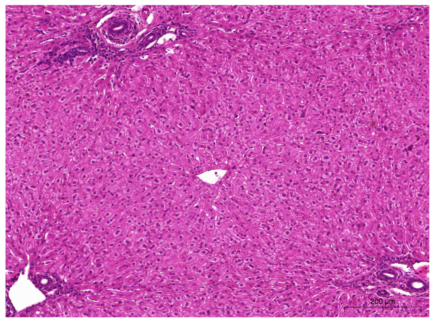
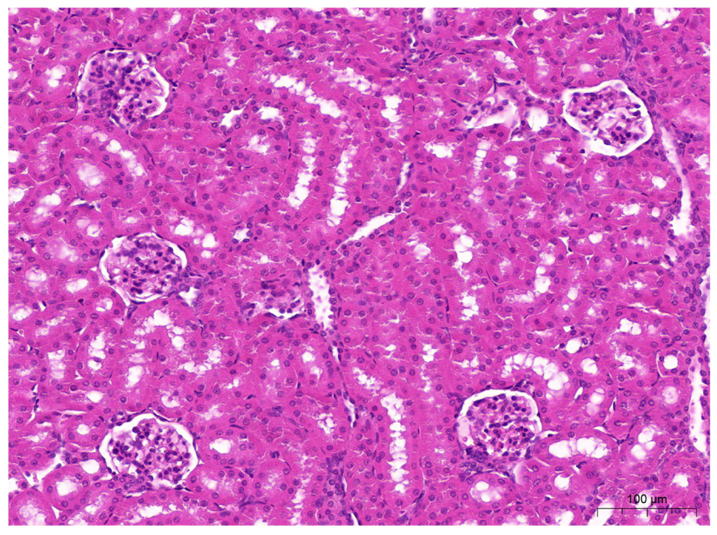
Microscopic examination of liver sections from animals in all groups revealed no significant differences in the structure of this organ. Similarly, no significant differences were observed in kidney sections from animals in the study groups (Figure 2 and Figure 3).

4. Discussion

The study confirmed that both biochar and Tribulus terrestris exert distinct yet partially complementary effects on rabbits, influencing metabolic, oxidative, and immunological balance without disturbing safety parameters. Both additives maintained hematological values within physiological limits, indicating no disruption of hematopoiesis or general nutritional status. Higher leukocyte counts, particularly neutrophils and monocytes, were observed in the control group, suggesting greater immune activation in the absence of supplementation. The more stable hematological profile in the biochar group may be attributed to its stabilizing influence on the intestinal environment, consistent with previous findings on the sorptive and microbiota-modulating properties of biochar [4,5,24]. Similar stabilizing effects on blood parameters in rabbits supplemented with biochar were reported by Ansah et al. [5]. The biochemical profile revealed distinct differences between the effects of T. terrestris and biochar. Supplementation with T. terrestris significantly increased total cholesterol concentration, which aligns with the known activity of steroidal saponins on lipid and steroid biosynthesis [11,14,15]. Despite changes in lipid metabolism, the stability of ALT, AST, and AP activities confirmed the absence of hepatotoxicity of T. terrestris at the applied doses, in agreement with Ismaiel et al. [25]. The increased CK activity could result from enhanced anabolic metabolism, consistent with the known effects of phytosteroids in T. terrestris [11,14,15]. Biochar exhibited a stabilizing and supportive influence on metabolic processes, as evidenced by elevated albumin levels and unchanged liver enzyme activities. This effect may be related to improved nutrient bioavailability due to its sorptive properties, as previously demonstrated by Qomariyah et al. [4] and in fermentation and metabolic studies involving biochar derived from wine products [24]. The increase in GGTP activity observed across all supplemented groups should be interpreted as a marker of intensified cell membrane turnover rather than pathology, consistent with earlier observations regarding biochar [5,24,26]. Marked effects of supplementation were observed in mineral metabolism. The significant increases in phosphorus, magnesium, and iron concentrations in groups E1, E2, and E3 may result from both the T. terrestris-induced enhancement of mineral bioavailability and the physicochemical effects of biochar within the intestinal environment. The particularly large rise in Fe observed in the biochar group aligns with findings on bamboo biochar’s influence on lipid metabolism and LDL oxidation [26]. The oxidative profile demonstrated that biochar acted as the most potent modulator of redox homeostasis. The highest FRAP and MDA values in group E1 may indicate activation of compensatory mechanisms enhancing antioxidant potential. This suggests that the supplement positively influences iron levels, reflected in the increased FRAP values [24,26]. T. terrestris, on the other hand, primarily elevated SOD, CAT, and GPx activities, but did not reduce MDA to the same extent as biochar, likely due to its impact on lipid metabolism. This observation agrees with previous reports on the immunomodulatory and antioxidant properties of T. terrestris [11,14,15]. In the combined E3 group, partial normalization of MDA was observed alongside an increase in FRAP, suggesting a synergistic interaction between T. terrestris and biochar in supporting intestinal and systemic redox balance. The concurrent increase in testosterone in this group may reflect the dual action of T. terrestris as a modulator of steroid hormones and biochar as a redox stabilizer and enhancer of nutrient absorption. All these findings are consistent with the histopathological results, which showed no organ damage. The absence of degenerative changes in the liver, kidneys, and gastrointestinal tract confirms the safety of the applied doses of biochar and T. terrestris and supports the agreement between biochemical and tissue-level observations. Overall, biochar primarily acts by stabilizing the intestinal microenvironment, reducing oxidative stress, and improving nutrient and mineral bioavailability. Tribulus terrestris, in turn, modulates lipid metabolism and hormonal activity while enhancing endogenous antioxidant defenses. Combining both additives appears more advantageous than their individual use, as biochar reduces oxidative stress and supports nutrient absorption, whereas T. terrestris enhances metabolic and hormonal responses. These results align with modern nutritional strategies that promote animal health and immunity, consistent with sustainable production principles and preventive nutrition aimed at maintaining systemic homeostasis.
Supplementing rabbit feed with natural additives represents a promising direction in the context of increasing consumer demand for sustainably produced animal products and efforts to reduce the environmental footprint of animal farming. As indicated in Khaleel et al. [27], natural feed additives offer a safe and effective alternative that enhances animal welfare and mitigates oxidative stress, a key factor in antibiotic resistance development. Properly selected natural substances can strengthen antioxidant mechanisms and limit inflammation. In this regard, it is important to emphasize that the biochar used in this study—derived from beechwood under strictly controlled technological conditions—serves as an example of a natural additive with the potential to support antioxidant processes and overall animal health. However, the physicochemical characteristics and biological activity of biochar depend strongly on feedstock type and pyrolysis parameters, such as temperature and residence time. Consequently, results may differ from those obtained using biochars produced from other materials. Future research should explore the influence of different biochar sources and production parameters to better understand their roles and mechanisms in animal nutrition.
Despite these limitations, the present results highlight the potential for developing standardized biochar formulations with defined composition and functionality, enabling safe and effective application in sustainable livestock production. Tribulus terrestris, known for its steroidal saponins that positively influence metabolism and immunity, also shows promising potential, though its effects may vary depending on phytochemical composition, plant source, dosage, and supplementation duration. Further studies are required to optimize its use in animal production systems.

5. Conclusions

The combined use of biochar and Tribulus terrestris in rabbit nutrition demonstrates synergistic potential for enhancing animal health and supporting sustainable production. The observed increase in antioxidant capacity indicates improved resilience against oxidative stress, which may strengthen immune mechanisms, stabilize physiological functions, and enhance overall productivity.
The elevated testosterone levels observed in rabbits receiving both biochar and T. terrestris may reflect stimulated anabolic processes and favorable shifts in muscle, fat, and bone proportions, contributing to improved growth performance and carcass quality. Although further research is required to clarify effects on production and reproduction, the results suggest that combining biochar with T. terrestris represents a promising natural strategy to enhance health, performance, and welfare in rabbits within sustainable production systems.

Author Contributions

Conceptualization, B.N.-D. and K.K.; methodology, K.K., B.N.-D. and D.K.; supervision, P.B., Ł.W. and A.C.; investigation, K.K., D.K. and P.B.; data curation, D.K., P.B. and A.C.; writing—original draft preparation, B.N.-D. and K.K.; writing—review and editing, D.K., P.B., Ł.W. and A.C. All authors have read and agreed to the published version of the manuscript.

Funding

This research received no external funding.

Institutional Review Board Statement

All research methods were approved by the Local Ethics Committee for Experiments on Animals of the University of Life Sciences in Lublin, Poland (Decision no. 62/2023 of 11 September 2023), and the study was in compliance with European Union guidelines (Directive 2010/63/EU of the European Parliament and of the Council on the protection of animals used for scientific or educational purposes, adopted in Poland by Legislative Decree 266/2015).

Informed Consent Statement

All methods were carried out in accordance with relevant guidelines and regulations. Informed consent was obtained from the animal’s owner. Signed the consent of the ethical approval and has the right to look after animals.

Data Availability Statement

The datasets presented in this article are not readily available because the data are part of an ongoing study. Requests to access the datasets should be directed to katarzyna.karpinska@up.lublin.pl.

Conflicts of Interest

The authors declare no conflicts of interest.

Abbreviations

The following abbreviations are used in this manuscript:
TTTribulus terrestris
NZWNew Zealand White
RBCred blood cells
HGBhemoglobin
MCV mean corpuscular volume
RDW-CVcoefficient of variation in red cell distribution width
RDW-SDred cell distribution width—standard deviation
PLTplatelet count
PDWplatelet distribution width
PCTplateletcrit
WBCwhite blood cells
EOSeosinophils
NEUneutrophils
LYMlymphocytes
MONmonocytes
CKcreatine kinase
GGTPgamma-glutamyl transpeptidase
MDAmalondialdehyde
FRAPferric reducing ability of plasma
CATcatalase
SODsuperoxide dismutase
HCThematocrit
ALTalanine aminotransferase
APalkaline phosphatase
ASTaspartate aminotransferase
GSHglutathione concentration

References

  1. Siddiqui, S.A.; Gerini, F.; Ikram, A.; Saeed, F.; Feng, X.; Chen, Y. Rabbit meat—Production, consumption and consumers’ attitudes and behavior. Sustainability 2023, 15, 2008. [Google Scholar] [CrossRef]
  2. Molenda, J.; Drabik, J. Study of the energetic properties of biochars from waste plant biomass using differential scanning calorimetry. Przem. Chem. 2024, 103, 525–528. [Google Scholar] [CrossRef]
  3. Medyńska-Juraszek, A.; Ćwieląg-Piasecka, I. Effect of biochar application on heavy metal mobility in soils impacted by copper smelting processes. Pol. J. Environ. Stud. 2020, 29, 1749–1757. [Google Scholar] [CrossRef] [PubMed]
  4. Qomariyah, N.; Ella, A.; Ahmad, S.N.; Yusriani, Y.; Sholikin, M.M.; Prihambodo, T.R.; Retnani, Y.; Jayanegara, A.; Wina, E.; Permana, I.G. Dietary biochar as a feed additive for increasing livestock performance: A meta-analysis of in vitro and in vivo experiment. Czech J. Anim. Sci. 2023, 68, 2. [Google Scholar] [CrossRef]
  5. Ansah, K.O.; Osafo, E.L.K.; Osman, A.; Opoku, G.C.; Ansah, A.S. Dose-dependent effects of biochar inclusion on growth, hematological, manure and carcass characteristics of rabbits. Ghana J. Anim. Sci. 2024, 15, 60–76. [Google Scholar] [CrossRef]
  6. Graves, C.; Kolar, P.; Shah, S.; Grimes, J.; Sharara, M. Can biochar improve the sustainability of animal production? Appl. Sci. 2022, 12, 5042. [Google Scholar] [CrossRef]
  7. Prasai, T.P.; Walsh, K.B.; Bhattarai, S.P.; Midmore, D.J.; Van, T.T.; Moore, R.J.; Stanley, D. Biochar, bentonite and zeolite supplemented feeding of layer chickens alters intestinal microbiota and reduces Campylobacter load. PLoS ONE 2016, 11, e0154061. [Google Scholar] [CrossRef]
  8. Placha, I.; Gai, F.; Pogány Simonová, M. Natural feed additives in animal nutrition—Their potential as functional feed. Front. Vet. Sci. 2022, 9, 1062724. [Google Scholar] [CrossRef]
  9. Abdelsalam, M.; Fathi, M. Improving productivity in rabbits by using some natural feed additives under hot environmental conditions—A review. Anim. Biosci. 2023, 36, 540. [Google Scholar] [CrossRef]
  10. Belhassen, T. Feed additives in rabbit nutrition to ensure sustainability. In Sustainable Use of Feed Additives in Livestock: Novel Ways for Animal Production; Springer International Publishing: Cham, Switzerland, 2023; pp. 859–871. [Google Scholar] [CrossRef]
  11. Saeed, M.; Munawar, M.; Bi, J.B.; Ahmed, S.; Ahmad, M.Z.; Kamboh, A.A.; Chen, H. Promising phytopharmacology, nutritional potential, health benefits, and traditional usage of Tribulus terrestris L. herb. Heliyon 2024, 10, 4. [Google Scholar] [CrossRef]
  12. Lee, B.; Jung, J.M.; Song, J.G.; Gwon, H.; Shin, H.; Tsang, Y.F.; Kim, H.W.; Kwon, E.E. Hazardous potential evaluation of biochar exposure on mice through analyses of gut-microbiome and fatty acids in brain. Chem. Eng. J. 2023, 461, 142006. [Google Scholar] [CrossRef]
  13. Reggi, S.; Frazzini, S.; Fusi, E.; Guagliano, M.; Cristiani, C.; Onelli, E.; Moscatelli, A.; Rossi, L. Biochar’s adsorption of Escherichia coli and probiotics Lactiplantibacillus plantarum and Limosilactobacillus reuteri and its impact on bacterial growth post in vitro digestion. Appl. Sci. 2025, 15, 5090. [Google Scholar] [CrossRef]
  14. Tuncer, M.A.; Yaymaci, B.; Sati, L.; Cayli, S.; Acar, G.; Altug, T.; Demir, R. Influence of Tribulus terrestris extract on lipid profile and endothelial structure in developing atherosclerotic lesions in the aorta of rabbits on a high-cholesterol diet. Acta Histochem. 2009, 111, 488–500. [Google Scholar] [CrossRef] [PubMed]
  15. Abadjieva, D.; Abadjiev, M.; Ribarski, S.; Penchev, P. Dose-dependent effect of Tribulus terrestris dry extract on reproductive organs of growing male rabbits. Maced. J. Anim. Sci. 2019, 9, 19–23. [Google Scholar] [CrossRef]
  16. Gugołek, A. (Ed.) Nutritional Recommendations and Feed Value for Fur Animals; Polish Society of Animal Production: Jabłonna, Poland, 2011. [Google Scholar]
  17. Wlazło, Ł.; Kowalska, D.; Bielański, P.; Chmielowiec-Korzeniowska, A.; Ossowski, M.; Łukaszewicz, M.; Czech, A.; Nowakowicz-Dębek, B. Effect of fermented rapeseed meal on the gastrointestinal microbiota and immune status of rabbit (Oryctolagus cuniculus). Animals 2021, 11, 716. [Google Scholar] [CrossRef]
  18. Esterbauer, H.; Cheeseman, K.H. Determination of aldehydic lipid peroxidation products malonaldehyde and 4-hydroxynonenal. Methods Enzymol. 1990, 186, 407–421. [Google Scholar] [CrossRef]
  19. Benzie, I.F.F.; Strain, J.J. The ferric reducing ability of plasma (FRAP) as a measure of “antioxidant power”: The FRAP assay. Anal. Biochem. 1996, 239, 70–76. [Google Scholar] [CrossRef]
  20. Aebi, H. Catalase in vitro. Methods Enzymol. 1984, 105, 121–126. [Google Scholar] [CrossRef]
  21. Beauchamp, C.; Fridovich, I. Superoxide dismutase: Improved assays and an assay applicable to acrylamide gels. Anal. Biochem. 1971, 44, 276–287. [Google Scholar] [CrossRef]
  22. Zawistowski, S. Histological Techniques, Histology, and Foundations of Histopathology; PZWL Medical Publishing: Warsaw, Poland, 1986. [Google Scholar]
  23. Winnicka, A. Wartości Referencyjne Podstawowych Badań Laboratoryjnych w Weterynarii, 1st ed.; Wydawnictwo SGGW: Warszawa, Poland, 1997. [Google Scholar]
  24. Schmidt, H.P.; Hagemann, N.; Draper, K.; Kammann, C. The use of biochar in animal feeding. PeerJ 2019, 7, e7373. [Google Scholar] [CrossRef]
  25. Ismaiel, S.I.; Farouk, S.M.; El-Ramady, R.A.; Khalil, W.F. Ameliorative impacts of Tribulus terrestris against ivermectin-induced hepato-renal toxicity in rabbit: Pharmacological and histopathological study. Am. J. Anim. Vet. Sci. 2017, 12, 8–16. [Google Scholar] [CrossRef]
  26. Cui, C.; Yang, W.; Dang, W.; Chen, R.; García-Caparrós, P.; Yang, G.; Huang, L.J. Bamboo biochar and sodium silicate alleviate oxybenzone-induced phytotoxicity via distinct mechanisms for sustainable plant protection. Plants 2025, 14, 2382. [Google Scholar] [CrossRef]
  27. Khaleel, H.K.; Abdelnour, S.A.; Osailan, R.; El-Kholy, K.H.; El-Haroun, E.; El-Nagar, H.A.; Mehilp, S.T.; El-Raghi, A.A.; Hassan, M.A.; Moustafa, M.; et al. Improving heat resilience in fattening rabbits: Nutritional strategies for mitigation via regulating blood physiology, inflammation and antioxidant pathways. Front. Vet. Sci. 2025, 12, 1677144. [Google Scholar] [CrossRef]
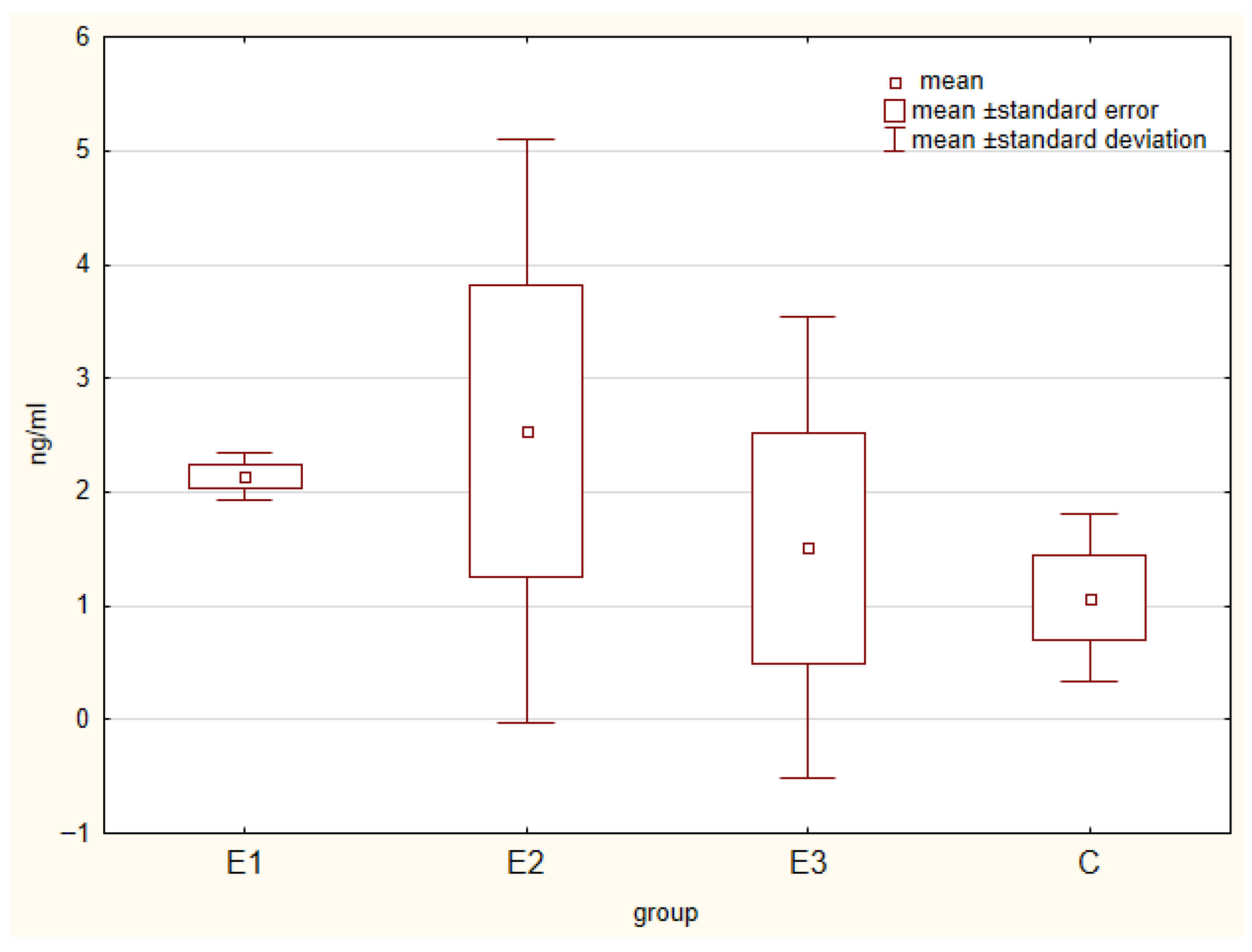
Figure 1. Testosterone levels in the plasma of rabbits [ng/mL].
Applsci 15 12515 g001
Figure 2. Liver sample of rabbits from E1 group. Hepatocytes with abundant granular cytoplasm and an oval, centrally located nucleus form trabeculae radiating from the central vein towards the portal-biliary spaces. HE. Bar = 200 µm.
Applsci 15 12515 g002
Figure 3. Kidney sample of rabbits from E1 group. Glomeruli with a visible filtration space, adjacent to the proximal and distal convoluted tubules. HE. Bar = 100.
Applsci 15 12515 g003
Table 1. Nutritional composition of the two main feed additives (biochar and T. terrestris).
ComponentBiocharTribulus terrestris Extract
Dry matter (%)95–9690
Crude protein (%)-<1
Crude fiber (%)77–805–8
Crude ash (%)25–281–2
Fat (%)-<1
Volatile matter (Vdaf, %)5.3-
Key bioactive compounds-Saponins (protodioscin 47%, dioscin 14%)
Table 2. Means for red blood cell parameters in the blood of rabbits.
ParameterGroupSEMp-Value
CE1E2E3
RBC [106/uL]6.666.235.806.140.1220.085
HGB [g/dL]13.6313.5512.0013.250.1850.090
HCT [%]40.63 a39.70 ab35.00 b39.40 ab0.5990.001
MCV [fL]61.1763.8560.9064.200.5890.077
MCH [pg]20.5321.8020.9021.650.2130.102
MCHC [g/dL]33.60 a34.15 ab34.25 b33.70 ab0.0920.015
MPV [um3]6.836.456.006.500.1430.234
RDW-CV [%]12.90 a13.60 ab14.15 b13.45 ab0.1640.047
RDW-SD [um3]31.77 a35.05 b34.90 b34.70 b0.353<0.001
PLT [106/uL]252.00 ab364.00 ab455.50 a227.50 b32.3720.035
PDW 16.0716.0015.8016.050.0400.056
PCT [%]0.160.240.280.160.0210.142
SEM—standard error of mean, a, b—means with different superscripts within a row are significantly different at p < 0.05; RBC—red blood cells; HGB—hemoglobin; HCT—hematocrit; MCV—mean corpuscular volume; MCH—Mean Corpuscular Hemoglobin; MCHC—Mean Corpuscular Hemoglobin Concentration; MPV—Mean Platelet Volume; RDW-CV—coefficient of variation in red cell distribution width; RDW-SD—red cell distribution width—standard deviation; PLT—platelet count; PDW—platelet distribution width.
Table 3. Mean leukocyte parameters in the blood of rabbits (103/µL).
ParameterGroupSEMp-Value
CE1E2E3
WBC12.10 a8.87 b6.14 c5.19 c0.614<0.001
EOS0.290.270.150.130.0370.278
NEU4.70 a1.85 b1.93 b1.08 b0.341<0.001
LYM5.366.283.403.280.2880.199
MON1.43 a0.62 b0.73 b0.48 b0.100<0.001
SEM—standard error of mean; a, b, c—means with different superscripts within a row are significantly different at p < 0.05; WBC—white blood cells; EOS—eosinophils; NEU—neutrophils; LYM—lymphocytes; MON—monocytes.
Table 4. Mean values for biochemical parameters and minerals in the plasma of rabbits.
ParameterGroupSEMp-Value
CE1E2E3
Albumins
[g/dL]
5.74 a6.60 b5.74 a6.07 ab0.1130.010
ALT [U/L]14.0424.1223.7122.281.1400.339
AP [U/L]200.93310.37240.44225.3115.5970.066
AST [U/L]15.2623.3218.9716.751.4010.195
Total protein [g/dL]6.026.135.655.540.0830.263
Total cholesterol [mg/dL]34.19 a90.28 b105.08 b89.24 b7.7320.001
CK [U/L]1246.921941.472548.982045.35172.3990.051
GGTP [U/L]5.84 a15.54 b12.03 b11.36 b0.838<0.001
Glucose [mg/dL]113.35123.30111.17121.041.7940.065
Urea [mg/dL]36.0039.2834.0133.181.0650.181
Triglycerides [mg/dL]128.60 ab198.67 b85.33 a60.20 a14.052<0.001
Phosphorus [mg/dL]4.55 a6.62 b6.82 b7.31 b0.234<0.001
Iron [ug/dL]134.15 a253.17 ab186.65 b216.10 b10.6660.000
Magnesium [mg/dL]2.33 a2.47 a2.78 b2.91 b0.0610.000
Calcium [mg/dL]15.2815.9415.3115.420.1400.312
SEM—standard error of mean; a, b—means with different superscripts within a row are significantly different at p < 0.05; ALT—alanine aminotransferase; AP—alkaline phosphatase; AST—aspartate aminotransferase; CK—creatine kinase; GGTP—gamma-glutamyl transpeptidase.
Table 5. Plasma concentrations of immunoglobulins, interleukins, and lysozyme in rabbits receiving natural feed additives.
ParameterGroupSEMp-Value
CE1E2E3
IgA [ng/mL]5.536.457.867.850.2330.450
IgG [ng/mL]31.66 a36.73 b44.72 bc37.82 b1.0490.036
IgM [ng/mL]12.2916.4913.5513.210.3590.972
IL-2 [pg/mL]8.006.876.787.350.1050.201
IL-6 [pg/mL]117.5684.9697.5886.992.8390.396
IL-8 [pg/mL]22.7033.0925.5427.390.9680.673
LZM [ng/mL]20.5924.9625.8520.740.6990.771
SEM—standard error of mean; a, b, c—means with different superscripts within a row are significantly different at p < 0.05; LZM—lysozyme.
Table 6. Indicators of redox status in the blood of rabbits.
ParameterGroupSEMp-Value
CE1E2E3
MDA [umol/L]1.83 bc2.02 b1.38 a1.59 c0.068<0.001
SOD [U/mL]27.29 a26.02 b25.82 bc21.72 c0.5580.009
CAT [U/mL]20.12 ac17.49 a16.89 b21.19 b0.511<0.001
GSH [umol/L]5.35 ac6.34 a6.79 b4.69 b0.230<0.001
FRAP [umol/L]7.46 a8.16 b7.97 ab5.33 b0.310<0.001
SEM—standard error of mean; a, b, c—means with different superscripts within a row are significantly different at p < 0.05; MDA—malondialdehyde; SOD—superoxide dismutase; CAT—catalase; GSH—reduced glutathione; FRAP—ferric reducing ability of plasma.
Table 7. Pearson correlation coefficients between oxidative stress markers and serum levels of immunoglobulins and interleukins.
Oxidative Stress MarkerExperimental GroupIgAIgGIgMIL2IL6IL8
MDAC−0.25110.49990.49710.8576−0.6632−0.1528
p = 0.631p = 0.313p = 0.316p = 0.029p = 0.151p = 0.773
E10.6956−0.1906−0.25680.44030.3512−0.3272
p = 0.125p = 0.718p = 0.623p = 0.382p = 0.495p = 0.527
E20.43460.74910.8115−0.7104−0.43240.3014
p = 0.389p = 0.087p = 0.050p = 0.114p = 0.392p = 0.562
E3−0.27960.63060.8166−0.2820.05530.0855
p = 0.591p = 0.179p = 0.047p = 0.588p = 0.917p = 0.872
SODC−0.17380.46670.66960.7121−0.273−0.1925
p = 0.742p = 0.351p = 0.146p = 0.112p = 0.601p = 0.715
E1−0.54490.0179−0.45640.4834−0.17280.449
p = 0.264p = 0.973p = 0.363p = 0.331p = 0.743p = 0.372
E2−0.4864−0.2282−0.72370.48810.57480.0573
p = 0.328p = 0.664p = 0.104p = 0.326p = 0.233p = 0.914
E30.2169−0.3725−0.72840.70010.27410.0352
p = 0.680p = 0.467p = 0.101p = 0.121p = 0.599p = 0.947
CATC−0.6161−0.04050.16760.5012−0.9658−0.2864
p = 0.193p = 0.939p = 0.751p = 0.311p = 0.002p = 0.582
E10.48340.7340.3477−0.43290.13490.5003
p = 0.331p = 0.097p = 0.500p = 0.391p = 0.799p = 0.312
E20.75050.4990.7532−0.3811−0.61020.0992
p = 0.086p = 0.314p = 0.084p = 0.456p = 0.198p = 0.852
E3−0.2810.57260.8266−0.5562−0.11730.1359
p = 0.590p = 0.235p = 0.043p = 0.252p = 0.825p = 0.797
GSHC−0.3470.30440.27310.7181−0.8255−0.2196
p = 0.500p = 0.558p = 0.601p = 0.108p = 0.043p = 0.676
E10.45640.75230.1794−0.38160.12540.6112
p = 0.363p = 0.084p = 0.734p = 0.455p = 0.813p = 0.197
E20.70740.25070.6622−0.0997−0.69060.0438
p = 0.116p = 0.632p = 0.152p = 0.851p = 0.129p = 0.934
E30.7377−0.3552−0.31080.4090.6122−0.0347
p = 0.094p = 0.490p = 0.549p = 0.421p = 0.196p = 0.948
FRAPC0.3854−0.2507−0.4449−0.74330.71640.4271
p = 0.451p = 0.632p = 0.377p = 0.090p = 0.109p = 0.398
E10.64670.650.3824−0.39250.20660.3343
p = 0.165p = 0.162p = 0.454p = 0.441p = 0.694p = 0.517
E2−0.3061−0.7307−0.6830.75480.2637−0.2761
p = 0.555p = 0.099p = 0.135p = 0.083p = 0.614p = 0.596
E30.1912−0.7091−0.90310.4915−0.0626−0.3743
p = 0.717p = 0.115p = 0.014p = 0.322p = 0.906p = 0.465
Disclaimer/Publisher’s Note: The statements, opinions and data contained in all publications are solely those of the individual author(s) and contributor(s) and not of MDPI and/or the editor(s). MDPI and/or the editor(s) disclaim responsibility for any injury to people or property resulting from any ideas, methods, instructions or products referred to in the content.