Article

The Bactericidal Effect of Calcium Hydroxide and Triple Antibiotic Paste During Regenerative Endodontic Procedures

1
Department of Public Health and Pediatrics, University of Turin, 10126 Turin, Italy
2
Department of Surgical Sciences, Dental School, Endodontics, University of Turin, 10126 Turin, Italy
3
Department of Molecular Biotechnology and Health Science, MBC—Molecular Biotechnology Center, University of Turin, 10126 Turin, Italy
*
Author to whom correspondence should be addressed.
Appl. Sci. 2025, 15(23), 12478; https://doi.org/10.3390/app152312478
Submission received: 11 October 2025 / Revised: 14 November 2025 / Accepted: 22 November 2025 / Published: 25 November 2025
(This article belongs to the Special Issue Research on Endodontic Treatment Methods and Materials—2nd Edition)

Featured Application

Regenerative endodontic procedures (REPs) are a modern treatment approach for managing immature teeth with pulp necrosis, designed to restore the pulp–dentin complex. Unlike traditional therapies, these techniques rely on tissue engineering principles, involving stem cells, growth factors, and biological scaffolds. However, the success of regenerative protocols may be affected by the presence of a scaffold within the root canal. This study examines how the blood clot used as a scaffold during REPs influences the antibacterial effectiveness of two commonly used disinfecting agents.

Abstract

This study investigates the bactericidal efficacy and penetration ability inside dentinal tubules of calcium hydroxide (CH) and a modified tri-antibiotic paste (ciprofloxacin, metronidazole, clarithromycin) (TAP) during regenerative endodontic procedures (REPs). The blood clot serving as a biological scaffold was introduced into the root canal to assess its influence on bacterial regrowth. Forty-four human extracted teeth were infected with E. faecalis and divided in four experimental groups (N = 10) with positive and negative controls (N = 4). Samples were treated with either CH or TAP as intracanal dressing. Bacterial viability and depth of penetration were evaluated using confocal laser scanning microscopy (CLSM) after fluorescent vital staining. The same analysis was performed with or without blood clot exposure and the data were analyzed by one-way ANOVA and a post hoc Bonferroni test (p < 0.05). TAP demonstrated significantly stronger bactericidal activity than CH (p = 0.008). However, its efficacy significantly decreased in the presence of blood clot (p = 0.032). CH showed a moderate antibacterial effect, with its efficacy reduced in the presence of blood. Blood clot exposure consistently reduced the bactericidal efficacy in both groups (p = 0.01) and the dye penetration in CH group (p = 0.041). In conclusion, TAP demonstrated superior antibacterial performance compared to CH and blood clot exposure seemed to decrease antimicrobial efficacy and depth of disinfection during REPs.

1. Introduction

Traumatic dental injuries and caries are common causes of pulp necrosis for immature permanent teeth [1]. Pulp necrosis stops further root development, leading to wide canals, open apices, and thin dentinal walls [2]. Teeth with these anatomical features are challenging to manage using conventional endodontic techniques, and their long-term clinical success rates are low [3]. The apexification with calcium hydroxide, mineral trioxide aggregate (MTA), or calcium silicate sealers is advocated as a reliable clinical approach for treating such conditions [4]. However, these procedures do not resume tooth development, leaving the dentinal walls thin, fragile, and prone to fracture, which greatly compromise the long-term clinical prognosis [5]. Thus, apexification has a relatively high failure rate—typically 15–20% after two years—creating a significant clinical challenge in managing these cases [4,5].
Recently, regenerative endodontic procedures (REPs) have been introduced as complementary clinical alternatives to apexification for managing necrotic permanent immature teeth [6]. The American Endodontic Association defines REPs as follows: “Biologically based procedures designed to physiologically replace damaged tooth structures, including dentin and root structures, as well as cells of the pulp-dentin complex” [7]. These procedures lead to the formation of a vascularized tissue within the root canal through the synergism of stem cells, scaffold, and signaling molecules. Therefore, the goal is to encourage the ingrowth of new vascularized tissue within the root canal, supporting continued root elongation and wall thickening, thereby increasing fracture resistance and improving the long-term prognosis [8]. The REP’s clinical protocol is divided into two main phases: the root canal disinfection using antimicrobial solutions, and the successive recruitment of stem cells from the apical papilla (SCAP) through the induction of intracanal bleeding from the periapical portion. Afterwards, it is recommended to stabilize the scaffold and then create a coronal sealing with a biocompatible material at the level of the cementum enamel junction (CEJ) before the establishment of a composite restoration [9].
The root canal disinfection is the most critical phase during REPs [9]. Several studies indicated that various medicaments were proposed for canal disinfection, but clinical guidelines generally recommend using CH or triple antibiotic paste (TAP) [10,11]. Double antibiotic pastes (DAPs) are also used for root canal disinfection during REPs, though they are generally considered less effective than TAP [10,11]. However, some studies reported the persistence of residual intracanal bacteria in the dentinal tubules in teeth treated with TAP or CH, with a consequent detrimental effect on the tissue regeneration [12]. In particular, some studies noted instances of clinical failure after treatment completion, suggesting that an intracanal infection may recur in teeth that initially showed resolution of signs and symptoms [10,11,12]. Thus, the hypothesis is that residual bacteria—negatively impacting the success of REPs—may exploit the blood clot as a growth medium, ultimately compromising long-term outcomes despite initial clinical and radiographic improvement. The primary aim of this study was to evaluate the bactericidal effect of CH and TAP during REPs using confocal laser scanning microscopy (CLSM) and fluorescent staining. The secondary aim was to analyze the impact of a blood clot scaffold on the viability of residual endodontic bacteria after the root canal disinfection during REPs.

2. Materials and Methods

2.1. Specimen Preparation

A sample size of 10 per group was calculated with G*Power 3.1.4 (Kiel University, Kiel, Germany) to set the study power at 80%. Forty-four single-rooted teeth with mature apices, similar roots and canals, free of caries, and extracted for periodontal reasons were collected and stored in 0.01% sodium hypochlorite. Teeth with prior endodontic treatment, extensive calcifications, pronounced isthmuses, open apices, root resorption, or fractures were excluded. The soft tissues, bone fragments, and cementum were removed from the root surface using a hand curette and Sof-lex discs (3M ESPE, Seefeld, Germany). Following conventional access, canals were scouted with a size 10 K-File at working length, then shaped using ProTaper Ultimate up to F3 (30/0.09) (Dentsply Sirona), alternating 10 mL of 5% NaOCl (Niclor 5, OGNA, Lombardia, Italy) and 10 mL of 10% EDTA (Tubuliclean, OGNA) per sample. A 6 mm dentin block was sectioned 2 mm below the CEJ with a precision diamond saw (Isomet 5000, Buehler Ltd., Lake Bluff, IL, USA) at 1000 rpm under water cooling, and two grooves were made for later specimen separation. Root canal diameters were standardized with Largo Peeso reamer #5 (Ø 1.50 mm) (Dentsply Sirona) at 300 rpm under water cooling. All blocks were ultrasonically cleaned with 5% NaOCl and 10% EDTA, rinsed with sterile water for 10 min, and checked microscopically for cleanliness and cracks. Specimens were stored in saline and sterilized by autoclaving (20 min at 121 °C). Then, sterility was checked by incubating two specimens in 5 mL of Brain Heart Infusion (BHI) medium broth (Oxoid) at 37 °C for 24 h. The sterilized specimens were transferred to a multi-well test plate within a laminar flow biohazard cabinet (CLANLAF—VFR). For dentin infection, under laminar flow, each root canal was suspended in 3.0 mL BHI medium broth and inoculated with 200 µL of Enterococcus faecalis ATCC 29,212, with an optical density (OD) of 50–55 [3–5 × 10⁷ colony forming units/mL (CFU/mL)]. The inoculum was confirmed by triplicate colony counts in BHI agar. The specimens were incubated aerobically at 37 °C for three weeks, with periodic centrifugation to enhance bacterial penetration into dentinal tubules. E. faecalis was selected due to its ability to resist to disinfection procedures, to penetrate deep into dentinal tubules, and to create biofilm [13,14]. The culture medium was refreshed every four days, and culture purity was routinely verified. After the incubation period of 21 days, the samples were randomly divided into four experimental groups (N = 10), with positive (C+) (N = 2) and negative (C−) (N = 2) controls. In the CH group, the samples were exposed to CH, while in the TAP group, the samples were exposed to a modified TAP containing ciprofloxacin 2 µg/mL, metronidazole 8 µg/mL, and clarithromycin 2 µg/mL for three weeks [14]. The antibiotic formulation was mixed with hyaluronic acid (HA) as a vehicle [14]. In the CH-BC and TAP-BC groups, specimens disinfected with CH or TAP for 3 weeks were subsequently irrigated with 10% EDTA and saline, then inoculated with human blood as a scaffold for an additional 7 days. Before CLMS analysis, all samples were profusely irrigated with saline and stained with BacLight Dead-Live (SYTO9 + PI—Sigma Aldrich (St. Louis, MI, USA)). The LIVE/DEAD (L/D) BacLight Bacteria Viability Kit (TermoFisher Scientific, Waltham, MA, USA) was used to detect viable and dead bacteria using a 1:1 ratio of SYTO 9 (20/1) and propidium iodine (PI, 120/1). After staining with the L/D viability test solution, the specimens were positioned in 100 µL of saline solution in a multi-well and stored within a microscopy chamber in a dark room. The specimens were washed in sterile water for 1 min and were then vertically fractured—through the root canal—into 2 halves to expose fresh and flat surfaces of longitudinally visible dentin canals for CLSM examination (Figure 1).

2.2. CLSM Analysis

Dentin segments were analyzed using a confocal laser scanning microscope (Leica Stellaris 8, Leica Microsystems GmbH, Wetzlar, Germany). SYTO-9 and Propidium Iodide (PI) served as viability fluorescent dyes with absorption/emission at 494/518 nm and 536/617 nm, respectively. Sequential frame scan mode minimized crosstalk. Specimens were observed under 40× and 63× oil lenses with 3× zoom; 40× images used 23 sections at 1 µm steps in 1024 × 1024-pixel format. Images were collected via Leica Application Suite-Advanced Fluorescence software (LAS AF 3.0). For the tested samples, the mean antibiotic depth of action was calculated from 10 separate measurements for each single image, adjusting for the red color channel. Data were recorded and differences were analyzed with one-way ANOVA and a post hoc Bonferroni test (p < 0.05). The ratio of red fluorescence to green-and-red fluorescence (indicating the proportion of dead cells for each group) was calculated from merged images and three-dimensional reconstructions. This measurement was a surrogate marker of bactericidal efficacy. The Kolmogorov–Smirnov test for normality was used to analyze the data distribution. The data were collected and the differences among the groups were analyzed by using Kruskal–Wallis and Dunn’s post hoc tests (p < 0.05).

3. Results

The mean depth of action and the mean proportion of dead cells volume for each tested group and subgroup have been summarized in Table 1.
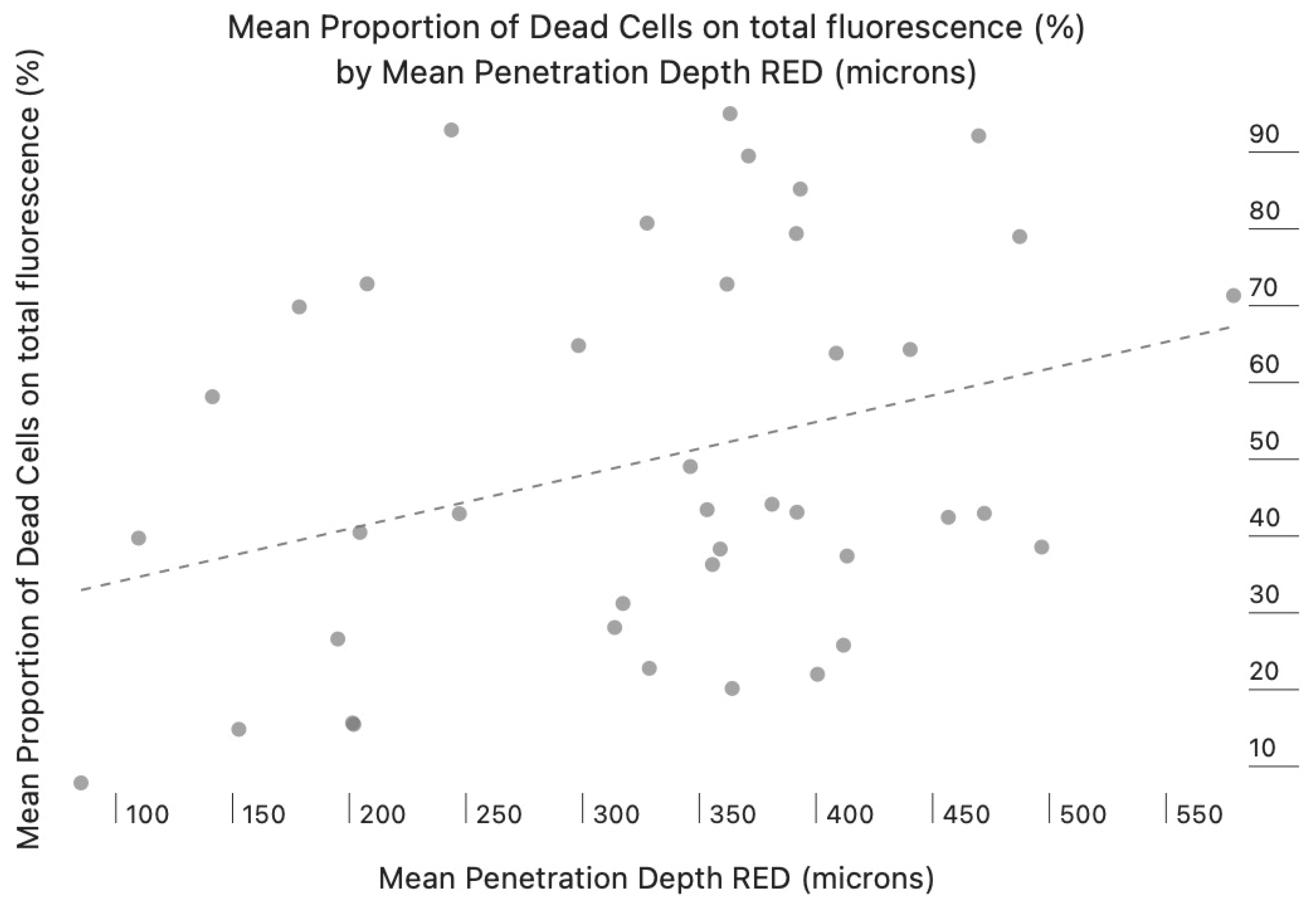
The analysis of the mean proportion of dead cells (red fluorescence) relative to total fluorescence reveals significant differences between the experimental groups and a positive correlation with the mean penetration depth (Figure 2).
The groups treated with calcium hydroxide (CH and CH-BC) showed a lower cell mortality rate compared to the groups treated with TAP (p = 0.023). In particular, CH-BC exhibited the lowest bactericidal effect compared to other groups (p = 0.012). The TAP-BC group displayed a moderate to high bactericidal effect, with a significantly lower cell mortality rate compared to TAP (p = 0.032), but still higher than the groups treated with calcium hydroxide (p = 0.026). This indicates that TAP appeared to be more effective than CH (p = 0.008) and that the groups exposed to blood clot showed an increase in viable bacteria inside dentinal tubules (p = 0.01). This highlights that the presence of blood seemed to reduce the bactericidal effect of both antibacterial compounds.
The TAP group showed the highest cell mortality rate, with almost overlapping values (78.998%). This indicates that TAP exhibits a strong bactericidal effect. The C+ group (positive control) recorded the lowest percentage of cell death, confirming the validity of the experimental model and serving as a reference for interpreting the results.
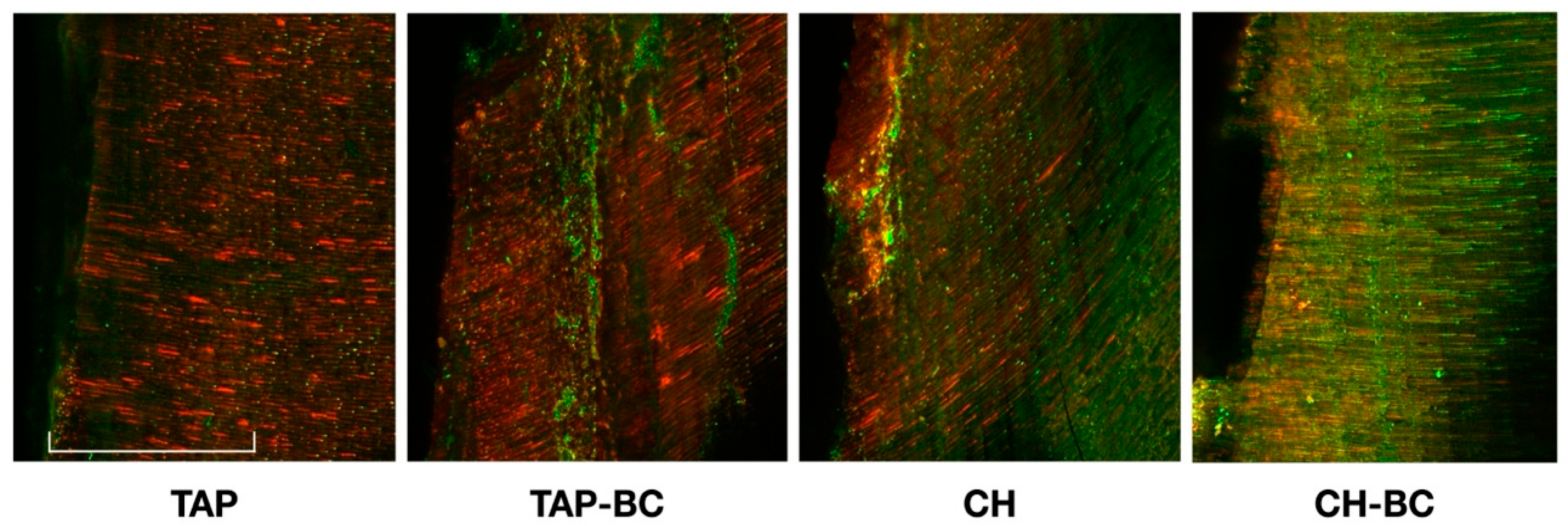
The highest penetration values were observed in the CH (402.57 µm) and TAP (372.78 µm) groups, suggesting greater diffusion in the samples treated without the presence of a blood clot (p = 0.065). The CH-BC group showed the lowest penetrations among all groups (313.732 µm) (p = 0.041); meanwhile, TAP-BC showed similar results compared to TAP (355.63 µm) (Table 1). Thus, the exposure to blood clot seemed to decrease the disinfection depth inside dentinal tubules (Figure 3).

4. Discussion

Regenerative endodontic procedures can help reduce pain and inflammation while promoting the healing of periapical lesions [15]. In addition, REPs promote continued root development—such as increased root length and dentinal wall thickness—which can improve the structural integrity of the root and enhance the long-term prognosis [9].
Persistent infection is the leading cause of REP failures, accounting for approximately 79% of unsuccessful cases [16]. Verma et al. reported that residual bacterial biofilms and their by-products are associated with a lack of periapical bone healing [17]. Furthermore, the residual intracanal infection seems to negatively influence the attachment, proliferation, and differentiation of SCAPs into the root canal [18]. In particular, bacterial lipopolysaccharides (LPS) can influence stem cell proliferation and alter their post-differentiation phenotype [18,19]. Therefore, it is widely reported that a high-level disinfection is fundamental to improve REP outcomes, but the complete sterilization of the root canal is not clinically achievable [19]. However, no data are still available on the behavior of the residual bacteria into dentinal tubules over time.
The use of different irrigation solutions and antibiotic medicaments was proposed to obtain a deep disinfection during REPs [20]. Nevertheless, an ideal concentration of intracanal medicaments was recommended to preserve SCAP viability [11]. The traditional TAP containing ciprofloxacin, metronidazole, and minocycline demonstrated high efficacy against a broad range of endodontic pathogens [21]. Despite its effectiveness, this formulation was associated with significant dentinal staining, largely attributed to minocycline’s chelating interaction with calcium dentinal ions [22]. This adverse effect was reported even when a dental adhesive was applied in the pulp chamber to isolate the dentin from chemical interaction, as recommended in clinical guidelines [22]. Consequently, alternative intracanal medicaments such as CH and DAP were widely proposed in previous studies [9]. However, CH showed limited effectiveness against root canal biofilms, and may weaken the dentinal walls of immature teeth, potentially compromising their structural integrity [23]. Additionally, DAP formulations combining ciprofloxacin and metronidazole were proposed in the clinical guidelines [10]. A previous clinical study tested the use of DAP during revitalization of necrotic permanent immature teeth, with acceptable results [24]. However, DAP demonstrated lower antimicrobial activity than TAP, likely due to weaker molecular synergy and limited penetration into dentinal tubules [14,25,26]. Thus, to avoid tooth discoloration, modified TAP formulations incorporating clindamycin or cefaclor were proposed and showed encouraging results [22]. Besides these combinations, a revised TAP formulation containing ciprofloxacin, metronidazole, and clarithromycin demonstrated significant in vitro antimicrobial efficacy without inducing tooth discoloration [26]. Clarithromycin is a second-generation macrolide, well-known for its broad-spectrum antimicrobial properties and strong anti-inflammatory action in both intracellular and extracellular environments [27].
In this study, the modified TAP containing clarithromycin was compared with CH to align with clinical guidelines while avoiding the risk of tooth discoloration. The root canal specimens were inoculated with Enterococcus faecalis, a Gram-positive facultative anaerobe commonly associated with persistent endodontic infections. This microorganism was selected basing on evidence in the literature describing its ability to resist against endodontic disinfection procedures and to create a stable biofilm dep into dentinal tubules [28]. The use of sodium hypochlorite (NaOCl) before applying the medicaments was omitted to specifically evaluate the effects of the medicaments on infected dentin without introducing confounding factors [10]. However, EDTA irrigation prior to blood clot formation was retained, as recommended in clinical guidelines [10].
Confocal laser scanning microscopy (CLSM) combined with viability staining is regarded as a reliable method for assessing bacterial biofilm development within dentinal tubules after incubation [29]. Furthermore, CLSM enables quantitative assessment of bacterial survival following disinfection and is widely regarded as a sensitive technique for detecting residual bacteria within dentin [13]. The viability staining with SYTO-9/PI combination is often used in Live/Dead assays, where live cells are seen as green fluorescence (SYTO-9) and dead cells are seen as red fluorescence (PI, which excludes SYTO-9)
To improve imaging quality, the specimens were longitudinally split along pre-made grooves with a scalpel to create flat, debris-free surfaces suitable for CLSM analysis [29]. Moreover, a three-week aerobic incubation period, combined with centrifugation, was used to facilitate deep bacterial penetration into the dentinal tubules [13]. The mean depth of action and the ratio of dead cells to total fluorescence were used as indicators to evaluate how effectively the tested solutions penetrated the dentinal tubules.
In the present study, TAP showed a stronger antibacterial effect compared to CH, and this observation seems to be in agreement with the existing literature [14]. Moreover, the presence of the blood clot decreased the depth of action and the percentage of dead microorganisms in both experimental subgroups. The rationale is that the blood clot may serve as a growth medium for E. faecalis over time, and its presence could negatively influence REP outcomes [30]. Studies showing greater efficacy of alternative scaffolds compared with blood clots may further support this assumption [31,32,33]. Nevertheless, developing alternative scaffolds—or adopting single-visit REPs using an artificial scaffold with sustained antibacterial activity and the capacity to recruit and promote stem cell differentiation—should be considered to reduce the blood clot’s impact on persistent infections. The limitations of this study include the small sample size and the short follow-up period for assessing bacterial growth over time. Nonetheless, further clinical research with longer follow-up is encouraged on this topic.
In conclusion, the TAP demonstrated higher antibacterial activity compared to CH deep into dentinal tubules. However, exposure to the blood clot appeared to gradually reduce the antimicrobial effectiveness achieved during REP, likely due to the potential regrowth of residual intracanal bacteria. Thus, the presence of a non-circulating blood clot may function as a bacterial culture medium over time, potentially undermining the success of regenerative endodontic therapy. Moreover, the toxic effects of the enzymes released by the hematopoietic cells could hamper the regenerative steps stimulating bacterial growth [34]. Therefore, a single visit REP approach with a continuous antibacterial and regenerative effect inside root canal could be ideal for future studies.

Author Contributions

Conceptualization: M.A. and N.M. Methodology: N.P., J.R., G.B., and V.A. Software and Validation: V.F. and M.G. Investigation: N.P., D.P., and V.F. Resources: M.A., D.P., and N.S. Writing—Original Draft Preparation: V.F., N.P., and N.M. Review and Editing: M.A. and N.S. Visualization: G.B., V.A., J.R., and D.P. Supervision: M.A. and N.S. All authors have read and agreed to the published version of the manuscript.

Funding

This research did not receive any specific grant from funding agencies in the public, commercial, or not-for-profit sectors.

Data Availability Statement

The raw data supporting the conclusions of this article will be made available by the authors on request.

Conflicts of Interest

The authors declare no conflicts of interest.

References

  1. Abbott, P.V.; Yu, C. A clinical classification of the status of the pulp and the root canal system. Aust. Dent. J. 2007, 52 (Suppl. 1), S17–S31. [Google Scholar] [CrossRef]
  2. Nicoloso, G.F.; Pötter, I.G.; Rocha, R.O.; Montagner, F.; Casagrande, L. A comparative evaluation of endodontic treatments for immature necrotic permanent teeth based on clinical and radiographic outcomes: A systematic review and meta-analysis. Int J Paediatr. Dent. 2017, 27, 217–227. [Google Scholar] [CrossRef]
  3. Bonte, E.; Beslot, A.; Boukpessi, T.; Lasfargues, J.J. MTA versus Ca(OH)2 in apexification of non-vital immature permanent teeth: A randomized clinical trial comparison. Clin. Oral Investig. 2015, 19, 1381–1388. [Google Scholar] [CrossRef]
  4. Rafter, M. Apexification: A review. Dent. Traumatol. 2005, 21, 1–8. [Google Scholar] [CrossRef]
  5. Mente, J.; Leo, M.; Panagidis, D.; Ohle, M.; Schneider, S.; Bermejo, J.L.; Pfefferle, T. Treatment outcome of mineral trioxide aggregate in open apex teeth. J. Endod. 2013, 39, 20–26. [Google Scholar] [CrossRef]
  6. Pulyodan, M.K.; Paramel Mohan, S.; Valsan, D.; Divakar, N.; Moyin, S.; Thayyil, S. Regenerative Endodontics: A Paradigm Shift in Clinical Endodontics. J. Pharm. Bioallied Sci. 2020, 12 (Suppl. 1), S20–S26. [Google Scholar] [CrossRef] [PubMed]
  7. American Association of Endodontists. Glossary of Endodontic Terms, 10th ed.; American Association of Endodontists: Chicago, IL, USA, 2020. [Google Scholar]
  8. Takimoto, K.; Widbiller, M.; Diogenes, A. Expression of Toll-like Receptors in Stem Cells of the Apical Papilla and Its Implication for Regenerative Endodontics. Cells 2023, 12, 2502. [Google Scholar] [CrossRef]
  9. Galler, K.M.; Krastl, G.; Simon, S.; Van Gorp, G.; Meschi, N.; Vahedi, B.; Lambrechts, P.; Dummer, P.M. European Society of Endodontology position statement: Revitalization procedures. Int. Endod. J. 2016, 49, 717–723. [Google Scholar] [CrossRef] [PubMed]
  10. American Association of Endodontists (AAE). Clinical Considerations for a Regenerative Procedure. Available online: https://f3f142zs0k2w1kg84k5p9i1o-wpengine.netdna-ssl.com/specialty/wpcontent/uploads/sites/2/2018/06/ConsiderationsForRegEndo_AsOfApril2018.pdf (accessed on 13 April 2018).
  11. Yoo, Y.-J.; Perinpanayagam, H.; Choi, Y.; Gu, Y.; Chang, S.-W.; Baek, S.-H.; Zhu, Q.; Fouad, A.F.; Kum, K.-Y. Characterization of Histopathology and Microbiota in Contemporary Regenerative Endodontic Procedures: Still Coming up Short. J. Endod. 2021, 47, 1285–1293.e1. [Google Scholar] [CrossRef]
  12. Verma, R.; Sharma, D.S.; Pathak, A.K. Antibacterial Efficacy of Pastes Against E Faecalis in Primary Root Dentin: A Confocal Microscope Study. J. Clin. Pediatr. Dent. 2015, 39, 247–254. [Google Scholar] [CrossRef]
  13. Ma, J.; Wang, Z.; Shen, Y.; Haapasalo, M. A new noninvasive model to study the effectiveness of dentin disinfection by using confocal laser scanning microscopy. J. Endod. 2011, 37, 1380–1385. [Google Scholar] [CrossRef]
  14. Mandras, N.; Alovisi, M.; Roana, J.; Crosasso, P.; Luganini, A.; Pasqualini, D.; Genta, E.; Arpicco, S.; Banche, G.; Cuffini, A.; et al. Evaluation of the Bactericidal Activity of a Hyaluronic Acid-Vehicled Clarithromycin Antibiotic Mixture by Confocal Laser Scanning Microscopy. Appl. Sci. 2020, 10, 761. [Google Scholar] [CrossRef]
  15. Galler, K.M. Clinical procedures for revitalization: Current knowledge and considerations. Int. Endod. J. 2016, 49, 926–936. [Google Scholar] [CrossRef]
  16. Almutairi, W.; Yassen, G.H.; Aminoshariae, A.; Williams, K.A.; Mickel, A. Regenerative Endodontics: A Systematic Analysis of the Failed Cases. J. Endod. 2019, 45, 567–577. [Google Scholar] [CrossRef] [PubMed]
  17. Verma, P.; Nosrat, A.; Kim, J.R.; Price, J.B.; Wang, P.; Bair, E.; Xu, H.H.; Fouad, A.F. Effect of Residual Bacteria on the Outcome of Pulp Regeneration In Vivo. J. Dent. Res. 2017, 96, 100–106. [Google Scholar] [CrossRef]
  18. Vishwanat, L.; Duong, R.; Takimoto, K.; Phillips, L.; Espitia, C.O.; Diogenes, A.; Ruparel, S.B.; Kolodrubetz, D.; Ruparel, N.B. Effect of bacterial biofilm on the osteogenic differentiation of stem cells of apical papilla. J. Endod. 2017, 43, 916–922. [Google Scholar] [CrossRef]
  19. Van Gorp, G.; Declerck, D. Long-term Outcome of Endodontically Treated Traumatized Immature Upper Incisors. J. Endod. 2023, 49, 1106–1119. [Google Scholar] [CrossRef]
  20. Tagelsir, A.; Yassen, G.H.; Gomez, G.F.; Gregory, R.L. Effect of Antimicrobials Used in Regenerative Endodontic Procedures on 3-week-old Enterococcus faecalis Biofilm. J. Endod. 2016, 42, 258–262. [Google Scholar] [CrossRef] [PubMed]
  21. Panchal, K.G.; Virani, K.; Patel, V.; Khan, A.A.; Pettiwala, A.; Puranik, S.S.; Joshi, S. Triple Antibiotic Paste: A Game Changer in Endodontics. J. Pharm. Bioallied Sci. 2024, 16 (Suppl. 3), S1913–S1915. [Google Scholar] [CrossRef] [PubMed]
  22. Montero-Miralles, P.; Martín-González, J.; Alonso-Ezpeleta, O.; Jiménez-Sánchez, M.D.C.; Velasco-Ortega, E.; Segura-Egea, J.J. Effectiveness and clinical implications of the use of topical antibiotics in regenerative endodontic procedures: A review. Int. Endod. J. 2018, 51, 981–988. [Google Scholar] [CrossRef] [PubMed]
  23. Yassen, G.H.; Vail, M.M.; Chu, T.G.; Platt, J.A. The effect of medicaments used in endodontic regeneration on root fracture and microhardness of radicular dentine. Int. Endod. J. 2013, 46, 688–695. [Google Scholar] [CrossRef]
  24. Ragab, R.A.; El Lattif, A.E.A.; Dokky, N.A.E.W.E. Comparative Study between Revitalization of Necrotic Immature Permanent Anterior Teeth with and without Platelet Rich Fibrin: A Randomized Controlled Trial. J. Clin. Pediatr. Dent. 2019, 43, 78–85. [Google Scholar] [CrossRef] [PubMed]
  25. Latham, J.; Fong, H.; Jewett, A.; Johnson, J.D.; Paranjpe, A. Disinfection Efficacy of Current Regenerative Endodontic Protocols in Simulated Necrotic Immature Permanent Teeth. J. Endod. 2016, 42, 1218–1225. [Google Scholar] [CrossRef]
  26. Mandras, N.; Roana, J.; Allizond, V.; Pasqualini, D.; Crosasso, P.; Burlando, M.; Banche, G.; Denisova, T.; Berutti, E.; Cuffini, A. Antibacterial efficacy and drug-induced tooth discolouration of antibiotic combinations for endodontic regenerative procedures. Int. J. Immunopathol. Pharmacol. 2013, 26, 557–563. [Google Scholar] [CrossRef]
  27. Amsden, G.W. Advanced-generation macrolides: Tissue-directed antibiotics. Int. J. Antimicrob. Agents 2001, 18 (Suppl. 1), S11–S15. [Google Scholar] [CrossRef] [PubMed]
  28. Zhang, C.; Du, J.; Peng, Z. Correlation between Enterococcus faecalis and Persistent Intraradicular Infection Compared with Primary Intraradicular Infection: A Systematic Review. J. Endod. 2015, 41, 1207–1213. [Google Scholar] [CrossRef]
  29. Kishen, A.; Shrestha, A.; Del Carpio-Perochena, A. Validation of Biofilm Assays to Assess Antibiofilm Efficacy in Instrumented Root Canals after Syringe Irrigation and Sonic Agitation. J. Endod. 2018, 44, 292–298. [Google Scholar] [CrossRef] [PubMed]
  30. Hage, W.; Karam Sarkis, D.; Kallassy, M.; Mallah, M.; Zogheib, C. In vitro evaluation of enterococcus faecalis growth in different conditions on dentinal substrate. Biomater. Investig. Dent. 2023, 10, 2287668. [Google Scholar] [CrossRef]
  31. de Araújo, L.; Goulart, T.S.; Gil, A.C.K.; Schuldt, D.P.V.; Coelho, B.S.; Figueiredo, D.d.R.; Garcia, L.d.F.R.; de Almeida, J. Do alternative scaffolds used in regenerative endodontics promote better root development than that achieved with blood clots? Braz. Dent. J. 2022, 33, 22–32. [Google Scholar] [CrossRef]
  32. Altaii, M.; Richards, L.; Rossi-Fedele, G. Histological assessment of regenerative endodontic treatment in animal studies with different scaffolds: A systematic review. Dent. Traumatol. 2017, 33, 235–244. [Google Scholar] [CrossRef]
  33. Ulusoy, A.T.; Turedi, I.; Cimen, M.; Cehreli, Z.C. Evaluation of Blood Clot, Platelet-rich Plasma, Platelet-rich Fibrin, and Platelet Pellet as Scaffolds in Regenerative Endodontic Treatment: A Prospective Randomized Trial. J. Endod. 2019, 45, 560–566. [Google Scholar] [CrossRef] [PubMed]
  34. Said, M.S.; Tirthani, E.; Lesho, E. Enterococcus Infections. 12 February 2024. In StatPearls; StatPearls Publishing: Treasure Island, FL, USA, 2025. [Google Scholar] [PubMed]
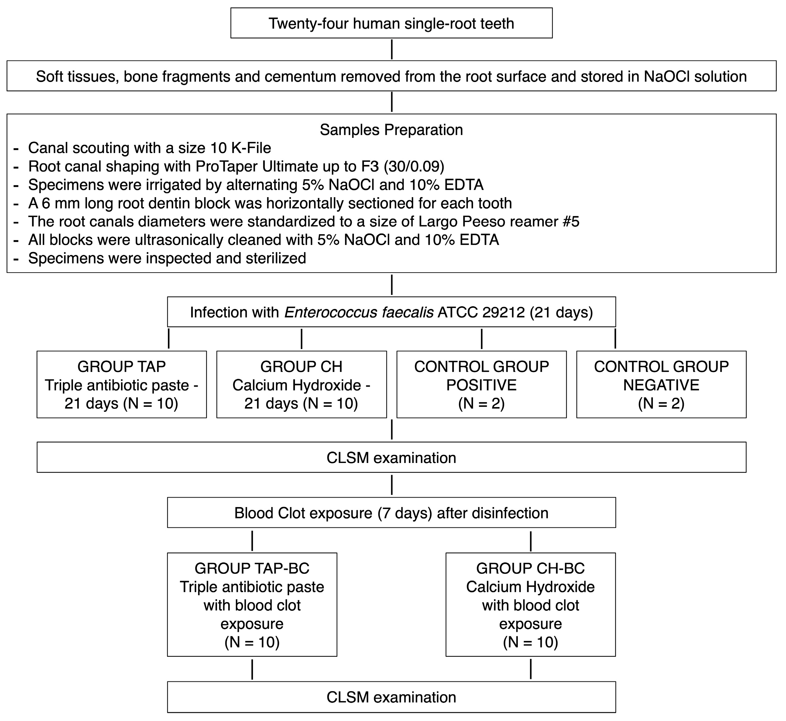
Figure 1. Flow diagram of the study design. TAP: triple antibiotic paste; CH: calcium hydroxide; CLSM: confocal laser scanning microscope.
Applsci 15 12478 g001
Figure 2. The mean proportion of dead cells on total fluorescence by mean penetration depth of the red fluorescence is represented. The correlation highlights the positive correspondence between depth of action and bactericidal effect (dotted line).
Applsci 15 12478 g002
Figure 3. The confocal laser scanning microscopy of the Enterococcus faecalis-infected dentinal tubules, treated with the different antibacterial pastes, after viability staining. The scale line length is 300 µm. Calcium hydroxide (CH), calcium hydroxide and blood clot (CH-BC), triple antibiotic paste (TAP), triple antibiotic paste and blood clot (TAP-BC).
Applsci 15 12478 g003
Table 1. The mean depth of action and the mean proportion of dead cell volume for each tested group. The different superscript letters indicate the statistically significant differences between the groups (p < 0.05). Positive controls (C+), negative controls (C−), calcium hydroxide (CH), calcium hydroxide and blood clot (CH-BC), triple antibiotic paste (TAP), triple antibiotic paste and blood clot (TAP-BC).
GroupsMean Depth of Action (µm)Mean Proportion of Dead Cells Volume (Red Fluorescence Ratio)
CH402.5 ± 58.2 a0.41 ± 0.08 a
TAP372.8 ± 41.6 a0.81 ± 0.06 b
CH-BC313.7 ± 37.4 b0.23 ± 0.11 c
TAP-BC355.63 ± 32.6 a0.45 ± 0.12 d
Positive controls0.5 ± 00.01 ± 0
Negative controlsNdNd
Disclaimer/Publisher’s Note: The statements, opinions and data contained in all publications are solely those of the individual author(s) and contributor(s) and not of MDPI and/or the editor(s). MDPI and/or the editor(s) disclaim responsibility for any injury to people or property resulting from any ideas, methods, instructions or products referred to in the content.