Article

Color Stability of a Composite Containing Hydroxyapatite, Fluorine, and Silver Fillers After Artificial Aging

by
Zofia Kula
1,*,
Cristina Bettencourt Neves
2,3,
Ana Bettencourt
2,
Sara Oliveira
3 and
João Carlos Roque
3,*
1
Department of Dental Technology, Medical University of Lodz, 251 Pomorska Street, 92-213 Lodz, Poland
2
Research Institute for Medicine (iMed.ULisboa), Faculdade de Farmácia, Universidade de Lisboa, 1649-003 Lisbon, Portugal
3
Dental Biomaterials Research Group (BIOMAT), Biomedical and Oral Sciences Research Unit (UICOB), Faculdade de Medicina Dentária, Universidade de Lisboa, 1600-277 Lisbon, Portugal
*
Authors to whom correspondence should be addressed.
Appl. Sci. 2025, 15(23), 12426; https://doi.org/10.3390/app152312426 (registering DOI)
Submission received: 14 October 2025 / Revised: 6 November 2025 / Accepted: 18 November 2025 / Published: 23 November 2025

Abstract

This work concerns composite materials containing hydroxyapatite, fluorine, and nanosilver fillers. These composites are intended for the reconstruction of lost hard tooth tissues. The aim of the work was to evaluate the color stability of a flow composite containing hydroxyapatite, fluorine, and silver fillers after artificial thermal, chemical, and a combination of thermal and chemical aging processes. The samples were prepared from a commercial flow-type composite material (Arkona Flow Art, Niemcy, Poland) color A2 VITA (Vita Classical, Vita Zahnfabrick, Bad Sackingen, Germany) original and a modified composite material containing a filler additive of 2 wt.% hydroxyapatite powder containing fluorine (calcium fluoride) and nanosilver (n = 15). An Optishade (Style Italiano, Italy) colorimeter was used to measure color against a black background. Samples were submitted to thermal aging (T) using thermocycling equipment; to chemical aging (C) by immersion on artificial saliva at pH7, 37 °C; and to constant agitation or thermal–chemical aging using a combination of the previous two methods. The modified composite showed reduced color differences compared to the original composite. The results also show that thermal aging has a stronger influence on ΔE increase than chemical and combined aging, but only for the modified composite. Cumulative aging processes had an influence below the acceptability threshold for the modified composite.

1. Introduction

At present, beauty is very important aspect in society. That is why most patients expect very natural and aesthetic dental solutions, sometimes at the expense of functionality [1,2]. To rebuild lost tooth tissue, dentists and dental technicians have a wide range of methods and materials available. The most popular materials are dental composites due to their quick and easy application as well as their low toxicity [3]. However, achieving lasting color stability using these materials is a big challenge [4,5]. Any change in the color of a tooth or filling can be defined as discoloration, which is caused by the aging process in the oral cavity environment. Color changes in composites may be a result of internal factors (physicochemical reactions) and external factors (dyes contained in beverages) [6,7]. Color changes in composite fillings may occur because of degradation, penetration, or the adsorption of dyes into composite layers [8]. Discoloration is also influenced by the surface texture, i.e., the degree of smoothness or roughness of the composite material [9,10,11,12]. Therefore, achieving color stability in a given material is crucial to achieving very good aesthetic effects in dental restorations [13].
To obtain very good aesthetics, the optical and chromatic properties of the material are important. The morphology of tissues affects the optical properties that determine the appearance of the tooth. An aesthetic composite filling should reflect the natural features of the tooth, i.e., translucency, opalescence, and fluorescence. The aesthetics of materials are closely linked to their composition. This is particularly true for the organic matrix and fillers [14,15]. Filler materials play an important role in color stability. In the literature, fillers such as titanium dioxide (TiO2), zirconium dioxide (ZrO2), and hydroxyapatite (HAp) are classified as functional aesthetic fillers [16,17]. Reference [18] confirms the potential use of TiO2 particles as a filler in restorative dentistry. TiO2 nanoparticles also influence working time, and improve the setting time and compressive strength of cements [19,20]. HAp nanoparticles are similar to structures in tooth enamel. Adding mineral fillers such as hydroxyapatite can improve resistance to discoloration [21,22]. The modification of dental composite fillers by introducing nanoparticles into the polymer matrix is a modern approach to improve the quality and effectiveness of materials for the reconstruction of hard dental tissues. There are reports in the literature that reveal that a reduced filler size would increase resistance to discoloration [22,23,24]. The use of nanoparticles also improves the aesthetic properties of dental composites [23,24]. They can scatter light in a similar way to natural tooth tissue [23]. Therefore, there is still a need to search for materials that will have very good mechanical and biological properties and, at the same time, have good aesthetic properties.
Dental spectrophotometers and colorimeters, equipped with advanced software for color analysis, are used for shade-taking, color measurements, and also for research purposes [25]. The use of this equipment in either Lab or LCh color spaces was first introduced by the Commission Internationale de l’Eclairage (CIE); the first was used to measure lightness (L*), red (+a*), and yellow (+b*), as well as the cartesian coordinates, and the second was used to measure the color parameters of brightness (lightness, L*), color intensity (chroma, C*), and hue (hº), as well as the cylindrical coordinates [26]. Color differences between paired samples under standardized conditions are quantified using ΔE formulas [27].
The most recent formula presented by CIE is ΔE00, updated from the previous ΔE94, both using the LCh* color space coordinates [26]. The smallest perceptible color difference—known as the perceptibility threshold—has been reported as 0.67 ΔE94 for adjacent uniform samples. Rizzi et al. [28] supported the use of the ΔE94 Graphics Arts formula due to its near-uniform perceptual spacing within the dental color range, while Paravina et al. [29] recommended adopting ΔE00 for dental applications. Reported perceptibility thresholds for ΔE00 in dentistry typically range between 2.6 and 3.5 units [30]. According to several studies, the perceptibility threshold obtained using this formula may have the range 2.6–3.5 [30].
The aim of this work was to evaluate the color stability of a flow composite containing hydroxyapatite, fluorine, and silver fillers after artificial, thermal, and chemical aging. The null hypothesis was as follows: (1) the inclusion of HAp, CaF2, and nAg as additives in a dental composite will not affect the color stability of the modified composite; (2) the different aging processes have no influence on the color stability of the composites tested.

2. Materials and Methods

2.1. Sample Fabrication and Preparation

The sample size (n = 15) was estimated with a power analysis to provide statistical significance (α = 0.05) at a power of 80%, based on pilot study data.
Fifteen specimens were made from a flow-type dental commercial composite (Arkona Flow Art, Niemcy, Poland), color A2 VITA (Vita Classical, Vita Zahnfabrick, Bad Sackingen, Germany), and the other fifteen were made from the modified composite material containing a filler additive of hydroxyapatite, fluorine (calcium fluoride), and nanosilver powder. A proprietary composite containing the following components was used in this study: bisphenol A diglycidyl ether dimethacrylate, diurethane dimethacrylate, triethylene glycol dimethacrylate, barium–aluminum–silica glass, titanium dioxide, silica, and camphorquinone. Fillers were used to modify this composite. Hydroxyapatite (HAp) was used at a concentration of 2 wt.%, with a hydroxyapatite grain size of up to 30 μm. The next filler was anhydrous calcium fluoride (Sigma-Aldrich, Saint Louis, MO, USA) of 99.99% purity in the amount of 0.2% by weight. The third filler was nanosilver (Sigma-Aldrich, Saint Louis, MO, USA) in the form of nanopowder at 1 wt%. The nanosilver grain size was less than 100 nm.
To obtain the hydroxyapatite filler, it was synthesized using the wet method. This process required the use of calcium hydroxide (Ca(OH)2) (Sigma-Aldrich, Saint Louis, MO, USA). Orthophosphoric acid (Sigma-Aldrich, Saint Louis, MO, USA) was also used. Calcium hydroxide was added to water heated to 40 °C and then stirred vigorously for an hour at a constant temperature using a magnetic stirrer. After this time, orthophosphoric acid was added dropwise at a fixed rate, while constantly stirring the solution. During this time, the precipitation process takes place. It is very important to maintain a temperature of around 40 °C (not higher than 42 °C) and a pH above 9. To avoid acidification of the solution, ammonia water was added in small portions. The last step is to filter the sediment using paper filters. The hydroxyapatite crystals obtained from the synthesis were then fractionated using an LPzE-3e laboratory shaker (MULTISERW-Morek, Brzeźnica, Poland).
The next step in obtaining the hydroxyapatite filler was to obtain the appropriate particles. The hydroxyapatite was ground using a planetary mill (Retsch PM 100, Retsch GmbH, Haan, Germany). This process resulted in HAp grains with a size of 30 μm. The fillers were combined using a Roti-Speed mixer (Carl Roth GmbH + Co., KG, Karlsruhe, Germany). Mixing was performed at 5000 rpm for approximately 5 min.
In the subsequent step, the two sets of samples (n = 15) underwent polymerization for 20 s using a curing unit, with a diode light intensity of 1400 mW/cm2 in the wavelength range of 450–490 nm (Elipar S10, 3M ESPE, St. Paul, MN, USA).
Each layer of the material was polymerized to a thickness of 1 mm. A celluloid strip and glass were used to obtain a smooth sample surface.
Using a manual polisher (Struers S.A.S), one of the sample surfaces was submitted to four movements over three different sheets of silicon carbide sandpaper (120 µm, 500 µm and 1000 µm), followed by mechanical polishing (Bench Top Dust Collector & Dental Lab Polishing Machine KK478, KaVo, Belgium) with pumice for 2 min and shining paste (Universal polishing paste, Ivoclar, Lichenstein) for 10 s, at 1500 rpm. Samples were randomly assigned to two groups (n = 15) and then marked with a tungsten bur from 1 to 15 before being submitted to aging processes (Thermal-T; Chemical-C or Combination Thermal and Chemical-TC), allowing for each sample to be tracked after each aging process (Table 1).

2.2. Color Measurements

Color measurements were made with a portable colorimeter (Optishade-Styleitaliano, Itália), coupled with a phone with iOS operating system (Figure 1a), using the Optishade App (https://www.styleitaliano.org/OPTISHADE/app/) (Figure 1b).
Each sample was placed against a black background, using glycerol as the contact medium, and two copy samples were positioned on each side of the original sample to ensure the equipment was at the right focal distance from the sample. Five consecutive images of the polished side of each sample were made, and measurements were registered from the center of the image, at the crossing point of two perpendicular lines positioned on the image using an overlaying transparent matrix. All the measurements were made by the same calibrated researcher, before and after the aging procedures, ensuring consistency in the data collection. The equipment automatically provided ΔE94 to the closest color match (D2 VITA = Lab: 69.3, 2.5, 11.2) and the CIELCh color space was used to register L*, C*, and h* color values from the image (Figure 1b). Equipment calibration was performed according to instructions through the App calibration mode, using the instrument’s white inner side of the lens cover cap. Calibration was performed every time the equipment was turned on and after five consecutive measurements of each sample. The measurements made for each group were set as the initial reference before the aging processes.
Once the samples underwent the aging processes, new color measurements were made, using the same methodology. Color measurements after the aging process were also taken to calculate the color differences (ΔE00) (ΔE94) within each group, using the CIEDE formulas.
Δ E 94 * = Δ L * K L 2 + Δ C a b * 1 + K 1 C 1 * 2 + Δ H a b * 1 + K 2 C 1 * 2
Δ Ε 00 = L K L S L 2 + C K C S C 2 + H K H S H 2 + R T C K C S C H K H S H
To calculate color differences (ΔE00; ΔE94) after thermal aging, L*, C*, and h* values at baseline and after thermal aging were used. For color differences (ΔE00; ΔE94) after chemical aging, values after thermal aging and after thermal and chemical aging were used, and finally for the two combined aging processes, values at base line and after thermal and chemical aging were used.

2.3. Aging Processes

After baseline measurements, samples from the two groups of resins were submitted to thermal aging (T) using thermocycling equipment (Refri 200E-Arab, Cascais, Portugal). It consisted of 5000 cycles of thermal fluctuations between a hot bath (55 °C) and a cold bath (5 °C); each bath lasted 20 s with a dwell time of 5 s, simulating six months of intraoral temperature fluctuations associated with mouth breathing and food or beverage intake [31].
After thermal aging, color measurements were performed. Samples were then submitted to chemical aging (C), characterized by six months of immersion in artificial saliva with a 1 g/5 mL ratio. The artificial saliva used was prepared with the following quantitative composition (% w/v): xanthan gum (0.05), calcium chloride dihydrate (0.04), sodium chloride (0.08), potassium chloride (0.08), and propylene glycol (15) in 100 mL of phosphate buffer pH = 7; all reagents were acquired from Sigma-Aldrich (Madrid, Spain). Immersion used an incubation thermostat bath (Memmert, Schwabach, Germany), at 37 °C, 300 rpm [32,33].
After thermal and chemical aging (TC), final color measurements were performed.

2.4. Statistics

Data analyses were performed using IBM SPSS Statistics v.31 (Statistical Package for the Social Sciences) software. Descriptive statistics were established for dependent variables (ΔE00 and ΔE94) within independent variables (resin group and aging processes). Shapiro–Wilk normality test and Levene homogeneity test were carried out to evaluate the application of the parametric tests (p > 0.05).
Following a negative outcome, groups of specimens were compared using non-parametric Mann–Whitney and Kruskal–Wallis tests, followed by multiple comparisons, before being corrected by Bonferroni. The analysis adopted a significance level of α = 0.05

3. Results

Descriptive statistics are presented in Table 2, including mean, standard deviation (SD), median, and inter-quartile range (IR) for ΔE00 and ΔE94 for the two groups tested after the different aging processes.
For the original composite (1—Arkona Original No Fillers), after the first aging process (T), ΔE00 and ΔE94 were similar, with a registered difference of just 0.2 values, and ΔE00 > ΔE94, and these values remained similar after the second aging process (C), with a registered difference of just 0.1, although both ΔE00 and ΔE94 increased 1.1 and 1.2, respectively, from the first (T) to the second (C) aging test. Finally, the combined (T + C) aging tests produced a similar result to ΔE00 and ΔE94, being approximately 0.1 higher for ΔE94.
On the modified composite (2—Arkona Modified with Fillers), after the first aging process (T), ΔE00 and ΔE94 were similar, with a registered difference of just 0.2 values, but showed opposite results to the original composite, since ΔE00 < ΔE94, and remained similar after the second aging process (C), with a registered difference of just 0.1, in the opposite direction to the original composite, since ΔE00 decreased by 1.3 and ΔE94 decreased by 1.6. Finally, the combined (T + C) aging tests produced a similar result to ΔE00 and ΔE94, being approximately 0.2 higher for ΔE94.
Table 3 presents the mean values obtained after the different aging processes with thresholds for in vitro tests and in vivo tests. The original composite showed ΔE00 values above the acceptability threshold for the three aging tests, with the maximum value being obtained after the chemical test. In contrast, the modified composite only registered a value above the acceptability threshold right after the thermal aging, registering a lower value after chemical aging and a value slightly above the limit after the thermal + chemical aging test.
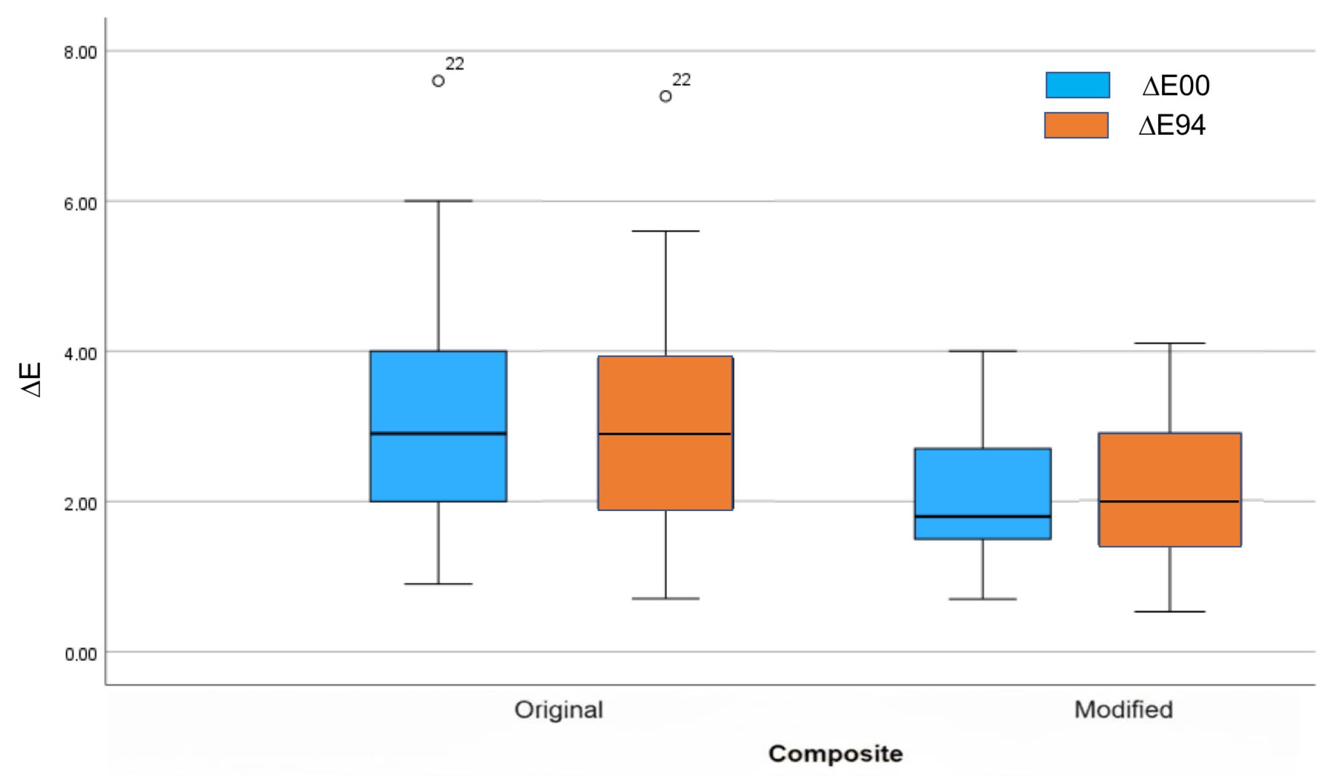
Comparing the two composites for ΔE00, specimens from the modified group (median = 1.8; IR = 1.20) are statistically lower (p < 0.001) than specimens from the original group (median = 2.9; IR = 2.05) (Figure 2). For ΔE94, specimens from the modified group (median = 2.0; IR = 1.56) are statistically lower (p < 0.003) than specimens from the original group (median = 2.9; IR = 2.10) (Figure 2).
Comparing the aging processes for ΔE00, the original composite registered no statistical differences (p = 0.261) (while, in contrast, for the modified composite, statistically significant differences (p < 0.001) were found (Figure 3). The thermal aging (T) produced higher values than the chemical aging (C) (p < 0.001) and the combination of thermal and chemical aging (TC) (p = 0.012), with no statistically significant differences observed between the last two (p = 0.198).
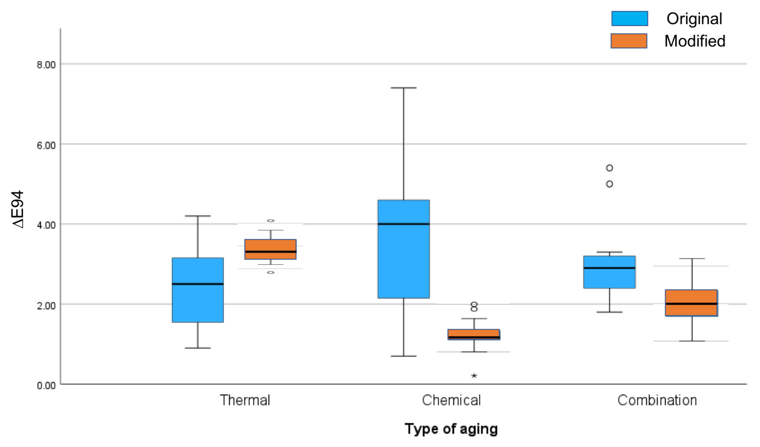
Comparing the aging processes for ΔE94, on the original composite, no statistical differences were found (p = 0.100) (while, in contrast, for the modified composite, statistically significant differences (p < 0.001) were found (Figure 4). The thermal aging (T) produced higher values than the chemical aging (C) (p < 0.001) and the combination of thermal and chemical aging (TC) (p = 0.042), with chemical aging (C) resulting in less statistically significant differences compared to the combined processes (TC) (p = 0.022).

4. Discussion

Dental aesthetics is a field that is developing very quickly and has a lot of solutions to offer to patients. However, we continue to search for new solutions that will combine both aesthetics and functionality. The proposed solutions are time-limited, such as composites, which discolor over time. Therefore, it is important to improve composite materials.
In vitro studies report improved color camouflage and color stability after nHAp treatments or when nHAp is included in the design of formulations to remineralize or mimic enamel [23]. Extrinsic stain penetration can be reduced because the material can deposit Ca2+/P043− into interfacial defects or micro-porosities and integrate with tooth apatite, reducing pathways that allow for chromogenic molecules to penetrate the composite–tooth interface. Also, because nHAp particles have a refractive index closer to natural enamel than many glass fillers, if they are properly sized/dispersed in a material they can reduce the obvious mismatch between fillers and the composite, additionally lowering the perceptible contrast changes that occur in resin during the ongoing aging processes [17].
Recently, HAp has been used in prevention. It is used in remineralizing and whitening toothpastes [35,36,37,38,39]. Additionally, its purpose is to minimize tooth hypersensitivity. It achieves very good anti-caries effects when combined with fluoride [38]. There is a noticeable increase in the microhardness pattern of the enamel [38]. The current literature reports indicate that hydroxyapatite may have whitening properties. Researchers have developed a HAp-based gel that showed whitening properties compared to other preparations [40]. Hydroxyapatite, in this case, was used as an active ingredient. The current literature indicates that adding HAp to the enamel surface can hide the dark color of teeth (or yellow tones) but also minimizes development [39,41]. Scientists believe that using HAp toothpaste causes a white, opaque, hydroxyapatite mineral layer to form. This layer stops beams of light from entering and reflecting white light back. The light beam that falls on the dentin is killed by the enamel tissues. The hydroxyapatite layer reflects and scatters light, which creates a brightening effect [39]. Applying toothpaste containing hydroxyapatite provides immediate results in terms of whitening effect by incorporating HAp microcrystals, which fill micro-cavities on the enamel surface and reconstruct damaged apatite on the surface [40,41,42]. To achieve better aesthetic results, a hydroxyapatite filler was introduced to eliminate the possibility of discoloration of the composite material. Due to the fact that obtaining better aesthetics in fillings may be associated with a deterioration of mechanical properties, fluorine and nanosilver were also added.
A common problem in conservative treatment is the leakage (microfissure) of dental fillings. They are often found under a filling and provide an excellent route through which bacteria can re-enter, causing secondary caries. The micro-gap is mainly caused by differences in the modulus of elasticity between the composite material and the tooth. Secondary caries is a very serious problem. Therefore, the introduction of nanosilver into the composite matrix was intended to limit bacterial growth. Silver is widely known for its antibacterial properties. Pathogenic microorganisms can adhere to composite materials intended for dental restorations. Therefore, it is extremely important to develop a highly aesthetic composite material with properties that limit the formation of bacterial biofilm, which will prevent colonization at the interface between the tooth and the filling by cariogenic bacteria such as Streptococcus mutans.
For this purpose, Jesús Alberto Garibay-Alvarado and co-authors prepared a composite with enhanced antibacterial activity for dental applications, which contained HA-NpsAg (RHN) in two concentrations (0.5% and 1%). The authors indicate that the RHN powder at a concentration of 1% showed maximum inhibition against S. aureus and S. mutans bacterial strains. The authors did not conduct tests related to color stability [43]. There are reports of similar fillers but used separately. The most similar studies are presented by the researchers Raiesa M M Hashem, Cherif A Mohsen, and Manal R Abu-Eittah, [44] who incorporated silver nanoparticles and silver hydroxyapatite nanoparticles at a concentration of 40 µg into dental ceramics. The authors observed that the addition of silver nanoparticles increased the fracture resistance of dental ceramics. The combination of nanosilver fillers and silver nanohydroxyapatite adversely affected the color of the dental ceramics. It is important to remember that these are completely different materials. Imran Alam Moheet and co-authors, in their research, also used glass ionomer cement (cGIC), which they enriched with fluoride and nanohydroxyapatite particles, along with silica. The results obtained by these scientists indicate better color stability of nanoHAp-Si-GIC compared to GIC. NanoHAp-Si-GIC also showed a significant increase in fluoride ion release capacity [45].
Due to clinical reports related to the whitening and antibacterial properties of HAp, scientists have investigated its use as a composite filler. Aldhuwayhi et al. [46] added hydroxyapatite to conventional glass ionomer cement (GIC) to compare its color stability with other materials. The applied GIC 5%nano ZrO2-SiO2-HA showed improved color stability compared to cGIC. The authors also showed that HA-containing composites can change their optical properties depending on the particle system and processing. Moreover, Kula et al. [47] reported that adding HAp tends to change the optical properties (opacity/translucency) of the resin matrix, and larger/macro-particles and loadings increase scattering (reduced translucency), and therefore can change the perceived shade. HAp itself is not uniformly protective against staining and may even increase visible changes if poor dispersion or large particles increase surface roughness.
Fluoride can act as a barrier because fluoride ions (F) released from fillers can drive the formation of fluorapatite-like species at the composite surface or adjacent enamel (Ca10(PO4)6F2), producing a harder, less soluble, less permeable surface layer that resists penetration by staining molecules [48]. Fluoride-doped nano-zirconia fillers incorporated into composite resins were reported to improve color stability and wear resistance during their aging protocols compared with control composites, as shown by Zheng et al. [49]. The authors attribute this to good filler dispersion, a high refractive index match (ZrO2), and reduced water sorption/solubility. Fluoride doping can deliver anti-caries benefits (release) and, when combined with appropriate high-refractive index fillers and good dispersion, can also improve color stability under thermal and chemical aging.
The use of silver (metallic silver or silver nanoparticles) can offer antimicrobial properties but also leads to uncertainty regarding material color. Silver brings antimicrobial benefits but carries a risk of discoloration, especially after accelerated aging or at high concentrations. Ag+ release suppresses the growth of chromogenic oral bacteria and reduces the biofilm, leading to less extrinsic staining over time. Thus, this provides an indirect color stability benefit via microbiology [50]. However, in oral environments, silver can react with sulfide species (HS, S2 from saliva or bacterial metabolism) to form Ag2S (black/tarnish) or other dark silver compounds, producing intrinsic darkening, especially exposed metallic silver or poorly stabilized AgNPs [51]. Strategies in the literature therefore aim to use low-dose, well-stabilized AgNPs or ionic silver formulations that minimize exposed metallic silver, and to combine these with protective surface layers [52].
Several factors can affect the color stability of composites, including their composition, fabrication, and the environment in which they are placed. Frequent variations in pH, stress, and temperature may have a major impact on the color stability of aesthetic and bioactive restorative materials, depending on the dynamic character of the oral environment. Testing the influence of aging procedures is essential to understand material behavior over time. Thermocycling reliably produces measurable color changes (ΔE) in restorative materials: the material type, filler type/size/loading, resin matrix, and surface finish are primary determinants of how large the ΔE will be [52,53].
There are currently no studies on the effect on the color of composite materials containing hydroxyapatite, fluoride, and silver; therefore, this study was needed. The authors propose that their simultaneous inclusion is beneficial for antibacterial and color stability properties, and that aging processes will not have a significant effect on the material. The inclusion of nanosilver, although representing a potential problem in terms of color, is believed to provide acceptable results. The present study complements previous studies on the mechanical and tribological properties of materials, such as those already published by the research team, with favorable results. However, not including nano-scale HAp fillers in the composition groups and other commercial composite materials for benchmarking is considered a limitation to a more comprehensive understanding of the results by the authors and should be addressed in future research.
Thermocycling was used as a standardized short-term thermal aging protocol. Although such an approach is frequently described in the literature, this equivalence is only approximate because thermocycling reproduces repeated thermal stress, without other clinically relevant factors (dietary staining, pH fluctuations, pellicle formation, mechanical abrasion, or UV exposure). Specimens were also stored in an artificial saliva solution, with daily renewal, to permit pellicle-like adsorption; the exact formulation and renewal schedule are reported to improve reproducibility. However, thermocycling was not combined with a cyclic staining protocol (coffee/tea exposure) or simulated toothbrushing to increase ecological validity. In future research, these factors should be considered, and in situ or clinical confirmation is still necessary to establish whether the observed ΔE00 changes are clinically perceptible or acceptable.
In the present study, two CIEDE formulas were used to calculate ΔE values. By measuring the samples with the colorimeter Optishade (Style Italiano, Italy), ΔE94 was automatically provided for the closest color match of the VITA classical shade guide—in this case, D2 (Lab: 69.3, 2.5, 11.2). Although the composite for the specimens tested was the color VITA A2, measurements were taken against a black background, which influenced the color identified by the equipment and meant that D2 was the closest match (Table S1 included in Supplementary Material).
To have a correct understanding of color behavior after aging procedures, formulas CIEDE94 and CIEDE00 were used with LCh* specimen values against a black background, registered at three stages (baseline, after thermal aging and after thermal and chemical aging). The ΔE94 formula adjusts color differences with weighting factors for lightness (L*), chroma (C*), and hue (h*), because the human eye is more sensitive to some differences than others. Although better than the previous ΔEab, it still does not match visual perception well when using low-chroma and near-neutral colors (very relevant in dental shades, which are low-chroma yellow/browns). The ΔE00 refines the earlier formula by incorporating hue–chroma interactions, improved weighting functions, and corrections for near-neutral colors, leading to a closer match with human visual perception. This makes ΔE00 the preferred metric in dentistry for evaluating color differences in teeth and restorative materials. According to ISO/TR 28642:2016 [54,55,56], it is the recommended formula for evaluating color stability, water sorption effects, and restorative matching. To ensure interpretative consistency, perceptibility and acceptability thresholds were predefined according to published dental standards: a ΔE00 value of approximately 1.0 represents the 50% perceptibility threshold, while a ΔE00 of approximately 2.0 corresponds to the clinical acceptability limit in vitro conditions. Ref. [29] For in vivo evaluations, slightly higher thresholds (ΔE00 ≈ 1.2 for perceptibility and 2.7 for acceptability) are typically considered appropriate [34]. Use of the ΔE00 metric avoids the interpretative inconsistencies that can arise when older formulas such as ΔE94 are applied, since ΔE00 yields numerically smaller values for equivalent visual differences and provides a more accurate reflection of clinically relevant color mismatches.
Statistical analyses revealed significant differences between the two different material composition tested, confirming null hypotheses 1. ΔE00 and ΔE94 were higher in the original composite compared to the modified material. Hydroxyapatite is a white-colored mineral and may increase lightness when incorporated into other materials. In the present study, L*, C*, and h* values were registered at three moments (Table 2) and it was observed that the modified composite (group 2—Arkona modified with fillers) initially registered a higher value (ΔL* ≅ 2), showing a lighter color when compared to the original composite (group 1—Arkona with no fillers). Upon the combination of the two aging processes (TC), both material formulations decreased lightness approximately equally (ΔL* ≅ 2). However, while the decrease in the original composite was observed just after the chemical aging (it remained constant upon thermal aging), in the modified composite the decrease was pronounced right after the thermal aging and increased slightly after the chemical aging. The increased lightness of the modified composite may be attributed to an increase in light reflection and scattering effects induced by the dense white opaque particles of hydroxyapatite. The diminishing of the observed initial lightness of the materials after the aging processes may be caused by the material water sorption induced by the thermal and chemical aging procedures, producing a defuse scattering effect of light through the water molecules and reducing the refraction of light to the material surface [57,58]. Alrefaie et al. [59] recently reported that water accumulation changed the light transmission and transparency parameters of nanofilled liquid composites. The changes in light transmission and transparency parameters (TPs) are consistent with the observed decrease in L* after water aging, inducing internal scattering and transmitted light, which lowers perceived lightness. This effect was observed in all groups tested. Other tests could lead to a better understanding of the results, namely, interfacial water uptake and hydrolysis (water sorption/solubility (ISO 4049 method) to confirm water sorption and polymer degradation as contributors to color changes; gravimetric kinetics; FTIR/Raman spectroscopy); RI mismatch and micro-void formation (ellipsometry; Micro-CT) for direct evidence that thermal cycling creates RI discontinuities and scattering effects; Ag particle optical changes (UV-Vis spectroscopy; TEM) to confirm that plasmonic and chemical changes in Ag particles directly alter optical response; and also surface roughness (optical/contact profilometry; AFM) to confirm that surface changes and extrinsic staining amplify color differences. These research paths should be explored in further research.
It was also observed that, contrary to the results obtained using the original composite, the decrease in lightness was more pronounced after thermal aging in the modified composite. The presence of hydroxyapatite and other fillers enhances the discontinuity of the composite matrix and opens space for the water to be absorbed [60], therefore achieving the scattering effect in the material more quickly, while the original resin matrix needs a longer period of water exposition to absorb it and for it to have an influence on the observable material lightness. Some experimental studies [61,62,63] have demonstrated that filler characteristics (type, size, loading, surface treatment) significantly influence water uptake and diffusion coefficients. Some studies show a higher filler loading reduces total matrix volume (reducing bulk sorption) but increases interfacial area (more sites for interfacial hydrolysis and localized water accumulation). Despite these different material behaviors, the amount of lightness lost in both materials is equivalent and may be attributed to the water absorption and filler content of the materials. Statistical analyses revealed no significant differences between the aging processes, using both ΔE00 and ΔE94 formulas, for the original composite but differences were observed in the modified composite. In the second formula, thermal aging produced significantly higher values than chemical aging for ΔE00 and ΔE94, and thermal aging produced higher values than combined thermal and chemical aging for ΔE94. For these reasons, null hypotheses 2 was rejected.
Color changes were previously reported for different commercial composites. Abduljadayel et al. also reported color changes in non-modified different bioactive restorative materials (ACTIVA, Beautifil II, Fuji II, Filtek Z350 XT) after 5000 thermocycles (5–55 °C) in different staining solutions [52,53]. As for modified composites, Zheng et al. tested different concentrations of fluoride-doped nano-ZrO2 fillers (25 wt%–50 wt%) submitted to accelerated thermal aging (10000 cycles of 5–55 °C) and reported a decrease in ΔE00 (Control ≈ 1.69; 25 wt% ≈ 1.20; 50 wt% ≈ 0.78). The authors state that a fluoride-doped high-index filler improved color stability (lower ΔE00) vs. control and cited the good optical match and reduced water uptake as reasons for this [49]. Testing composites with different staining solutions provides a more comprehensive understanding of the effects on composite colors when using materials submitted to different processes, and the authors consider this a limitation of the presented work that should be addressed in further research.
This research obtained promising results for the proposed modified composite in relation to its color stability. In future strategies related to improving composite formulation, ensuring color stabilization, preventing stain infiltration, improving interface stability, and reducing micro-void formations, the use of nano-Hap as a small fraction of filler load should be considered and silane coupling should be ensured [17]. Also, to help reduce demineralization and extrinsic staining, the inclusion of controlled fluoride-releasing phases should be considered to assist in superficial fluorapatite formation [64]. Finally, to ensure that sulfidation/tarnish does not occur, stabilized silver complexes could be included or a nano-Ag surface coating could be used [55]. The authors are confident that, similarly to other commercially available composite materials, a new version of the original composite may be released in the future, with improved mechanical, color stability, and antibacterial properties.
The color difference thresholds in dentistry have been well established by Paravina et al. [29] and, according to the authors, ΔE00 provides somewhat lower (more stringent) thresholds that more closely match human visual perception. For the ΔE00 formula, the perceptibility threshold was ≈0.8–1.00 and the acceptability threshold was ≈1.8–2.25, which are more realistic than the higher ΔE94 thresholds. In the present study, the difference between ΔE00 and ΔE94 values varied according to the aging processes and it is impossible to confirm the tendency for ΔE00 values to be lower than those of ΔE94. However, the modified composite registered values within the acceptability threshold after both chemical (C) and combined (T&C) aging processes for both ΔE00 and ΔE94 formulas. According to the obtained results, the modified composite demonstrated a satisfactory result using the ΔE00 acceptability threshold after the aging processes and a ΔL* equal to the original composite before the aging processes. The incorporation of the filler in the modified composite somehow helps the material to better withstand aging process, helping it to maintain a more acceptable color behavior over time. Considering that the reference thresholds for in vivo tests are higher than those for the in vitro tests, the modified composite shows a more stable behavior than the original, better withstanding aging stress tests.
In a study with five resin composites and accelerated artificial aging (AAA) for 300 h, Fidan [57] obtained similar results, with ΔE00 above clinically acceptable thresholds after aging for all groups. Notably, material type influenced magnitude. Also, after AAA, there were decreases in L* (lightness). Also, in an in vitro study with composites, Salles de Oliveira et al. [65] reported that ΔE00 tends to provide somewhat lower absolute magnitudes than ΔEab, but shows similar trends.
Thanks to this type of scientific research, we are able to directly help our patients. The search for a better solution for the reconstruction of cavities is still ongoing, as the available materials do not meet all the expectations of doctors, and especially patients. In addition to the fact that these materials must have appropriate strength properties, they should also be highly aesthetic. Unfortunately, in the case of composite fillings, discoloration occurs, which significantly deteriorates the aesthetic properties, especially in the anterior section of the dental arch. It is then necessary to replace the filling with a new one, which involves re-preparing the tooth and thus wearing away more of the tooth’s own tissues. The development of a non-staining composite material would allow for the avoidance of repeat procedures associated with conservative or prosthetic treatment, such as the use of veneers, which also involve the preparation of tooth tissue. The use of a new dental filling containing hydroxyapatite, silver, and fluoride could improve patient comfort and aesthetics, and eliminate the development of secondary caries.

5. Conclusions

Within the limitations of the study, the following conclusions can be obtained:
-
The modified composite showed reduced color differences compared to the original one.
-
Differences were observed between aging processes for the modified composite, but not for the original composite.
-
The incorporation of hydroxyapatite results in an increase in lightness in the composite material.
-
A modified composite with hydroxyapatite, silver, and fluorine could better withstand color changes when submitted to cumulative thermal and saliva aging processes.

6. Patents

Modified light-cured composite for dental fillings and its application, Pat.243701.

Supplementary Materials

The following supporting information can be downloaded at: https://www.mdpi.com/article/10.3390/app152312426/s1, Table S1: Mean and Standard Deviation (SD) values of group samples for ΔE94 to color D2 VITA and L*, C*, h* values of the two different composite compositions at the three points of measurement.

Author Contributions

Conceptualization, Z.K., J.C.R., and C.B.N.; methodology, Z.K., J.C.R. and C.B.N.; software, J.C.R.; validation, J.C.R., C.B.N. and Z.K.; formal analysis, Z.K., J.C.R. and C.B.N.; investigation, Z.K., J.C.R., A.B. and S.O.; resources, J.C.R. data curation, J.C.R. and C.B.N.; writing—original draft preparation, Z.K., J.C.R. and C.B.N.; writing—review and editing, Z.K., J.C.R. and C.B.N.; visualization, J.C.R., and C.B.N.; supervision, Z.K.; project administration, Z.K. All authors have read and agreed to the published version of the manuscript.

Funding

This research received no external funding.

Institutional Review Board Statement

No applicable.

Informed Consent Statement

No applicable.

Data Availability Statement

The original contributions presented in this study are included in the article. Further inquiries can be directed to the corresponding author.

Conflicts of Interest

The authors declare no conflicts of interest.

References

  1. Newton, J.T.; Subramanian, S.S.; Westland, S.; Gupta, A.K.; Luo, W.; Joiner, A. The impact of tooth colour on the perceptions of age and social judgements. J. Dent. 2021, 112, 103771. [Google Scholar] [CrossRef]
  2. Dellepiane, E.; Pera, F.; Zunino, P.; Mugno, M.G.; Pesce, P.; Menini, M. Oral Health-Related Quality of Life and Full-Arch Immediate Loading Rehabilitation: An Evaluation of Preoperative, Intermediate, and Posttreatment Assessments of Patients Using a Modification of the OHIP Questionnaire. J. Oral Implantol. 2020, 46, 540–549. [Google Scholar] [CrossRef]
  3. Zhang, N.; Ma, Y.; Weir, M.D.; Xu, H.H.K.; Bai, Y.; Melo, M.A.S. Current Insights into the Modulation of Oral Bacterial Degradation of Dental Polymeric Restorative Materials. Materials 2017, 10, 507. [Google Scholar] [CrossRef]
  4. Hajdu, A.I.; Dumitrescu, R.; Balean, O.; Lalescu, D.V.; Buzatu, B.L.R.; Bolchis, V.; Floare, L.; Utu, D.; Jumanca, D.; Galuscan, A. Enhancing Esthetics in Direct Dental Resin Composite: Investigating Surface Roughness and Color Stability. J. Funct. Biomater. 2024, 15, 208. [Google Scholar] [CrossRef] [PubMed]
  5. Schulze, K.A.; Marshall, S.J.; A Gansky, S.; Marshall, G.W. Color stability and hardness in dental composites after accelerated aging. Dent. Mater. 2003, 19, 612–619. [Google Scholar] [CrossRef] [PubMed]
  6. Bagheri, R.; Burrow, M.F.; Tyas, M. Influence of food simulating solutions and surface finish on susceptibility to staining of aesthetic restorative materials. J. Dent. 2005, 33, 389–398. [Google Scholar] [CrossRef] [PubMed]
  7. Poggio, C.; Vialba, L.; Berardengo, A.; Federico, R.; Colombo, M.; Beltrami, R.; Scribante, A. Color Stability of New Esthetic Restorative Materials: A Spectrophotometric Analysis. J. Funct. Biomater. 2017, 8, 26. [Google Scholar] [CrossRef]
  8. Dimitrova, M.; Chuchulska, B.; Zlatev, S.; Kazakova, R. Colour Stability of 3D-Printed and Prefabricated Denture Teeth After Immersion in Different Colouring Agents—An In Vitro Study. Polymers 2022, 14, 3125. [Google Scholar] [CrossRef]
  9. Alkhadim, Y.K.; Hulbah, M.J.; Nassar, H.M. Color Shift, Color Stability, and Post-Polishing Surface Roughness of Esthetic Resin Composites. Materials 2020, 13, 1376. [Google Scholar] [CrossRef]
  10. Bahbishi, N.; Mzain, W.; Badeeb, B.; Nassar, H.M. Color Stability and Micro-Hardness of Bulk-Fill Composite Materials After Exposure to Common Beverages. Materials 2020, 13, 787. [Google Scholar] [CrossRef]
  11. Ilie, N.; Ionescu, A.C.; Huth, K.C.; Moldovan, M. Light transmission characteristics and cytotoxicity within a dental composite color palette. Materials 2023, 16, 3773. [Google Scholar] [CrossRef]
  12. Paolone, G.; Mazzitelli, C.; Boggio, F.; Breschi, L.; Vichi, A.; Gherlone, E.; Cantatore, G. Effect of Different Artificial Staining Procedures on the Color Stability and Translucency of a Nano-Hybrid Resin-Based Composite. Materials 2023, 16, 2336. [Google Scholar] [CrossRef]
  13. Paulraj, J.; Maiti, S.; Shanmugam, R. Comparative analysis of color stability and its impact on artificial aging: An in vitro study of bioactive chitosan, titanium, zirconia, and hydroxyapatite nanoparticle-reinforced glass ionomer cement compared with conventional glass ionomer cement. Cureus 2024, 16, e54517. [Google Scholar] [CrossRef]
  14. Hajdu, A.I.; Dumitrescu, R.; Balean, O.; Jumanca, D.; Sava-Rosianu, R.; Floare, L.; Bolchis, V.; Vlase, T.; Galuscan, A. Microscopic and Color Changes in Direct Dental Restorative Composite Resins upon Immersion in Beverages: Characterization by Scanning Electron Microscopy (SEM) and Energy-Dispersive X-ray Spectroscopy (EDS). Biomedicines 2024, 12, 1740. [Google Scholar] [CrossRef]
  15. Habib, E.; Wang, R.; Wang, Y.; Zhu, M.; Zhu, X.X. Inorganic Fillers for Dental Resin Composites: Present and Future. ACS Biomater. Sci. Eng. 2016, 2, 1–11. [Google Scholar]
  16. Mansoor, A.; Khurshid, Z.; Khan, M.T.; Mansoor, E.; Butt, F.A.; Jamal, A.; Palma, P.J. Medical and Dental Applications of Titania Nanoparticles: An Overview. Nanomaterials 2022, 12, 3670. [Google Scholar] [CrossRef] [PubMed]
  17. Moldovan, M.; Dudea, D.; Cuc, S.; Sarosi, C.; Prodan, D.; Petean, I.; Furtos, G.; Ionescu, A.; Ilie, N. Chemical and Structural Assessment of New Dental Composites with Graphene Exposed to Staining Agents. J. Funct. Biomater. 2023, 14, 163. [Google Scholar] [CrossRef]
  18. Raorane, D.V.; Chaughule, R.S.; Pednekar, S.R.; Lokur, A. Experimental synthesis of size-controlled TiO2 nanofillers and their possible use as composites in restorative dentistry. Saudi Dent. J. 2019, 31, 194–203. [Google Scholar] [CrossRef] [PubMed]
  19. Khurshid, Z.; Zafar, M.; Qasim, S.; Shahab, S.; Naseem, M.; AbuReqaiba, A. Advances in nanotechnology for restorative dentistry. Materials 2015, 8, 717–731. [Google Scholar] [CrossRef] [PubMed]
  20. Samiei, M.; Janani, M.; Asl-Aminabadi, N.; Ghasemi, N.; Divband, B.; Shirazi, S.; Kafili, K. Effect of the Titanium dioxide (TiO2) nanoparticles on the selected physical properties of mineral trioxide aggregate. J. Clin. Exp. Dent. 2017, 9, 191–195. [Google Scholar]
  21. Gu, X.; Yang, L.; Yang, D.; Gao, Y.; Duan, X.; Zhu, X.; Yuan, H.; Li, J. Esthetic Improvements of Postorthodontic White-Spot Lesions Treated with Resin Infiltration and Microabrasion: A Split-Mouth, Randomized Clinical Trial. Angle Orthod. 2019, 89, 372–377. [Google Scholar] [CrossRef] [PubMed]
  22. Campus, G.; Cocco, F.; Wierichs, R.J.; Wolf, T.G.; Salerno, C.; Arghittu, A.; Dettori, M.; Cagetti, M.G. Effects of Hydroxyapatite-Containing Toothpastes on Some Caries-Related Variables: A Randomised Clinical Trial. Int. Dent. J. 2024, 74, 754–761. [Google Scholar] [CrossRef] [PubMed]
  23. Novozhilova, N.; Mun, A.; Polyakova, M.; Mikheikina, A.; Zaytsev, A.; Babina, K. Color Change and Color Stability of White Spot Lesions Treated with Resin Infiltration, Microabrasion, or Nano-Hydroxyapatite Remineralization: An In Vitro Study. Dent. J. 2025, 13, 112. [Google Scholar] [CrossRef]
  24. Yadav, R.; Lee, H.; Lee, J.H.; Singh, J.K.; Lee, H.H. A comprehensive review: Physical, mechanical, and tribological characterization of dental resin composite materials. Tribol. Int. 2023, 179, 108102. [Google Scholar] [CrossRef]
  25. Awdaljan, M.W.; Roque, J.C.; Choi, J.; Rondón, L.F. Introducing a novel approach to dental color reproduction using AI technology. J. Esthet. Restor. Dent. 2024, 36, 1623–1637. [Google Scholar] [CrossRef]
  26. Menini, M.; Rivolta, L.; Manauta, J.; Nuvina, M.; Kovacs-Vajna, Z.M.; Pesce, P. Dental Color-Matching Ability: Comparison between Visual Determination and Technology. Dent. J. 2024, 12, 284. [Google Scholar] [CrossRef]
  27. Fonseca, V.; Neves, C.B.; Portugal, J.; Anes, V.; Chasqueira, F.; Roque, J.C. Color Evaluation of Pre-Shaded Monolithic Zirconia Restorations on Different Substrates and Resin Cements. Appl. Sci. 2025, 15, 4160. [Google Scholar] [CrossRef]
  28. Rizzi, A.; Bonanomi, C.; Brazzoli, S.; Cerutti, A.; Kovacs-Vajna, Z.M. Assessing appearance in human dental colour space. Comput. Methods Biomech. Biomed. Eng. Imaging Vis. 2018, 6, 59–67. [Google Scholar]
  29. Paravina, R.D.; Perez, M.M.; Ghinea, R. Acceptability and perceptibility thresholds in dentistry: A comprehensive review of clinical and research applications. J. Esthet. Restor. Dent. 2019, 31, 103–112. [Google Scholar] [CrossRef]
  30. Douglas, R.D.; Steinhauer, T.J.; Wee, A.G. Intraoral determination of the tolerance of dentists for perceptibility and acceptability of shade mismatch. J. Prosthet. Dent. 2007, 97, 200–208. [Google Scholar] [CrossRef]
  31. Gale, M.S.; Darvell, B.W. Thermal cycling procedures for laboratory testing of dental restorations. J. Dent. 1999, 27, 89–99. [Google Scholar] [CrossRef] [PubMed]
  32. Costa, J.; Bettencourt, A.; Madeira, A.; Nepomuceno, L.; Portugal, J.; Neves, C.B. Surface properties after Chemical Aging of chlorhexidine delivery systems based on acrylic resin. Rev. Port. Estomatol. Med. Dent. Cir. Maxilofac. 2019, 60, 155–162. [Google Scholar] [CrossRef]
  33. Preetha, A.; Banerjee, R. Comparison of artificial saliva substitutes. Trends Biomater. Artif. Organs 2005, 18, 178–186. [Google Scholar]
  34. Ghinea, R.; Pérez, M.M.; Herrera, L.J.; Rivas, M.J.; Yebra, A.; Paravina, R.D. Color difference thresholds in dental ceramics. J. Dent. 2010, 38 (Suppl. 2), e57–e64. [Google Scholar] [CrossRef] [PubMed]
  35. Florea, A.D.; Pop, L.C.; Benea, H.-R.-C.; Tomoaia, G.; Racz, C.-P.; Mocanu, A.; Dobrota, C.-T.; Balint, R.; Soritau, O.; Tomoaia-Cotisel, M. Remineralization Induced by Biomimetic Hydroxyapatite Toothpastes on Human Enamel. Biomimetics 2023, 8, 450. [Google Scholar] [CrossRef]
  36. O’Hagan-Wong, K.; Enax, J.; Meyer, F.; Ganss, B. The use of hydroxyapatite toothpaste to prevent dental caries. Odontology 2022, 110, 223–230. [Google Scholar] [CrossRef]
  37. Degli Esposti, L.; Ionescu, A.C.; Brambilla, E.; Tampieri, A.; Iafisco, M. Characterization of a toothpaste containing bioactive hydroxyapatites and In Vitro evaluation of its efficacy to remineralize enamel and to occlude dentinal tubules. Materials 2020, 13, 2928. [Google Scholar] [CrossRef]
  38. Amaechi, B.T.; AbdulAzees, P.A.; Alshareif, D.O.; Shehata, M.A.; Lima, P.P.C.S.; Abdollahi, A.; Kalkhorani, P.S.; Evans, V. Comparative efficacy of a hydroxyapatite and a fluoride toothpaste for prevention and remineralization of dental caries in children. BDJ Open 2019, 5, 18. [Google Scholar] [CrossRef]
  39. Limeback, H.; Meyer, F.; Enax, J. Tooth whitening with hydroxyapatite: A systematic review. Dent. J. 2023, 11, 50. [Google Scholar] [CrossRef]
  40. Sarembe, S.; Enax, J.; Morawietz, M.; Kiesow, A.; Meyer, F. In vitro whitening effect of a hydroxyapatite-based oral care gel. Eur. J. Dent. 2020, 14, 335–341. [Google Scholar] [CrossRef]
  41. Epple, M.; Meyer, F.; Enax, J. A critical review of modern concepts for teeth whitening. Dent. J. 2019, 7, 79. [Google Scholar] [CrossRef]
  42. Hojabri, N.; Kaisarly, D.; Kunzelmann, K.H. Adhesion and whitening effects of P11-4 self-assembling peptide and HAP suspension on bovine enamel. Clin. Oral Investig. 2021, 25, 3237–3247. [Google Scholar] [CrossRef] [PubMed]
  43. Garibay-Alvarado, J.A.; Garcia-Zamarron, D.J.; Silva-Holguín, P.N.; Donohue-Cornejo, A.; Cuevas-González, J.C.; Espinosa-Cristóbal, L.F.; Ruíz-Baltazar, Á.d.J.; Reyes-López, S.Y. Polymer-Based Hydroxyapatite–Silver Composite Resin with Enhanced Antibacterial Activity for Dental Applications. Polymers 2024, 16, 2017. [Google Scholar] [CrossRef]
  44. Hashem, R.; Mohsen, C.A.; Abu-Eittah, M.R. Effect of silver nanoparticles and silver hydroxyapatite nanoparticles on color and fracture strength of dental ceramic. Mater. Sci. Med. 2015, 61, 2–7. [Google Scholar]
  45. Moheet, I.A.; Luddin, N.; Ab Rahman, I.; Masudi, S.A.; Kannan, T.P.; Abd Ghani, N.R. Evaluation of fluoride ion release and color stability of nano-hydroxyapatite-silica added glass ionomer cement for dental application. Fluoride 2020, 53, 100–111. [Google Scholar]
  46. Aldhuwayhi, S.D. Evaluation of fracture toughness, color stability, and sorption solubility of a fabricated novel glass ionomer nano zirconia-silica-hydroxyapatite hybrid composite material. Int. J. Polym. Sci. 2021, 1, 6626712. [Google Scholar] [CrossRef]
  47. Kula, Z.; Neves, C.B.; Dąbrowska, K.; Roque, J.C.; Klimek, L. Evaluation of the Impact of Various Functional Fillers on Key Properties of Dental Composites. Appl. Sci. 2025, 15, 4961. [Google Scholar] [CrossRef]
  48. Yago, R.; Kawamoto, C.; Islam, R.; Kaneko, H.; Yamauti, M.; Otsuki, M.; Sano, H.; Tomokiyo, A. Prevention of Tooth Discoloration Using Fluoride Varnish Immediately After Bleaching. J. Funct. Biomater. 2025, 16, 245. [Google Scholar] [CrossRef]
  49. Zheng, L.; Zhang, Y.; Bai, Y.; Zhang, Z.; Wu, Q. Study on the mechanical and aging properties of an antibacterial composite resin loaded with fluoride-doped nano-zirconia fillers. Front. Bioeng. Biotechnol. 2024, 12, 1397459. [Google Scholar] [CrossRef]
  50. Fernandez, C.C.; Sokolonski, A.R.; Fonseca, M.S.; Stanisic, D.; Araújo, D.B.; Azevedo, V.; Portela, R.D.; Tasic, L. Applications of Silver Nanoparticles in Dentistry: Advances and Technological Innovation. Int. J. Mol. Sci. 2021, 22, 2485. [Google Scholar] [CrossRef] [PubMed]
  51. Liu, J.; Pennell, K.G.; Hurt, R.H. Kinetics and Mechanisms of Nanosilver Oxysulfidation. Environ. Sci. Technol. 2011, 45, 7345–7353. [Google Scholar] [CrossRef]
  52. Xu, G.Y.; Yin, I.X.; Zhao, I.S.; Lung, C.Y.; Lo, E.C.; Chu, C.H. Minimizing tooth discoloration caused by topical silver diamine fluoride application: A systematic review. J. Dent. 2024, 150, 105353. [Google Scholar] [CrossRef] [PubMed]
  53. Abuljadayel, R.; Mushayt, A.; Al Mutairi, T.; Sajini, S.; Almutairi, T. Evaluation of Bioactive Restorative Materials’ Color Stability: Effect of Immersion Media and Thermocycling. Cureus 2023, 15, e43038. [Google Scholar] [CrossRef] [PubMed]
  54. ISO/TR 28642:2016; Dentistry—Guidance on Colour Measurement. International Organization for Standardization: Geneva, Switzerland, 2016.
  55. Ruiz-López, J.; Melgosa, M.; Ghinea, R.; Tejada-Casado, M.; Pop-Ciutrila, I.-S.; Pérez, M.M. Effect of White Light-Emitting Diode Illuminants Recommended by the CIE on Colors of Dental Ceramic Materials. Appl. Sci. 2023, 13, 1518. [Google Scholar] [CrossRef]
  56. Salas, M.; Lucena, C.; Herrera, L.J.; Yebra, A.; Della Bona, A.; Pérez, M.M. Translucency thresholds for dental materials. Dent. Mater. 2018, 34, 1168–1174. [Google Scholar] [CrossRef]
  57. Fidan, M. Accelerated Aging Effects on Color Change, Translucency Parameter, and Surface Hardness of Resin Composites. BioMed Res. Int. 2022, 2022, 6468281. [Google Scholar] [CrossRef]
  58. Ardu, S.; Gutemberg, D.; Krejci, I.; Feilzer, A.J.; Di Bella, E.; Dietschi, D. Influence of Water Sorption on Resin Composite Color and Color Variation Amongst Various Composite Brands with Identical Shade Code: An In Vitro Evaluation. J. Dent. 2011, 39 (Suppl. 1), e37–e44. [Google Scholar] [CrossRef] [PubMed]
  59. Alrefaie, T.; Abdou, A.; Almasabi, W.; Qi, F.; Nakamoto, A.; Nakajima, M.; Otsuki, M.; Shimada, Y. Effect of Water Storage and Bleaching on Light Transmission Properties and Translucency of Nanofilled Flowable Composite. Materials 2023, 16, 10. [Google Scholar] [CrossRef]
  60. ElEmbaby, A.E.; Nassar, A.E.; Elawsya, M.E. Impact of silica nanoparticles incorporation on the properties of resin infiltration: An in vitro study. BMC Oral Health 2024, 24, 1484. [Google Scholar] [CrossRef] [PubMed] [PubMed Central]
  61. Kalachandra, S. Influence of fillers on the water sorption of composites. Dent Mater. 1989, 5, 283–288. [Google Scholar] [CrossRef]
  62. Santos, C.; Ferracane, J.L. Water absorption characteristics of dental composites: Influence of filler type and silanization. J. Biomed. Mater. Res. 2002, 60, 620–626. [Google Scholar] [CrossRef]
  63. Berger, S.B.; Costa, R.M. Characterization of water sorption, solubility and filler content in resin composites. Braz. Dent. J. 2009, 20, 125–131. [Google Scholar] [CrossRef] [PubMed]
  64. Iazzetti, G.; Burgess, J.O.; Gardiner, D.; Ripps, A. Color stability of fluoride-containing restorative materials. Oper. Dent. 2000, 25, 520–525. [Google Scholar] [PubMed]
  65. Salles de Oliveira, D.C.R.; Ayres, A.P.A.; Ferracane, J.L.; Sinhoreti, M.A.C.; Rocha, M.G.; Giannini, M.; Rontani, R.M.P. Effect of Different In Vitro Aging Methods on Color Stability of a Dental Resin-Based Composite Using CIELAB and CIEDE2000 Color-Difference Formulas. J. Esthet. Restor. Dent. 2015, 27, 322–330. [Google Scholar] [CrossRef]
Figure 1. (a) Optishade—cone shaped equipment, with calibration cover white cap, connected to iPhone displaying the Optishade App. (b) Optishade App measuring mode, displaying sample color measurement at center point junction of perpendicular cross lines.
Applsci 15 12426 g001
Figure 2. Box plots of ΔE00 data of the two resin groups (Mann–Whitney test: ΔE00 p < 0.001 (° means outliers)) and box plots of ΔE94 data of the two resin groups (Mann–Whitney test: ΔE94 p < 0.003 (o means outliers)).
Applsci 15 12426 g002
Figure 3. Box plots of ΔE00 data of the original composite submitted to different aging processes (Kruskal–Wallis: p = 0.261) (° means outliers) and box plots of ΔE00 data of the modified composite submitted to different aging processes (Kruskal–Wallis: p < 0.001; multiple comparisons: thermal vs. chemical p < 0.001; thermal vs. combination p = 0.012; chemical vs. combination p = 0.198) (o means outliers).
Applsci 15 12426 g003
Figure 4. Box plots of ΔE94 data of the original composite submitted to different aging processes (Kruskal–Wallis: p = 0.100 (° means outliers) and box plots of ΔE94 data of the modified composite submitted to different aging processes (Kruskal–Wallis * p < 0.001; multiple comparisons: Thermal vs. Chemical).
Applsci 15 12426 g004
Table 1. Characteristics of the researched groups. (Bis-GMA—bisphenol A glycidyl ether dimethacrylate; Hap—hydroxyapatite; F—fluorine; Ag—nanosilver).
Sample GroupComposite TypeResin
Type
Filler ContentAging Processes
HAp [wt%]F [wt%]Ag [wt%]
1ArconaFlow originalBis-GMA---Thermal (T)
+
Chemical (C)
+
Combination (TC)
2ArconaFlow modifiedBis-GMA20.21Thermal (T)
+
Chemical (C)
+
Combination (TC)
Table 2. Mean (±standard deviation) and median (interquartile range) values of ΔE00 and ΔE94, after aging processes using different composite compositions.
After
Aging Test
Values
Thermal (T)
Mean (SD)
Median (IR)
Chemical (C)
Mean (SD)
Median (IR)
Thermal & Chemical (T + C)
Mean (SD)
Median (IR)
GroupΔE00ΔE94ΔE00ΔE94ΔE00ΔE94
1-Arkona
Original No Fillers
2.6 (1.14)
2.4 (1.50)
2.4 (1.04)
2.5 (1.80)
3.7 (1.91)
1.5 (4.00)
3.6 (1.91)
4.0 (3.30)
2.9 (0.93)
2.8 (1.10)
3.0 (1.00)
2.9 (0.90)
2-Arkona
Modified With Fillers
2.8 (0.56)
2.7 (0.70)
3.0 (0.58)
2.9 (0.67)
1.5 (0.32)
1.5 (0.60)
1.4 (0.36)
1.3 (0.30)
1.9 (0.62)
1.8 (0.80)
2.1 (0.67)
2.0 (0.90)
Table 3. Mean (±standard deviation) values of ΔE00 after aging processes of different composite compositions, in relation to in vitro [29] and in vivo [34] perceptibility and acceptability thresholds.
AfterThermal (T)Chemical (C)Thermal + Chemical (T + C)
Aging Test
ValuesMean (SD)Mean (SD)Mean (SD)
GroupΔE00ΔE00ΔE00
1-Arkona Original2.6 (1.14)3.7 (1.91)2.9 (0.93)
No Fillers
2-Arkona Modified 2.8 (0.56)1.5 (0.32)1.9 (0.62)
With Fillers
in vitroΔE00 ≈ 1ΔE00 ≈ 2ΔE00 ≈ 1ΔE00 ≈ 2ΔE00 ≈ 1ΔE00 ≈ 2
in vivoΔE00 ≈ 1.2ΔE00 ≈ 2.7ΔE00 ≈ 1.2ΔE00 ≈ 2.7ΔE00 ≈ 1.2ΔE00 ≈ 2.7
Tresholdsperceptabilityacceptabilityperceptabilityacceptabilityperceptabilityacceptability
Disclaimer/Publisher’s Note: The statements, opinions and data contained in all publications are solely those of the individual author(s) and contributor(s) and not of MDPI and/or the editor(s). MDPI and/or the editor(s) disclaim responsibility for any injury to people or property resulting from any ideas, methods, instructions or products referred to in the content.