Article

Terbinafine-Loaded PLGA Nanoparticles Applicable to the Treatment of Tinea Fungus

1
Department of Medical Pharmacy, Graduate School of Pharmaceutical Sciences, Josai International University, 1 Gumyo Togane, Chiba 283-0002, Japan
2
Faculty of Pharmaceutical Sciences, Tokyo University of Science, 2641, Yamazaki, Noda 278-8510, Japan
3
Faculty of Pharmaceutical Sciences, Josai International University, 1 Gumyo Togane, Chiba 283-0002, Japan
4
Faculty of Pharmacy and Pharmaceutical Sciences, Josai University, 1-1 Keyakidai Sakado, Saitama 350-0295, Japan
*
Author to whom correspondence should be addressed.
Appl. Sci. 2025, 15(23), 12357; https://doi.org/10.3390/app152312357
Submission received: 29 August 2025 / Revised: 30 October 2025 / Accepted: 31 October 2025 / Published: 21 November 2025

Abstract

Tinea is a superficial fungal infection of keratinized structures caused by specific filamentous fungi called dermatophytes. Terbinafine, a drug used to treat tinea, is poorly soluble in water, and its delivery into the skin via nanoparticle formulation usingpoly(lactic-co-glycolic acid) (PLGA) has been demonstrated. In this study, we investigated the preparation conditions for nanoparticles (NPs) to achieve efficient intradermal delivery of terbinafine. Terbinafine-loaded PLGA NPs were prepared using the nanoprecipitation method, and the particle size distribution and average particle size were measured using dynamic light scattering. Skin permeability tests were conducted using mouse dorsal skin, and the amount of terbinafine delivered into the skin was measured to evaluate the release behavior in the skin. In the preparation of terbinafine-loaded PLGA NPs, under conditions where the external solution was purified water, the mean volume diameter was 40.49 ± 15.63 nm, the terbinafine-loaded content was 3.31 ± 0.29%, and the entrapment efficiency was 55.08 ± 4.88%. Under conditions of an external solution containing 1.0 × 10⁻³ w/v% arginine(Arg) aq. solution, the mean volume diameter was 41.71 ± 16.08 nm, the terbinafine-loaded content was 5.17 ± 0.37%, and the entrapment efficiency was 86.48 ± 6.01%. The entrapment efficiency and content were higher under the condition using 1.0 × 10⁻³ w/v% Arg aq. solution compared to purified water. In addition, in the skin permeability test, no drug was detected in the receptor solution sampled from both the NPs suspension group and the simple solution group, and no drug was detected in the intradermal solution in the simple solution group. The intradermal drug concentration was 77.94 ± 10.66 µg/g under conditions where purified water was used as the dialysate, and 96.42 ± 61.62 µg/g under conditions using 1.0 × 10⁻³ w/v% arginine, exceeding the reported minimum inhibitory concentration (MIC) of 8.87 µg/g, suggesting the efficacy of terbinafine-loaded PLGA NPs for the treatment of tinea versicolor. Since tinea treatment is a long-term process, it is desirable to deliver a stable amount of drug to the treatment site at all times. Therefore, the nanoparticle preparation conditions using purified water as the external solution, where the intradermal drug concentration exceeded the MIC and remained stable in the skin permeability test, were suggested to be suitable for tinea treatment.

1. Introduction

Tinea is a superficial fungal infection of the keratinized structures caused by specific filamentous fungi known as dermatophytes. Dermatophytes have the ability to break down keratin on the skin surface, invading the outermost layer of the skin, the stratum corneum, and causing infection in the skin and nails [1,2]. Dermatophytosis is the most common fungal disease, accounting for 3–4% of dermatological conditions, and it is estimated that approximately 20–25% of the global population is affected by superficial fungal skin diseases [3,4]. Infection occurs through contact with infected humans, animals, soil, or contaminated objects, and secondary infections from other infected areas may also occur. Treatment for tinea pedis primarily involves the application of topical antifungal agents from the azole class, while for tinea unguium and tinea capitis, topical antifungal agents are used in combination with oral antifungal agents such as terbinafine or itraconazole. Itraconazole has many contraindications for concomitant use, so terbinafine is often used for treatment [5,6,7]. However, topical antifungal agents must be applied for a long period of time, at least two months [8,9,10,11], and oral antifungal agents can burden the liver, so if liver function deteriorates, it becomes necessary to discontinue the medication [12,13]. Regarding previous studies on tinea treatment, research investigating the interaction between stratum corneum components and terbinafine, as well as studies using various drug carriers such as lipid-based vesicular nanocarriers and niosomes, have been reported [14,15,16]. These studies have revealed that the poor skin penetration of hydrophilic antifungal agents and the high administration frequency of conventional antifungal formulations result in reduced efficacy against skin fungal pathogens. The stratum corneum is the target area for topical antifungal drug therapy, and for terbinafine topical therapy to be effective, terbinafine must remain in the stratum corneum at a sufficient concentration for an adequate duration to eliminate fungi. In tinea treatment, ensuring that the drug penetrates the stratum corneum and remains at a sufficient concentration to eliminate fungi is also a challenge.
Transdermal absorption formulations are known as a local administration route that can avoid the effects of first-pass metabolism and allow for the continuous administration of therapeutic drugs in a safe manner [17,18]. The skin is composed of the epidermis, dermis, and subcutaneous tissue, and the stratum corneum, the outermost layer of the epidermis, serves as an important barrier that limits the delivery of many drugs [19]. To date, various skin penetration enhancement approaches have been studied to deliver sufficient amounts of drugs to deeper layers. In particular, transdermal drug delivery systems using various types of nano-sized carriers have been reported [20,21,22]. In transdermal drug delivery, the use of chemical penetration enhancers or physical enhancement techniques has been studied; however, these can cause unwanted reduction in skin barrier function due to the irritation they induce in the skin. Therefore, drug delivery systems using polymeric nanoparticles (NPs) have garnered attention for delivering therapeutic drugs into the skin while maintaining skin barrier function [23,24]. Poly(lactide-co-glycolide) (PLGA) is widely studied and used as a biodegradable polymer for medical applications due to its biocompatibility and biodegradability. In the field of transdermal delivery, PLGA NPs enhance the permeability of medical drugs, protect unstable drugs within the particles, and control the release of active drugs from the carrier [25,26,27,28,29]. It has been reported that PLGA NPs pass through the stratum corneum but do not migrate into the dermis or subcutaneous tissue [30]. These characteristics are thought to be highly compatible with the objectives of this study, which aims to develop formulations suitable for overcoming challenges in tinea treatment. Therefore, in this study, we planned to develop a nanoparticle formulation for tinea treatment with high drug delivery efficiency into the skin by using PLGA NPs as a drug carrier for terbinafine. First, we investigated four different preparation methods by varying the combination of the poor solvent and external solution during the nanoparticle formation process for terbinafine, a poorly water-soluble drug. Subsequently, we conducted a release test at 32 °C to evaluate the release behavior of terbinafine from the terbinafine-loaded PLGA NPs prepared under the optimal conditions. Furthermore, using skin excised from the backs of normal mice, we performed skin permeability tests on terbinafine-loaded PLGA NPs to evaluate the intradermal delivery of terbinafine.

2. Materials and Methods

2.1. Materials

PLGA (molecular weight: 10,000, lactic acid: glycolic acid = 75:25) was purchased from Takagi Chemical Co., Ltd. (Hyogo, Japan). Terbinafine (C21H25N, purity > 98.0%) and tetramethylammonium hydroxide (C4H13NO) were purchased from Tokyo Chemical Industry Co., Ltd. (Tokyo, Japan). Acetone (CH3COCH3, purity > 99.5%), acetonitrile (ACN, CH3CN, purity > 99.0%), tetrahydrofuran (THF, C4H8O, purity > 99.8%), and phosphoric acid (H3PO4, purity > 85.0%) were purchased from Kanto Kagaku Co., Ltd. (Tokyo, Japan). L(+)-arginine (Arg, C6H14N4O2, purity ≥ 98.0%), Darbecco’s PBS (-) powder “Nissui,” and polysorbate 80 were purchased from Fujifilm Wako Pure Chemical Corp. (Osaka, Japan). Other chemicals were purchased as commercially available reagent grade products.

2.2. Preparation of Terbinafine-Loaded PLGA NPs

Terbinafine-loaded PLGA NPs were prepared using the nanoprecipitation method [31]. PLGA 75.2 mg and terbinafine 4.8 mg were weighed and dissolved in 3.0 mL of acetone, a good solvent. This solution was injected into 20 mL of purified water or 1.0 × 10⁻¹ w/v% Arg aq., which are poor solvent. The resulting suspension of terbinafine-loaded PLGA NPs was enclosed in a dialysis membrane (UC36-32, molecular weight cut-off (MWCO): 14,000, Sekisui Material Solutions Co., Ltd., Tokyo, Japan) and dialyzed for 24 h while stirring at 300 RPM using a stirrer (RS-6DN, AS ONE Corp., Osaka, Japan) with 1 L of purified water or 1.0 × 10⁻³ w/v% Arg aq. The external solution was replaced after 1, 2, and 3 h from the start of dialysis washing. This was to prevent the concentration gradient between the suspension of terbinafine-loaded PLGA NPs in the dialysis membrane and the external solution from becoming smaller during dialysis. It is presumed that this dialysis washing causes PLGA that did not form NPs and the terbinafine that was not contained in the NPs to flow out of the dialysis membrane, leaving pure terbinafine-loaded PLGA NPs in the dialysis membrane. The MWCO of the dialysis membrane used was considered sufficient for this purpose. The combination of good solvent, poor solvent, and external solution used during nanoparticle preparation is shown in Table 1.

2.3. Physicochemical Evaluation of Terbinafine-Loaded PLGA NPs

2.3.1. Particle Size and Polydispersity Index

Terbinafine-loaded PLGA NPs were measured using a zeta-potential & particle size analyzer (ELSZneo, Otsuka Electronics Co., Ltd., Osaka, Japan) and dynamic light scattering to determine particle size and polydispersity index (PDI) at 25 °C. The particle morphology was observed using a transmission electron microscope (TEM, H-7650, Hitachi High-Technologies Corporation, Tokyo, Japan). Hydrophilic treatment was applied to the membrane surface of the colloid-supported membrane fixed grid by subjecting it to plasma irradiation for 10 s using a plasma ion bombardment system. The grid was then immersed for 3 min in a 25% dilution of a terbinafine-loaded PLGA NPs suspension in purified water. It was subsequently dried for 24 h in a desiccator to completely remove the solvent. Observations were performed at an acceleration voltage of 100 kV and a magnification of 20,000×. To confirm the formulation stability of these terbinafine-loaded PLGA NPs, samples were stored at 4 °C and 32 °C for 24 h each. Samples were collected after 1, 2, 3, 15, and 24 h, and the average particle size and polydispersity index were measured using ELSZneo.

2.3.2. Yield, Content, and Entrapment Efficiency

The quantification of terbinafine contained in terbinafine-loaded PLGA NPs was measured using high-performance liquid chromatography (HPLC, SIL-10AF, SPD-10Avp, LD-10ADvp, CTO-10Avp, SCL-10Avp, DGU-12A, Shimadzu Corp., Kyoto, Japan). The columns used were ODS columns: Wakosil®-II 5C18 AR (4.6 × 250 mm, Fujifilm Wako Pure Chemical Corp., Osaka, Japan), TSKgel® ODS-120A (Tosoh Corp., Tokyo, Japan), and COSMOSIL 5C18-MS-II Packed Column (Nacalai Tesque, Inc., Kyoto, Japan). The mobile phase for HPLC was first prepared by mixing 600 mL of purified water with 2.7 mL of tetrahydro ammonium hydroxide. Phosphoric acid aq. (1→25) was then added dropwise, and the solution was adjusted to pH 8.0 using a pH meter (HM-30G, DKK-TOA Corp., Tokyo, Japan). The pH tolerance range was set to pH 7.9–8.1. Subsequently, the prepared solution was mixed with THF in a 2:1 ratio. Finally, the prepared solution was mixed with ACN in a 2:3 ratio, and the mobile phase was completed by degassing. Measured 5.0 mL of the prepared suspension of terbinafine-loaded PLGA NPs, pre-freeze at −30 °C for 24 h, and then used a freeze dryer (FDU-1200, Tokyo Rika Kikai Co., Ltd., Tokyo, Japan) for 24 h to remove the solvent, yielding terbinafine-loaded PLGA nanoparticle powder. The freeze-dried sample was dissolved in 5.0 mL of mobile phase, and terbinafine quantification was performed using HPLC. Yield, content, and entrapment efficiency were calculated using the following formulas:
Yield (%) = (Total nanoparticle weight/Initial weight of drug and polymer used in preparation) × 100
Content (%) = (Terbinafine content in NPs/Total nanoparticle weight) × 100
Entrapment efficiency (%) = (Experimental terbinafine loading/Theoretical terbinafine loading) × 100

2.3.3. Release Test

To evaluate the release properties of the terbinafine-loaded PLGA NPs prepared in this study, a tabletop shaking incubator (PERSONAL-11, Taitec Corp., Koshigaya, Saitama, Japan) was used to measure the release amount of terbinafine while shaking at a speed of 100 rpm in warm water at 32 °C [32]. The temperature was set to 32 °C because human skin surface temperature is reported to be approximately 32 °C [33]. A 3 mL suspension of terbinafine-loaded PLGA NPs prepared in Section 2.2 was enclosed in a dialysis membrane (UC20-32, molecular weight cut-off: 14,000, Sekisui Material Solutions Co., Ltd., Tokyo, Japan), and 95 mL of purified water was added to the external solution for the experiment. The purified water in the external solution was preheated to 32 °C prior to the experiment. At 0.5, 1, 2, 4, 6, 8, and 24 h, 2 mL of the external solution was sampled and analyzed by HPLC to calculate the release rate. When sampling the external solution, the same volume of purified water (2 mL) preheated to 32 °C was added to maintain the total volume of the external solution at 95 mL.

2.4. Animal Experiments

In this study, male BALB/cCrslc mice (15 weeks old, Japan SLC Inc., Hamamatsu, Shizuoka, Japan) weighing 20–26 g were used in the skin application test. All animal experiments were conducted in accordance with a protocol approved by the Animal Experiment Ethics Committee of Josai International University and based on the Josai International University Animal Experimental Guidelines.

2.5. Determination of Skin Penetration and Intradermal Concentration of Terbinafine

A skin permeability test was conducted to confirm the release behavior of terbinafine in the skin. Shaved mouse back skin was placed in a two-chamber diffusion cell, and the experiment was conducted at 32 °C. The skin area in contact with the solution at this time is approximately 3.8 cm2. To maintain the temperature at 32 °C, a low-temperature constant-temperature water bath (NCB-1200, Tokyo Rika Kikai Co., Ltd., Tokyo, Japan) was used to circulate warm water at 34 °C. The donor solution contained 3.8 mL of terbinafine-loaded PLGA NPs suspension prepared in Section 2.2, and the receptor solution contained 3.8 mL of PBS solution. Each is set to 3.8 mL to match the maximum volume of the chamber cell used in the experiment. Receptor solutions were sampled at 0.5, 1, 2, 4, 6, and 8 h, and donor solutions were also sampled at 8 h, each at 1.5 mL, and terbinafine was quantified using HPLC. At this time, 1.5 mL of PBS solution preheated to 32 °C was added to the receptor solution to compensate for the sampled volume. After 8 h, the skin was gently wiped with cotton moistened with purified water and then homogenized using a hand homogenizer (MH-1000, AS ONE Corp.) in a solution with a water to mobile phase ratio of 1: 4. The supernatant was extracted, and the amount of terbinafine in the skin was quantified by HPLC. As a comparison, a similar experiment was performed using a simple solution of terbinafine dissolved in polysorbate 80. The terbinafine simple solution was prepared by calculating the average terbinafine concentration contained within the nanoparticles based on the terbinafine content of the terbinafine-loaded PLGA NPs suspension prepared in Section 2.2 and then dissolving terbinafine in a Tween 80 solution at that concentration.

2.6. Data Analysis

All data are presented as mean ± standard deviation (S.D.). Statistical significance was confirmed using t-test at two significance levels: p < 0.01 and p < 0.05. Furthermore, the tests were conducted after performing multiple comparison corrections and power analysis.

3. Results

3.1. Physicochemical Evaluation of Terbinafine-Loaded PLGA NPs

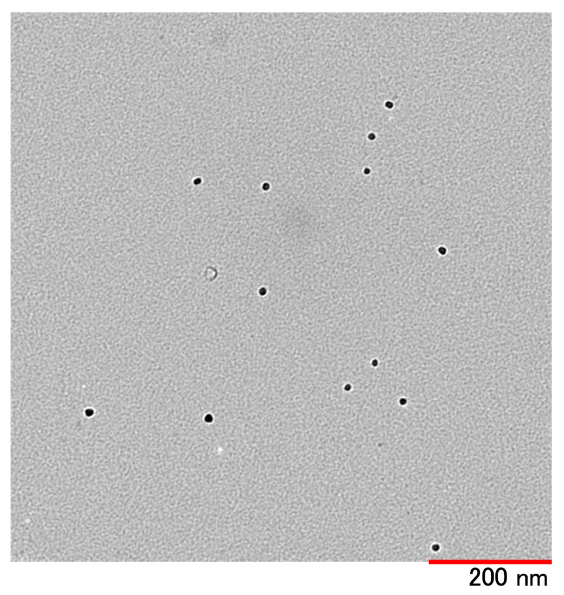
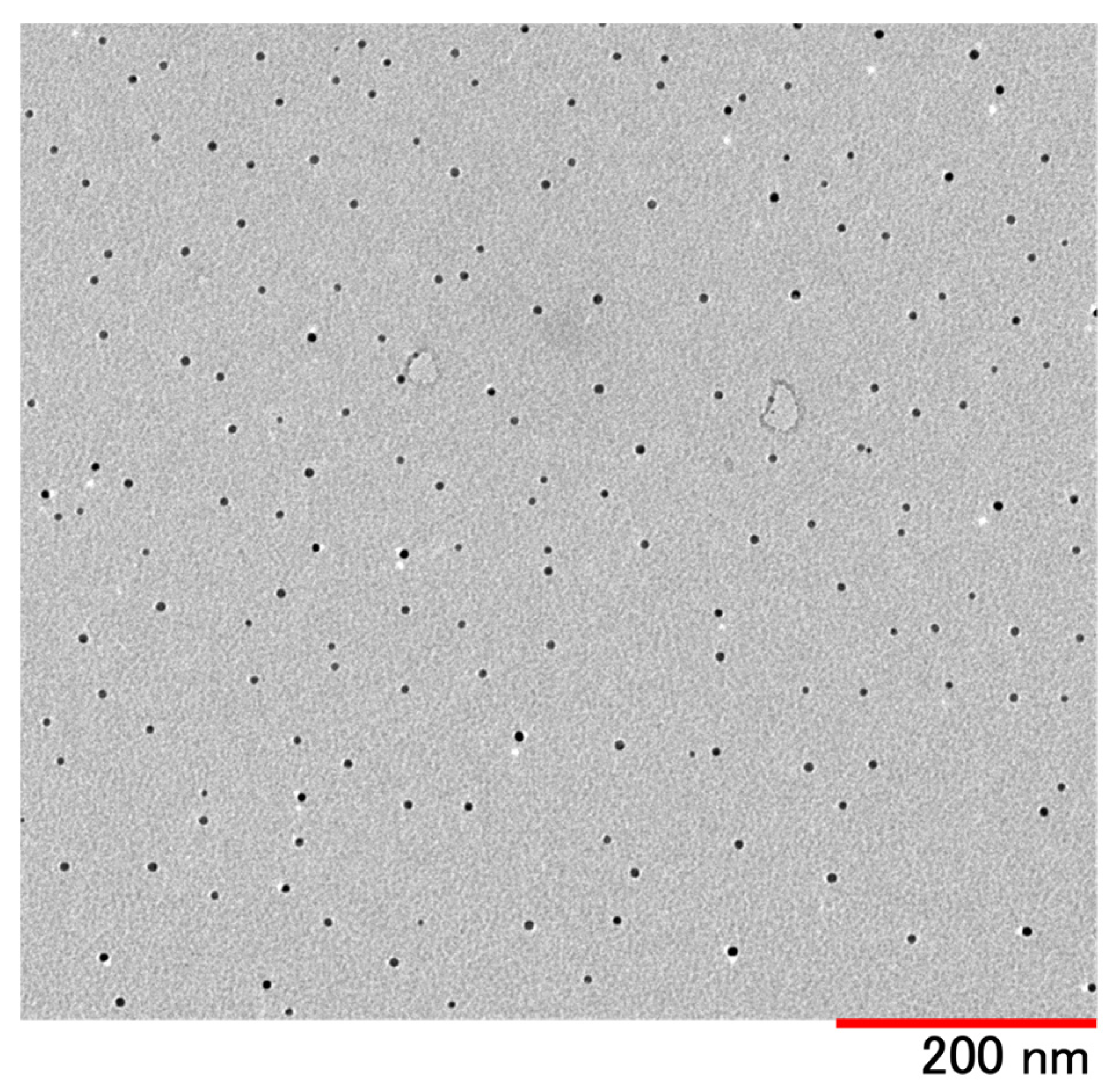
The particle size distribution of terbinafine-loaded PLGA NPs is shown in Figure 1. Table 2 shows the average particle size, polydispersity index, yield, content, and content efficiency of terbinafine-loaded PLGA NPs prepared under each condition. When the poor solvent was purified water and the external solvent was purified water, the average particle size was 40.6 ± 16.3 nm, the polydispersity index was 0.168 ± 0.036, the yield was 79.02 ± 5.47%, the content was 3.53 ± 0.16%, and the loading efficiency was 58.83 ± 2.78%. When the poor solvent was purified water and the external solution was 1.0 × 10⁻³ w/v% Arg aq., the average particle size was 41.3 ± 15.7 nm, the polydispersity index was 0.164 ± 0.008, the yield was 83.21 ± 3.19%, the content was 5.27 ± 0.43%, and the content efficiency was 87.76 ± 7.16%. When the poor solvent was 1.0 × 10⁻¹ w/v%. Arg aq., and the external solution was purified water, the average particle size was 55.1 ± 19.5 nm, the polydispersity index was 0.278 ± 0.047, the yield was 79.29 ± 2.91%, the content was 5.24 ± 0.05%, and the content efficiency was 87.35 ± 0.84%. When the poor solvent was 1.0 × 10⁻¹ w/v% Arg aq. and the external solution was 1.0 × 10⁻³ w/v% Arg aq., the average particle size was 2754.0 ± 3650.3 nm, the polydispersity index was 0.294 ± 0.024, the yield was 78.08 ± 6.13%, the content was 5.22 ± 0.27%, and the content efficiency was 86.52 ± 4.52%. Particles larger than 1000 nm are thought to be agglomerated particles, so they have been excluded from this graph. Additionally, TEM images of Condition 1 and Condition 2 are shown in Figure 2 and Figure 3. The terbinafine-loaded PLGA NPs were monodisperse and existed without agglomeration.

3.2. Stability of Terbinafine-Loaded PLGA NPs

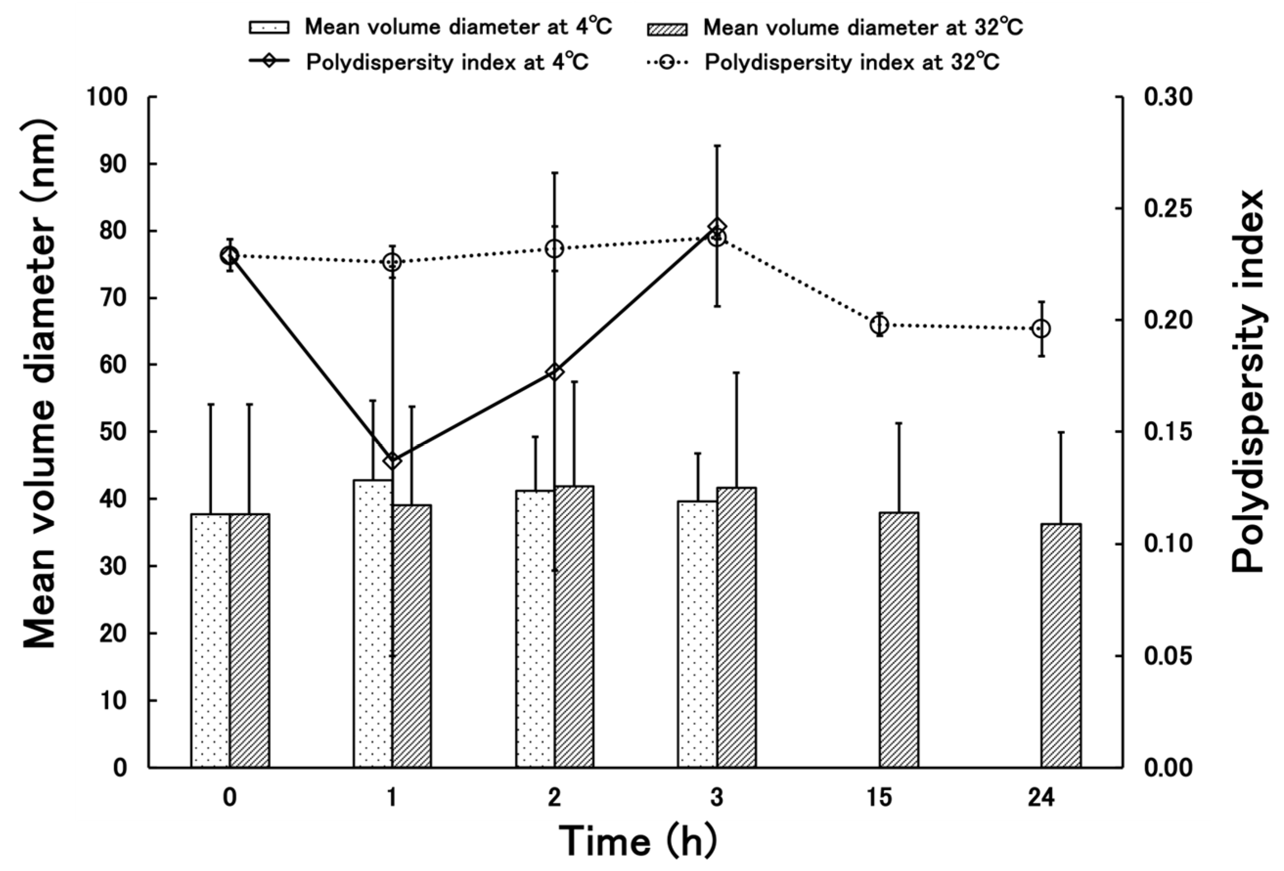
The stability test results for terbinafine-loaded PLGA NPs under Condition 1 are shown in Figure 4, and those for Condition 2 are shown in Figure 5. Under Condition 1 at 4 °C, the average particle size ranged from 60.7–72.9 nm with a standard deviation of 22.4–32.2, demonstrating stability without agglomeration. The polydispersity index ranged from 0.218–0.287, confirming the data showed no variation. At 32 °C, the average particle size ranged from 40.8–63.2 nm, with a standard deviation ranging from 14.9–29.9, demonstrating stability without agglomeration. The polydispersity index ranged from 0.144–0.274, confirming the data showed no variation. Condition 2 at 4 °C showed the average particle size ranging from 37.7–42.8 nm, with a standard deviation ranging from 7.2–16.4. The polydispersity index ranged from 0.137–0.242, confirming the data showed no variation. However, agglomeration was observed by 15 h, so the test was terminated. At 32 °C, the average particle size ranged from 36.3–41.9 nm, with a standard deviation ranging from 13.3–17.1 nm. The particles remained stable without agglomeration. The polydispersity index ranged from 0.196–0.237, confirming that data with low variability was obtained.

3.3. Release Behavior of Terbinafine-Loaded PLGA NPs

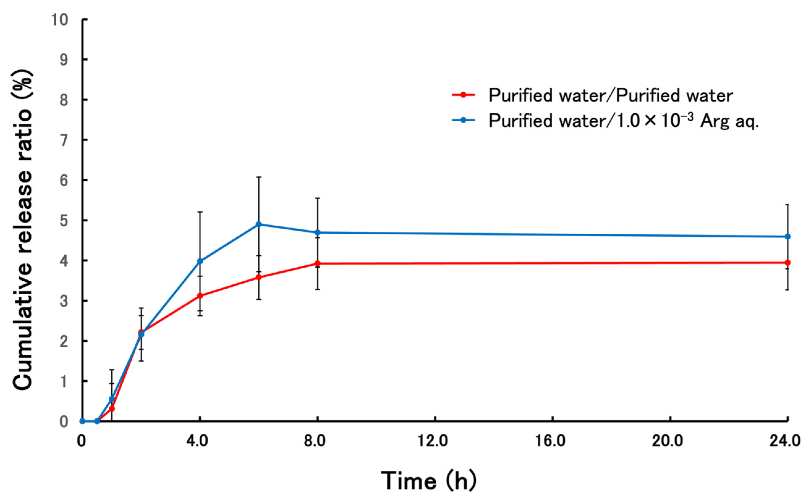
The release behavior of terbinafine from terbinafine-loaded PLGA NPs is shown in Figure 6. In the suspension of terbinafine-loaded PLGA NPs prepared under conditions where the poor solvent was purified water and the external solution was 1.0 × 10⁻³ w/v% Arg aq., the cumulative release ratio of terbinafine from the NPs 24 h after the start of the release test was 4.59 ± 0.79%. There was no significant difference in the release rate at any time point. It is estimated that the release of terbinafine from the NPs will have reached a plateau within 24 h.

3.4. Skin Permeability Test of PLGA NPs Terbinafine-Loaded

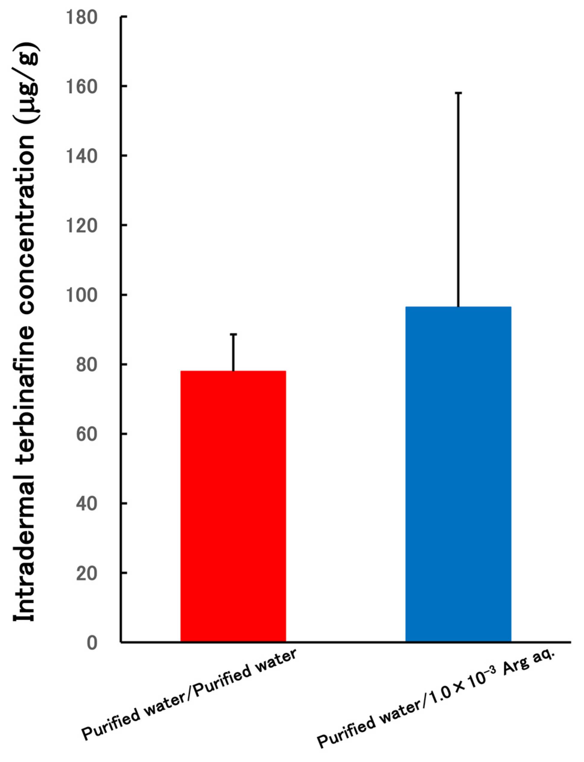
Figure 7 shows the amount of terbinafine delivered intradermally depending on the external fluid conditions during dialysis cleaning. When a suspension of terbinafine-loaded PLGA NPs prepared with purified water as the poor solvent and purified water as the external solution was used as the donor solution, the terbinafine concentration in the skin 8 h after the skin permeability test was 77.94 ± 10.66 μg/g of tissue. On the other hand, when a suspension of terbinafine-loaded PLGA NPs prepared with purified water as the poor solvent and 1.0 × 10⁻³ w/v% Arg aq. as the external solution was used as the donor solution, the terbinafine concentration in the skin 8 h after the skin permeability test was 96.42 ± 61.62 μg/g of tissue. In both receptor solutions, terbinafine was not detected in any of the samples taken at any time point. A t-test was performed to confirm statistical significance, but no significant difference was observed. Additionally, when the donor solution was a simple terbinafine solution, terbinafine was not detected in the skin 8 h after the start of the skin permeability test or in any of the receptor solutions sampled at any time point.

4. Discussion

In the preparation of terbinafine-loaded PLGA NPs, when prepared with purified water as the poor solvent, the average particle size was smaller and stable NPs were obtained compared to when prepared with 1.0 × 10⁻¹ w/v% Arg aq. (Figure 1). When prepared with a poor solvent of 1.0 × 10⁻¹ w/v% Arg aq., the suspension remained turbid even after dialysis washing. This is thought to be due to the aggregation and precipitation of some of the terbinafine and PLGA, and although the content in the NPs is high, it is presumed that some terbinafine that was not nanoparticulated was also detected. When preparing terbinafine-loaded PLGA NPs, purified water is considered preferable to 1.0 × 10⁻¹ w/v% arginine aqueous solution as the poor solvent. This is because the nanoparticle preparation yields a single-peak particle size distribution with particles approximately 40 nm in size. It is well established that particle skin permeability differs between 50 nm and 100 nm particles [34]. The isoelectric point of arginine molecules in Arg aq. is 10.76, and it is classified as a basic amino acid [35]. Arg is an amphiphilic molecule that has both positive and negative charges within the molecule, and when the pH is lower than its isoelectric point, it becomes positive charged. In aqueous solution, PLGA NPs possess a negative charge due to the terminal carboxyl groups. Therefore, a method for preparing PLGA NPs using Arg has been reported [36], with the expectation that stabilization would be achieved by forming an electric double layer. However, these results revealed that using Arg aq. as a poor solvent was not effective for terbinafine-loaded PLGA NPs.
We conducted stability and release, skin permeability tests only on the two types of NPs prepared under conditions 1 and 2, which used purified water as the poor solvent. As shown in Figure 4 and Figure 5, during the stability testing, only the Condition 2 at 4 °C was terminated prematurely; otherwise, no stability issues were observed. Condition 1 showed fluctuations in particle size during the test, but since this was not due to aggregation, it was not considered problematic this time. However, these particle size fluctuations may have caused changes in the drug content, necessitating further investigation. Although the polydispersity index also fluctuated significantly, it remained within the range where monodispersity can be estimated, so it is considered acceptable. As shown in Figure 6, in the release test, no significant difference was observed in the cumulative release ratio of terbinafine from NPs under conditions 1 and 2 at any time point after the start of the test, and release reached a plateau after 24 h. Although we had expected differences in release ratio due to differences in the terbinafine concentration in the NPs, the results may be attributed to the use of purified water as the external solution. Since terbinafine is a poorly water-soluble drug, it is speculated that it did not release smoothly from the NPs. Although the release rate from the NPs was not particularly high, PLGA NPs undergo hydrolysis and gradually degrade within the skin, so it is thought that terbinafine leaks out from the NPs during this degradation process. Therefore, it is anticipated that the actual terbinafine release from terbinafine-loaded PLGA NPs within the skin would be greater than the measured value. The minimum inhibitory concentration (MIC) of terbinafine has been reported as 8.87 μg/g [37]. In the skin permeability test, both NPs prepared under conditions 1 and 2 showed intradermal terbinafine concentrations higher than the MIC. This is likely because nanoparticle formulation enhances penetration through the stratum corneum, which is the rate-limiting step for skin penetration, while also improving intradermal retention through sustained drug release from the NPs. The medication remains within the skin, and as the skin undergoes its natural turnover process, the stratum corneum gradually sheds, allowing new skin to form and thereby achieving treatment. Unless the medication’s retention within the skin improves, the dermatophyte tinea fungus will proliferate during the skin’s turnover cycle, preventing a complete cure. Therefore, it is hypothesized that enhancing the medication’s retention within the skin may shorten the treatment period for tinea fungus [38,39]. Although there was no significant difference, the intradermal delivery of terbinafine was slightly higher in the NPs prepared in condition 2 than in condition 1. However, the NPs prepared in condition 2 showed variability in results and were inferior in terms of reproducibility.
Based on these results, it is thought that terbinafine-loaded PLGA NPs prepared under conditions where the poor solvent is purified water, and the external solution is purified water are the optimal NPs. Although the physicochemical properties of the terbinafine-loaded PLGA NPs prepared under conditions where the poor solvent was purified water and the external solution was purified water were inferior, this preparation method is the simplest, and it yielded equivalent results in the release test and equivalent and stable results in the skin permeability test. Therefore, it is thought to be more suitable for therapeutic use.

5. Conclusions

Terbinafine-loaded PLGA NPs prepared under conditions where the poor solvent was purified water and the external solution was purified water showed skin penetration test results where the intradermal drug concentration exceeded the MIC and yielded stable results, suggesting that they are stable NPs suitable for the treatment of tinea. Since the treatment of tinea is a long-term process, it is desirable to continuously supply the drug to the treatment site in a stable manner. Given the nature of PLGA NPs, sustained-release properties are also anticipated, which may help suppress symptom progression and shorten treatment duration. A limitation of this research is that tinea versicolor is a commensal fungus, making it difficult to plan in vivo experiments. This necessitates evaluating in vitro antifungal efficacy tests and other approaches to compensate. Moving forward, we plan to conduct skin permeability tests using tape stripping technology to confirm the extent of penetration into the stratum corneum of the skin, as well as release tests using solutions such as PBS that mimic the biological environment as the external solution.

Author Contributions

Conceptualization, R.F. and I.T.; methodology, R.F. and I.T.; validation, R.F. and I.T.; formal analysis, R.F., I.T. and K.M.; investigation, R.F.; resources, K.M., A.S. and I.T.; data curation, R.F. and I.T.; writing—original draft preparation, R.F.; writing—review and editing, R.F., R.S., T.O., K.M., A.S., I.T. and K.S.; visualization, R.F., R.S. and I.T.; supervision, K.M., I.T. and K.S.; project administration, R.F. and I.T.; funding acquisition, K.M., I.T. and K.S. All authors have read and agreed to the published version of the manuscript.

Funding

No funding was received to assist with the preparation of this manuscript.

Institutional Review Board Statement

The study was conducted in accordance with the Declaration of Helsinki and approved by the Ethics Committee of Ethics Committee for Animal Experimentation of Josai International University (Ethics Code: 2400164 and date: 26 February 2024).

Data Availability Statement

The data presented in this study are available on request from the corresponding author.

Acknowledgments

We are deeply indebted to all members of our laboratory for their invaluable contributions. Special thanks are extended to Maki Sato and Mayu Hasegawa for their outstanding assistance during this research.

Conflicts of Interest

The authors declare no conflicts of interest.

References

  1. Sahoo, A.K.; Mahajan, R. Management of tinea corporis, tinea cruris, and tinea pedis: A comprehensive review. Indian Dermatol. Online J. 2016, 7, 77–86. [Google Scholar] [CrossRef]
  2. Brown, G.D.; Denning, D.W.; Gow, N.A.; Levitz, S.M.; Netea, M.G.; White, T.C. Hidden Killers: Human Fungal Infections. Sci. Transl. Med. 2012, 165, 165. [Google Scholar] [CrossRef]
  3. Havlickova, B.; Czaika, V.A.; Friedrich, M. Epidemiological trends in skin mycoses worldwide. Mycoses 2008, 51, 2–15. [Google Scholar] [CrossRef] [PubMed]
  4. Seebacher, C.; Bouchara, J.P.; Mignon, B. Updates on the epidemiology of dermatophyte infections. Mycopathologia 2008, 166, 335. [Google Scholar] [CrossRef]
  5. Panday, M.; Pandey, D.; Upadhyay, P.; Upadhyay, S. Terbinafine Preferred Antifungal with a Focus on Dermatophytes. Acta Sci. Microbiol. 2020, 3, 65–72. [Google Scholar] [CrossRef]
  6. Poirier, J.M.; Cheymol, G.J. Optimisation of itraconazole therapy using target drug concentrations. Clin. Pharmacokinet. 1998, 35, 461–473. [Google Scholar] [CrossRef] [PubMed]
  7. Glyn, E.; Evans, V.; Sigugeirsson, B. Double blind, randomised study of continuous terbinafine compared with intermittent itraconazole in treatment of toenail onychomycosis. Br. Med. J. 1999, 318, 1031–1035. [Google Scholar] [CrossRef]
  8. Elewski, B.E.; Rich, P.; Pollak, R.; Pariser, D.M.; Watanabe, S.; Senda, H.; Ieda, C.; Smith, K.; Pillai, R.; Ramakrishna, T.; et al. Efinaconazole 10% solution in the treatment of toenail onychomycosis: Two phase III multicenter, randomized, double-blind studies. J. Am. Acad. Dermatol. 2013, 68, 600–608. [Google Scholar] [CrossRef] [PubMed]
  9. Watanabe, S.; Kishida, H.; Okubo, A. Efficacy and safety of luliconazole 5% nail solution for the treatment of onychomycosis: A multicenter, double-blind, randomized phase III study. J. Dermatol. 2017, 44, 753–759. [Google Scholar] [CrossRef]
  10. Korting, H.C.; Tietz, H.J.; Bräutigam, M.; Mayser, P.; Rapatz, G.; Paul, C. One week terbinafine 1% cream (Lamisil) once daily is effective in the treatment of interdigital tinea pedis: A vehicle controlled study. LAS-INT-06 Study. Med. Mycol. 2001, 39, 335–340. [Google Scholar] [CrossRef]
  11. Berman, B.; Ellis, C.; Leyden, J.; Lowe, N.; Savin, R.; Shupack, J.; Stiller, M.; Tschen, E.; Zaias, N.; Birnbaum, J.E. Efficacy of a 1-week, twice-daily regimen of terbinafine 1% cream in the treatment of interdigital tinea pedis. Results of placebo-controlled, double-blind, multicenter trials. J. Am. Acad. Dermatol. 1992, 26, 956–960. [Google Scholar] [CrossRef]
  12. Iverson, S.L.; Uetrecht, J.P. Identification of a reactive metabolite of terbinafine: Insights into terbinafine-induced hepatotoxicity. Chem. Res. Toxicol. 2001, 14, 175–181. [Google Scholar] [CrossRef]
  13. Ajit, C.; Zaeri, N.; Munoz, S.J.; Suvannasankha, A. Terbinafine-Associated Hepatotoxicity. Am. J. Med. Sci. 2003, 325, 292–295. [Google Scholar] [CrossRef]
  14. Pyatski, Y.; Flach, C.R.; Mendelsohn, R. FT-IR investigation of Terbinafine interaction with stratum corneum constituents. Biochim. Biophys. Acta-Biomembr. 2020, 1862, 183335. [Google Scholar] [CrossRef] [PubMed]
  15. Verma, S.; Utreja, P. Vesicular nanocarrier based treatment of skin fungal infections: Potential and emerging trends in nanoscale pharmacotherapy. Asian J. Pharm. Sci. 2019, 14, 117–129. [Google Scholar] [CrossRef] [PubMed]
  16. Chatur, V.M.; Dhole, S.N. Development and Characterization of Niosomal Gel for Topical Delivery of Luliconazole. Int. J. Pharm. Res. Allied Sci. 2022, 11, 99–107. [Google Scholar] [CrossRef]
  17. Prausnitz, M.R.; Langer, R. Transdermal drug delivery. Nat. Biotechnol. 2008, 26, 1261–1268. [Google Scholar] [CrossRef]
  18. Takeuchi, I.; Kagawa, A.; Makino, K. Skin permeability and transdermal delivery route of 30-nm cyclosporin A-loaded nanoparticles using PLGA-PEG-PLGA triblock copolymer. Colloids Surf. A Physicochem. Eng. Asp. 2020, 600, 124866. [Google Scholar] [CrossRef]
  19. Moser, K.; Kriwet, K.; Naik, A.; Kalia, Y.; Guy, R. Passive skin penetration enhancement and its quantification in vitro. Eur. J. Pharm. 2001, 52, 103–112. [Google Scholar] [CrossRef]
  20. Palmer, B.C.; DeLouise, L.A. Nanoparticle-enabled transdermal drug delivery systems for enhanced dose control and tissue targeting. Molecules 2016, 21, 1719. [Google Scholar] [CrossRef]
  21. Desai, P.; Patlolla, R.R.; Singh, M. Interaction of nanoparticles and cell-penetrating peptides with skin for transdermal drug delivery. Mol. Membr. Biol. 2010, 27, 247–259. [Google Scholar] [CrossRef]
  22. Pearton, M.; Allender, C.; Brain, K.; Anstey, A.; Gateley, C.; Wilke, N.; Morrissey, A.; Birchall, J. Gene Delivery to the Epidermal Cells of Human Skin Explants Using Microfabricated Microneedles and Hydrogel Formulations. Pharm. Res. 2008, 25, 407–416. [Google Scholar] [CrossRef] [PubMed]
  23. Cerc, G.; Vierl, U. Nanotechnology and the transdermal route A state of the art review and critical appraisal. J. Control. Release 2010, 141, 277–299. [Google Scholar] [CrossRef] [PubMed]
  24. Khan, S.; Ullah, M.; Saeed, S.; Saleh, E.; Kassem, A.; Arbi, F.; Wanab, A.; Rehman, M.; Rehman, K.; Khan, D.; et al. Nanotherapeutic approaches for transdermal drug delivery systems and their biomedical applications. Eur. Polym. J. 2024, 207, 112819. [Google Scholar] [CrossRef]
  25. Brannon-Peppas, L. Recent advances on the use of biodegradable microparticles and nanoparticles in controlled drug delivery. Int. J. Pharm. 1995, 116, 1–9. [Google Scholar] [CrossRef]
  26. Mir, M.; Ahmed, N.; Rehman, A. Recent applications of PLGA based nanostructures in drug delivery. Colloids Surf. B Biointerfaces 2017, 159, 217–231. [Google Scholar] [CrossRef]
  27. Sun, L.; Liu, Z.; Wang, L.; Cun, D.; Tong, H.; Yan, R.; Chen, X.; Zheng, Y. Enhanced topical penetration, system exposure and anti-psoriasis activity of two particle-sized, curcumin-loaded PLGA nanoparticles in hydrogel. J. Control. Release 2017, 254, 44–54. [Google Scholar] [CrossRef]
  28. Zhang, W.; Gao, J.; Zhu, Q.; Zhang, M.; Ding, X.; Wang, X.; Hou, X.; Fan, W.; Ding, B.; Wu, X.; et al. Penetration and distribution of PLGA nanoparticles in the human skin treated with microneedles. Int. J. Pharm. 2010, 402, 205–212. [Google Scholar] [CrossRef] [PubMed]
  29. Jain, S.; Mittal, A.; Jain, A. Enhanced Topical Delivery of Cyclosporin—A Using PLGA Nanoparticles as Carrier. Curr. Nanosci. 2011, 7, 524–530. [Google Scholar] [CrossRef]
  30. Takeuchi, I.; Suzuki, T.; Makino, K. Iontophoretic transdermal delivery using chitosan-coated PLGA nanoparticles for transcutaneous immunization. Colloids Surf. A Physicochem. Eng. Asp. 2021, 608, 125607. [Google Scholar] [CrossRef]
  31. Tomoda, K.; Yabuki, N.; Terada, H.; Makino, K. Surfactant free preparation of PLGA nanoparticles: The combination of antisolvent diffusion with preferential solvation. Colloids Surf. A Physicochem. Eng. Asp. 2014, 457, 88–93. [Google Scholar] [CrossRef]
  32. Takeuchi, I.; Makino, K. Biocompatibility and effectiveness of paclitaxel-encapsulated micelle using phosphoester compounds as a carrier fir cancer trearment. Colloids Surf. B Biointerfaces 2019, 177, 356–361. [Google Scholar] [CrossRef] [PubMed]
  33. Dipasquale, D.; Buono, M.; Kolkhorst, F. Effect of Skin Temperature on the Cholinergic Sensitivity of the Human Eccrine Sweat Gland. Jpn. J. Physiol. 2003, 53, 427–430. [Google Scholar] [CrossRef]
  34. Takeuchi, I.; Suzuki, T.; Makino, K. Skin permeability and transdermal delivery route of 50-nm indomethacin-loaded PLGA nanoparticles. Colloids Surf. B Biointerfaces 2017, 159, 312–317. [Google Scholar] [CrossRef]
  35. Fitch, C.; Platzer, G.; Okon, M.; Garcia-Moreno, B.; McIntosh, L. Arginine: Its pKa value revisited. Protein Sci. 2015, 24, 752–761. [Google Scholar] [CrossRef]
  36. Fujisawa, R.; Sakurai, R.; Oshizaka, T.; Mori, K.; Saitoh, A.; Takeuchi, I.; Sugibayashi, K. Development of PSL-loaded PLGA nanoparticles for the treatment of allergic contact dermatitis. Colloids Interfaces 2024, 8, 39. [Google Scholar] [CrossRef]
  37. Nakashima, T.; Nozawa, A.; Majima, T. A novel method using micropig stratum corneum in vitro for the evaluation of anti-Trichophyton mentagrophytes activity. Microbiol. Immunol. 2002, 46, 521–525. [Google Scholar] [CrossRef]
  38. Yokouchi, M.; Kubo, A. Maintenance of tight junction barrier integrity in cell turnover and skin diseases. Exp. Dermatol. 2018, 27, 876–883. [Google Scholar] [CrossRef]
  39. Akhtar, N.; Verma, A.; Pathak, K. Topical Delivery of Drugs for the Effective Treatment of Fungal Infections of Skin. Curr. Pharm. Des. 2015, 21, 2892–2913. [Google Scholar] [CrossRef] [PubMed]
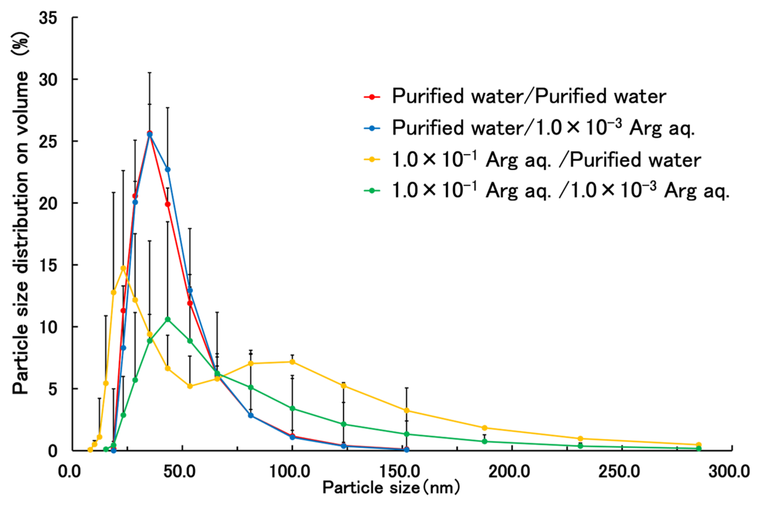
Figure 1. Volume size distribution of terbinafine-loaded PLGA NPs (n = 3).
Applsci 15 12357 g001
Figure 2. The TEM image of Condition 1 terbinafine-loaded PLGA NPs was made at an accelerating voltage of 100 kV (magnification: 20,000×).
Applsci 15 12357 g002
Figure 3. The TEM image of Condition 2 terbinafine-loaded PLGA NPs was made at an accelerating voltage of 100 kV (magnification: 20,000×).
Applsci 15 12357 g003
Figure 4. Time-department changes in mean volume diameter and polydispersity indexes of Condition 1 terbinafine-loaded PLGA NPs at 4 and 32 °C (n = 3, mean ± S.D.).
Applsci 15 12357 g004
Figure 5. Time-department changes in mean volume diameter and polydispersity indexes of Condition 2 terbinafine-loaded PLGA NPs at 4 and 32 °C (n = 3, mean ± S.D.).
Applsci 15 12357 g005
Figure 6. The cumulative release ratio of terbinafine from terbinafine-loaded PLGA NPs (n = 3, mean ± S.D.).
Applsci 15 12357 g006
Figure 7. Intradermal delivery amount of terbinafine (n = 3, mean ± S.D.).
Applsci 15 12357 g007
Table 1. Preparation conditions for terbinafine-loaded PLGA NPs.
Good SolventPoor SolventExternal Solution
Condition 1AcetonePurified waterPurified water
Condition 2AcetonePurified water1.0 × 10−3 w/v% Arg aq.
Condition 3Acetone1.0 × 10−1 w/v% Arg aq.Purified water
Condition 4Acetone1.0 × 10−1 w/v% Arg aq.1.0 × 10−3 w/v% Arg aq.
Table 2. Physicochemical property of terbinafine-loaded PLGA NPs prepared under different conditions (n = 3, mean ± S.D.).
Condition 1Condition 2Condition 3 Condition 4
Mean volume diameter
(nm)
40.6 ± 16.341.3 ± 15.755.1 ± 19.52754.0 ± 3650.3
PDI0.168 ± 0.0360.164 ± 0.0080.278 ± 0.0470.294 ± 0.024
Yield (%)79.02 ± 5.4783.21 ± 3.1979.29 ± 2.9178.08 ± 6.13
Content (%)3.53 ± 0.165.27 ± 0.435.24 ± 0.055.22 ± 0.27
Entrapment efficiency (%)58.83 ± 2.7887.76 ± 7.1687.35 ± 0.8486.52 ± 4.52
Disclaimer/Publisher’s Note: The statements, opinions and data contained in all publications are solely those of the individual author(s) and contributor(s) and not of MDPI and/or the editor(s). MDPI and/or the editor(s) disclaim responsibility for any injury to people or property resulting from any ideas, methods, instructions or products referred to in the content.