Article

Solid Pseudopapillary Neoplasm of the Pancreas: EUS Features and Diagnostic Accuracy of EUS-Guided Fine Needle Biopsy Using a 22-Gauge Fork-Tip Needle in a High Volume Center

by
Nicolò de Pretis
1,*,
Pietro Mastella
1,
Roberto Baldan
1,
Luigi Martinelli
2,
William Mantovani
2,
Federico Caldart
1,
Salvatore Crucillà
1,
Claudio Luchini
3,
Paola Mattiolo
3,
Aldo Scarpa
3,
Stefano Francesco Crinò
1,
Maria Cristina Conti Bellocchi
1,
Riccardo De Robertis
4,
Salvatore Paiella
5,
Antonio Pea
5,
Antonio Amodio
1,
Giulia De Marchi
1 and
Luca Frulloni
1
1
Gastroenterology Unit, University of Verona, 37134 Verona, Italy
2
Clinical Epidemiology Unit, Azienda Provinciale Servizi Sanitari, 38123 Trento, Italy
3
Department of Diagnostics and Public Health, Section of Pathology, University of Verona, 37134 Verona, Italy
4
Department of Radiology, University of Verona, 37134 Verona, Italy
5
Pancreas Surgery, University of Verona, 37134 Verona, Italy
*
Author to whom correspondence should be addressed.
Appl. Sci. 2025, 15(22), 12313; https://doi.org/10.3390/app152212313
Submission received: 9 October 2025 / Revised: 7 November 2025 / Accepted: 17 November 2025 / Published: 20 November 2025
(This article belongs to the Section Biomedical Engineering)

Abstract

Background and Aims: Solid pseudopapillary neoplasms (SPN) are rare neoplasms of the pancreatic gland. Despite the indolent behavior, surgical resection is required according to the risk of metastasis development. Few data are available on endoscopic ultrasound (EUS) features, comparison between the features described at EUS and contrast-enhanced, and diagnostic accuracy and safety of EUS-guided FNB in these tumors. Patients and Methods: All consecutive patients with a EUS-guided FNB-based pathological diagnosis of SPN were extrapolated from a prospectively maintained database. Demographic, radiologic, and echo-endoscopic features were collected. FNB specimens were re-evaluated from two expert pathologist and the main histological features of SPN were investigated. Results: Thirty-seven patients were included (32 females and 5 males), with a mean age of 35.8 ± 15.8 years. Contrast-enhanced imaging based diagnosis was accurate in 20 patients (54.1%). EUS features were significantly different compared to contrast-enhanced imaging in terms of cystic appearance (40.5% vs. 16.2%; p = 0.03) and vascular pattern (p = 0.01). FNB-based diagnosis of SPN was confirmed on surgical specimen in all, 37 patients, resulting in a diagnostic accuracy of EUS-guided FNB of 100%. Only one patient (2.6%) experienced a mild procedure-related adverse event. Discussion: Contrast-enhanced imaging based diagnosis of SPN is difficult. Despite the rarity of the disease, EUS-guided FNB with 22-gauge fork-tip needle has a very high diagnostic accuracy for SPN, with rare and mild adverse events.

1. Backgrounds and Aims

Solid pseudopapillary neoplasms (SPNs) are rare neoplasms of the pancreas with low-grade malignant behavior and a higher incidence in young women [1,2,3,4]. Although these neoplasms typically exhibit indolent biological behavior, metastasis, and recurrence following surgical resection have been reported in up to 2% of cases [2,5,6]. SPNs may appear as solid or cystic lesions, with histopathological examination revealing pseudopapillary architecture. However, the exact pancreatic epithelial cell lineage from which SPNs originate remains unidentified [7]. Consequently, there are no definitive immunohistochemical markers for SPNs, and the pathological diagnosis relies on the recognition of classic features, such as hypotrophic papillae, foam cells, proteinaceous material, and calcifications [8].
A definitive radiologic diagnosis of SPNs can be challenging due to their low incidence—accounting only 1% of all resected solid pancreatic lesions—and the limited accuracy of pre-operative diagnostic modalities [9]. In a large retrospective study involving 131 surgically resected patients, the pre-operative misdiagnosis rate was 34.4% [5]. However, as the majority of the published literature is based on cases with a definitive pathological diagnosis following surgical resection, there is a paucity of data regarding the true pre-operative diagnostic accuracy for SPNs.
Endoscopic ultrasound (EUS)-guided tissue acquisition has become a standard technique worldwide for the characterization of solid pancreatic lesions [10]. Tissue samples are obtained with either fine-needle aspiration (FNA) or fine-needle biopsy (FNB). FNA utilizes a conventional straight needle to aspirate cellular material from the target lesion, allowing cytological assessment and, occasionally, evaluation of small tissue fragments. In contrast, FNB employs specifically designed needles to obtain intact histological core samples, thereby preserving tissue architecture [11].
Only a limited number of studies have investigated the diagnostic accuracy of EUS-guided tissue acquisition in SPNs, with most of the available data involving EUS-guided FNA. Reports on EUS-guided FNB in SPNs are restricted to small case series, despite the technique being considered superior to FNA for diagnostic accuracy in other solid pancreatic lesions [12]. Moreover, the entire available literature on EUS-guided FNA and FNB evaluated patients with definitive pathological diagnosis of SPN and FNA/FNB data were retrospectively collected. Therefore, no data are available on possible incorrect SPN diagnosis on FNA and FNB. Finally, the safety of EUS-guided FNB has never extensively investigated.
The largest available study on EUS-guided tissue acquisition for SPNs is a recent multicenter study including 104 patients with a definitive surgical diagnosis of SPN between 2010 and 2022 [13]. Only 37 patients underwent EUS-guided FNB, but no data are available on needle type, and the diameter ranged from 20 to 25 gauge. The authors reported 97.2% of diagnostic EUS-guided tissue acquisition, but data are not differentiated between FNA and FNB, without data on diagnostic accuracy. Another large study on SPN is a European multicenter study including 149 patients with a surgical diagnosis of SPN between 2000 and 2018 [14]. However, only 78 underwent pre-surgery EUS-guided tissue acquisition, and only 9 underwent FNB using different needle types. An additional study from the US included 17 cases of resected SPNs between 2007 and 2021, which underwent EUS-guided FNA, but none underwent EUS-guided FNB [15].
Finally, a large multicenter radiological study from China compared imaging features between 277 SPNs and 354 neuroendocrine tumors (NETs) evaluated between 2006 and 2021 [16]. Only 26 patients of the SPN group underwent EUS-guided tissue acquisition, without available data on the FNA-FNB proportion, on diagnostic accuracy, and the safety of FNB. The authors concluded that younger age, larger size, and presence of calcification indicate a higher possibility of SPN diagnosis in this setting.
The main aims of the present paper were to investigate the diagnostic accuracy of EUS-guided FNB for SPNs, the pathological features of FNB specimens, and the safety of EUS-guided.
The secondary aim was to evaluate the diagnostic accuracy of contrast-enhanced imaging, as well as the concordance between imaging and EUS in terms of tumor features and vascular pattern.

2. Patients and Methods

All patients with a pathological diagnosis of SPN at EUS-guided FNB specimens between 1 June 2019 and 30 June 2024 were retrospectively identified on a prospectively maintained pathological database. Demographic, clinical, radiological, endoscopic, pathological, and surgical data were evaluated and analyzed.
Exclusion criteria were as follows:
-
EUS-guided FNB performed at another hospital;
-
Absence of EUS-guided FNB;
-
Performance of EUS-guided FNA;
-
Absence of an available contrast-enhanced imaging procedure (contrast-enhanced CT scan and/or contrast-enhanced MRI scan) performed before the EUS-guided FNB;
-
Lack of a surgical specimen with definitive confirmation of the diagnosis of SPN.
Every single FNB specimen was re-evaluated by two expert pathologists (CL and PM) and the percentage cellular, stromal, and necro-hemorrhagic components, respectively. Diagnostic accuracy of EUS-guided FNB was defined as the proportion of SPN diagnosis at FNB confirmed by surgical pathology. Similarly, the diagnostic accuracy of imaging was calculated as the proportion of SPN diagnoses made at imaging that were confirmed on surgical specimens. FNB was performed with a 22-gauge fork-tip needle after contrast medium injection on the solid component of the lesion with a slow-pull technique. EUS-guided FNB was classified as non-diagnostic if a definitive pathological diagnosis could not be established. Adverse events, classified according to the AGREE criteria [17], were categorized as “early” if they occurred during the procedure and as “late” if they developed post-procedurally.
The pancreatic lesion was considered “solid” if no clear cystic component was detected or if a minimal cystic component was described (<20% of the entire volume) and “cystic” if the cystic component was >20% of the entire lesion volume. The vascular pattern observed on imaging and EUS was classified as hypovascular, isovascular, or hypervascular by comparison with the surrounding pancreatic parenchyma. Follow-up duration was calculated from the date of EUS-guided FNB to the date of the most recent clinical evaluation. Patients were considered lost to follow-up if no clinical assessment was available for more than 12 months.
Of the 3821 EUS-guided FNB procedures performed during the study period, 37 patients (0.97%) were diagnosed with SPN and included in the study.

Statistical Analysis

The comparison between patients with and without pre-endoscopic suspicion of SPN based on radiologic imaging was conducted using appropriate univariable tests. Specifically, group differences for categorical variables were assessed using Fisher’s exact test, while continuous variables were evaluated for distributional assumptions and compared using the Wilcoxon–Mann–Whitney test.
In addition, multivariable logistic regression was employed to estimate odds ratios (ORs) and corresponding 95% confidence intervals (CIs) to explore associations between radiological suspicion of TSP and patient’s age, tumor location, and tumor size.
All p-values of less than 0.05 were considered statistically significant.
All statistical analyses were performed using Stata version 16.1 (StataCorp, College Station, TX, USA).

3. Results

Among the 37 patients included in the study, the mean age at the time of EUS-guided FNB was 35.8 ± 15.8 years and 32 patients (86.5%) were female. Fifteen patients (40.5%) were asymptomatic at diagnosis, while only two patients (5.4%) presented with non-biliary pancreatitis. Elevated Ca19.9 levels (699 U/mL) were observed in one patient (2.7%), with normalization following pancreatic resection (24 U/mL).
None of the 3782 patients excluded from the study who underwent surgery were found to have SPN on histological examination of the surgical specimen.
The main epidemiological and clinical features of the included patients are reported in Table 1.

3.1. Endoscopic Ultrasound (EUS)

In all patients EUS was performed using a linear echoendoscope under deep sedation administered by an anesthesiologist, without endotracheal intubation. No sedation-related adverse events were observed.
At EUS, the mean size of the pancreatic lesion was 35.8 ± 15.8 mm. The tumor was located in the head of the pancreas in 8 patients (21.6%), in the body in 15 patients (40.5%), and in the tail in 14 patients (37.8%). Main pancreatic duct dilation was observed in five patients (13.5%), while common bile duct dilation was present in one patient (2.7%).
The pancreatic lesion was described as solid in 31 patients (83.9%) and cystic in 6 patients (16.1%) on EUS, whereas on imaging, the lesion was characterized as solid in 22 patients (59.5%) and cystic in 15 patients (40.5%). This difference was statistically significant (p = 0.03).
Following injection of contrast medium (SonoVue®, Bracco, Milan, Italy, 4,8 mL), the vascular pattern on EUS was categorized as hypovascular in 9 patients (24.2%), isovascular in 9 patients (24.3%), and hypervascular in 19 patients (51.5%). These findings were significantly different from the vascular patterns observed on imaging, which were hypovascular in 19 patients (51.4%), isovascular in 10 patients (27.0%), and hypervascular in 8 patients (21.6%) (p = 0.01).
Comparison between imaging and EUS features is presented in Table 2. Figure 1 shows a case of SPN at MRI and EUS.

3.2. EUS-Guided FNB

EUS-guided FNB was performed in all 37 included patients using a 22-gauge fork-tip FNB needle (SharkCore™, Medtronic Co., Boston, MA, USA) with a mean of 2.8 ± 0.6 passes per procedure. FNB was considered diagnostic in 100% of cases and no patient required an additional EUS-guided FNB.
All 37 patients with a pathological diagnosis of SPN on FNB had a definitive diagnosis of SPN confirmed on surgical specimens, resulting in a diagnostic accuracy of 100%. No early adverse events were observed, while one patient (2.7%) experienced a late adverse event classified as grade II according to the AGREE classification (mild acute pancreatitis requiring five days of hospitalization).
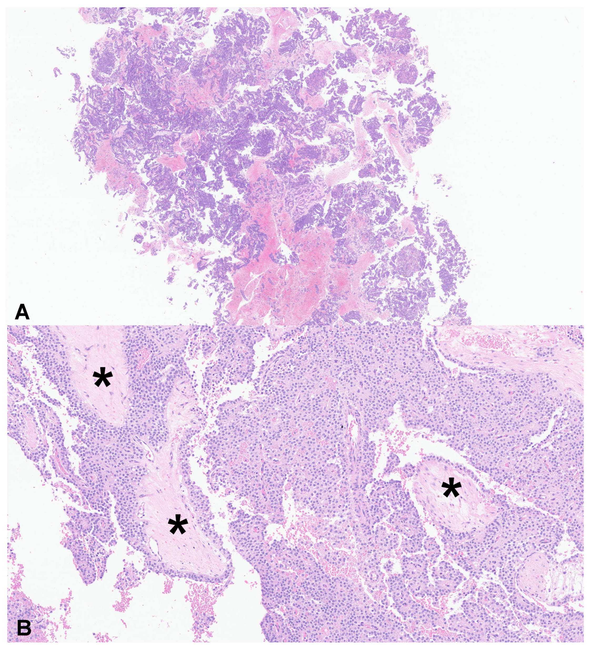
Pathological evaluation of FNB specimens revealed a mean cellular component of 41.2 ± 21.5%, a mean stromal component of 10.0 ± 5.0%, and a mean necro-hemorrhagic component of 46.0 ± 21.2%. There was no significant association between these histological features and the probability of correct diagnosis at imaging (p = 0.173). Typical SPN pathological features, such as hypotrophic papillae, foam cells, proteinaceous material, and calcifications, were identified in FNB specimens in 10 (25.6%), 7 (17.9%), 7 (17.9%), and 2 (5.1%) patients, respectively. At least one of these features was present in 24 patients (61.5%). Figure 2 shows material obtained through FNB.

3.3. Radiology

Thirty-five patients underwent MRI, two patients underwent CT, and five patients underwent both contrast-enhanced imaging modalities. On imaging, the mean size of the pancreatic lesion was 41.4 ± 15.1 mm. Main pancreatic duct dilation was observed in five patients (13.5%), while common bile duct dilation was present in one patient (2.7%).
The pancreatic lesions were characterized as solid in 22 patients (59.5%) and cystic in 15 patients (40.5%). The vascular pattern was categorized as hypovascular in 19 patients (51.4%), isovascular in 10 patients (27.0%), and hypervascular in 8 patients (21.6%). No pseudocapsule was identified in any case.
In 20 cases (54.1%), there was agreement between the radiological suspicion and the surgical diagnosis, while in 17 patients (45.9%) the histological diagnosis was necessary to obtain the correct diagnosis. The alternative radiological diagnoses included NET (12 patients), mucinous cystic neoplasm (1 patient), post-pancreatitis scar (1 patient), pancreatic cancer (1 patient), IPMN (1 patient), and other diagnoses (1 patient). Therefore, the diagnostic accuracy of contrast-enhanced imaging in this cohort was 54.1%.
At multivariable analysis, age was the only parameter significantly different between patients with a correct versus incorrect diagnosis of SPN at imaging (26.7 ± 11.2 years vs. 43.9 ± 14.7 years, respectively). Results of the univariable and multivariable analyses comparing patients with and without correct radiological diagnosis of SPN are presented in Table 3 and Table 4.

3.4. Long-Term Follow-Up

The median follow-up was 37 months (IQR 30–49), and no patients were lost at follow-up. All patients underwent surgical resection and were alive at the end of the follow-up period. The surgical procedures performed included pylorus-preserving duodeno-pancreatectomy in 8 patients (21.6%), intermediate pancreatic resection in 8 patients (21.6%), distal pancreatectomy in 20 patients (54.1%), and retro-pancreatic resection with splenectomy in 1 patient (2.7%).
No patients had metastases at the time of diagnosis; however, one patient (2.7%) developed liver metastasis during follow-up, and one additional patient (2.7%) experienced local recurrence of SPN. Consequently, 35 patients (94.6%) remained disease-free after surgical resection.

4. Discussion

Despite the indolent behavior of SPNs surgical resection is considered mandatory due to the risk of hepatic metastasis and local recurrence [1,2,3]. Given that SPNs are frequently diagnosed in young patients, and considering the significant morbidity and mortality associated with pancreatic surgery, establishing an accurate diagnosis is crucial for optimal clinical management. Imaging has limited diagnostic accuracy for SPNs, with a considerable risk of misdiagnosis and possible inappropriate clinical management [5,16]. Although EUS-guided fine-needle biopsy (FNB) is an internationally accepted diagnostic tool for pancreatic lesions, data regarding its diagnostic accuracy and safety in patients with SPNs remain limited.
This study represents the largest series of SPN patients undergoing EUS-guided FNB and demonstrates excellent diagnostic accuracy alongside a favorable safety profile. The demographic and clinical characteristics of the included patients are consistent with the previously published literature. Specifically, SPNs predominantly affect females (87%), of a young age (mean < 36 years), who are frequently diagnosed incidentally (>40%) [1,2,3]. Furthermore, our findings confirm that contrast-enhanced imaging has limited diagnostic accuracy, even in high-volume centers. These results are similar to those reported by Marchegiani et al. and De Robertis et al., who described correct preoperative diagnoses in fewer than two-thirds of cases [5,18]. Similarly, a large study from China highlighted the diagnostic challenges in differentiating SPNs from NETs on imaging and observed a higher likelihood of correct SPN diagnosis in younger patients [16]. Notably, our data confirm that younger age is associated with a higher probability of correct preoperative SPN diagnosis on imaging.
Additionally, we confirmed significant differences in the features of SPNs as assessed by contrast-enhanced imaging versus EUS [19,20]. On contrast-enhanced imaging, SPNs more frequently appeared cystic compared to EUS, possibly due to EUS’s superior capacity to distinguish between liquid and necrotic components. Moreover, the vascular patterns differed significantly between the two modalities. EUS more often identified a hypervascular pattern, likely attributable to its ability to visualize real-time perfusion and detect early enhancement.
The diagnostic accuracy of EUS-guided FNB using a 22-gauge fork-tip needle was 100%, confirming the high diagnostic accuracy (>97%) reported by Pawlak et al. in a mixed cohort of FNA and FNB procedures retrospectively evaluated between 2010 and 2022 [13]. Remarkably, the diagnostic accuracy of EUS-guided FNB with a 22-gauge fork-tip needle remained high despite the rarity of the disease (<1% of all FNBs performed for solid pancreatic lesions) and the notable proportion of necro-hemorrhagic and stromal components in the biopsy material (46% and 10%, respectively).
Unlike previous studies, the present study included a highly homogeneous cohort of consecutive patients who all underwent the same procedure (EUS-guided FNB), with the same needle type (22-gauge fork-tip), over a relatively short period (5 years), in a high-volume pancreatic center. Furthermore, whereas prior studies focused exclusively on patients with a definitive surgical diagnosis of SPN [4,13,14,15,16], our cohort included patients with an FNB-based diagnosis. Consequently, this investigation assessed the reliability of FNB-based diagnosis, including the risk of incorrect preoperative diagnosis. Notably, none of the patients with a definitive SPN diagnosis on surgical pathology had a non-diagnostic or inaccurate FNB-based diagnosis, suggesting that the risk of misdiagnosing SPN as a different pancreatic tumor via FNB is extremely low.
Finally, EUS-guided FNB appears to be safe with low and mild adverse events. However, the risk of pancreatitis should be considered.
The present study has certain limitations, including its retrospective design, although this was mitigated by the use of a prospectively maintained database. Despite representing the largest series on this subject, the sample size remains limited due to the rarity of SPNs. Therefore, prospective multicenter studies are warranted to validate these findings.
In conclusion, this study represents the largest investigation to date on the role of EUS-guided FNB in the diagnosis of SPNs. Despite the rarity of the disease, diagnostic accuracy is very high when using a 22-gauge fork-tip needle, with only rare and mild adverse events observed.

Author Contributions

Conceptualization, N.d.P., R.B. and S.P.; methodology, N.d.P., W.M. and L.M.; validation, L.F., C.L., A.S.; formal analysis, W.M. and L.M.; investigation, N.d.P., P.M. (Pietro Mastella), S.F.C. and M.C.C.B.; data curation, A.P., P.M. (Paola Mattiolo), F.C., S.C., G.D.M. and A.A.; writing—original draft preparation, N.d.P., L.F. and R.D.R.; writing—review and editing, N.d.P., P.M. (Pietro Mastella) and R.B. All authors have read and agreed to the published version of the manuscript.

Funding

This research received no external funding.

Institutional Review Board Statement

The study was conducted in accordance with the Declaration of Helsinki, and approved by the Ethics Committee of the Province of Verona and Rovigo (1271CESC). This research followed the Strengthening the Reporting of Observational Studies in Epidemiology (STROBE) statement of observational studies.

Informed Consent Statement

Written informed consent was obtained from all patients for the procedure following the Declaration of Helsinki.

Data Availability Statement

The data that supports the findings of this study are available from the corresponding authors upon reasonable request.

Conflicts of Interest

The authors declare no conflict of interest.

References

  1. Law, J.K.; Ahmed, A.; Singh, V.K.; Akshintala, V.S.; Olson, M.T.; Raman, S.P.; Ali, S.Z.; Fishman, E.K.; Kamel, I.; Canto, M.I.; et al. A systematic review of solid-pseudopapillary neoplasms: Are these rare lesions? Pancreas 2014, 43, 331–337. [Google Scholar] [CrossRef] [PubMed]
  2. Papavramidis, T.; Papavramidis, S. Solid pseudopapillary tumors of the pancreas: Review of 718 patients reported in English literature. J. Am. Coll. Surg. 2005, 200, 965–972. [Google Scholar] [CrossRef] [PubMed]
  3. Yu, P.F.; Hu, Z.H.; Wang, X.B.; Guo, J.M.; Cheng, X.D.; Zhang, Y.L.; Xu, Q. Solid pseudopapillary tumor of the pancreas: A review of 553 cases in Chinese literature. World J. Gastroenterol. 2010, 16, 1209–1214. [Google Scholar] [CrossRef] [PubMed]
  4. Klimstra, D.S.; Hruban, R.H.; Sigel, C.S.; Klöppel, G. Tumors of the Pancreas; American Registry of Pathology: Arlington, VI, USA, 2023. [Google Scholar] [CrossRef]
  5. WHO Classification of Tumours Editorial Board. Digestive System Tumours, 5th ed.; International Agency for Research on Cancer: Lyon, France, 2019; Available online: https://publications.iarc.who.int/Book-And-Report-Series/Iac-Iarc-Who-Cytopathology-Reporting-Systems/WHO-Reporting-System-For-Pancreaticobiliary-Cytopathology-2022 (accessed on 16 July 2025).
  6. Marchegiani, G.; Andrianello, S.; Massignani, M.; Malleo, G.; Maggino, L.; Paiella, S.; Ferrone, C.R.; Luchini, C.; Scarpa, A.; Capelli, P.; et al. Solid pseudopapillary tumors of the pancreas: Specific pathological features predict the likelihood of postoperative recurrence. J. Surg. Oncol. 2016, 114, 597–601. [Google Scholar] [CrossRef] [PubMed]
  7. Amato, E.; Mafficini, A.; Hirabayashi, K.; Lawlor, R.T.; Fassan, M.; Vicentini, C.; Barbi, S.; Delfino, P.; Sikora, K.; Rusev, B.; et al. Molecular alterations associated with metastases of solid pseudopapillary neoplasms of the pancreas. J. Pathol. 2019, 247, 123–134. [Google Scholar] [CrossRef] [PubMed] [PubMed Central]
  8. La Rosa, S.; Bongiovanni, M. Pancreatic Solid Pseudopapillary Neoplasm: Key Pathologic and Genetic Features. Arch. Pathol. Lab. Med. 2020, 144, 829–837. [Google Scholar] [CrossRef] [PubMed]
  9. Samad, A.; Shah, A.A.; Stelow, E.B.; Alsharif, M.; Cameron, S.E.; Pambuccian, S.E. Cercariform cells: Another cytologic feature distinguishing solid pseudopapillary neoplasms from pancreatic endocrine neoplasms and acinar cell carcinomas in endoscopic ultrasound-guided fine-needle aspirates. Cancer Cytopathol. 2013, 121, 298–310. [Google Scholar] [CrossRef] [PubMed]
  10. Zhao, M.; Wang, J.; Lai, J.; Liu, F.; Zhang, Y.; Cao, L.; Liu, L.; Ma, K.; Li, J.; Deng, Q. Solid pseudopapillary neoplasms of the pancreas (SPNs): Diagnostic accuracy of CT and CT imaging features. World J. Surg. Oncol. 2024, 22, 225. [Google Scholar] [CrossRef] [PubMed]
  11. Crinò, S.F.; Di Mitri, R.; Nguyen, N.Q.; Tarantino, I.; de Nucci, G.; Deprez, P.H.; Carrara, S.; Kitano, M.; Shami, V.M.; Fernández-Esparrach, G.; et al. Endoscopic Ultrasound-guided Fine-needle Biopsy with or Without Rapid On-site Evaluation for Diagnosis of Solid Pancreatic Lesions: A Randomized Controlled Non-Inferiority Trial. Gastroenterology 2021, 161, 899–909. [Google Scholar] [CrossRef] [PubMed]
  12. de Pretis, N.; Crinò, S.F.; Frulloni, L. The Role of EUS-Guided FNA and FNB in Autoimmune Pancreatitis. Diagnostics 2021, 11, 1653. [Google Scholar] [CrossRef] [PubMed]
  13. Yao, D.W.; Qin, M.Z.; Jiang, H.X.; Qin, S.Y. Comparison of EUS-FNA and EUS-FNB for diagnosis of solid pancreatic mass lesions: A meta-analysis of prospective studies. Scand. J. Gastroenterol. 2024, 59, 972–979. [Google Scholar] [CrossRef] [PubMed]
  14. Pawlak, K.M.; Tehami, N.; Maher, B.; Asif, S.; Rawal, K.K.; Balaban, D.V.; Tag-Adeen, M.; Ghalim, F.; Abbas, W.A.; Ghoneem, E.; et al. Role of endoscopic ultrasound in the characterization of solid pseudopapillary neoplasm of the pancreas. World J. Gastrointest. Endosc. 2023, 15, 273–284. [Google Scholar] [CrossRef] [PubMed]
  15. Karsenti, D.; Caillol, F.; Chaput, U.; Perrot, B.; Koch, S.; Vuitton, L.; Jacques, J.; Valats, J.C.; Poincloux, L.; Subtil, C.; et al. Safety of Endoscopic Ultrasound-Guided Fine-Needle Aspiration for Pancreatic Solid Pseudopapillary Neoplasm Before Surgical Resection: A European Multicenter Registry-Based Study on 149 Patients. Pancreas 2020, 49, 34–38. [Google Scholar] [CrossRef] [PubMed]
  16. Wang, B.G.; Mani, H.; Wang, Z.Q.; Li, W. Cytological Diagnosis of Pancreatic Solid-Pseudopapillary Neoplasm: A Single-Institution Community Practice Experience. Diagnostics 2022, 12, 449. [Google Scholar] [CrossRef] [PubMed]
  17. Hu, H.F.; Li, Z.; Chen, K.; Liu, M.Q.; Ye, Z.; Chen, X.M.; Zhang, Y.; Yu, X.J.; Xu, X.W.; Ji, S.R. Multimodality imaging differentiation of pancreatic neuroendocrine tumors and solid pseudopapillary tumors with a nomogram model: A large single-center study. Front. Surg. 2022, 9, 970178. [Google Scholar] [CrossRef] [PubMed]
  18. Nass, K.J.; Zwager, L.W.; van der Vlugt, M.; Dekker, E.; Bossuyt, P.M.M.; Ravindran, S.; Thomas-Gibson, S.; Fockens, P. Novel classification for adverse evvents in GI endoscopy: The AGREE classification. Gastrointest. Endosc. 2022, 95, 1078–1085.e8. [Google Scholar] [CrossRef] [PubMed]
  19. De Robertis, R.; Marchegiani, G.; Catania, M.; Ambrosetti, M.C.; Capelli, P.; Salvia, R.; D'Onofrio, M. Solid Pseudopapillary Neoplasms of the Pancreas: Clinicopathologic and Radiologic Features According to Size. AJR Am. J. Roentgenol. 2019, 213, 1073–1080. [Google Scholar] [CrossRef] [PubMed]
  20. Raman, S.P.; Kawamoto, S.; Law, J.K.; Blackford, A.; Lennon, A.M.; Wolfgang, C.L.; Hruban, R.H.; Cameron, J.L.; Fishman, E.K. Institutional experience with solid pseudopapillary neoplasms: Focus on computed tomography, magnetic resonance imaging, conventional ultrasound, endoscopic ultrasound, and predictors of aggressive histology. J. Comput. Assist. Tomogr. 2013, 37, 824–833. [Google Scholar] [CrossRef] [PubMed]
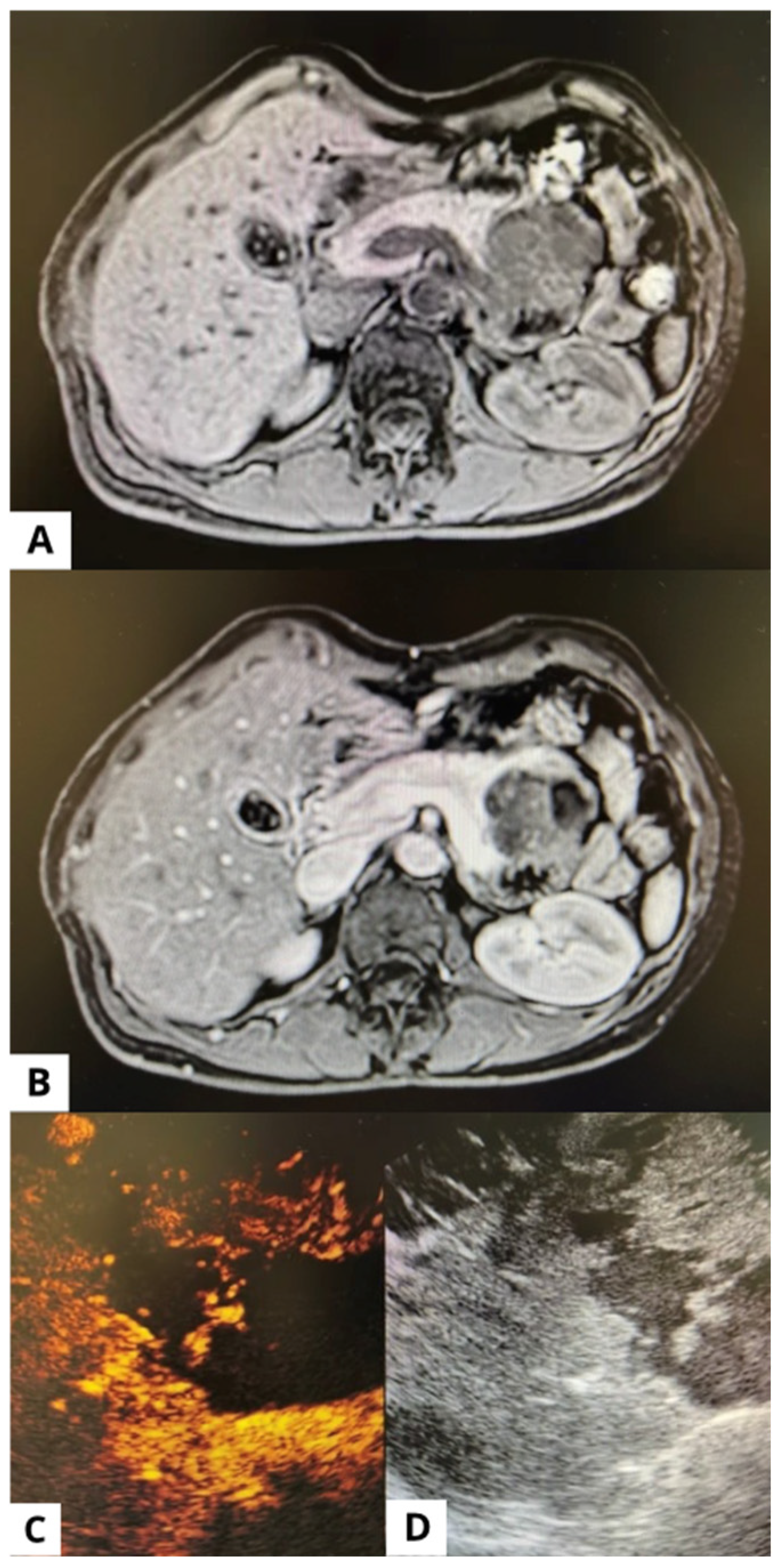
Figure 1. A case of solid pseudopapillary neoplasm. The same lesion shown in (A) MRI without contrast, (B) MRI with contrast, (C) EUS post contrast medium, and (D) EUS before contrast medium.
Applsci 15 12313 g001
Figure 2. Microscopic pathology images of solid pseudopapillary neoplasma hematoxylin and eosin staining 4× (A). Relationship between solid and cystic portions 20× (B) showing that some areas manage to maintain adequate vascular support with the formation of solid structures, even in close proximity to areas in fibromyxoid degeneration (asterisk).
Applsci 15 12313 g002
Table 1. General demographical and clinical features of the included patients. Nr. is number, SD is standard deviation.
Total patients nr. (%)37 (100)
Females nr. (%)32 (86.5)
Mean age years (SD)35.8 (15.8)
Symptoms nr. (%)22 (59.5)
         Abdominal pain nr. (%)15 (40.5)
         Acute pancreatitis nr. (%)2 (5.4)
         Weight loss nr. (%)1 (2.7)
         Vomiting nr. (%)1 (2.7)
         Itching nr. (%)1 (2.7)
Family history with pancreatic disease nr. (%)1 (2.7)
Ca19.9 > 37 U/mL nr. (%)1 (2.7)
Diabetes nr. (%)3 (8.1)
Table 2. Comparison between solid pseudopapillary neoplasm appearance at contrast-enhanced imaging (imaging) and endoscopic ultrasound (EUS). SD is standard deviation, nr. is number, CBD is common bile duct, MPD is main pancreatic duct.
ImagingEUSp
Size, median (SD) mm41.4 (15.1)35.8 (15.8)0.12
Contrast enhancement
     Hypo-enhancing nr. (%)19 (51.4)9 (24.3)
     Iso-enhancing nr. (%)10 (27.0)9 (24.3)0.01
     Hyper-enhancing nr. (%)8 (21.6)19 (51.4)
Cystic appearance nr. (%)6 (16.2)15 (40.5)0.03
Dilation of CBD nr. (%)1 (2.7)1 (2.7)>0.99
Dilation of MPD nr. (%)5 (13.5)5 (13.5)>0.99
Calcifications nr. (%)8 (21.6)6 (16.2)0.77
Pseudocapsule nr. (%)0 (0)0 (0)>0.99
Table 3. Univariable analysis comparing patients with correct and incorrect diagnosis of solid pseudopapillary neoplasm (SPN) at contrast-enhanced imaging. Nr. is number, SD is standard deviation, Hypo is hypo-enhancing, Iso is iso-enhancing, and hyper is hyper-enhancing compared to the surrounding pancreatic parenchyma, MPD is the main pancreatic duct, and CBD is the common bile duct.
CorrectNot-Correctp
Total nr. (%)20 (54.1)17 (45.9)/
Females nr. (%)19 (95.0)13 (76.5)0.159
Mean age years (SD)26.7 (11.2)43.9 (14.7)<0.001
Location
         Head nr (%)5 (25.0)3 (17.7)0.006
         Body nr (%)12 (60.0)3 (17.7)
         Tail nr (%)3 (15.0)11 (64.7)
Size mm (SD)46.4 (26.9)36.7 (21.1)0.08
Main cystic aspect nr. (%)3 (15.0)3 (17.6)>0.99
Enhancement
         Hypo nr. (%)10 (50.0)9 (52.9)
         Iso nr. (%)6 (30.0)4 (23.5)>0.99
         Hyper nr. (%)4 (20.0)5 (29.44)
Dilation of MPD nr. (%)5 (25.0)0 (0)0.05
Dilation of CBD nr. (%)1 (5.0)0 (0.0)>0.99
Table 4. Multivariable analysis comparing patients with correct and incorrect diagnosis of solid pseudopapillary neoplasm (SPN) at contrast-enhanced imaging. OR is Odds ratio, CI is confidence interval.
OR (95% CI)p
Age0.90 (0.83–0.98)0.012
Location
Head1 #
Body 8.99 (0.58–139.31)0.116
Tail0.31 (0.03–3.78)0.358
Size1.05 (1.00–1.10)0.053
# indicates the OR calculated for the body and tail.
Disclaimer/Publisher’s Note: The statements, opinions and data contained in all publications are solely those of the individual author(s) and contributor(s) and not of MDPI and/or the editor(s). MDPI and/or the editor(s) disclaim responsibility for any injury to people or property resulting from any ideas, methods, instructions or products referred to in the content.