Review

From Light to Insight: Hemodynamic Models for Optical Monitoring of the Brain in Cardiac Arrest

by
Nima Soltani
1,2 and
Vladislav Toronov
1,2,*
1
Department of Physics, Toronto Metropolitan University, Toronto, ON M5B 2K3, Canada
2
Institute of Biomedical Engineering, Science and Technology (iBEST), Li Ka-Shing Knowledge Institute, Toronto, ON M5B 1T8, Canada
*
Author to whom correspondence should be addressed.
Appl. Sci. 2025, 15(22), 12260; https://doi.org/10.3390/app152212260
Submission received: 31 August 2025 / Revised: 13 November 2025 / Accepted: 16 November 2025 / Published: 19 November 2025

Abstract

Optical neuromonitoring has matured from descriptive oxygenation trends to model-informed quantification of cerebral physiology. This review synthesizes evidence on near-infrared spectroscopy (NIRS), diffuse correlation spectroscopy (DCS), and laser Doppler flowmetry (LDF) for monitoring cerebral blood flow (CBF), blood volume (CBV), and cerebral metabolic rate of oxygen (CMRO2) during cardiac arrest (CA) and cardiopulmonary resuscitation (CPR). We focus on using hemo-metabolic models, especially Coherent Hemodynamic Spectroscopy (CHS) and the BrainSignals models, as a framework to explain what optical signals do (and do not) tell us about microvascular oxygen transport and mitochondrial metabolism. We compare linear vs. non-linear CHS formulations for large perturbations (e.g., CA/CPR), summarize emerging depth-sensitivity and extracerebral-signal suppression strategies, and outline how DCS pairs with NIRS to link oxygen delivery with use. Across animal and human studies, we highlight convergent patterns (rapid oxygenation collapse, partial reperfusion during CPR, lagging metabolic recovery), recurring limitations (extracerebral contamination, calibration to absolutes, motion), and standardization efforts required for translation. We conclude with a pragmatic roadmap for bedside implementation: harmonized physiological endpoints (CBF, CMRO2, rCCO), reporting standards, and model-informed thresholds to guide resuscitation. This review aims to bridge instrumentation, physiology, and modeling to enhance neuroprotective care in CA/CPR.

1. Introduction

Cardiac arrest (CA) remains a major cause of death and neurological disability, with out-of-hospital survival still low despite improvements in resuscitation science [1]. Neurological injury reflects a cascade from global cerebral ischemia to reperfusion and secondary metabolic failure, underscoring the need to monitor cerebral blood flow (CBF), oxygen delivery, and mitochondrial function during CA and cardiopulmonary resuscitation (CPR) and into post-return of spontaneous circulation (ROSC) care [2,3]
Optical methods—near-infrared spectroscopy (NIRS), diffuse correlation spectroscopy (DCS), and laser Doppler flowmetry (LDF)—offer continuous assessment of cerebral microvascular oxygenation and flow. Foundational work demonstrated that near-infrared light can track hemoglobin oxygenation and cytochrome-c-oxidase redox changes in vivo [4]. Subsequent developments combined NIRS and DCS to estimate hemoglobin changes and CBF in brain [5,6], while LDF enabled microvascular flow indexing, albeit with shallow penetration [7]. Yet, interpretation in CA/CPR is challenging, because signals are confounded by extracerebral layers and device variability, and mitochondrial measurements remain technically demanding [8,9,10].
Over the past three decades, optical monitoring technologies have evolved from experimental laboratory tools to increasingly viable options for real-time, bedside neuromonitoring in acute settings. Early continuous-wave NIRS systems provided relative changes in chromophore concentrations, which, while valuable, were insufficient for absolute quantification. The introduction of frequency-domain and time-domain NIRS enabled depth discrimination and partial correction for scattering effects, improving the accuracy of cerebral oxygenation estimates [11,12]. DCS emerged in the late 1990s as a non-invasive technique for quantifying microvascular cerebral blood flow. Recent advances in hybrid instruments allowed simultaneous acquisition of DCS and NIRS signals from the same tissue volume, enabling integrated assessment of oxygen delivery and utilization [13].
Despite these advances, CA and CPR present a distinct physiological environment for optical monitoring compared to conditions such as traumatic brain injury, stroke, or cardiac surgery. During CA, the complete cessation of cerebral perfusion induces a rapid drop in oxygenated hemoglobin and oxidized cytochrome C oxidase (oxCCO), while reperfusion during CPR is highly non-uniform and influenced by chest compression depth, rate, and intrathoracic pressure dynamics. Unlike in focal brain injuries, the insult in CA is global, and optical signals reflect both systemic circulatory variables and localized microvascular responses. This creates both opportunities—because global changes are often easier to detect—and challenges, as systemic variables can confound cerebral-specific interpretations.
Microvascular physiology plays a central role in determining how optical signals relate to tissue health in CA/CPR. Capillary recruitment, pericyte-mediated constriction, and blood–brain barrier permeability changes can all alter the relationship between macroscopic hemodynamic variables (e.g., mean arterial pressure) and the microvascular flow measured by DCS. These processes are compounded by post-ROSC phenomena such as reactive hyperemia, delayed hypoperfusion, and cerebral edema, all of which influence oxygen delivery–utilization balance and oxCCO dynamics.
Mathematical modeling can address these interpretive challenges by linking optical signals to underlying physiology through mechanistic relationships. The CHS framework, for example, describes the coupling between cerebral blood volume, cerebral blood flow, and oxygen metabolism in response to controlled perturbations, while the BrainSignals model integrates metabolic, vascular, and mitochondrial compartments to simulate observed changes in [HbO2], [HHb], and oxCCO. In CA/CPR research, such models can help isolate whether changes in optical signals are driven predominantly by flow deficits, metabolic suppression, or both, thereby improving specificity in clinical decision-making.
Hemodynamic- and physiologically based models provide a principled bridge from optical signals to underlying physiology—linking observed changes to CBF, cerebral blood volume (CBV), cerebral metabolic rate of oxygen (CMRO2), and redox and oxidation state of cytochrome-c-oxidase (rCCO and oxCCO, respectively). The Coherent Hemodynamics Spectroscopy framework formalizes the dynamics of oxy-/deoxyhemoglobin given perturbations in flow, volume, and oxygen consumption [12], with a non-linear extension to accommodate large deviations relevant to arrest and reperfusion [14]. The BrainSignals model and its simplified variants integrate circulation and metabolism, explicitly modeling oxCCO alongside hemoglobin signals [15,16], and Bayesian treatments have improved identifiability and uncertainty quantification for such models [17].
Translational implementation of optical monitoring in CA also demands consideration of logistical and practical barriers. Prehospital environments—such as ambulance transport or on-scene resuscitation—pose challenges for device portability, probe stability, and motion artifact suppression. Data interpretation must also be rapid and robust to variations in patient anatomy, skin pigmentation, and extracerebral contamination. These limitations are active areas of research, with prototype systems now incorporating real-time artifact detection algorithms, automated calibration routines, and wireless data transmission to facilitate use by emergency medical services.
The integration of modeling and optical monitoring into CA care pathways has the potential to transform both prehospital triage and in-hospital management. In the field, real-time optical data could identify patients with preserved microvascular reactivity or metabolic activity, guiding transport and advanced care decisions. In the intensive care unit, model-informed interpretation could tailor post-ROSC interventions such as targeted temperature management, ventilation strategies, and blood pressure optimization to individual patients’ cerebral physiology. The convergence of these technologies represents a critical frontier for resuscitation science.
Although optical monitoring and modeling have each matured, a unified synthesis focused on their integration for cardiac arrest and CPR is lacking. This review brings these strands together—summarizing optical techniques, detailing modeling frameworks, and highlighting how model-informed interpretation can improve physiological specificity and translational potential in CA/CPR.
Table 1 presents short summaries on each experimental paper from the list of references.

2. Cardiac Arrest

Cardiac arrest is the abrupt cessation of effective cardiac output with immediate loss of cerebral perfusion and oxygen delivery. Despite improvements in systems of care, survival with favorable neurological outcome remains modest, which motivates physiology-informed monitoring across arrest, CPR, and the early post-ROSC period [1,2,3]. The cerebral insult is global and rapidly evolving: oxygen supply collapses at arrest; chest compressions provide only partial and heterogeneous reperfusion; and early post-ROSC recovery is complicated by vascular and mitochondrial dysfunction, impaired autoregulation, and delayed metabolic normalization that together influence neurological outcome [2,3].
Because these dynamics reflect coupled changes in CBF, CBV, oxygen extraction, and mitochondrial redox state, there is strong rationale to continuously track tissue oxygenation (e.g., [HbO2], [HHb], regional cerebral oxygen saturation (rSO2)), microvascular flow, and—when feasible—indices of metabolism (CMRO2, ΔoxCCO) in real-time during arrest and resuscitation. Optical approaches are uniquely positioned to do this at the bedside: NIRS provides hemoglobin-based oxygenation changes; DCS yields a microvascular flow index; and LDF offers high-rate perfusion signals in superficial or invasive contexts [7,8]. Importantly, optical readouts during arrest are not static values but trajectories—rapid desaturation with arrest, partial rebound during compressions, and heterogeneous recovery after ROSC—that can be linked to mechanisms and prognosis when interpreted with attention to depth-sensitivity and model-informed analysis [18,60].
Translational studies have mapped these trajectories with high temporal resolution. In large-animal ventricular fibrillation models with CPR, invasive and non-invasive broadband/hyperspectral NIRS show precipitous declines in hemoglobin oxygenation and ΔoxCCO at the moment of arrest, partial restoration during compressions, and a tendency for ΔoxCCO recovery to lag behind restored delivery after ROSC—consistent with persistent mitochondrial stress despite improved perfusion [19,20,21,61]. These observations support a pragmatic distinction between delivery-limited and utilization-limited states during and after resuscitation, and they motivate combining oxygenation and flow measurements whenever possible.
Clinical data align with these mechanistic patterns. Intra-arrest cerebral oxygenation measured by NIRS has been associated with ROSC and, in several cohorts, with survival and neurological outcomes, although effect sizes vary across settings and age groups [22,60]. In pediatric survivors, NIRS-derived autoregulation metrics correlate with outcome, underscoring the relevance of individualized perfusion targets in the early post-ROSC period [62]. Beyond oxygenation, DCS-based neuromonitoring during CPR has demonstrated predictive value for ROSC, highlighting the complementary role of a direct microvascular flow index [18]. Preclinical work has also shown that rapid, transcutaneous optical monitoring can detect hemodynamic collapse promptly in arrest models, supporting its feasibility for time-critical environments [23].
The interaction between systemic interventions and brain microcirculation is not always intuitive. In porcine arrest with CPR, epinephrine augmented systemic perfusion yet adversely affected cerebro-microcirculatory and metabolic markers, emphasizing the need for brain-specific endpoints rather than reliance on systemic surrogates alone [19,63]. Early post-ROSC monitoring reveals additional non-steady-state features—reactive hyperemia, delayed hypoperfusion, edema, and evolving oxygen extraction—that reinforce the requirement for continuous, depth-resolved assessment to guide titration of ventilation, perfusion, temperature, and sedation in the immediate hours after ROSC [3,8].
Practical deployment spans prehospital, emergency department (ED), operating room (OR), intensive care unit (ICU) settings. Portability, probe stability, motion robustness, and rapid interpretation are essential for field use. Engineering progress toward wearable/portable platforms and hybrid instruments (co-registered NIRS&DCS) responds directly to these constraints and enables simultaneous assessment of oxygen delivery and bedside use [24,64,65]. Depth sensitivity and absolute quantification matter: extracerebral contamination and optical-property variability confound naïve interpretation, motivating short-separation regression, dual-slope FD-NIRS, and TD-NIRS photon time-gating to preferentially weighted cortical paths and stabilize estimates across subjects and devices [25,66,67]. Community-level benchmarking further addresses inter-device variability and supports harmonized reporting that is crucial for multi-center adoption [10].
Together, the literature on arrests motivates continuous, depth-resolved monitoring of CBF, CBV, CMRO2, rSO2, and ΔoxCCO during CPR and early post-ROSC care [1,2,3]. This frames the next section, which focuses on optical modalities, specifically in arrest and resuscitation, followed by model-based frameworks that map these signals onto physiological drivers with quantitative specificity [12,14,15,16,17].

3. Optical Modalities in Cardiac Arrest

Optical monitoring during arrest targets delivery–utilization physiology through complementary signals: hemoglobin oxygenation and ΔoxCCO from NIRS, microvascular flow from DCS, and high-rate superficial perfusion with LDF. These signals are most informative when co-registered and interpreted with depth sensitivity, because extracerebral layers, motion, and optical-property variability can mask cortical changes [8,10].

3.1. Near-Infrared Spectroscopy (NIRS)

NIRS is employed throughout this review to resolve changes in [HbO2], [HHb], [tHb], and oxCCO during brain insults and interventions. In highly scattered tissue, multi-wavelength attenuation changes are related to chromophore concentration dynamics through the modified Beer–Lambert law (MBLL), while diffusion-theory models enable estimation of absolute absorption and scattering to stabilize quantification and improve interpretability [68,69]. Using these relations, optical densities recorded at multiple wavelengths are inverted to recover [HbO2], [HHb], [tHb], and—given sufficient spectral bandwidth—ΔoxCCO. Because tissue CCO concentrations are markedly lower than hemoglobin, isolating ΔoxCCO requires broadband (hyperspectral) acquisition and careful spectral unmixing algorithms to separate overlapping hemoglobin and water signatures [26,70,71].
Within the electron transport chain, oxCCO is a mitochondrial marker sensitive to ischemia, hypoxia, and brain injury; hence, broadband/hyperspectral NIRS (hNIRS) seeks to disentangle ΔoxCCO to provide a metabolic complement to hemoglobin-based oxygenation (Bale, Journal of Biomedical Optics, 2016 [9]). Many instruments use discrete near-infrared wavelengths from lamps, LEDs, or lasers with PMT/photodiode detection; hNIRS extends this by employing white-light sources and spectrometer-based detection to sample hundreds of wavelengths per time point, increasing specificity for ΔoxCCO, improving depth selectivity, and enabling more robust chromophore separation [27,28,29]. Multi-laboratory status reports synthesize these hardware and analysis advances and outline standardization challenges for ΔoxCCO [72,73].
Figure 1 explicitly underpins these analyses: Figure 1a shows the molar absorption spectrum of CCO, with rCCO defined as the oxidized–reduced difference; Figure 1b reports specific extinction coefficients of [HHb], water, and rCCO—together illustrating why broadband/hyperspectral sampling is required to isolate ΔoxCCO [30,74]. Hyperspectral NIRS established ΔoxCCO as a non-invasive metabolic marker alongside hemoglobin signals [31].
Depth specificity and motion handling are essential in CA/CPR; short-separation regression, dual-slope FD-NIRS, and TD-NIRS time-gating improve cortical selectivity and stabilize estimates across subjects and devices [8,10,25,66,67]. Blaney et al. implemented dual-slope FD-NIRS to enhance depth sensitivity and reduce extracerebral contamination, validating improved cerebral oxygenation/volume estimates and providing statistical thresholds for significant time–frequency coherence [66].
Across CPR after cardiac arrest, animal work demonstrates precipitous declines in oxygenation and ΔoxCCO with partial recovery during reperfusion, with ΔoxCCO behaving as a more brain-specific signal than scalp-weighted rSO2 when depth and spectral strategies are used [19,20,61]. Cerebral NIRS continues to advance toward depth-enhanced, mitochondrial-sensitive monitoring of function in hypoxia and brain injury, while multi-laboratory benchmarking addresses inter-device variability to support translation toward a clinical biomarker [10,72].
In recent years, the number of studies applying cerebral NIRS in CA increased exponentially: over 60 papers since 2015 compared with less than 20 papers in 20 years between 1995 and 2015 (according to NIH National Library of Medicine). Most of these studies used cerebral oximetry (NIRS measuring only rSO2) simply for brain monitoring [75]. In ref. [76] based on the review of recent literature a conclusion was made that rSO2 has the potential to serve multiple roles as a neuromonitoring tool during CPR and to guide neuroprotective strategies. Many studies were focused on the relationships between cerebral rSO2 and other physiological parameters. For example, the possibility to determine the optimal mean arterial pressure from rSO2 was studied in [32,33]. In ref. [34,35,77] to assess brain metabolism and mitochondrial function cerebral oxCCO was measured during cardiac arrests using advanced multispectral and hyperspectral NIRS systems.
While it is only tangentially relevant to the main theme of this review, it is worth mentioning that several interesting recent studies were focused on the use of near-infrared light as a neuroprotective therapy for a variety of pathological conditions, including ischemia/reperfusion injury of the brain, which can be caused by cardiac arrest [36,37,38]. This therapeutic effect is believed to work through modulation of oxCCO affecting the activity of mitochondrial electron transport chain.

3.2. Laser Doppler Flowmetry (LDF)

LDF derives an index of superficial microvascular perfusion from the Doppler shift in coherent light backscattered by moving red blood cells. The technique offers high temporal resolution but is limited by shallow penetration depth and sensitivity to motion and optical-property heterogeneity, constraining applicability for cortical monitoring unless used invasively or in superficial beds [7]. Calibration protocols and optical-property corrections have been proposed to mitigate variability and improve reliability of LDF-derived indices. Calibration procedures can improve stability of LDF-derived indices in practice [78].

3.3. Diffuse Correlation Spectroscopy (DCS)

DCS quantifies a blood-flow index (BFI) from temporal speckle fluctuations governed by the correlation diffusion equation, enabling non-invasive assessment of cortical microvascular CBF at centimeter scales [79]. Calibration procedures for DCS flow measurements have been demonstrated and are relevant for translational validity [39]. Neither LDF nor DCS intrinsically provide absolute flow without calibration; BFI is typically reported as a relative index unless converted using physiological or optical calibration approaches, and both modalities are influenced by optical properties, probe geometry, and motion. Multi-distance sampling and short-separation channels reduce extracerebral contamination and stabilize inter-subject comparisons [78]. In CA/CPR, non-invasive DCS neuromonitoring predicted ROSC [18], and pairing DCS with NIRS links delivery (CBF) to use (oxygenation/ΔoxCCO) in real-time.
Intravascular or confined-space implementations of DCS have been investigated to increase spatial specificity, illustrating the feasibility of catheter-style probes for microcirculatory monitoring [7]. More broadly, wearable and handheld DCS platforms have expanded bedside feasibility in perioperative and neurocritical environments and have demonstrated sensitivity to clinical perturbations, including decreases in CBF that coincide with declines in rCCO in procedures such as transcatheter aortic valve implantation (TAVI) [24,64]. Frequency-domain NIRS (FD-NIRS) enhances quantification of absorption and scattering via phase and amplitude modulation analysis, and time-domain NIRS (TD-NIRS) further advances brain specificity by exploiting photon time-of-flight distributions to separate superficial from deeper paths. Integrating DCS with FD-/TD-NIRS yields a complementary picture—delivery from DCS and oxygenation/mitochondrial metabolism from NIRS—supporting assessment of delivery–utilization coupling in CA/CPR and related contexts [66,67]. Wearable/portable DCS and co-registered fast multi-distance TD-NIRS&DCS platforms extend feasibility to peri-arrest and transport settings [24,64,65].
Co-registered NIRS&DCS streams are most interpretable with hemodynamic modeling [13], which separates delivery, volume, and metabolic contributions—setting up CHS [12] and BrainSignals [15] in the next section. In ref. [40,41,42,43,80], DCS was used together with NIRS to monitor CBF changes during CA.

3.4. Impacts of Extracerebral Tissues on NIRS and DCS Measurements of the Brain

Epidermal melanin, dermal vasculature, skull, and CSF alter apparent absorption and scattering properties of the brain and bias hemoglobin/oxygenation estimates—effects that vary with skin color and wavelength [81,82]. In addition, scalp/skin hemodynamics can mimic or mask cortical changes. In DCS, the scalp and skin layers contribute a large portion of detected speckle fluctuations because photon paths are weighted toward these superficial tissues, especially at conventional source–detector separations (20–30 mm). As a result, DCS-derived CBF indices can be overestimated or dominated by extracerebral dynamics, such as thermoregulatory skin perfusion or autonomic changes [13,83]. Also, high scattering and relatively slow flow in skin compared to brain tissue alter the temporal autocorrelation decay of DCS signals. This creates fitting ambiguities unless multilayer models or spatially varying sensitivity functions are used [84,85]. Approaches that reduce extracerebral influence include:
  • Short-separation channel regression (~5–10 mm) to capture superficial dynamics and regress them from longer-separation signals—standardized in [44,83].
  • Time-domain NIRS and DCS using late-photon or moment analysis to emphasize deeper photons [67,86].
  • Explicit Multilayer analytical modeling and fitting incorporating scalp/skull explicitly [84,87,88].
  • Frequency-domain/multi-distance fitting to better constrain the values of absorption and scattering properties [89], such as the dual-slope method in frequency-domain near-infrared spectroscopy (FD-NIRS) [45,66].
  • Shifting to 1064 nm and employing separations ≥30 mm improves photon penetration and cerebral sensitivity while reducing absorption by melanin and skin chromophores [84,85].
  • Combining DCS with high-density time-resolved NIRS (TR-NIRS) or hybrid devices provides joint estimation of absorption, scattering, and flow with better discrimination of cortical versus extracerebral signals [46].
  • Using applied pressure modulation of NIRS and DCS signals [47,48].
  • Proper coupling, minimizing pressure artifacts, and accounting for pigmentation effects [71].

3.5. CBF/CMRO2 Measurements

The gold-standard method for measuring absolute CBF (in animals) is using microspheres (radioactive or fluorescent) with reference blood [90,91]. 15O-oxygen PET with arterial input and kinetic modeling is the benchmark for absolute CMRO2 [92]. Optical methods (NIRS, DCS, or combined) measure relative CBF and CMRO2 (percent changes from a baseline). Relative CBF/CMRO2 are appropriate when you only need to track direction and magnitude of physiological responses within the same subject and setup—e.g., hypercapnia/hypocapnia, pharmacologic challenges, or intra-subject longitudinal monitoring. They avoid calibration burden and inter-subject variability and are often the only practical option at bedside without reference standards. Absolute CBF/CMRO2 (mL·100 g−1·min−1; mL O2·100 g−1·min−1) are necessary for
  • Inter-subject or cross-study comparisons, normative ranges, and effect sizes.
  • Diagnostics/triage thresholds (e.g., hypoperfusion or metabolic failure).
  • Model validation and multimodal integration (e.g., comparing optical estimates to gold-standard PET or microspheres).
  • Therapy titration where dose targets rely on absolute levels.
Absolute CBF via DCS requires a scaling factor; ICG bolus (with TR-NIRS or frequency-domain NIRS) provides an optical analog of an arterial input function to calibrate flow, yielding credible absolute CBF and, combined with OEF (from NIRS), absolute CMRO2 [35,49,50,51,93]. The main drawbacks of this method are that it involves dye injection (not purely non-invasive) and depends on pathlength/tissue optics modeling. The alternative is the model-based (no dye) optical method using CHS calibration [35,93]. This method can yield absolute CBF/CMRO2 from NIRS-measured absorption changes in addition to DCS or LDF flow indexes. Model-based absolute CBF/CMRO2 in pigs are closely aligned with empirical optical/PET magnitudes. This model-based method can be reliable with (i) robust priors (hemoglobin concentration, scattering/pathlength), (ii) multimodal constraints (e.g., LDF/DCS), and (iii) physiological perturbations that excite model observability.

4. Hemodynamic Modeling Frameworks

Optical measurements during CA and CPR capture dynamic changes in hemoglobin oxygenation, CBF, and in some cases, oxCCO. While these raw signals are valuable, their physiological interpretation can be challenging due to the combined influence of blood volume changes, oxygen delivery, and metabolic consumption. Hemodynamic modeling frameworks provide a quantitative approach to disentangling these effects and estimating underlying physiological parameters.
Interpreting the optical measurements is most effective with a forward–inverse framing: the forward problem maps the physiological drivers (flow, volume, metabolism) through light transport and vascular/mitochondrial dynamics to the observed signals; the inverse problem estimates those drivers from the data under realistic constraints [8,10].

4.1. Coherent Hemodynamics Spectroscopy Model

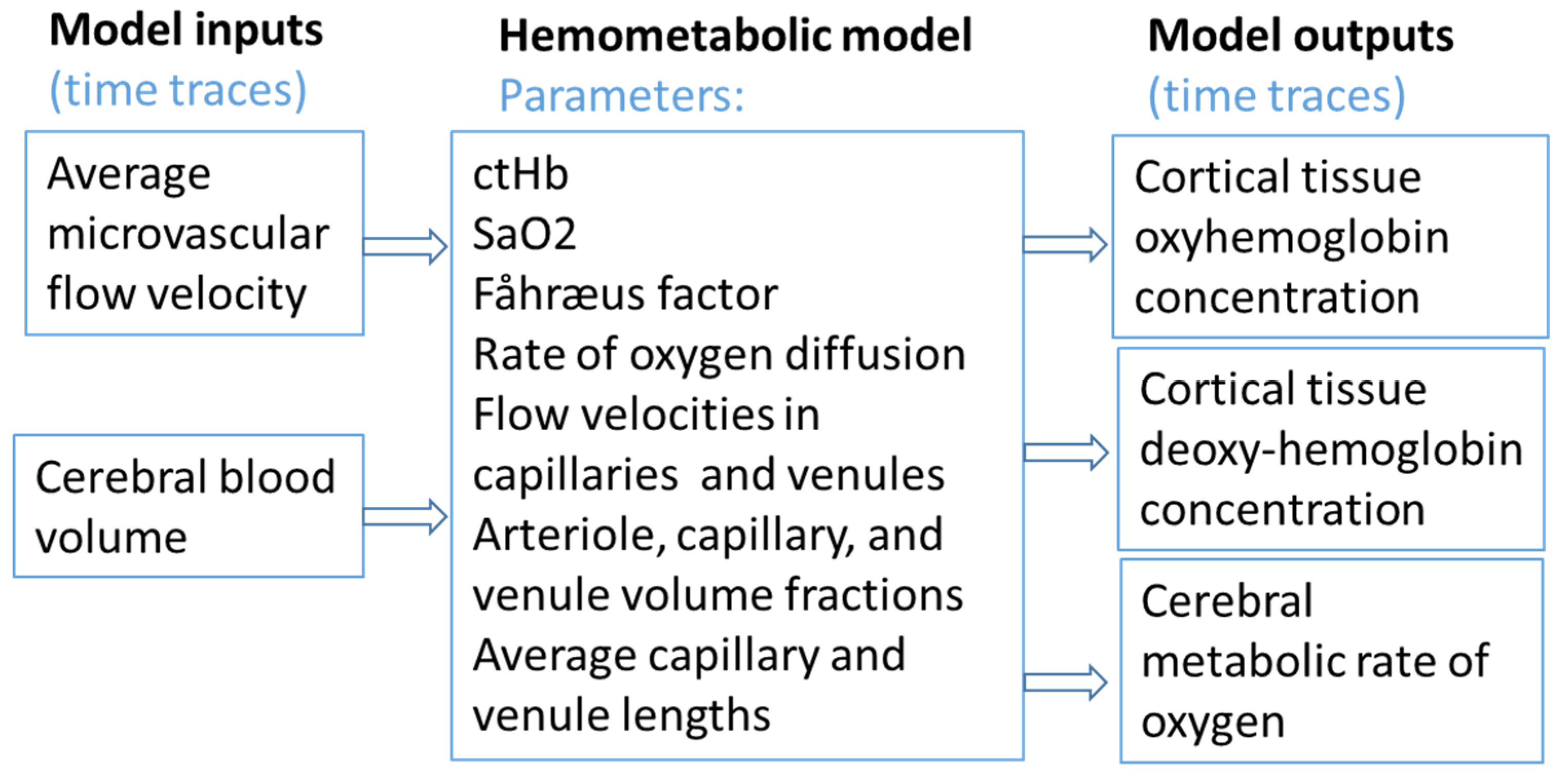
Coherent Hemodynamics Spectroscopy (CHS) model (Figure 2) describes the relationship between changes in hemoglobin oxygenation and the physiological drivers of these changes: CBF, CBV, and CMRO2 [12]. By analyzing oscillatory or perturbation-driven changes in the optical signals, CHS can estimate absolute and relative changes in flow, volume, and metabolism. A non-linear extension of the CHS model accommodates large deviations from baseline, making it suitable for arrest and reperfusion conditions [14]. Originally validated with small (~≤10%) perturbations in capillary flow velocity, the linear formulation is appropriate for mild transients and oscillatory stimuli. For arrest/reperfusion, where deviations are large and drivers are coupled, the non-linear CHS variant is preferred to preserve physiological fidelity [12].
Extensions of CHS have been used to differentiate impaired autoregulation and to estimate microcirculatory parameters [21]. Xu et al. [94] introduced the PIPE model (refers to a simplified representation of blood circulation where vessels are modeled as pipes), which quantifies dynamic changes in blood volume, flow velocity, and autoregulation, and applies to CHS outputs.
In linear CHS, small perturbations justify a linearized mapping between measured changes and the underlying drivers, making it suitable for controlled oscillations and mild transients [12]. For arrest and reperfusion, non-linear CHS captures large deviations and coupling among drivers [81]. Constraining parameters within physiologic ranges, CHS estimates the time series of parameter values as outputs of a compact, physiology-based model that represents vascular-compartment behavior and tissue oxygen transport [12,15,16].
Inverse non-linear CHS models have been used to estimate arterial, capillary, and venous oxygen saturations; the relative volume fractions of arterioles, capillaries, and venules; and the hematocrit partitioning described by the Fåhraeus factor; and the rate of oxygen diffusion in tissue by fitting NIRS hemoglobin time courses together with DCS/LDF signals to the model equations [21]. Combining these recovered variables with measured flow enables calculation of the CMRO2 based on calculated CBF using CHS outputs [35].

4.2. The BrainSignals Model

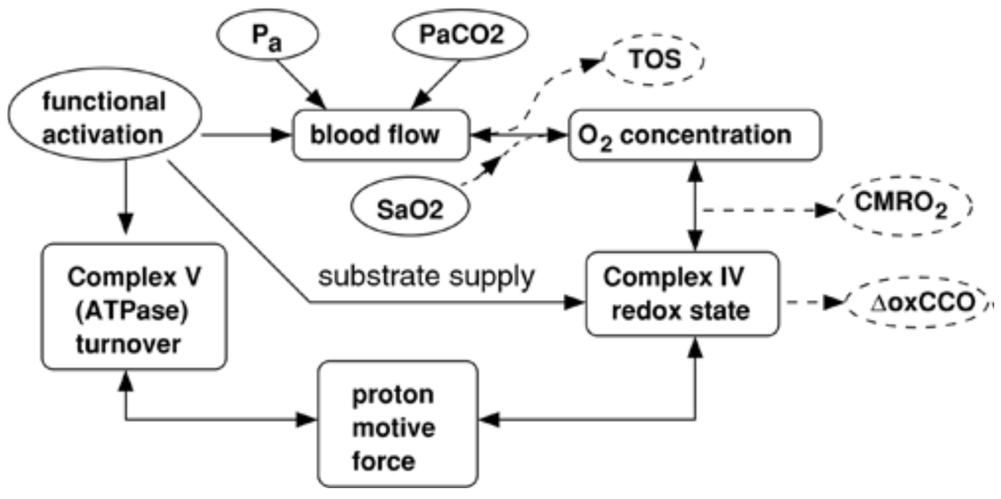
The BrainSignals model (Figure 3) and related physiology-based frameworks simulate the dynamics of oxygen transport and metabolism in the brain, integrating hemoglobin oxygenation, CBF, CBV, CMRO2, and oxCCO [15]. These models account for both vascular and mitochondrial compartments, enabling estimation of mitochondrial function from optical data. Bayesian inference approaches have further enhanced these models by quantifying parameter uncertainty and improving robustness to measurement noise [17]. Extensions include temperature-aware simulations assessing hypothermia effects on cerebral flow and metabolism [83]. Related computational work demonstrates physiology-based simulation frameworks applicable to neonatal brain metabolism [84].
This physiology-based model links arterial–capillary–venous compartments with tissue and mitochondrial modules, allowing fits of the optical signals (±oxCCO) alongside systemic covariates and enabling scenario testing—e.g., reduced delivery versus reduced utilization—under arrest/reperfusion conditions [15]. Relative to CHS, which compactly maps drivers to optical outputs, BrainSignals trades compactness for mechanistic breadth by representing vascular compartments, tissue oxygen transport, and mitochondrial redox explicitly [15]. Table 2 presents a comparative overview of CHS and BrainSignals models parameters.
In the context of CA/CPR, modeling frameworks offer three key advantages:
(1)
Physiological specificity—separating overlapping contributions of flow, volume, and metabolism to observed optical signals.
(2)
Quantification of hidden variables—estimating parameters like CMRO2 and oxCCO that cannot be measured directly at the bedside.
(3)
Adaptability to non-steady-state conditions—extending to large, rapid changes characteristic of cardiac arrest and resuscitation.
In practice, extracerebral contributions, calibration differences, and cross-platform variability must be handled explicitly to avoid biased physiology in CA/CPR datasets [8,10,13].

4.3. Model Inversion, Identifiability and Parameter Sensitivity Issues

To date, no published studies have systematically produced a sensitivity/observability table that explicitly maps which physiological parameters are constrained by which observables for the hemo-metabolic models discussed in this review. The existing partial qualitative insights are reviewed in this subsection.
For CHS, priors constrained by physiological ranges improve stability [12,14]. Analytical studies [12,14] show that CBF and CMRO2, although entering CHS additively, are practically non-identifiable because their sensitivities to hemoglobin signals are nearly collinear—independent measurements such as DCS or LDF are required. In the foundational CHS works [12,14,95]—the authors did not measure CBF directly. Instead, they used the Grubb’s exponent relationship between CBV and CBF:
V V 0 = ( F F 0 ) α ,
with α 0.38 , as originally determined by Grubb et al. [96] This empirical law allowed inference of vascular-volume changes from modeled flow perturbations, maintaining internal consistency without independent flow measurements. However, the Grubb relationship is empirical and steady-state, derived from global PET data under hypercapnia, and not generally valid for dynamic or regional phenomena. The exponent α varies with baseline vascular tone and CO2 reactivity (≈0.3–0.6), conflates arterial, capillary, and venous compartments, and lacks a mechanistic basis in compliance or autoregulation. Consequently, while Grubb’s law provided a convenient closure for early CHS formulations, it limits quantitative interpretation, particularly in transient or pathological states. Later studies such as [21,93] addressed this by incorporating LDF-derived CBF, empirically validating CHS predictions beyond the Grubb approximation. Although these studies did not conduct formal sensitivity analyses, they demonstrated empirically that multimodal inputs—LDF for CBF and hNIRS for absolute Hb—are crucial for stable inversion and physiologically realistic estimation of CMRO2 and OEF during rapid transients.
With over 100 parameters, BrainSignals model [16]. Caldwell et al. [15] used global eFAST analysis to identify dominant factors (venous compliance, autoregulatory gain). Caldwell, Scholkmann and Tachtsidis [8] incorporated extracerebral contamination, revealing further observability loss. Russell-Buckland et al. [17] implemented Bayesian Approximate Computation, exposing broad posterior uncertainties for metabolic parameters despite screening.
Complementary Balloon/BOLD models [97,98,99] reached the same conclusion: single-modality optical or MRI data are under-determined. Reliable inference of cerebral hemo-metabolic parameters therefore demands multimodal, multi-scale measurement sets, for example, hNIRS or TD NIRS + DCS + systemic covariates (ABP, PETCO2, SpO2).
Classical least-squares inversion often fails in “sloppy” hemodynamic models where parameters co-vary strongly. Bayesian inversion instead estimates posterior distributions rather than point fits, integrates physiological priors, and quantifies credible intervals, thereby revealing degeneracy explicitly. For fNIRS and optical modeling, Bayesian approaches within Dynamic Causal Modeling (DCM) improve robustness and interpretability of connectivity and flow estimates [100]. Multilevel Bayesian mixed-effects methods achieve more realistic population-level inference than simple curve fits [101]. Hierarchical Bayesian formulations stabilize diffuse optical tomography and layered NIRS reconstruction [52], while for BrainSignals, Approximate Bayesian Computation (ABC) exposes multimodal posterior landscapes and uncertainty correlations [17]. Collectively, Bayesian methods yield better-conditioned inversions, provide explicit uncertainty quantification, and integrate multimodal priors naturally making them superior for CHS or BrainSignals parameter estimation.
Recent methodological work by Colebank et al. [102] introduced spectral surrogate modeling and active-subspace dimension reduction to evaluate parameter identifiability in complex hemodynamic PDE systems. Although applied to pulmonary and systemic circulation, the approach is conceptually parallel to CHS and BrainSignals: all share large parameter spaces and limited observables. By approximating full PDE solutions with frequency-domain surrogates, Colebank’s method [95,97,102] efficiently computed global sensitivities and revealed low-dimensional manifolds governing observable behavior. Such techniques could directly benefit cerebral modeling—e.g., constructing spectral sensitivity maps for CHS or BrainSignals, quantifying parameter degeneracy, and guiding multimodal experimental design for improved identifiability.

4.4. Common Observability Gaps

Common observability gaps for the discussed models include
  • CBF—CMRO2 ambiguity [95,97].
  • Venous–capillary indistinguishability: [14].
  • Baseline dependence [95,97].
  • Superficial contamination [8].
  • Autoregulation–compliance coupling: [16,103,104].
  • Metabolic unobservability [17].
These define the practical observability envelope: reliable estimation requires multimodal datasets combining complementary sensitivities and timescales.

4.5. Recommended Minimum Measurement Sets for Stable Inversion

A time-domain or broadband NIRS (TD-NIRS) system providing absolute [HbO2]/[HHb] enables baseline Hb and OEF0 estimation [53,95]. Adding diffuse correlation spectroscopy (DCS) gives an independent CBF measure, breaking the CBF—CMRO2 degeneracy [54,105].
Systemic covariates—continuous arterial blood pressure (ABP), end-tidal CO2 (PETCO2), and finger pulse oximetry for SpO2—should be recorded to separate vascular from metabolic drivers [15,106].
Alternatively, multi-distance TD-NIRS with short-separation regression and long-separation late-gate signals provides comparable stability [107,108,109].
The emerging consensus is that NIRS (broadband or TD) + DCS + continuous pulse oximetry + systemic monitoring constitutes the minimal multimodal set that stably constrains non-linear CHS or BrainSignals inversions and reliably recovers CBF, CBV, and CMRO2 trajectories.

5. Integrated Optical-Modeling Approaches in Cardiac Arrest

By combining NIRS, DCS, or LDF data with the above modeling frameworks, it becomes possible to track not only whether cerebral oxygenation changes, but why—for example, distinguishing between oxygen delivery failure versus metabolic suppression. Integrated optical-modeling approaches are well-suited to the rapidly evolving physiology of cardiac arrest and resuscitation, where delivery and utilization can change on second-to-second timescales. By combining continuous optical signals with model-based interpretation (as established in Section 4), it becomes possible to disentangle delivery-driven from extraction-driven components and to derive physiologically specific trajectories rather than relying on raw intensity trends alone [8,15,16]. Model-informed analyses using CHS separate delivery, volume, and metabolism contributions under large perturbations relevant to CA/CPR [12,14].
A recent study advanced this framework by focusing on the algorithmic implementation of the non-linear CHS model, emphasizing how parameter fitting and model stability can be achieved in practice [93]. By detailing the optimization workflow, parameter constraints, and strategies for applying the model to long recordings or sliding windows, the work addressed the gap between theoretical formulations and reproducible application. This contribution strengthens the methodological foundation of CHS, ensuring that its use in extreme physiological settings such as arrest and reperfusion is both transparent and extendable.
A consistent empirical picture has emerged in which intra-arrest optical patterns reflect abrupt collapse at onset, partial restoration during compressions, and heterogeneous recovery after return of spontaneous circulation. Multiple clinical studies report that higher intra-arrest optical indices and favorable early recovery dynamics are associated with return of spontaneous circulation and early neurological outcomes in both prehospital and in-hospital cohorts [19,20,21,22,60,61,62]. These observations motivate linking the measurement stream to physiology-based inference so that delivery, volume, and metabolic components can be resolved, rather than inferred indirectly.
In animal work focused on hemodynamics during CA, post-arrest reperfusion and CPR, hNIRS have been used to track redox-related changes alongside hemoglobin, with the most brain-specific signals providing insight into the coupling between perfusion and metabolism during the early recovery window. These studies document large decreases during arrest, partial restoration under compressions, and lagging metabolic recovery despite improved delivery, aligning with the expectation that reperfusion does not immediately normalize cellular demand and mitochondrial function [19,20,21,61]. Results from these models reinforce the need to jointly interpret delivery and utilization to avoid misattribution when only a single optical observable is considered.
Where paired flow and oxygenation data are available, integrated analysis improves interpretability. In ref. [35], by applying CHS to hNIRS and LDF data, we found a non-linear relationship between CMRO2, and ΔoxCCO during CA in pigs. Studies combining NIRS and DCS established joint estimation of hemoglobin dynamics and CBF in vivo [5,6], and this pairing is now central to CA/CPR monitoring. Practical implementations increasingly favor fast, multi-distance acquisition with motion-resilient hardware designed for emergency and prehospital settings so that co-registered signals can drive model inference in near-real-time. Transcranial NIRS detects hemodynamic collapse promptly in arrest models, supporting prehospital triggers for acquisition and modeling [23]. Engineering efforts demonstrate feasibility of such integrated, portable monitoring, emphasizing stability during movement and rapid physiological transitions [65]. Intra-arrest DCS neuromonitoring has predicted ROSC in human cohorts [18]. When available, broadband/hyperspectral data that include mitochondrial-sensitive ΔoxCCO provide additional constraints on models and help distinguish delivery limitation from metabolic suppression [9,55].
A typical analysis pipeline begins with artifact-robust preprocessing, followed by forward simulation and constrained inversion. Physiological models such as PIPE quantify dynamic blood volume, flow velocity, and autoregulation and integrate with CHS outputs for CA/CPR analysis [94]. Time–frequency analysis with significance thresholds provides objective detection of coherent oscillatory content and improves robustness in non-steady-state arrest signals [110]. Physiological priors and bounds stabilize estimation during large perturbations, while hierarchical or Bayesian formulations quantify parameters and state uncertainty to guard against over-interpretation of noisy or incomplete data [8,15,16]. This structure is essential for real-time or near-real-time deployment where decisions must be supported despite motion, extracerebral contamination, and non-steady-state dynamics intrinsic to arrest and resuscitation. Depth specificity and motion handling are improved by short-separation regression, dual-slope FD-NIRS, and TD-NIRS time-gating, which stabilize inter-subject comparisons and model inversions [8,10,25,66,67].
Translational data across out-of-hospital, emergency department, operating room, and intensive care environments show that integrated optical-modeling outputs align with intra-arrest predictors and early post-ROSC neurological assessments, supporting their potential role in triage, titration of compressions and ventilation, and post-ROSC management [19,22,60,62]. These studies also underscore operational constraints—speed, robustness to movement, and harmonized reporting—that must be addressed for multi-center adoption [8]. Cross-site comparability further depends on community benchmarking and transparent performance metrics [10]. See also a recent status report summarizing instrumentation and standardization efforts [72].
Finally, validation remains crucial. Controlled perturbations and convergent modalities provide testbeds for checking that model-derived delivery and extraction trajectories agree with independent flow and systemic trends, and for stress-testing identifiability when signals are limited or noisy. Ongoing research continues to refine hardware and algorithms for use during transport and resuscitation, with the aim of making physiologically explicit indicators available at the bedside in clinically actionable time frames [8,16,65]. Co-registered NIRS&DCS streams are the most interpretable with hemodynamic modeling, which partitions delivery, volume, and metabolic components for CA/CPR decision support [13].

6. Clinical Translation, Limitations, Implementation, and Future Directions

Translating optical-modeling approaches into resuscitation and early post-ROSC care requires a shift from descriptive trends to physiology-anchored endpoints that can be acted at the bedside. The arrest trajectory—abrupt collapse in delivery, partial restoration during compressions, and heterogeneous post-ROSC recovery—maps naturally onto a small set of targets that are observable with current instruments: CBF (by DCS), oxygenation indices and CBV changes (by NIRS/DCS), and mitochondrial redox dynamics where broadband sensitivity enables ΔoxCCO estimation.
A practical translational pipeline begins with artifact-robust acquisition and preprocessing; paired NIRS&DCS is preferred because it couples delivery and use, improving interpretability over either signal alone [8,13]. Intra-arrest DCS has predicted ROSC in human cohorts, strengthening the case for co-registered oxygenation–flow streams during compressions [18]. In pediatric models and cohorts, flow–oxygenation dynamics and derived autoregulation metrics align with outcome, underscoring the value of individualized targets during the vulnerable post-ROSC period [56,62]. When available, broadband/hyperspectral data that include mitochondrial-sensitive ΔoxCCO add constraints that help distinguish delivery limitation from metabolic suppression [9].
Across preclinical and clinical reports, higher intra-arrest cerebral oxygenation relates to ROSC and sometimes to survival and neurological outcomes; animal experiments demonstrated that ΔoxCCO tends to recover more slowly than delivery surrogates after ROSC, consistent with lingering mitochondrial stress [111]. In ref. [55], Ko et al. used ΔoxCCO capable FD-NIRS to assess the therapeutic effect of carbon monoxide on swine model of CA. They found overall improvement in mitochondrial respiration and ATP concentrations in the brains of animals treaded with CO after CA. Nosrati et al. [19] used ΔoxCCO capable hNIRS to study the effect of epinephrine injections on CPR and ROSC in a swine CA model. They found that epinephrine administration by discrete boluses resulted in transient improvements in cerebral oxygenation and metabolism, whereas continuous epinephrine infusion did not, compared with placebo. Mavroudis et al. [63] used a combination of ΔoxCCO capable NIRS with DCS and LDF to determine if epinephrine doses have a significant effect on CBF and cerebral tissue oxygenation during CPR and if the effect of each subsequent dose of epinephrine differs significantly from the effect of the first dose.
Depth specificity and motion handling are improved by short-separation regression, dual-slope FD-NIRS, and TD-NIRS time-gating, stabilizing inter-subject comparisons and model inversions; multi-distance DCS further reduces extracerebral contamination [78]. Field-ready systems emphasize portability and speed; wearable/portable DCS and co-registered fast multi-distance TD-NIRS&DCS platforms extend feasibility to peri-arrest and transport settings, while rapid transcutaneous NIRS detects hemodynamic collapse promptly in arrest models and supports prehospital triggers for acquisition and modeling [23,24,64,65].
Model-informed interpretation is central to translation. CHS offers a compact forward model linking drivers to optical outputs; non-linear CHS maintains physiological fidelity under large perturbations characteristic of arrest and reperfusion. BrainSignals (and simplified/Bayesian variants) represents vascular compartments, oxygen transport, and mitochondrial redox explicitly, with outputs that include StO2, CMRO2, and ΔoxCCO [35]. Bayesian formulations improve identifiability and uncertainty quantification [12,14,15,16,17].
The following limitations apply across all devices and models. Extracerebral contamination, optical-property variability, motion, and differences in algorithms can bias estimates; rigorous use of short-separation channels, dual-slope FD-NIRS, and TD-NIRS photon time-gating mitigates these issues and should be reported explicitly [8,10,25,66,67]. Absolute calibration is desirable where feasible (FD/TD-NIRS) to stabilize interpretation across subjects and over time [68,69]. Spectral unmixing for ΔoxCCO requires broadband acquisition and appropriate priors, given the low abundance of CCO and spectral overlap with hemoglobin and water [9,27,28,29,72,73,74]. Pharmacologic interventions that improve systemic perfusion can diverge at the cerebro-microcirculatory level; optical-modeling integration can separate these effects and prevent misleading inferences from systemic surrogates alone [19,63]. Finally, inter-device variability remains a barrier to multi-center adoption; community benchmarking and transparent performance metrics are needed for harmonization [10,66].
Implementation priorities follow directly. First, standardize reporting: specify probe geometry, source–detector separations (including short-separation channels), wavelengths or time-domain settings, calibration approach (FD/TD vs. continuous-wave), and motion-handling. Second, report model settings: CHS linear vs. non-linear, BrainSignals parameter priors and constraints, identifiability checks, and uncertainty quantification (credible intervals or ensemble spreads). Third, harmonize endpoints that map to decision points in resuscitation and neurocritical care: CBF by DCS, rSO2 and hemoglobin pool changes by NIRS/DCS, ΔoxCCO where available, and model-derived CMRO2 [12,13,14,15,16]. Prospective protocols should align capture windows to the CA/CPR phases (arrest onset, early compressions, pre-ROSC, immediate post-ROSC, early ICU hours), log interventions (vasoactives, ventilation, temperature), and include concurrent neurologic endpoints where possible. In perioperative contexts, joint NIRS&DCS dynamics during inflow/outflow manipulations (e.g., valve interventions) illustrate delivery–use coupling and can be interpreted alongside ΔoxCCO trends when broadband is available [24,64,89].
Future directions center on closed-loop readiness. On-device preprocessing and model updates must be fast enough for compressions-scale decisions; statistical thresholds (time–frequency coherence; change-point detection) should be pre-specified; and outputs should be framed as actionable, model-informed indicators rather than raw intensities [66,82]. Expanded evaluation across prehospital, ED, OR, and ICU settings—with harmonized protocols and benchmarking—will be necessary to establish generalizability. As time-resolved systems mature and portable DCS advances, clinical translation can prioritize combined delivery–utilization endpoints with uncertainty bounds, enabling rational titration of compressions, ventilation, perfusion pressure, and temperature in CA/CPR and early post-ROSC care [8,13,65].

7. Discussion

Optical neuromonitoring for cardiac arrest has progressed from descriptive traces toward model-informed, depth-resolved quantification of cerebral delivery and use. The literature synthesized in this review converges on a consistent physiological narrative: the arrest–resuscitation–post-ROSC sequence is marked by a rapid collapse of cerebral oxygenation and flow, partial and heterogeneous restoration during compressions, and frequently lagging metabolic recovery, especially at the mitochondrial level. Broader systems-level work on breakdown and repair of brain metabolism motivates incorporating recovery trajectories into model-informed endpoints [112]. Pairing hemoglobin-based signals (NIRSs) with microvascular flow (DCS) increases interpretability in this non-steady-state setting and supports the translation from “numbers” to mechanistic drivers that are clinically actionable. The CHS model explicitly couples hemodynamics, blood flow, OEF, and vascular compartments. Inversion of CHS with NIRS/DCS inputs can produce absolute CBF/CMRO2 without exogenous tracers, but accuracy depends on identifiability and the fidelity of optical forward models (scattering/pathlength).
The first implication is methodological: evidence integration must recognize heterogeneity in devices, analysis, and patient populations. Reviews and meta-analyses outside cardiac arrest consistently show both feasibility and variability for NIRS-guided care in perioperative and critical-care settings [113,114,115,116]. Interpreting pooled effects requires careful selection, bias assessment, and summary statistics [117], and, for cardiac arrest specifically, accurate mapping of optical epochs (arrest, CPR, immediate post-ROSC, early ICU) to clinical endpoints. Within this landscape, time-domain innovations have improved depth specificity and absolute estimation, strengthening cross-study comparability [67].
Second, physiological anchoring is essential. Preclinical data demonstrate canonical ischemia–reperfusion cascades that align with optical observations in arrest: kinase-mediated neuronal injury and endothelial/mitochondrial dysfunction during and after CBF interruption [57,58]. Optical platforms have captured these transitions across disease contexts, including closed-head injury and cardiopulmonary bypass, underscoring generalizability of the delivery–utilization framing [118]. Intra-arrest simultaneous recording of hemoglobin and flow further validates the integrated approach in resuscitation [61], while studies of CBF–oxygenation coupling clarify how perfusion changes propagate to tissue saturation under hemodynamic perturbations [42]. Complementary bilateral cerebral/somatic comparisons help contextualize central versus peripheral perfusion pressures when systemic and cerebral goals diverge [59].
Third, DCS measurements enhances prognostic leverage. Low-frequency content in DCS-derived CBF during the acute post-arrest period has been associated with neurological injury signatures, suggesting an early, non-invasive biomarker for risk stratification [56]. Pharmacologic choices during resuscitation can dissociate systemic from cerebro-microcirculatory responses; epinephrine in particular has been linked to adverse cerebral microcirculation despite systemic augmentation, reinforcing the need for brain-specific optical endpoints within an integrated model [63]. Together with cardiac-surgery and neuro-trauma experience [51,118], these results argue for paired NIRS&DCS as the minimal multimodal core for arrest/CPR monitoring, particularly when mitochondrial-sensitive ΔoxCCO is available.
Fourth, implementation must follow the constraints of field and bedside care. Time-resolved methods, multi-distance sampling, and motion-resilient optodes are enabling data quality during compressions, transport, and early ICU care; the emergence of fast multi-distance TD-NIRS&DCS prototypes directly targets this need [119]. Clinical feasibility across cardiopulmonary bypass [34] and other controlled perturbations provides a practical sandbox for delivery–utilization calibration and for validating analysis pipelines before deployment in arrest [51]. When mitochondrial specificity is required, broadband/hyperspectral sampling and careful unmixing remain prerequisites; depth-resolved acquisition and standardized reporting are the parallel engineering and methodological pillars that keep estimates interpretable across centers [113,114,115,116,117].
Finally, model-informed approach ties all methods together. The arrest problem is non-steady-state and multi-driver; PIPE, CHS and BrainSignals models formalize how delivery, volume, and mitochondrial use combine to shape optical outputs, while inverse frameworks and frequency-domain tools stabilize estimates and make uncertainty explicit [12,14,16,17,94,110,120]. Programmatic translation and proceedings-level reports continue to align modeling narratives with observed CA/CPR dynamics and practical workflows [61]. With paired NIRS&DCS as the measurement backbone, time-domain depth control and broadband ΔoxCCO, and model-based inversion as the analytic scaffold, optical neuromonitoring is positioned to deliver actionable, physiology-specific endpoints for neuroprotective resuscitation.

8. Conclusions

Optical monitoring during cardiac arrest can quantify absolute values for cerebral blood flow and oxygen delivery and consumption in real-time. The synthesis presented here—rooted in NIRS, DCS, and hemodynamic modeling—supports a pragmatic roadmap: (i) co-registered NIRS&DCS with depth control and, where feasible, ΔoxCCO; (ii) standardized acquisition/reporting to ensure cross-site comparability; and (iii) model-informed thresholds for titrating the CPR compressions, perfusion pressure, ventilation, and temperature in the early post-ROSC window. Continued evaluation across prehospital, ED, OR, and ICU settings, with harmonized endpoints and benchmarking, is expected to consolidate the evidence base and accelerate bedside implementation.

Author Contributions

Conceptualization and methodology, V.T.; validation, N.S., formal analysis, V.T. and N.S.; writing—original draft preparation, N.S.; writing—review and editing, V.T.; supervision, V.T. All authors have read and agreed to the published version of the manuscript.

Funding

This review creation received no external funding.

Data Availability Statement

Not applicable.

Conflicts of Interest

The authors declare no conflicts of interest.

References

  1. Yan, S.; Gan, Y.; Jiang, N.; Wang, R.; Chen, Y.; Luo, Z.; Zong, Q.; Chen, S.; Lv, C. The global survival rate among adult out-of-hospital cardiac arrest patients who received cardiopulmonary resuscitation: A systematic review and meta-analysis. Crit. Care 2020, 24, 61. [Google Scholar] [CrossRef] [PubMed]
  2. Callaway, C.W. Cerebral oximetry and cardiopulmonary resuscitation. J. Am. Heart Assoc. 2015, 4, e002373. [Google Scholar] [CrossRef] [PubMed]
  3. Takegawa, R.; Hayashida, K.; Rolston, D.M. Near-Infrared Spectroscopy Assessments of Regional Cerebral Oxygen Saturation for the Prediction of Clinical Outcomes in Patients With Cardiac Arrest: A Review of Clinical Impact, Evolution, and Future Directions. Front. Med. 2020, 7, 587930. [Google Scholar] [CrossRef] [PubMed]
  4. Jöbsis, F.F. Noninvasive, Infrared Monitoring of Cerebral and Myocardial Oxygen Sufficiency and Circulatory Parameters. Science (1979) 1977, 198, 1264–1267. [Google Scholar] [CrossRef]
  5. Cheung, C.; Culver, J.P.; Takahashi, K. In vivocerebrovascular measurement combining diffuse near-infrared absorption and correlation spectroscopies. Phys. Med. Biol. 2001, 46, 2053–2065. [Google Scholar] [CrossRef]
  6. Culver, J.P.; Durduran, T.; Furuya, D. Diffuse Optical Tomography of Cerebral Blood Flow, Oxygenation, and Metabolism in Rat during Focal Ischemia. J. Cereb. Blood Flow Metab. 2003, 23, 911–924. [Google Scholar] [CrossRef]
  7. Dirnagl, U.; Kaplan, B.; Jacewicz, M. Continuous Measurement of Cerebral Cortical Blood Flow by Laser-Doppler Flowmetry in a Rat Stroke Model. J. Cereb. Blood Flow Metab. 1989, 9, 589–596. [Google Scholar] [CrossRef]
  8. Caldwell, M.; Scholkmann, F.; Wolf, U.; Wolf, M.; Elwell, C.; Tachtsidis, I. Modelling confounding effects from extracerebral contamination and systemic factors on functional near-infrared spectroscopy. Neuroimage 2016, 143, 91–105. [Google Scholar] [CrossRef]
  9. Bale, G.; Elwell, C.E.; Tachtsidis, I. From Jöbsis to the present day: A review of clinical near-infrared spectroscopy measurements of cerebral cytochrome-c-oxidase. J. Biomed. Opt. 2016, 21, 91307. [Google Scholar]
  10. Lanka, P.; Yang, L.; Orive-Miguel, D. Multi-laboratory performance assessment of diffuse optics instruments: The BitMap exercise. J. Biomed. Opt. 2022, 27, 074716. [Google Scholar] [CrossRef]
  11. Delpy, D.T.; Cope, M. Quantification in tissue near-infrared spectroscopy. Philos. Trans. R. Soc. Lon. Ser. B Biol. Sci. 1997, 352, 649–659. [Google Scholar] [CrossRef]
  12. Fantini, S. Dynamic model for the tissue concentration and oxygen saturation of hemoglobin in relation to blood volume, flow velocity, and oxygen consumption: Implications for functional neuroimaging and coherent hemodynamics spectroscopy (CHS). Neuroimage 2014, 85, 202–221. [Google Scholar] [CrossRef]
  13. Durduran, T.; Yodh, A.G. Diffuse correlation spectroscopy for non-invasive, micro-vascular cerebral blood flow measurement. Neuroimage 2014, 85, 51–63. [Google Scholar] [CrossRef]
  14. Sassaroli, A.; Kainerstorfer, J.M.; Fantini, S. Nonlinear extension of a hemodynamic linear model for coherent hemodynamics spectroscopy. J. Theor. Biol. 2016, 389, 132–145. [Google Scholar] [CrossRef]
  15. Caldwell, M.; Hapuarachchi, T.; Highton, D. BrainSignals revisited: Simplifying a computational model of cerebral physiology. PLoS ONE 2015, 10, e0126695. [Google Scholar] [CrossRef] [PubMed]
  16. Banaji, M.; Mallet, A.; Elwell, C.E. A Model of Brain Circulation and Metabolism: NIRS Signal Changes during Physiological Challenges. PLoS Comput. Biol. 2008, 4, e1000212. [Google Scholar] [CrossRef] [PubMed]
  17. Russell-Buckland, J.; Barnes, C.P.; Tachtsidis, I. A Bayesian framework for the analysis of systems biology models of the brain. PLoS Comput. Biol. 2019, 15, e1006631. [Google Scholar] [CrossRef] [PubMed]
  18. Ko, T.S.; Mavroudis, C.D. Non-invasive diffuse optical neuromonitoring during cardiopulmonary resuscitation predicts return of spontaneous circulation. Sci. Rep. 2021, 11, 3828. [Google Scholar] [CrossRef]
  19. Nosrati, R.; Lin, S.; Ramadeen, A. Cerebral Hemodynamics and Metabolism During Cardiac Arrest and Cardiopulmonary Resuscitation Using Hyperspectral Near Infrared Spectroscopy. Circ. J. 2017, 81, 879–887. [Google Scholar] [CrossRef]
  20. Nosrati, R.; Lin, S.; Mohindra, R. Study of the Effects of Epinephrine on Cerebral Oxygenation and Metabolism During Cardiac Arrest and Resuscitation by Hyperspectral Near-Infrared Spectroscopy. Crit. Care Med. 2019, 47, 349–357. [Google Scholar] [CrossRef]
  21. Khalifehsoltani, N.; Rennie, O.; Mohindra, R.; Lin, S.; Toronov, V. Tracking Cerebral Microvascular and Metabolic Parameters during Cardiac Arrest and Cardiopulmonary Resuscitation. Appl. Sci. 2023, 13, 12303. [Google Scholar] [CrossRef]
  22. Francoeur, C.; Landis, W.P.; Winters, M. Near-infrared spectroscopy during cardiopulmonary resuscitation for pediatric cardiac arrest: A prospective, observational study. Resuscitation 2022, 174, 35–41. [Google Scholar] [CrossRef] [PubMed]
  23. Raschdorf, K.; Mohseni, A.; Hogle, K. Evaluation of transcutaneous near-infrared spectroscopy for early detection of cardiac arrest in an animal model. Sci. Rep. 2023, 13, 4537. [Google Scholar] [CrossRef] [PubMed]
  24. Nguyen, T.N.; Wu, W.; Woldemichael, E. Hyperspectral near-infrared spectroscopy assessment of the brain during hypoperfusion. J. Biomed. Opt. 2019, 24, 035007. [Google Scholar] [CrossRef] [PubMed]
  25. Shoemaker, L.N.; Milej, D.; Mistry, J. Using depth-enhanced diffuse correlation spectroscopy and near-infrared spectroscopy to isolate cerebral hemodynamics during transient hypotension. Neurophotonics 2023, 10, 025013. [Google Scholar] [CrossRef]
  26. Nosrati, R.; Vesely, K.; Schweizer, T.A. Event-related changes of the prefrontal cortex oxygen delivery and metabolism during driving measured by hyperspectral fNIRS. Biomed. Opt. Express 2016, 7, 1323. [Google Scholar] [CrossRef]
  27. Kovacsova, Z.; Bale, G.; Mitra, S. Absolute quantification of cerebral tissue oxygen saturation with multidistance broadband NIRS in newborn brain. Biomed. Opt. Express 2021, 12, 907. [Google Scholar] [CrossRef]
  28. Giannoni, L.; Lange, F.; Sajic, M. A Hyperspectral Imaging System for Mapping Haemoglobin and Cytochrome-c-Oxidase Concentration Changes in the Exposed Cerebral Cortex. IEEE J. Sel. Top. Quantum Electron. 2021, 27, 7400411. [Google Scholar] [CrossRef]
  29. Lange, F.; Peyrin, F.; Montcel, B. Broadband time-resolved multi-channel functional near-infrared spectroscopy system to monitor in vivo physiological changes of human brain activity. Appl. Opt. 2018, 57, 6417. [Google Scholar] [CrossRef]
  30. Kaynezhad, P.; Mitra, S.; Bale, G. Quantification of the severity of hypoxic-ischemic brain injury in a neonatal preclinical model using measurements of cytochrome-c-oxidase from a miniature broadband-near-infrared spectroscopy system. Neurophotonics 2019, 6, 045009. [Google Scholar] [CrossRef]
  31. Siddiqui, M.F.; Lloyd-Fox, S.; Kaynezhad, P. Non-invasive measurement of a metabolic marker of infant brain function. Sci. Rep. 2017, 7, 1330. [Google Scholar] [CrossRef]
  32. Sekhon, M.S.; Smielewski, P.; Bhate, T.D. Using the relationship between brain tissue regional saturation of oxygen and mean arterial pressure to determine the optimal mean arterial pressure in patients following cardiac arrest: A pilot proof-of-concept study. Resuscitation 2016, 106, 120–125. [Google Scholar] [CrossRef]
  33. Hoiland, R.L.; Sekhon, M.S.; Cardim, D. Lack of agreement between optimal mean arterial pressure determination using pressure reactivity index versus cerebral oximetry index in hypoxic ischemic brain injury after cardiac arrest. Resuscitation 2020, 152, 184–191. [Google Scholar] [CrossRef]
  34. Rajaram, A.; Milej, D.; Suwalski, M. Optical monitoring of cerebral perfusion and metabolism in adults during cardiac surgery with cardiopulmonary bypass. Biomed. Opt. Express 2020, 11, 5967. [Google Scholar] [CrossRef]
  35. Soltani, N.; Mohindra, R.; Lin, S.; Toronov, V. Assessing the Relationship Between Cerebral Metabolic Rate of Oxygen and Redox Cytochrome C Oxidase During Cardiac Arrest and Cardiopulmonary Resuscitation. Appl. Sci. 2025, 15, 1542. [Google Scholar] [CrossRef]
  36. Morse, P.T.; Goebel, D.J.; Wan, J. Cytochrome c oxidase-modulatory near-infrared light penetration into the human brain: Implications for the noninvasive treatment of ischemia/reperfusion injury. IUBMB Life 2021, 73, 554–567. [Google Scholar] [CrossRef] [PubMed]
  37. Wider, J.M.; Gruley, E.; Morse, P.T. Modulation of mitochondrial function with near-infrared light reduces brain injury in a translational model of cardiac arrest. Crit. Care 2023, 27, 491. [Google Scholar] [CrossRef] [PubMed]
  38. Morse, P.T.; Tuck, S.; Kerns, M. Non-invasive treatment of ischemia/reperfusion injury: Effective transmission of therapeutic near-infrared light into the human brain through soft skin-conforming silicone waveguides. Bioeng. Transl. Med. 2024, 8, e10496. [Google Scholar] [CrossRef]
  39. Li, Z.; Baker, W.B.; Parthasarathy, A.B. Calibration of diffuse correlation spectroscopy blood flow index with venous-occlusion diffuse optical spectroscopy in skeletal muscle. J. Biomed. Opt. 2015, 20, 125005. [Google Scholar] [CrossRef]
  40. Ferradal, S.L.; Yuki, K.; Vyas, R. Non-invasive assessment of cerebral blood flow and oxygen metabolism in neonates during hypothermic cardiopulmonary bypass: Feasibility and clinical implications. Sci. Rep. 2017, 7, srep44117. [Google Scholar] [CrossRef]
  41. Zavriyev, A.I.; Kaya, K.; Farzam, P. The role of diffuse correlation spectroscopy and frequency-domain near-infrared spectroscopy in monitoring cerebral hemodynamics during hypothermic circulatory arrests. JTCVS Tech. 2021, 7, 161–177. [Google Scholar] [CrossRef] [PubMed]
  42. Johnson, T.W.; Dar, I.A.; Donohue, K.L. Cerebral Blood Flow Hemispheric Asymmetry in Comatose Adults Receiving Extracorporeal Membrane Oxygenation. Front. Neurosci. 2022, 16, 858404. [Google Scholar] [CrossRef] [PubMed]
  43. Shaw, K.; Mavroudis, C.D.; Ko, T.S. The use of novel diffuse optical spectroscopies for improved neuromonitoring during neonatal cardiac surgery requiring antegrade cerebral perfusion. Front. Pediatr. 2023, 11, 1125985. [Google Scholar] [CrossRef] [PubMed]
  44. Yücel, M.A.; Selb, J.; Aasted, C.M.; Petkov, M.P.; Becerra, L.; Borsook, D.; Boas, D.A. Short separation regression improves statistical significance and better localizes the hemodynamic response obtained by near-infrared spectroscopy for tasks with differing autonomic responses. Neurophotonics 2015, 2, 35005. [Google Scholar] [CrossRef]
  45. Blaney, G.; Sassaroli, A.; Pham, T.; Fernandez, C.; Fantini, S. Phase dual-slopes in frequency-domain near-infrared spectroscopy for enhanced sensitivity to brain tissue: First applications to human subjects. J. Biophotonics 2020, 13, 201960018. [Google Scholar] [CrossRef]
  46. Kamar, F.; Shoemaker, L.N.; Eskandari, R.; Milej, D.; Drosdowech, D.; Murkin, J.M.; Lawrence, K.S.; Chui, J.; Diop, M. Assessing changes in regional cerebral hemodynamics in adults with a high-density full-head coverage time-resolved NIRS device. J. Biomed. Opt. 2024, 29, S33302. [Google Scholar] [CrossRef]
  47. Cugmas, B.; Bürmen, M.; Bregar, M.; Pernuš, F.; Likar, B. Pressure-induced near infrared spectra response as a valuable source of information for soft tissue classification. J. Biomed. Opt. 2013, 18, 047002. [Google Scholar] [CrossRef]
  48. Baker, W.B.; Parthasarathy, A.B.; Ko, T.S.; Busch, D.R.; Abramson, K.; Tzeng, S.Y.; Mesquita, R.C.; Durduran, T.; Greenberg, J.H.; Kung, D.K.; et al. Pressure modulation algorithm to separate cerebral hemodynamic signals from extracerebral artifacts. Neurophotonics 2015, 2, 035004. [Google Scholar] [CrossRef]
  49. Tichauer, K.M.; Elliott, J.T.; Hadway, J.A.; Lee, D.S.; Lee, T.-Y.; Lawrence, K.S. Using near-infrared spectroscopy to measure cerebral metabolic rate of oxygen under multiple levels of arterial oxygenation in piglets. J. Appl. Physiol. 2010, 109, 878–885. [Google Scholar] [CrossRef]
  50. Verdecchia, K.; Diop, M.; Lee, T.-Y.; Lawrence, K.S. Quantifying the cerebral metabolic rate of oxygen by combining diffuse correlation spectroscopy and time-resolved near-infrared spectroscopy. J. Biomed. Opt. 2013, 18, 27007. [Google Scholar] [CrossRef]
  51. Milej, D.; Shahid, M.; Abdalmalak, A. Characterizing dynamic cerebral vascular reactivity using a hybrid system combining time-resolved near-infrared and diffuse correlation spectroscopy. Biomed. Opt. Express 2020, 11, 4571. [Google Scholar] [CrossRef]
  52. Yamashita, O.; Shimokawa, T.; Kosaka, T.; Amita, T.; Inoue, Y.; Sato, M.-A. Hierarchical Bayesian model for diffuse optical tomography of the human brain: Human experimental study. J. Adv. Comput. Intell. Intell. Inform. 2014, 18, 1026–1033. [Google Scholar] [CrossRef]
  53. Liebert, A.; Wabnitz, H.; Steinbrink, J.; Obrig, H.; Möller, M.; Macdonald, R.; Villringer, A.; Rinneberg, H. Time-resolved multidistance near-infrared spectroscopy of the adult head: Intracerebral and extracerebral absorption changes from moments of distribution of times of flight of photons. Appl. Opt. 2004, 43, 3037–3047. [Google Scholar] [CrossRef] [PubMed]
  54. Milej, D.; Rajaram, A.; Suwalski, M.; Morrison, L.B.; Shoemaker, L.N.; Lawrence, K.S. Assessing the relationship between the cerebral metabolic rate of oxygen and the oxidation state of cytochrome-c-oxidase. Neurophotonics 2022, 9, 35001. [Google Scholar] [CrossRef]
  55. Ko, T.S. Attenuation of mitochondrial dysfunction in a ventricular fibrillation swine model of cardiac arrest treated with carbon monoxide. Resuscitation 2024, 213, 110647. [Google Scholar] [CrossRef]
  56. White, B.R.; Ko, T.S.; Morgan, R.W. Low frequency power in cerebral blood flow is a biomarker of neurologic injury in the acute period after cardiac arrest. Resuscitation 2022, 178, 12–18. [Google Scholar] [CrossRef]
  57. Lin, H.W.; Gresia, V.L.; Stradecki, H.M. Protein kinase C delta modulates endothelial nitric oxide synthase after cardiac arrest. J. Cereb. Blood Flow Metab. 2014, 34, 613–620. [Google Scholar] [CrossRef]
  58. Larson, A.C.; Jamrogowicz, J.L.; Kulikowicz, E. Cerebrovascular autoregulation after rewarming from hypothermia in a neonatal swine model of asphyxic brain injury. J. Appl. Physiol. 2013, 115, 1433–1442. [Google Scholar] [CrossRef]
  59. Candan, T.; Candan, M.; Yildiz, C.E. Comparison of bilateral cerebral and somatic tissue oxygenation with near-infrared spectroscopy in cyanotic and acyanotic pediatric patients receiving cardiac surgery. Arch. Med. Sci. Atheroscler. Dis. 2020, 5, 320–331. [Google Scholar] [CrossRef]
  60. Cournoyer, A.; Iseppon, M.; Chauny, J.M. Near-infrared Spectroscopy Monitoring During Cardiac Arrest: A Systematic Review and Meta-analysis. Acad. Emerg. Med. 2016, 23, 851–862. [Google Scholar] [CrossRef]
  61. Nosrati, R.; Ramadeen, A.; Hu, X. Simultaneous measurement of cerebral and muscle tissue parameters during cardiac arrest and cardiopulmonary resuscitation. In Optical Techniques in Neurosurgery, Neurophotonics, and Optogenetics II; Hirschberg, H., Madsen, S.J., Jansen, E.D., Eds.; SPIE: Bellingham, WA, USA, 2015; p. 93051. [Google Scholar]
  62. Kirschen, M.P.; Majmudar, T.; Beaulieu, F. Deviations from NIRS-derived optimal blood pressure are associated with worse outcomes after pediatric cardiac arrest. Resuscitation 2021, 168, 110–118. [Google Scholar] [CrossRef]
  63. Mavroudis, C.D.; Ko, T.S.; Morgan, R.W. Epinephrine’s effects on cerebrovascular and systemic hemodynamics during cardiopulmonary resuscitation. Crit. Care 2020, 24, 583. [Google Scholar] [CrossRef]
  64. Hou, L.; Liu, Y.; Qian, L. Portable Near-Infrared Technologies and Devices for Noninvasive Assessment of Tissue Hemodynamics. J. Healthc. Eng. 2019, 12, 3750495. [Google Scholar] [CrossRef] [PubMed]
  65. Nabacino, M.; Amendola, C.; Contini, D. Fast Multi-Distance Time-Domain NIRS and DCS System for Clinical Applications. Sensors 2024, 24, 7375. [Google Scholar] [CrossRef] [PubMed]
  66. Blaney, G.; Fernandez, C.; Sassaroli, A. Dual-slope imaging of cerebral hemodynamics with frequency-domain near-infrared spectroscopy. Neurophotonics 2023, 10, 013508. [Google Scholar] [CrossRef]
  67. Lange, F.; Tachtsidis, I. Clinical brain monitoring with time domain NIRS: A review and future perspectives. Appl. Sci. 2019, 9, 1612. [Google Scholar] [CrossRef]
  68. Fallon, P.; Roberts, I.; Kirkham, F.J. Cerebral hemodynamics during cardiopulmonary bypass in children using near-infrared spectroscopy. Ann. Thorac. Surg. 1993, 56, 1473–1477. [Google Scholar] [CrossRef]
  69. Yeganeh, H.Z.; Toronov, V.; Elliott, J.T. Broadband continuous-wave technique to measure baseline values and changes in the tissue chromophore concentrations. Biomed. Opt. Express 2012, 3, 2761. [Google Scholar] [CrossRef]
  70. Smith, M. Shedding light on the adult brain: A review of the clinical applications of near-infrared spectroscopy. Philos. Trans. R. Soc. A Math. Phys. Eng. Sci. 2011, 369, 4452–4469. [Google Scholar] [CrossRef]
  71. Tachtsidis, I.; Scholkmann, F. False positives and false negatives in functional near-infrared spectroscopy: Issues, challenges, and the way forward. Neurophotonics 2016, 3, 31405. [Google Scholar] [CrossRef]
  72. Ayaz, H.; Baker, W.B.; Blaney, G. Optical imaging and spectroscopy for the study of the human brain: Status report. Neurophotonics 2022, 9, S24001. [Google Scholar] [CrossRef]
  73. Leadley, G.; Austin, T.; Bale, G. Review of measurements and imaging of cytochrome-c-oxidase in humans using near-infrared spectroscopy: An update. Biomed. Opt. Express 2023, 15, 162. [Google Scholar] [CrossRef]
  74. Denault, A.Y.; Shaaban-Ali, M.; Cournoyer, A. Near-Infrared Spectroscopy. In Neuromonitoring Techniques; Elsevier: Amsterdam, The Netherlands, 2018. [Google Scholar]
  75. Sandroni, C.; Skrifvars, M.B.; Taccone, F.S. Brain monitoring after cardiac arrest. Curr. Opin. Crit. Care 2023, 29, 68–74. [Google Scholar] [CrossRef] [PubMed]
  76. Genbrugge, C.; Dens, J.; Meex, I. Regional Cerebral Oximetry during Cardiopulmonary Resuscitation: Useful or Useless? J. Emerg. Med. 2016, 50, 198–207. [Google Scholar] [CrossRef] [PubMed]
  77. Okuma, Y.; Shinozaki, K.; Yagi, T.; Hayashida, K.; Aoki, T.; Yin, T.; Kiguchi, T.; Iwami, T.; Becker, L.B. Oxyhaemoglobin Level Measured Using Near-Infrared Spectrometer Is Associated with Brain Mitochondrial Dysfunction After Cardiac Arrest in Rats. In Oxygen Transport to Tissue XLIII; Felix, S., Joseph, L., Wolf, U., Eds.; Springer International Publishing: Cham, Switzerland, 2022; pp. 385–390. [Google Scholar]
  78. Fredriksson, I.; Larsson, M.; Salomonsson, F. Improved calibration procedure for laser Doppler perfusion monitors. In Optical Diagnostics and Sensing XI: Toward Point-of-Care Diagnostics; and Design and Performance Validation of Phantoms Used in Conjunction with Optical Measurement of Tissue III; SPIE: Bellingham, WA, USA, 2011; p. 790602. [Google Scholar]
  79. Culver, J.P.; Durduran, T.; Cheung, C. Diffuse Optical Measurement of Hemoglobin and Cerebral Blood Flow in Rat Brain During Hypercapnia, Hypoxia and Cardiac Arrest. In Oxygen Transport To Tissue XXIII; Springer: New York, NY, USA, 2003; pp. 293–297. [Google Scholar]
  80. Buckley, E.M.; Lynch, J.M.; Goff, D.A. Early postoperative changes in cerebral oxygen metabolism following neonatal cardiac surgery: Effects of surgical duration. J. Thorac. Cardiovasc. Surg. 2013, 145, 196–205.e1. [Google Scholar] [CrossRef]
  81. Setchfield, K.; Gorman, A.; Simpson, A.H.R.W.; Somekh, M.G.; Wright, A.J. Effect of skin color on optical properties and the implications for medical optical technologies: A review. J. Biomed. Opt. 2024, 29, 010901. [Google Scholar] [CrossRef] [PubMed]
  82. Strangman, G.E.; Zhang, Q.; Li, Z. Scalp and skull influence on near-infrared photon propagation in the Colin27 brain template. NeuroImage 2014, 85, 136–149. [Google Scholar] [CrossRef]
  83. Carp, S.A.; Robinson, M.B.; Franceschini, M.A. Diffuse correlation spectroscopy: Current status and future outlook. Neurophotonics 2023, 10, 013509. [Google Scholar] [CrossRef]
  84. James, E.; Munro, P.R.T. Diffuse Correlation Spectroscopy: A Review of Recent Advances in Parallelisation and Depth Discrimination Techniques. Sensors 2023, 23, 9338. [Google Scholar] [CrossRef]
  85. Wang, Q.; Pan, M.; Kreiss, L.; Samaei, S.; Carp, S.A.; Johansson, J.D.; Zhang, Y.; Wu, M.; Horstmeyer, R.; Diop, M.; et al. A comprehensive overview of diffuse correlation spectroscopy: Theoretical framework, recent advances in hardware, analysis, and applications. NeuroImage 2024, 298, 120793. [Google Scholar] [CrossRef]
  86. Ozana, N.; Lue, N.; Renna, M.; Robinson, M.B.; Martin, A.; Zavriyev, A.I.; Carr, B.; Mazumder, D.; Blackwell, M.H.; Franceschini, M.A.; et al. Functional Time-Domain Diffuse Correlation Spectroscopy. Front. Neurosci. 2022, 16, 932119. [Google Scholar] [CrossRef] [PubMed]
  87. Eleveld, N. The Influence of Extracerebral Tissue on Continuous-Wave NIRS. J. Clin. Med. 2023, 12, 2776. [Google Scholar] [CrossRef] [PubMed]
  88. Zhao, H.; Buckley, E.M. Influence of source–detector separation on diffuse correlation spectroscopy measurements of cerebral blood flow with a multilayered analytical model. Neurophotonics 2022, 9, 035002. [Google Scholar] [CrossRef] [PubMed]
  89. Pfeifer, M.D. Signal Processing in fNIRS: A Review. Front. Hum. Neurosci. 2018, 11, 641. [Google Scholar]
  90. Meadow, W.; Rudinsky, B.; Raju, T.; John, E.; Fornell, L.; Shankararao, R. Correlation of flow probe determinations of common carotid artery blood flow and internal carotid artery blood flow with microsphere determinations of cerebral blood flow in piglets. Pediatr. Res. 1999, 45, 324–330. [Google Scholar] [CrossRef]
  91. Powers, K.M.; Schimmel, C.; Glenny, R.W.; Bernards, C. Cerebral blood flow determinations using fluorescent microspheres: Variations on the sedimentation method validated. J. Neurosci. Methods 1999, 87, 159–165. [Google Scholar] [CrossRef]
  92. Mörtberg, E.; Cumming, P.; Wiklund, L.; Rubertsson, S. Cerebral metabolic rate of oxygen (CMRO2) in pig brain determined by PET after resuscitation from cardiac arrest. Resuscitation 2009, 80, 701–706. [Google Scholar] [CrossRef]
  93. Toronov, V.; Soltani, N.; Leung, L.; Mohindra, R.; Lin, S. Using Coherent Hemodynamic Spectroscopy Model to Investigate Cardiac Arrest. Algorithms 2025, 18, 128. [Google Scholar] [CrossRef]
  94. Xu, M.; Zheng, Y.; Chen, X. Dynamic microcirculation PIPE model for functional neuroimaging, non-neuroimaging, and coherent hemodynamics spectroscopy: Blood volume and flow velocity variations, and vascular autoregulation. Biomed. Opt. Express 2020, 11, 4602. [Google Scholar] [CrossRef]
  95. Fantini, S. A new hemodynamic model shows that temporal perturbations of cerebral blood flow and metabolic rate of oxygen cannot be measured individually using functional near-infrared spectroscopy. Physiol. Meas. 2013, 35, N1–N9. [Google Scholar] [CrossRef]
  96. Grubb, R.L., Jr.; Raichle, M.E.; Eichling, J.O.; Ter-Pogossian, T.-P. The effect of changes in PaCO2 on cerebral blood volume, blood flow, and vascular mean transit time. Stroke 1974, 5, 630–639. [Google Scholar] [CrossRef] [PubMed]
  97. Buxton, R.B.; Wong, E.C.; Frank, L.R. Dynamics of blood flow and oxygenation changes during brain activation: The balloon model. Magn. Reson. Med. 1998, 39, 855–864. [Google Scholar] [CrossRef] [PubMed]
  98. Friston, K.J.; Mechelli, A.; Turner, R.; Price, C. Nonlinear responses in fMRI: The Balloon model, Volterra kernels, and other hemodynamics. NeuroImage 2000, 12, 466–477. [Google Scholar] [CrossRef] [PubMed]
  99. Stephan, K.E.; Weiskopf, N.; Drysdale, P.M.; Robinson, P.A.; Friston, K.J. Comparing hemodynamic models with DCM. NeuroImage 2007, 38, 387–401. [Google Scholar] [CrossRef]
  100. Tak, S.; Kempny, A.M.; Friston, K.; Leff, A.; Penny, W. Dynamic causal modelling for functional near-infrared spectroscopy. NeuroImage 2015, 111, 338–349. [Google Scholar] [CrossRef]
  101. Ciftçi, K.; Sankur, B.; Kahya, Y.P.; Akin, A. Multilevel statistical inference from functional near-infrared spectroscopy data during Stroop interference. IEEE Trans. Biomed. Eng. 2008, 55, 1865–1872. [Google Scholar] [CrossRef]
  102. Colebank, M.J. Assessing parameter identifiability of a hemodynamics PDE model using spectral surrogates and dimension reduction. PLoS Comput. Biol. 2025, 21, 1013553. [Google Scholar] [CrossRef]
  103. Hapuarachchi, T.; Scholkmann, F.; Caldwell, M. Simulation of Preterm Neonatal Brain Metabolism During Functional Neuronal Activation Using a Computational Model. In Oxygen Transport to Tissue XXXVII; Springer: New York, NY, USA, 2016; pp. 111–120. [Google Scholar]
  104. Russell-Buckland, J.; Tachtsidis, I. Developing a Model to Simulate the Effect of Hypothermia on Cerebral Blood Flow and Metabolism. Adv. Exp. Med. Biol. 2020, 1232, 299–306. [Google Scholar]
  105. Durduran, T.; Choe, R.; Baker, W.B.; Yodh, A.G. Diffuse optics for tissue monitoring and tomography. Rep. Prog. Phys. 2010, 73, 76701. [Google Scholar] [CrossRef]
  106. Jubran, A. Pulse oximetry. Crit. Care 2015, 19, 272. [Google Scholar] [CrossRef]
  107. Brigadoi, S.; Cooper, R.J. How short is short? Optimum source–detector distance for short-separation channels in functional near-infrared spectroscopy. Neurophotonics 2015, 2, 25005. [Google Scholar] [CrossRef] [PubMed]
  108. Zimeo Morais, G.A.; Balardin, J.B.; Sato, J.R. fNIRS Optodes’ Location Decider (fOLD): A toolbox for probe arrangement guided by brain regions-of-interest. Sci. Rep. 2018, 8, 3341. [Google Scholar] [CrossRef] [PubMed]
  109. Yücel, M.A. Best practices for fNIRS publications. Neurophotonics 2021, 8, 012101. [Google Scholar] [CrossRef] [PubMed]
  110. Blaney, G.; Sassaroli, A.; Fantini, S. Algorithm for determination of thresholds of significant coherence in time-frequency analysis. Biomed. Signal Process. Control. 2020, 56, 101704. [Google Scholar] [CrossRef]
  111. Soltani, N.; Toronov, V.; Rennie, O. Investigating cerebral dynamics during cardiac arrest using an optical technique and a hemodynamic model. SPIE Int. Soc. Opt. Eng. 2024, 21, 19. [Google Scholar]
  112. Shichkova, P.; Coggan, J.S.; Kanari, L. Breakdown and repair of metabolism in the aging brain. Front. Sci. 2025, 3, 1441297. [Google Scholar] [CrossRef]
  113. Serraino, G.F.; Murphy, G.J. Effects of cerebral near-infrared spectroscopy on the outcome of patients undergoing cardiac surgery: A systematic review of randomised trials. BMJ Open 2017, 7, e016613. [Google Scholar] [CrossRef]
  114. Zheng, F.; Sheinberg, R.; Yee, M.S. Cerebral near-infrared spectroscopy monitoring and neurologic outcomes in adult cardiac surgery patients: A systematic review. Anesth. Analg. 2013, 116, 663–676. [Google Scholar] [CrossRef]
  115. Nenna, A.; Barbato, R.; Greco, S.M. Near-infrared spectroscopy in adult cardiac surgery: Between conflicting results and unexpected uses. J. Geriatr. Cardiol. 2017, 14, 659–661. [Google Scholar]
  116. Sun, Q.; Wu, W. Effect of near-infrared spectroscopy on postoperative delirium in cardiac surgery with cardiopulmonary bypass: A systematic review and meta-analysis. Front. Cardiovasc. Med. 2024, 11, 1404210. [Google Scholar] [CrossRef]
  117. Israel, H.; Richter, R.R. A guide to understanding meta-analysis. J. Orthop. Sports Phys. Ther. 2011, 41, 496–504. [Google Scholar] [CrossRef]
  118. Zhou, C.; Eucker, S.A.; Durduran, T. Diffuse optical monitoring of hemodynamic changes in piglet brain with closed head injury. J. Biomed. Opt. 2009, 14, 34015. [Google Scholar] [CrossRef]
  119. Caldwell, M.; Moroz, T.; Hapuarachchi, T. Modelling Blood Flow and Metabolism in the Preclinical Neonatal Brain during and Following Hypoxic-Ischaemia. PLoS ONE 2015, 10, 140171. [Google Scholar] [CrossRef]
  120. Eskandari, R. Quantification of oxidized and reduced cytochrome-c-oxidase by combining discrete-wavelength time-resolved and broadband continuous-wave near-infrared spectroscopy. Biomed. Opt. Express 2025, 16, 3797–3812. [Google Scholar] [CrossRef]
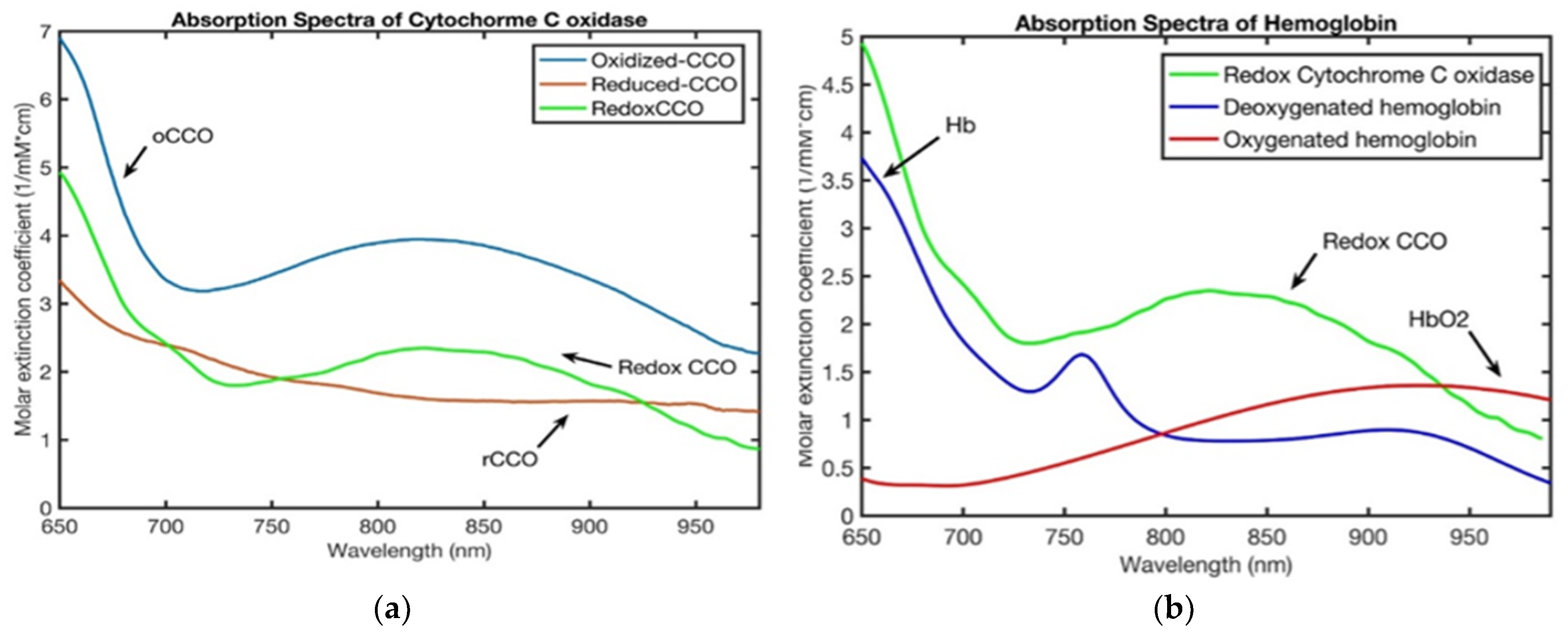
Figure 1. (a) Molar absorption spectrum of cytochrome C oxidase. rCCO is the difference between oxidized and reduced molar extinction coefficients. (b) Specific extinction coefficients of deoxygenated-hemoglobin, water and rCCO spectra.
Applsci 15 12260 g001
Figure 2. Block diagram of the CHS hemodynamic model showing the input dynamic parameters (cerebral blood volume and flow velocity) and the output dynamic parameters predicted by the model (tissue concentrations of deoxy-, oxy-, and total hemoglobin, tissue saturation, CMRO2).
Applsci 15 12260 g002
Figure 3. Summary of the main inputs, variables and processes in the BrainSignals model, https://doi.org/10.1371/journal.pcbi.1000212.
Applsci 15 12260 g003
Table 1. Summary of experimental studies.
Ref.
#
Species
and Sample Size
Device/ManufacturerNIRS Type and DCS (Y/N)Primary EndpointsKey FindingQuality/Limitations
[18]Human (CPR)
52
Imagent ISS Inc., Champaig, IL, USAFD NIRSOptical indices vs. ROSCOptical signals predicted ROSC during CPR.Exact device and n require full text.
[19]Animal (pigs; CA/CPR)
14
CustomhHIRSO2 delivery, ΔoxCCO, Hb specieshNIRS-tracked cerebral hemo-metabolic shifts during CA/CPR.Model-based deconvolution; motion in CPR.
[20]Animal (pigs; CA/CPR)
9
CustomhNIRSΔoxCCO, HbO/HbR, O2 deliveryEpinephrine bolus produced transient metabolic/oxygenation boosts.Small n; translation uncertain.
[21]Animal (pigs; CA/CPR)
10
CustomhNIRSΔoxCCO, Hb species;CHS inversion-tracked microvascular and metabolic parameters during CA/CPR; majority of cases fit adequately.Model dependence; preclinical instrumentation; modest n.
[22]Human (pediatric CPR)
21
Equanox 7600; (Nonin Medical, Plymouth, MN, USA)CW cerebral oximeterrSO2 during CPR; ROSC/survival linksHigher rSO2 associated with ROSC.Observational; motion/compression artifacts.
Human (pediatric post-arrest) 34(Nonin SenSmart Nonin Medical, Inc., Plymouth, MN, USA))CW cerebral oximeterDeviation from MAPopt (NIRS-COx) vs. outcomeTime below MAPopt → worse outcomes.Retrospective aspects; device-specific index.
[23]Animal
8
NX-BF/OF/E; Oxford Optronix, Oxford, UKCombined tissue pO2 and blood flow monitorTSI%, pulse detection in barbiturate CANIRS detected arrest early.Anesthetic CA model; small n.
[24]Animal/human (hypoperfusion)
10
Hyperspectral NIRS, customhNIRSHbO2/HHb/tHb; cortical mappingHyperspectral NIRS captured brain changes during hypoperfusion.Small cohorts; model dependence.
[25]Human (healthy adults)
9
CustomDepth-enhanced DCS + TR-NIRSΔCBF, rSO2 under hypotension; extracerebral removalDepth-enhanced DCS improved cerebral specificity.Experimental hypotension; lab system.
[26]Human (drivers)
16
CustomhNIRSO2 delivery (HbT × SaO2), metabolism indicesDriving task modulated prefrontal delivery/metabolism.Task-based fNIRS; algorithmic deconvolution.
[27]Human (term infant with HIE)
1
CustomMulti-distance hNIRS, TR-NIRSAbsolute StO2 (cerebral)Demonstrated absolute cerebral StO2 quantification in neonates.Algorithm/device specific; clinical validation pending.
[28]Phantom, Animal (mice, exposed cortex) 3CustomhNIRSHb, oxCCO mapsMapped Hb and oxCCO changes across cortex.Exposed cortex; through-skull translation pending.
[29]Human
5
CustomTD hNIRSFunctional responses (HbO/HbR)In vivo monitoring of brain activity with broadband TD-fNIRS.Research system; small cohorts.
[30]Animal (neonatal pig model)
27
CustomhNIRSΔoxCCO, Hb changes vs. injury severityoxCCO tracked hypoxic–ischemic injury severity.Preclinical model; miniature device.
[31]Human (infants)
33
CustomhNIRSΔoxCCO as metabolic markerDetected task-related oxCCO in infants.Signal-to-noise; motion; developmental variability.
[32]Human (post-CA)
20
Invos, (Covidien, Dublin, Ireland)CW cerebral oximeterMAP-rSO2 reactivity (COx), MAPoptDerived patient-specific MAPopt post-CA.Pilot; single-center.
[33]Human (post-CA)
10
INVOS, (Medtronic, Minneapolis, MN, USA)CW cerebral oximeterAgreement of MAPopt (COx vs. Prox)Poor agreement between COx- and PRx-derived MAPopt.Physiologic index comparison; modest n.
[34]Human (adults; cardiac surgery)
10
CustomhNIRS + DCSrSO2, rCBF, CMRO2 (indices)Continuous intra-op perfusion/metabolism tracking feasible.Single-center; research hardware.
[35]Animal (pigs; CA/CPR)
11
CustomhNIRSCMRO2 index vs. ΔoxCCO during CA/CPRoxCCO changes paralleled model-derived CMRO2 trends.Model dependence.
[36]Human cadavers/ex vivo
4
CustomPhotobiomodulation deliveryNIR penetration to brain; therapeutic feasibilityTransmission to cortex via silicone waveguides context.Mixed experimental/simulation; translational assumptions.
[37]Animal (pig cardiac arrest model)
30
CustomPBM (810–1064 nm) + physiologic monitoringNeurologic injury markers; survivalPBM reduced brain injury in translational CA model.Model selection; dosing regimen.
[38]Human cadavers/ex vivo
4
CustomSilicone waveguide PBM systemDelivered dose at scalp/skull/CSFEffective brain-directed PBM transmission demonstrated.Delivery study; not neuro-outcome trial.
[39]Human (skeletal muscle)
10
Imagent (ISS Medical, Champaign, IL, USA)FD NIRS
Custom DCS
DCS BFI calibration vs. DOS in muscleEstablished calibration between DCS and DOS.Muscle not brain; still physiologically experimental.
Human (neonates with CHD)
36
Imagent (ISS Medical, Champaign, IL, USA)FD NIRS +
custom DCS
rCBF, rCMRO2, rOEFQuantified post-op trends in rCBF/rCMRO2/rOEF.Observational; relative metrics; heterogeneous cases.
[40]Human neonates
9
Imagent (ISS Medical, Champaign, IL, USA)FD NIRS
custom DCS
CBF, CMRO2 (indices), rSO2CBF and CMRO2 decreased during deep hypothermic CPB; feasibility shown.Pilot; small n; custom device; motion/cooling confounds.
[41]Human (adults; HCA strategies)
12
MetaOx (ISS Inc., Champaign, IL, USA)FD NIRS
custom DCS
CBF index, CMRO2 index, rSO2DCS showed near-zero CBF in HCA; ACP restored flow.Indices; perfusion strategy heterogeneity.
[42]Human (comatose adults on VA-ECMO)
13
CustomBilateral DCSCBF asymmetry indexFrequent hemispheric rCBF asymmetry on ECMO.Single-center; relative CBF.
[43]Human (neonates; cardiac surgery)
~5
CustomFD-NIRS + DCSCBF index, rSO2 during ACPHybrid optics provided value beyond rSO2 alone.Small series; descriptive.
[44]Human (task fNIRS)
11
CW6 (TechEn Inc., Milford, MA, USA)CW NIRSHbO/HbR HRF significance, localizationShort-separation regression improved stats/localization.Task fNIRS; device-algorithm specificity.
[45]Human (adults)
4
Imagent, (ISS Medical, Champaign, IL, USA)FD dual-slope NIRSCortical sensitivity metricsEnhanced brain sensitivity with phase dual-slope FD-NIRS.First applications; modest n.
[46]Human (adults)
37
CustomTD NIRSRegional hemodynamics during tasksFull-head TR-NIRS mapped regional responses.Complex headgear; lab setup.
[47]Human (soft tissue)
4
CustomhNIRSPressure-induced spectral responsePressure modulates spectra; tissue classification aid.Non-cerebral tissue; lab conditions.
[48]Humans
9
Imagent (ISS Medical, Champaign, IL, USAFD-NIRS + custom
DCS
Cerebral Hb and CBF changesPressure modulation separates cerebral
hemodynamic signals from extracerebral artifacts
Pressure needs to be applied to the probe
[49]Animal (newborn piglets)
12
CustomTR NIRS + DCSAbsolute CBF, SvO2, CMRO2Validated NIRS-based CMRO2 across SaO2 levels.Catheter references; specialized setup.
[50]Human (adults)
7
CustomTD NIRS + DCSAbsolute/calibrated CBF, SvO2, CMRO2Enabled calibrated CMRO2 changes; SvO2 validated.Small cohort; invasive venous ref.
[51]Human (healthy adults)
9
CustomTD NIRS + DCSΔCBF, ΔHbO2, ΔHHbDecomposed static/dynamic CVR to CO2.Healthy cohort; research setup.
[52]Human
1
FOIRE3000 (Shimadzu Corp., Kyoto, Japan)High-density CW fNIRSDOT reconstruction of brain activityHierarchical Bayes DOT with human data.Single subject; computational focus.
[53]Human (adult)
1
CustomTD NIRSICG-based depth separation (DTOF moments)Separated intra- vs. extracerebral absorption.Single subject; ICG required.
[54]Animal (piglets)
6
CustomTD NIRS+
DCS
CMRO2 vs. oxCCO relationshipReported association between CMRO2 changes and oxCCO.Small n.
[55]Animal (swine VF CA)
11
CustomFD-NIRS + DCSMitochondrial/vascular outcomesCO-attenuated mitochondrial dysfunction post-CA.Translational gap to humans.
[56]Piglets (swine CA)
37
Imagent (ISS Medical, Champaign, IL, USA)FD NIRS+
DCS
Low-frequency CBF power vs. injuryLow-frequency CBF power linked to neurologic injury.Observational; small n.
[57]Animal (post-CA)
~30
CustomNo NIRS, two-photon laser scanning microscopy
, LDF
PKCδ ↔ eNOS modulation after CAProtein kinase C delta modulates endothelial nitric oxide synthase after cardiac arrestLDF used to monitor CA and recovery; molecular focus; not NIRS/DCS.
[58]Animal (neonatal swine)
48
Covidien (Boulder, CO, USA); LDF—Moor Instruments DRT4 (Devon, UK)CW cerebral oximeterrSO2, rTHb, COx, HVx, CBF (LDF)Autoregulation altered post-hypothermia.Model specificity.
[59]Human (pediatric cardiac surgery)
30
INVOS (Somanetics, Covidien Mansfield, MA, USA)CW cerebral oximeterBilateral cerebral vs. somatic rSO2Compared cyanotic vs. acyanotic patterns.Observational; specific cohort.
Table 2. Comparative overview of key variables used in the CHS and BrainSignals models.
VariableDefinitionUnitCHSBrainSignals
CMRO2Cerebral metabolic rate of oxygen m M   O 2 l   t i s s u e   s
CBFCerebral blood flow m l   b l o o d   m l   t i s s u e   s
StO2Absolute total oxygen saturation%
oxCCOChanges in oxidized state of cytochrome C oxidase μ M   l   t i s s u e
∆[tHB]Changes in total hemoglobin μ M   l   t i s s u e
∆[HbO2]Changes in oxyhemoglobin μ M   l   t i s s u e
∆[HHb]Changes in deoxyhemoglobin μ M   l   t i s s u e
S v Oxygen saturation of venous blood%
S c Oxygen saturation of capillary blood%
Disclaimer/Publisher’s Note: The statements, opinions and data contained in all publications are solely those of the individual author(s) and contributor(s) and not of MDPI and/or the editor(s). MDPI and/or the editor(s) disclaim responsibility for any injury to people or property resulting from any ideas, methods, instructions or products referred to in the content.