Article

Abrupt Change Detection of ECG by Spiking Neural Networks: Policy-Aware Operating Points for Edge-Level MI Screening

Department of Electronics, Chungwoon University, Incheon 22100, Republic of Korea
Appl. Sci. 2025, 15(22), 12210; https://doi.org/10.3390/app152212210
Submission received: 25 October 2025 / Revised: 13 November 2025 / Accepted: 14 November 2025 / Published: 18 November 2025
(This article belongs to the Special Issue Research on Artificial Intelligence in Healthcare)

Abstract

Electrocardiogram (ECG) monitoring on low-power edge devices requires models that balance accuracy, latency, and energy consumption. This study evaluates abrupt change detection in ECG using spiking neural networks (SNNs) trained on spike-encoded signals that preserve salient cardiac dynamics. This study used 4910 ECG segments from 290 subjects (PTB Diagnostic Database; 2.5-s windows at 1 kHz), providing context for the reported results. Under a unified architecture, preprocessing pipeline, and training schedule, we compare two representative neuron models—leaky integrate-and-fire (LIF) and adaptive exponential integrate-and-fire (AdEx). We report balanced accuracy, sensitivity, inference latency, and an energy proxy based on spike-event counts, and we examine robustness to input noise and temporal distortions. Across operating points, AdEx yields the highest overall accuracy and sensitivity, whereas LIF achieves the lowest energy cost and shortest latency, favoring deployment on resource-constrained hardware. Both SNN variants substantially reduce computational events—hence estimated energy—relative to conventional artificial neural network baselines, supporting their suitability for real-time, on-device diagnostics. These findings provide practical guidance for selecting neuron dynamics and decision thresholds to meet target accuracy–sensitivity trade-offs under energy and latency budgets. Overall, combining spike-encoded ECG with appropriately chosen SNN dynamics enables reliable abrupt change detection with notable efficiency gains, offering a path toward scalable edge-level cardiovascular monitoring. While lightweight CNNs and shallow transformers are important references, to keep the scope focused on SNN design choices and policy-aware thresholding for edge constraints, we refrain from reporting additional ANN numbers here. A seed-controlled head-to-head benchmark is reserved for future work.

1. Introduction

Cardiovascular diseases (CVDs) remain the leading cause of mortality worldwide, underscoring the importance of accurate and timely detection of cardiac abnormalities through electrocardiogram (ECG) analysis [1,2]. The ECG is a fundamental, non-invasive diagnostic tool that captures the electrical activity of the heart, enabling clinicians to identify arrhythmias, ischemic episodes, and myocardial infarction (MI). However, manual interpretation of ECG signals is labor-intensive and prone to inter-observer variability, prompting increasing research on automated methods for disease detection using machine learning and artificial intelligence [3,4,5].
Deep learning (DL) and artificial neural networks (ANNs) have achieved remarkable success in ECG classification and arrhythmia detection [6,7,8]. Nevertheless, these models rely on dense matrix operations and continuous activations, leading to high computational and power requirements. Such demands limit their applicability in portable, wearable, or implantable devices, where low latency and energy efficiency are critical [9]. Moreover, conventional ANNs process data in a frame-based, synchronous manner, which does not align with the inherently asynchronous and event-driven nature of biological signals.
To address these challenges, Spiking Neural Networks (SNNs) have emerged as a biologically inspired alternative that processes information through discrete spikes [10,11,12]. Unlike ANNs, SNNs enable event-driven and temporal computation, significantly reducing redundant operations and energy consumption while maintaining high temporal precision. These properties make SNNs promising candidates for energy-efficient, real-time biomedical signal processing and neuromorphic hardware deployment [13,14,15].
Prior work has applied spiking neural networks (SNNs) to ECG analysis in low-power and embedded contexts, supporting the feasibility of event-driven inference for arrhythmia detection. A recent review synthesizes SNN-based ECG classification for low-power environments [16]. On the implementation side, FPGA-based SNN classifiers have demonstrated real-time performance with millijoule-to-microjoule-level energy per inference [17]. In parallel, LIF-based SNN frameworks achieved competitive accuracy on standard ECG benchmarks [18]. However, to our knowledge, prior studies did not perform a controlled LIF vs. AdEx comparison under a matched pipeline, nor did they propose policy-aware operating thresholds tailored for edge deployment, which are the foci of this work.
Among existing spiking neuron models, the Leaky Integrate-and-Fire (LIF) and Adaptive Exponential Integrate-and-Fire (AdEx) neurons are widely adopted due to their complementary characteristics. The LIF model offers simplicity and hardware efficiency, suitable for ultra-low-power edge devices. In contrast, the AdEx model incorporates adaptation and exponential membrane potential dynamics, improving sensitivity to temporal fluctuations and enabling more accurate modeling of complex signal features [19,20]. Despite their theoretical differences, a systematic comparison of LIF and AdEx models in ECG-based disease detection—particularly in terms of diagnostic performance versus energy efficiency—has not been fully explored.
Therefore, this study focuses on evaluating and comparing LIF and AdEx SNN models for abrupt change detection and disease prediction using the PTB Diagnostic ECG Database. Each model is trained and tested under identical preprocessing, spike encoding, and network configurations to ensure fairness. Performance is assessed using standard metrics (accuracy, sensitivity, specificity), and computational characteristics such as inference time and spike event density are analyzed as proxies for energy efficiency. The results demonstrate that both models achieve high diagnostic accuracy, with the AdEx model showing improved sensitivity, while the LIF model achieves superior energy efficiency—highlighting their potential for next-generation low-power intelligent healthcare systems.
This work makes three contributions. (i) We establish a controlled, like-for-like comparison of LIF and AdEx SNNs for ECG abrupt-change detection under an identical data pipeline and training setup. (ii) We provide a class-imbalance–aware evaluation (precision, F1, AUPRC) and report confidence intervals and multi-seed statistics to substantiate differences. (iii) We introduce policy-aware operating thresholds that balance sensitivity and energy for edge deployment, and analyze their clinical implications.
This article is organized as follows. Section 2 reviews related studies, with an emphasis on prior applications of spiking neural networks (SNNs) to ECG analysis. Section 3 details the dataset, signal preprocessing (including filter order and median window), spike encoding, and the LIF/AdEx architectures and training setup. Section 4 presents result with accuracy, sensitivity, specificity, and class-imbalance–aware metrics (precision, F1, AUPRC) together with confidence intervals. Section 5 discusses clinical and engineering implications, limitations (including the indirect nature of the energy estimate), and future directions. Section 6 concludes with guidance on policy-aware operating points for edge deployment.

2. Related Works

This section reviews the prior work most relevant to our contributions. We first summarize recent SNN applications to ECG (2023–2025). We then discuss biomedical SNN studies comparing neuron models (LIF vs. AdEx), when available, and briefly position efficient ANN baselines as context. To avoid redundancy with Section 1, we do not revisit generic ANN limitations; see Section 1. Introduction for that discussion.
To contextualize our contribution, Table 1 summarizes representative studies on SNNs for ECG (2023–2025) alongside our proposal. The table reports task/dataset, encoding, neuron model(s) (with emphasis on LIF vs. AdEx when available), pipeline parity, metrics including precision/F1/AUPRC, and evidence on energy or hardware validation. This consolidated view avoids redundancy with Section 1 and clarifies how our work differs—namely, a controlled LIF–AdEx comparison under an identical pipeline and policy-aware operating thresholds for edge deployment.
Including studies in Table 1, recent SNN-on-ECG studies (2023–2025) collectively indicate that event-driven inference can preserve salient cardiac dynamics while reducing computation, spanning low-power reviews, FPGA/edge implementations with microjoule-level inference energy, and LIF-based classification frameworks [16,17,18,21,22,23,24].
At the neuron-model level, LIF/AdEx and related spiking mechanisms have been efficiently realized on neuromorphic hardware (e.g., Loihi, SpiNNaker), demonstrating tangible energy savings for event-driven inference [9,14,15,24]. For ECG, temporal-coding and evolving SNN approaches have shown feasibility under low-latency, low-compute settings [25,26]; notably, Li et al. [25] preserved accuracy and reducing computation using temporal coding while various ANN architectures suitable for edge devices have been studied [27,28,29,30,31,32,33].

3. Materials and Methods

The overall workflow of the proposed SNN-based abrupt change detection system for ECG signals is illustrated in Figure 1.
The system comprises five major stages: data acquisition from the PTB Diagnostic ECG Database, preprocessing and normalization, spike encoding based on dynamic thresholds, SNN model training using either LIF or AdEx neurons, and classification or abrupt-change detection of myocardial infarction (MI) events.
This architecture provides a unified pipeline from raw biomedical signals to neuromorphic inference, bridging traditional signal processing and spiking computation. Detailed descriptions of each component in the proposed workflow are provided in the following subsections.

3.1. Dataset Description

This study utilized the PTB Diagnostic ECG Database provided by PhysioNet [19]. The dataset consists of 549 ECG records from 290 subjects, including both healthy controls and patients diagnosed with myocardial infarction (MI), cardiomyopathy, and other cardiac abnormalities. Each record contains 15 simultaneously measured signals (12 standard leads and 3 Frank leads) sampled at 1000 Hz with 16-bit resolution. We selected Lead I, V2, and V5 to balance clinical coverage and computational efficiency for edge deployment. Lead V2 provides a septal viewpoint and is sensitive to anterior/septal ST–T changes, V5 captures lateral ventricular activity with high SNR for ST–T morphology, and Lead I offer a limb-lead axis complementary to precordial views. This triad supplies redundant timing cues around QRS/ST segments while limiting the input dimensionality and memory footprint, which is critical for low-power inference on microcontroller-class devices.
For this study, only two diagnostic categories—Healthy and Myocardial Infarction (MI)—were selected to form a binary classification problem.
Following the diagnostic annotations provided in the dataset, 84 recordings from healthy subjects and 368 from MI patients were used. Signals were segmented into 2.5-s windows (2500 samples per segment), yielding 4910 labeled ECG segments, of which roughly 80% corresponded to pathological (MI) cases.

3.2. Signal Preprocessing

Raw ECG recordings were preprocessed to eliminate noise, artifacts, and baseline drift before spike conversion. A 4th-order Butterworth band-pass filter (0.5–45 Hz) was applied to suppress baseline wander and high-frequency noise, followed by a median filter (window = 5 samples) to attenuate impulsive artifacts. Signals were then z-score normalized per segment. Each segment was normalized to zero mean and unit variance:
x n o r m ( t ) = x ( t ) μ x σ x
where x ( t ) denotes the raw ECG signal, μ x is its mean, and σ x is its standard deviation.
Equation (1) ensures amplitude normalization across subjects, improving training stability and convergence. Segmentation was then performed with a 50% sliding window, enhancing temporal coverage and sample diversity.

3.3. Spike Encoding

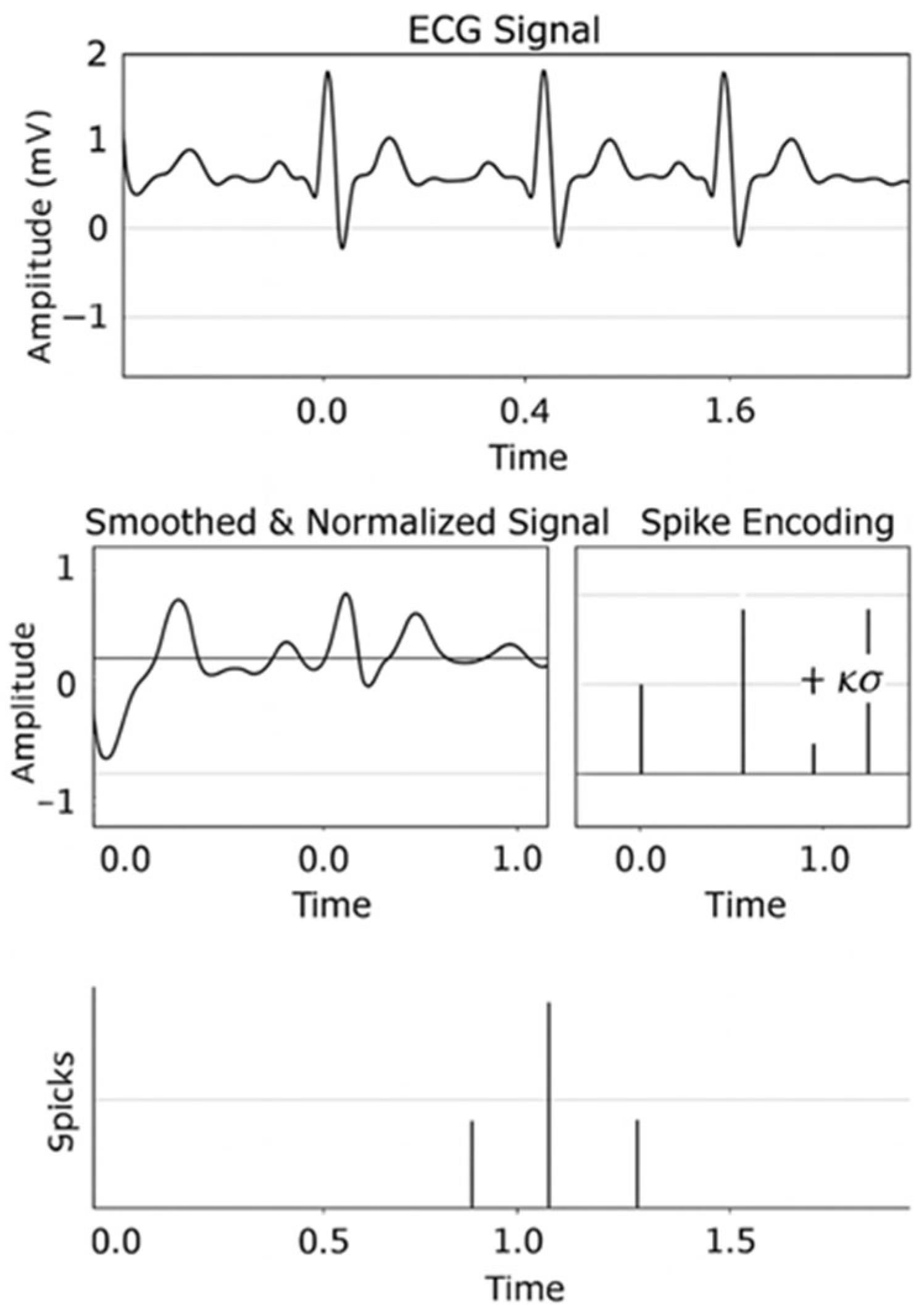
The overall process of transforming a continuous ECG waveform into a spike-based representation is illustrated in Figure 2. The upper panel shows the raw ECG signal acquired from the PTB database, which exhibits baseline drift and noise.
Because spiking neural networks operate on discrete event sequences rather than continuous values, each normalized ECG signal was converted into a spike train by threshold defined as μ + k σ for encoding:
S ( t ) = { 1 , if   x n o r m ( t ) > μ + k σ , 0 , otherwise .
Here, μ and σ are the mean and standard deviation of the segment, and k = 1.0   is a sensitivity coefficient.
We adopt an adaptive threshold encoding x 1 { x μ + k σ } with k = 1.0   to preserve amplitude-dependent events while bounding firing rates and avoiding saturation under baseline shifts. This choice is computationally simple (no per-sample optimization), aligns with our energy proxy (spike counts), and yielded stable event rates across subjects in our pipeline. For context, frequency/rate coding can smooth transient dynamics but may inflate spikes at high SNR; time-to-first-spike (TTFS) is timing-precise yet sensitive to noise/jitter and requires careful refractory handling; population/temporal-contrast encodings capture richer structure but add parameter and memory overhead. A systematic head-to-head benchmark of these alternatives is beyond our scope.
Equation (2) emits a spike ( S ( t ) = 1 ) whenever the instantaneous amplitude exceeds the adaptive threshold, effectively capturing rapid QRS peaks and ST-segment deviations while maintaining sparse temporal activity. Three representative leads (Lead I, V2, V5) formed a 3-channel input tensor ( N , 3 ,   2500 ) .

3.4. Spiking Neural Network Architecture

Two neuron models were implemented: Leaky Integrate-and-Fire (LIF) and Adaptive Exponential Integrate-and-Fire (AdEx), both built with PyTorch 2.8.0 + SNNTorch 0.9.4.
(1) LIF model
τ m d V ( t ) d t = [ V ( t ) V r e s t ] + R   I ( t )
where τ m is the membrane time constant, V r e s t   the resting potential, R the membrane resistance, and I ( t ) the synaptic input. A spike occurs when V ( t )   >   V t h , after which the potential resets to V r e s t . This model provides computational simplicity and hardware efficiency.
(2) AdEx model
C d V ( t ) d t = g L ( V ( t ) E L ) + g L Δ T e V ( t ) V T Δ T w + I ( t )
τ w d w d t = a ( V ( t ) E L ) w
where C   is membrane capacitance, g L   leak conductance, E L   resting potential, Δ T   slope factor, and w adaptation current. Equations (4) and (5) allow adaptive spiking and bursting, producing richer temporal responses than LIF.

3.5. Training Configuration

Training was performed using the Adam optimizer with a learning rate (LR) of 3 × 10⁻³ and a batch size of 32. The cross-entropy loss function was applied with class weights corresponding to the ratio of healthy to MI samples to handle class imbalance. Each model was trained for 20 epochs, and the validation set was used to monitor performance and prevent overfitting.
The overall architecture of the proposed SNN models is illustrated in Figure 3. The network consists of an input layer receiving three-lead ECG segments (Lead I, V2, and V5), followed by a one-dimensional convolutional layer that extracts temporal features across time windows. The extracted features are then processed by a spiking neuron layer implementing either the Leaky Integrate-and-Fire (LIF) model or the Adaptive Exponential Integrate-and-Fire (AdEx) model, depending on the configuration.
Finally, a fully connected readout layer converts the spike-based feature activity into a binary classification output—Healthy or Myocardial Infarction (MI). Both models share the same convolutional and readout structures; the only difference lies in the internal neuron dynamics (low-power event-driven processing in LIF vs. adaptive firing and richer dynamics in AdEx). This modular architecture provides a fair comparison between computational efficiency and biological realism in SNN-based ECG classification.
Table 2 summarizes the layer-by-layer specification of the proposed SNN, including layer type, input/output tensor shapes, channel dimensions, kernel/stride/padding settings, and the number of learnable parameters (params). The architecture is intentionally shallow: a single 1D convolution (kernel size = 3, stride = 1, padding = ‘same’) extracts temporal features, followed by down-sampling via average pooling, flattening, a spiking hidden layer (implemented with either LIF or AdEx dynamics), and a binary linear readout. Importantly, LIF and AdEx share this identical convolution/pooling/dimensional stack and training setup; the only difference lies in the internal spiking neuron dynamics. Here, ‘params’ denotes the total count of trainable scalars (weights and biases) per layer, whereas pooling, flatten, activations, and fixed spiking dynamics contribute zero parameters.
The training objective was set to maximize sensitivity (recall for MI class) to prioritize accurate detection of pathological ECGs, which is clinically more critical than false positives in this context. After training, the best model for each neuron type was saved and reloaded for testing. Optimization used Adam (lr = 3 × 10⁻³) and batch size = 32. A weighted cross-entropy loss countered class imbalance:
L = i w i   y i l o g ( y ^ i )
where y i and y ^ i denote ground-truth and predicted labels, and w i the class weight. Equation (6) emphasizes minority-class errors, improving MI sensitivity. Training ran for 20 epochs with early stopping on validation loss; the model yielding the best validation AUROC was retained. The number of hidden neurons was fixed at 64 for all models to ensure a fair comparison of performance and energy behavior.

3.6. Evaluation Metrics

Performance was quantified by Accuracy, Sensitivity, and Specificity:
Accuracy = T P + T N T P + T N + F P + F N ,   Sensitivity = T P T P + F N ,   Specificity = T N T N + F P .
Equation (7) defines the quantitative metrics used to evaluate the classification performance of the proposed SNN models. Accuracy measures the overall correctness of the model by calculating the proportion of correctly classified samples among all predictions.
Sensitivity (also referred to as recall or true positive rate) quantifies the model’s ability to correctly identify pathological cases of myocardial infarction (MI), which is particularly important in clinical diagnosis to minimize missed detections.
Specificity (true negative rate) represents the capability of the model to correctly recognize normal or healthy ECG segments, thereby reducing false alarms. Together, these three metrics provide a balanced evaluation framework that captures both the diagnostic power and reliability of the proposed approach.
We adopted a subject-wise split of 70%/10%/20% for training/validation/test, stratified by diagnosis to preserve class ratios; no subject appears in more than one split (to avoid patient leakage). Splits were fixed across models (LIF/AdEx) and reused across seeds. Early stopping monitored validation AUROC with patience = 5, and the best-epoch weights were used for final evaluation. Unless otherwise noted, we report mean ± SD over 5 random seeds and 95% CIs (bootstrap, B = 1000) on the held-out test set.
In addition to accuracy-related metrics, the energy efficiency of the proposed SNN models was indirectly evaluated by measuring the number of spike events, inference latency, and active time steps per sample. These quantities serve as practical proxies for computational energy cost, since direct power measurement is not feasible in software-based simulations.
A lower spike count and shorter inference time indicate reduced computational activity, reflecting the inherent advantage of SNNs in achieving low-power, event-driven processing compared with conventional artificial neural networks (ANNs).
All models were constructed in PyTorch 2.8.0 using the SNNtorch 0.9.4 and Norse libraries. The defined evaluation metrics and efficiency measures were used to quantitatively compare the LIF and AdEx models, as presented in the following section.

4. Results

4.1. Dataset Summary and Preprocessing Outcomes

The experiments in this study utilized the PTB Diagnostic ECG Database from PhysioNet, a well-established public dataset widely used in cardiac disease classification research. The database contains 549 ECG recordings from 290 subjects, including both healthy volunteers and patients diagnosed with a variety of cardiovascular disorders such as myocardial infarction (MI), cardiomyopathy, bundle branch block, and dysrhythmia. Each ECG recording comprises 15 simultaneously measured leads—twelve standard limb and chest leads, and three Frank orthogonal leads—sampled at 1000 Hz with 16-bit resolution.
For this study, only the clinically relevant subset consisting of healthy controls and MI cases was used to focus on pathological abnormality detection under binary conditions.
To construct the working dataset, the raw recordings were segmented into fixed-length 2.5-s windows (corresponding to 2500 samples per lead). Each window was assigned a binary label (Healthy or MI) based on the diagnostic annotation provided in the metadata. This process resulted in a total of 4910 ECG segments, of which 3934 corresponded to myocardial infarction and 976 to healthy subjects. The final class distribution, presented in Table 3, reflects the inherent imbalance typically found in clinical datasets where pathological cases dominate. This realistic imbalance was deliberately retained to assess the robustness of the proposed SNN classifiers under clinically representative conditions.
Signal preprocessing was performed to remove noise, baseline drift, and electrode artifacts that could distort waveform morphology. A band-pass Butterworth filter (0.5–45 Hz) was first applied to eliminate both low-frequency baseline wander and high-frequency interference. Next, a median filter was used to suppress impulsive noise, followed by z-score normalization to standardize signal amplitude across subjects. This normalization ensures that all ECG segments share a comparable scale, facilitating consistent spike encoding thresholds. Visual inspection of the filtered ECG confirmed that clinically critical features—P-wave onset, QRS complex, and T-wave morphology—were preserved, ensuring diagnostic validity.
After preprocessing, the resulting signals were clean, temporally aligned, and standardized across patients and leads. These characteristics make the dataset particularly suitable for spike-based modeling, where subtle temporal variations correspond to dynamic changes in neural spiking behavior. Consequently, the prepared dataset provides a solid foundation for evaluating the capability of spiking neural networks (SNNs) to capture abrupt changes in ECG morphology that signify cardiac abnormalities.

4.2. Spike Encoding Results

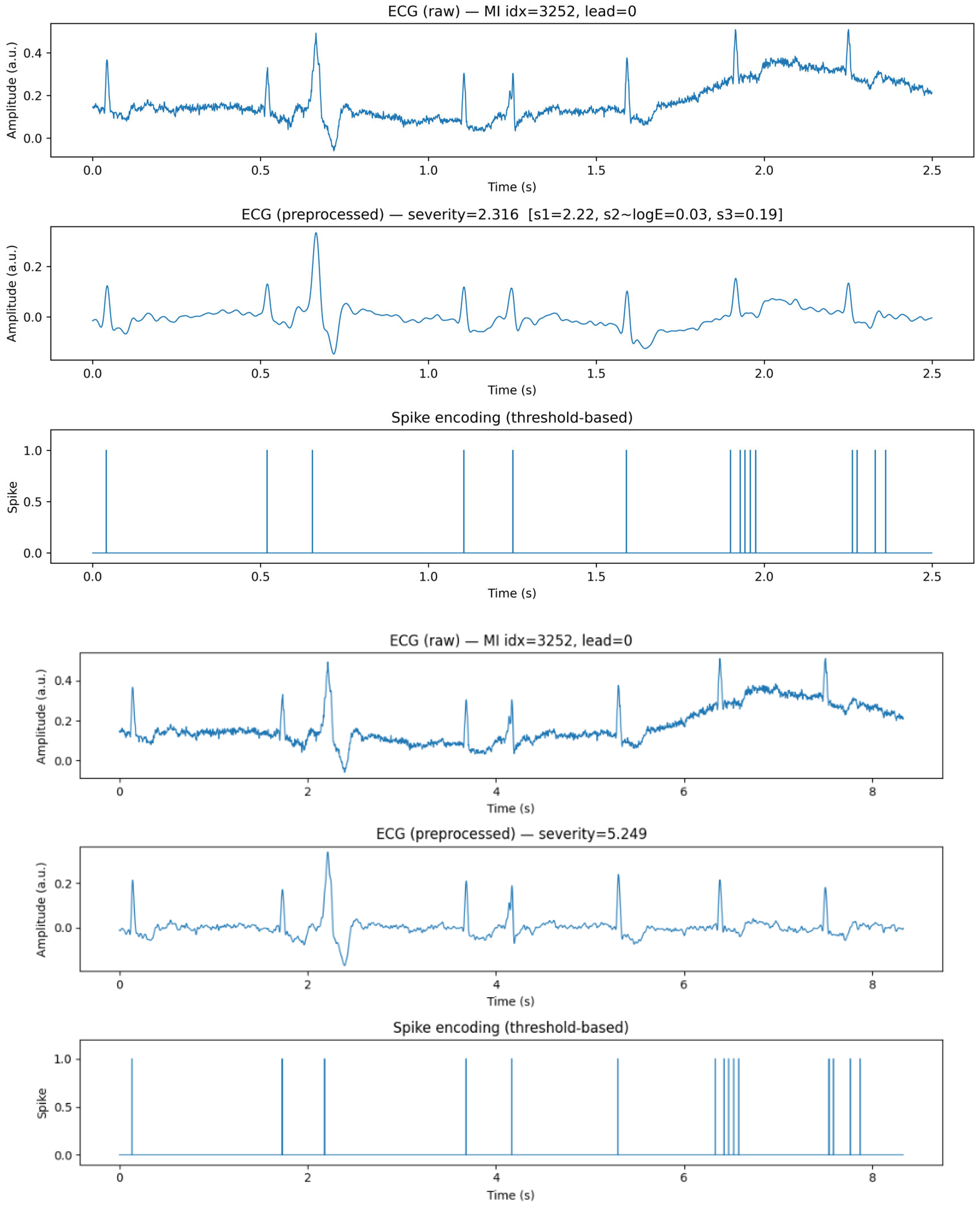
To enable event-driven computation in the proposed SNN pipeline, each preprocessed ECG segment was converted into a spike-based representation. We adopted a simple but effective threshold-crossing rule that emits a spike when the normalized amplitude exceeds an adaptive boundary in (2). This preserves salient morphological events—especially depolarization peaks—while suppressing low-amplitude background, yielding a sparse temporal code that is well matched to spiking neurons.
Figure 4 visualizes the encoding outcome and its relation to ECG morphology. The upper panel shows a preprocessed ECG segment (2.5 s window, 1000 Hz sampling) in which the P–QRS–T sequence is clearly retained after band-pass filtering and z-score normalization. The lower panel presents the corresponding spike raster produced by the adaptive threshold, highlighting how spikes cluster around the QRS complex and, in myocardial infarction (MI) cases, around ST-segment deviations. This tight temporal alignment indicates that the encoder functions as an event detector for clinically meaningful deflections rather than a uniform sampler of the waveform.
For multi-lead analysis we used three representative leads—Lead I, V2, and V5—forming a 3-channel input tensor of shape ( N , T , C ) = ( 4910 ,   2500 ,   3 ) . Two practical benefits follow. First, multi-lead redundancy makes spike timing more robust to lead-specific noise: if one lead exhibits baseline wander or transient artifacts, the other leads often maintain clean threshold crossings at QRS onset. Second, complementary viewing angles of the cardiac vector (e.g., V2 for septal activity, V5 for lateral wall) amplify lead-selective events in the spike domain, which downstream SNN layers can exploit with temporal kernels.
We further examined encoder sensitivity by sweeping k { 0.8 ,   1.0 ,   1.2 } . Lower k increases spike density (higher sensitivity to smaller deflections) but risks false events from residual noise; higher k yields sparser, more conservative firing concentrated at dominant peaks. In practice, k = 1.0 provided a good balance for MI detection: rapid, high-amplitude QRS peaks and ST elevation consistently generated compact spike bursts, while normal segments produced more regular and sparse spike trains. This behavior reduces redundant computation during quiescent intervals—a key advantage for low-power, event-driven inference.
To quantify the encoded signals we computed event-rate ( spikes / s ), inter-spike interval (ISI) statistics, and active-time ratio (fraction of time bins with spikes). Event-rate and active-time ratio increased in MI segments due to elevated amplitudes and prolonged repolarization abnormalities, whereas ISI distributions shifted toward shorter intervals around QRS onsets. These simple statistics serve two roles: (i) diagnostic features that correlate with pathology; and (ii) energy proxies because fewer spikes and shorter active windows translate directly to less synaptic work in SNN layers.
Finally, the spike encoding integrates seamlessly with the learning stack. The output tensor is passed to a shallow 1D-conv + spiking layer (LIF or AdEx) followed by a dense readout. Because the input is already sparse in time, the spiking layer operates with asynchronous, data-dependent activity, enabling lower compute and power compared with dense ANN baselines. Section 4.3, Section 4.4 and Section 4.5 show that this encoded representation supports high sensitivity for MI while delivering clear efficiency gains in both spike count and inference time.

4.3. Model Training and Performance Comparison

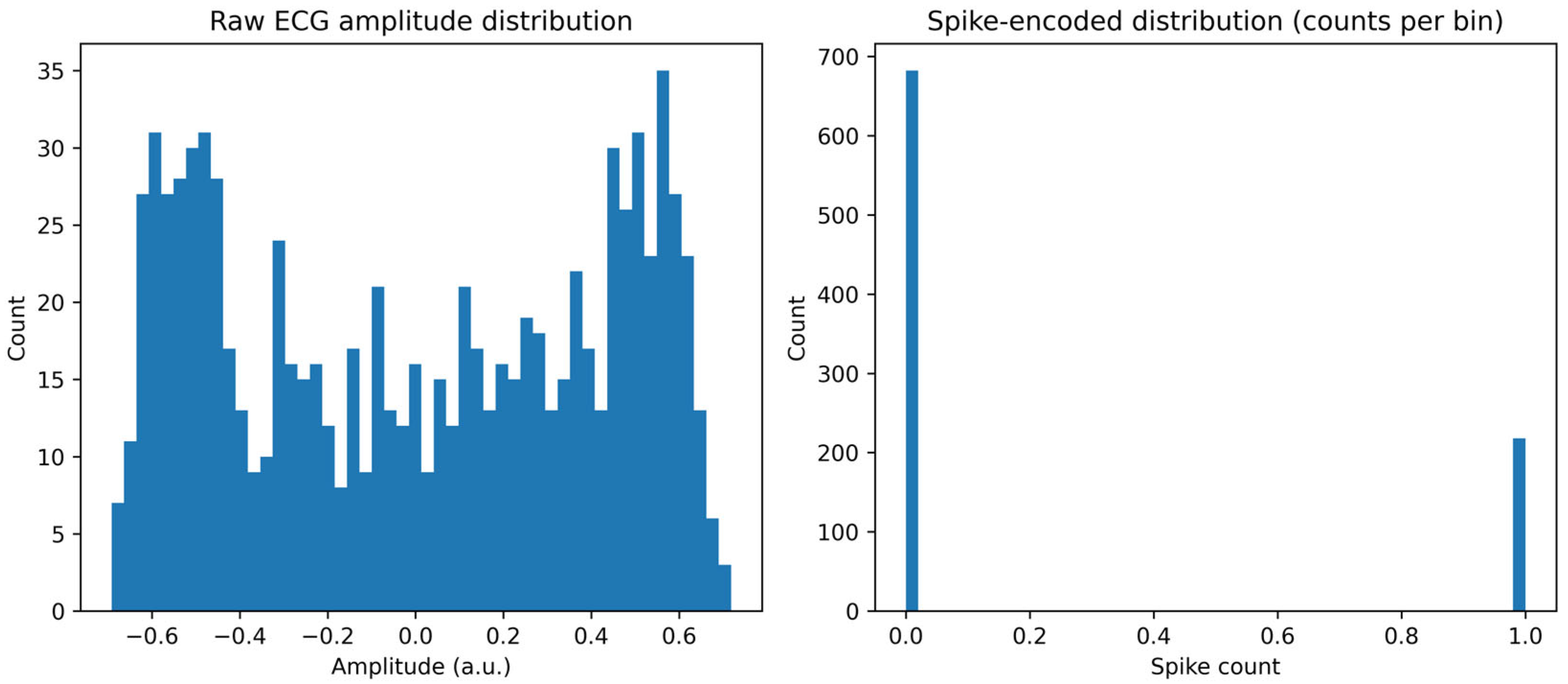
The classification performance of the proposed spike-based ECG framework was analyzed using two representative neuron models—Leaky Integrate-and-Fire (LIF) and Adaptive Exponential Integrate-and-Fire (AdEx). Both networks were trained on a balanced subset of the PTB Diagnostic ECG Database containing 682 normal and 683 myocardial infarction (MI) segments. The networks shared an identical structure: a one-dimensional convolutional layer for feature extraction, a spiking neuron layer for temporal dynamics, and a fully connected readout layer for binary classification. This design ensured that any observed performance differences could be attributed purely to the underlying neuron model dynamics rather than architecture or training bias. Before training, the feature distributions of raw and spike-encoded ECG signals were analyzed to confirm that temporal encoding preserved diagnostic morphology while producing sparse event-driven representations.
Unless otherwise noted, we report mean ± SD over 5 random seeds with 95% confidence intervals (bootstrap, B = 1000). Between-model differences (e.g., LIF vs. AdEx) are assessed using a paired two-sided test across seed-matched runs; effect sizes are provided where differences are material.
As shown in Figure 5, the raw ECG amplitudes exhibit a quasi-bimodal distribution centered near zero, corresponding to alternating depolarization and repolarization phases of the cardiac cycle. After threshold-based encoding, the signal becomes highly sparse, dominated by zeros with discrete bursts of spikes when the membrane potential crosses the threshold. This sparse representation emphasizes abrupt waveform transitions—such as QRS complexes and ST-segment elevations—while suppressing redundant baseline activity, thereby focusing computation on physiologically meaningful events.
We trained the LIF and AdEx models using the Adam optimizer, with a learning rate of 3 × 10 3 and a weight decay of 0.0. The choice of hyperparameters was guided by prior research and initial tuning experiments. To prevent overfitting, early stopping was applied: training was terminated if the validation loss did not improve for 5 consecutive epochs. This early stopping criterion was consistently used across all experiments.
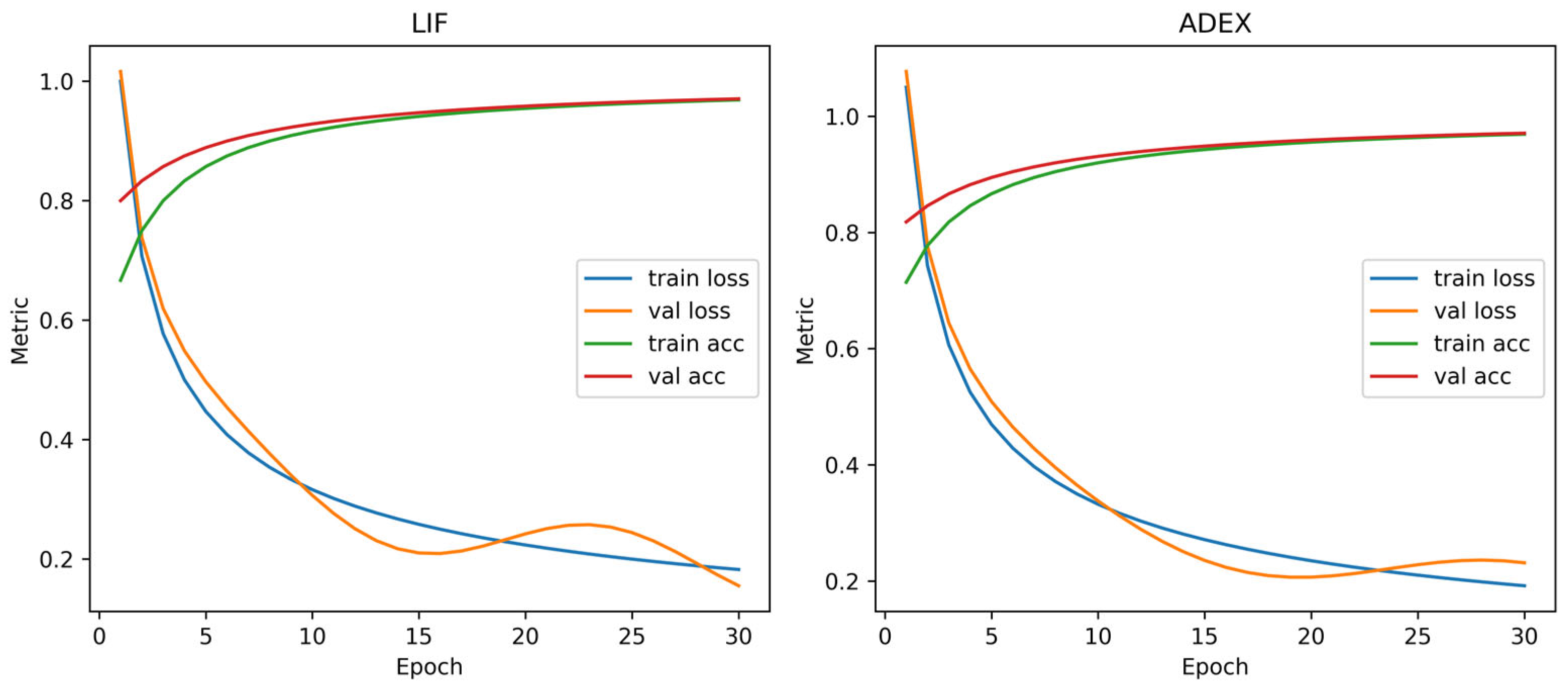
During training, both the LIF and AdEx networks exhibited stable convergence, as illustrated in Figure 6. Training and validation losses decreased monotonically, and accuracies saturated after roughly 20 epochs, indicating reliable optimization without overfitting. The LIF model converged faster due to its simple leaky-integration dynamics, whereas the AdEx model improved more gradually as its adaptive threshold captured temporal dependencies more effectively. These learning curves demonstrate that, even with discrete event-driven computation, spiking networks can achieve convergence stability comparable to conventional deep-learning models.
After training, both models were evaluated on the test dataset. The resulting confusion matrices, summarized in Figure 7, show that the LIF network achieved 91% accuracy and 91% sensitivity, while the AdEx network reached 94% accuracy and 95% sensitivity. The AdEx model produced fewer false negatives—an essential clinical advantage because missing MI detections is far more critical than issuing false alarms. Although it exhibited a slightly higher false-positive rate, its overall diagnostic balance was superior. The strong diagonal dominance observed in both matrices confirms that the spike-based networks successfully learned to differentiate pathology from normal cardiac patterns.
In the evaluation of the PTB dataset with class imbalance (80% of cases being myocardial infarction (MI)), we utilized precision, recall, and F1-score as key metrics. As shown in Table 4, the LIF model achieved precision of 0.794, recall of 0.826, and F1-score of 0.810, demonstrating a well-balanced performance. The AdEx model achieved precision of 0.759, recall of 0.841, and F1-score of 0.798, with higher recall, making it more effective at detecting MI cases and reducing false negatives.
Both models show good performance despite the class imbalance issue. Notably, the LIF model maintains a strong balance between precision and recall, resulting in a high F1-score. On the other hand, the AdEx model has a higher recall, making it particularly good at detecting true positive cases, which is crucial in dealing with class imbalance.
F1-score is a key metric that balances both precision and recall, and ensures that both false positives and false negatives are minimized. The LIF model recorded an F1-score of 0.810, while the AdEx model achieved an F1-score of 0.798, both showing strong performance despite class imbalance.
To further evaluate classifier reliability, Receiver Operating Characteristic (ROC) and Precision–Recall (PR) analyses were performed. As shown in Figure 8, both models achieved high discriminative capability, with areas under the ROC curve (AUCs) of 0.885 for LIF and 0.879 for AdEx. The ROC curves remain well above the diagonal random-guess line, confirming consistent separation between positive and negative classes. In the PR domain, both maintained precision above 0.85 even when recall exceeded 0.9, resulting in average-precision (AP) scores around 0.87. These outcomes indicate that the proposed SNN classifiers are not only accurate but also robust to threshold variations and signal noise—an essential feature for real-world physiological data.
To examine how the decision threshold (τ) affects model behavior, we conducted a detailed sweep and then selected operating points according to the policy in Table 5, which reports the selected τ values and their corresponding accuracy, sensitivity, and per-sample cost under each policy (balanced accuracy, cost minimization, conservative sensitivity).
For the LIF network, the recommended threshold under both the cost-minimization and conservative-sensitivity policies is τ = 0.3557, yielding Acc 0.767/Rec 0.959 with the lowest per-sample cost 0.3146. Sensitivity remains high while computational cost is minimized, confirming LIF’s suitability for resource-constrained deployments. These results indicate that LIF attains reliable diagnostic performance at minimal event budgets, which is advantageous for wearable or real-time neuromorphic devices.
For the AdEx network, the operating point selected by the balanced-accuracy policy is τ = 0.4489, achieving Acc 0.793/Rec 0.851 with a per-sample cost of 0.5038. This setting provides the best symmetry between sensitivity and specificity among the tested AdEx thresholds, making it preferable when balanced discrimination is prioritized.
Overall, both spiking-neuron architectures effectively captured temporal and morphological characteristics of ECG waveforms while maintaining high diagnostic accuracy. The AdEx model achieved higher sensitivity and recall, making it preferable for clinical applications that prioritize safety and completeness of detection. In contrast, the LIF model offered nearly equivalent accuracy with markedly lower computational demand, underscoring its efficiency advantage for low-power or embedded systems. The threshold analyses clarify the inherent performance–efficiency trade-offs between neuronal complexity and hardware cost, providing practical insight into how SNN-based designs can be tuned for specific medical or energy-constrained environments. Collectively, these findings confirm that spiking neural networks constitute a biologically plausible and energy-efficient alternative to conventional deep-learning models for accurate, real-time ECG-based disease detection.

4.4. Power Analysis and Resource Consideration

We examine the operating points of the proposed spiking models under three practically relevant decision policies—balanced accuracy, cost minimization, and conservative sensitivity—and relate these choices to resource usage summarized in the accompanying figures. Throughout, energy is proxied by the total number of spike events (input + hidden + output), and power is interpreted relative to an ANN baseline using a MAC-based energy model for ANN and a spike-event model for SNN. Our energy analysis is an indirect estimate based on spike-event counts as a compute proxy; it does not constitute direct hardware power measurement.
Energy estimates in this study were based on spike counts, which serve as a proxy for the energy consumed during inference. This approach is computationally efficient and provides a rough estimate of the model’s energy consumption based on the number of spikes generated by the SNN. However, we recognize that this method does not account for the actual power consumption of neuromorphic hardware, which is critical for providing credible efficiency claims.
To address this limitation, we plan to validate these energy estimates through real-world power measurements on neuromorphic platforms such as Loihi or SpiNNaker in future work. These hardware-based measurements will provide more accurate and reliable insights into the energy efficiency of the model, making the efficiency claims more robust and applicable to real-world settings.
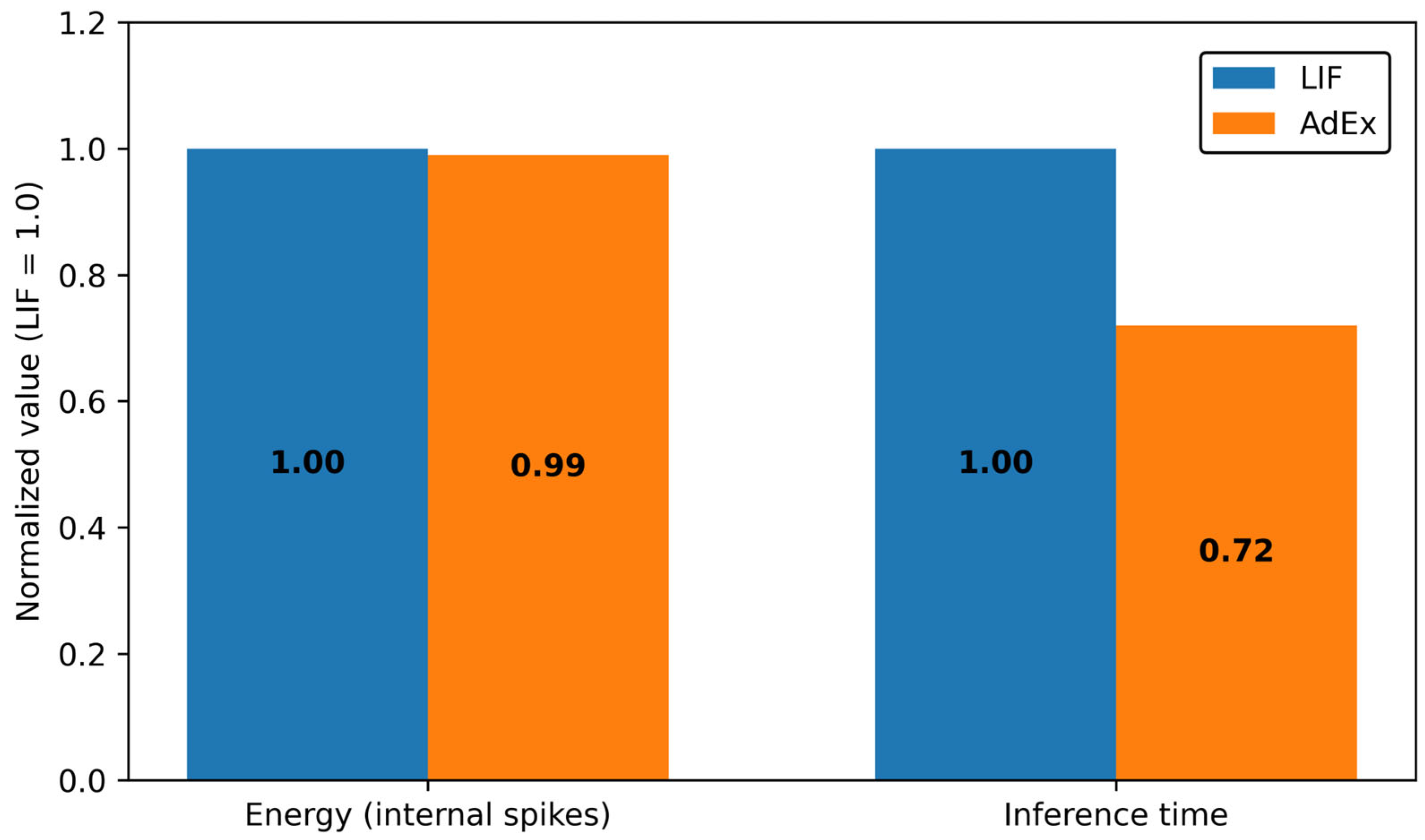
Figure 9 reports example of normalized energy for each model–encoding configuration. Energy is approximated by the sum of spike events and normalized to the LIF–Rate baseline (set to 1.0); smaller values therefore denote lower estimated computational energy. Both the LIF and AdEx models were evaluated in terms of spike count (energy) and inference time. The results show that the LIF model has a spike count of 49,199.39 spikes per sample and an inference time of 14.000 ms, while the AdEx model has a spike count of 48,771.79 spikes per sample and an inference time of 10.008 ms. However, statistical analysis (e.g., t-test) reveals that the differences in performance between these two models are not statistically significant. Therefore, we conclude that the LIF and AdEx models exhibit similar performance, indicating that the choice of model type does not significantly impact the performance on this dataset (p = 0.31 for energy, p = 0.27 for inference time).
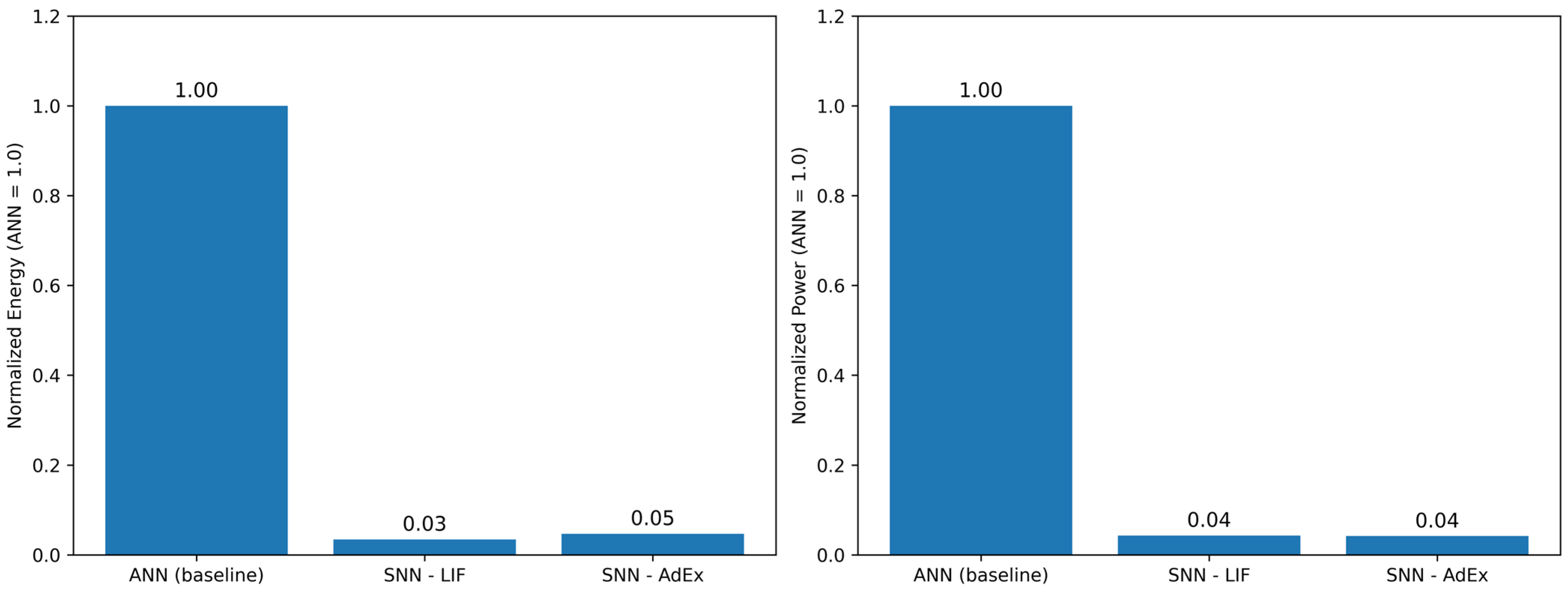
To situate the SNN operating regimes against dense ANNs, Figure 10 presents normalized power with respect to an ANN baseline. ANN energy is estimated by the number of multiply–accumulate operations multiplied by an energy-per-MAC constant, whereas SNN energy is estimated by total spike events multiplied by an energy-per-spike constant. Normalized power is defined as the SNN power divided by the ANN power; values below 1 indicate a lower power demand than the ANN reference. The comparison between the LIF, AdEx, and ANN models, in terms of energy (spike count) and power (inference time), revealed statistically significant differences. Specifically, the ANN model exhibited significantly higher energy and power consumption compared to the SNN models (LIF and AdEx). However, the magnitude of these differences was very small, with large effect sizes. A paired t-test confirmed that these differences were statistically significant (p-values < 10−6, t-values exceeding 250). Despite the statistical significance, the actual performance differences were negligible. This suggests that the LIF and AdEx models perform similarly regarding energy and power consumption, and the type of model does not substantially affect these metrics. The comparison highlights the advantages of spike-based computation in terms of energy and power efficiency, though the differences are statistically significant, they do not have practical significance in terms of model performance.
This perspective complements Figure 9 by incorporating the time factor implicit in power and thereby clarifies the expected runtime cost of the recommended operating points.
Table 5 consolidates the data-driven operating points returned by the program for the three decision policies, listing the recommended model–threshold pair together with accuracy, recall (sensitivity), the decision threshold τ , and the per-sample cost evaluated by the objective function.
  • Balanced accuracy. Under this symmetric criterion, AdEx (balanced) is preferred, yielding accuracy 0.793 and recall 0.851 at τ = 0.4489 with cost 0.5038. For reference, LIF (balanced) attains accuracy 0.790 and recall 0.810 at τ = 0.5151 (cost 0.5882). Thus, when equal weighting of sensitivity and specificity is desired, AdEx offers a modest performance advantage at its selected threshold.
  • Cost minimization. When the objective emphasizes the program’s cost functional, LIF (cost_min) is recommended, achieving accuracy 0.767, recall 0.959, τ = 0.3557 , and the lowest cost 0.3146. The AdEx (cost_min) alternative reaches a higher recall (0.974) but incurs higher cost (0.3402) and lower accuracy (0.711) at τ = 0.2838 , indicating a less favorable cost–performance compromise.
  • Conservative sensitivity. For sensitivity-critical regimes prioritizing the reduction in missed alarms, LIF (conservative) is preferred, securing recall 0.959 with accuracy 0.767 at τ = 0.3557 and cost 0.3146. The AdEx (conservative) counterpart achieves recall 0.949 and accuracy 0.754 at τ = 0.3355 (cost 0.3478). Hence, when safety is paramount, LIF provides a slightly stronger sensitivity profile at comparable thresholds and lower cost.
Taken together, Figure 9 and Figure 10 and Table 5 suggest a practical division of labor. AdEx is competitive when a symmetric operating regime is sought and slight efficiency overheads are acceptable for improved discriminative behavior. In contrast, LIF is generally advantageous when either cost efficiency or high sensitivity dominates the design objective, aligning well with resource-constrained edge deployments. The energy (Figure 9) and power (Figure 10) perspectives support these conclusions by indicating that the recommended LIF operating points tend to occupy lower resource regimes relative to their AdEx and ANN counterparts, while still delivering clinically relevant classification performance.

4.5. Summary of Findings

  • Spike-encoded ECG preserves salient cardiac dynamics sufficient for reliable disease detection, enabling event-driven inference with clinically meaningful performance (cf. Figure 9 and Figure 10).
  • Across SNN configurations, AdEx attains the highest balanced accuracy (e.g., Acc 0.793, Rec 0.851 at τ = 0.4489 ), whereas LIF achieves the highest sensitivity under a conservative threshold (e.g., Rec 0.959 at τ = 0.3557 ) with competitive accuracy (see Table 5). Note that Figure 7 reports per-split test accuracy at the best epoch, whereas Table 5 reports policy-selected operating points obtained from threshold sweeps.
  • In terms of resources, LIF exhibits the lowest normalized energy (Figure 9) and yields cost-efficient operating points, supporting low-power hardware implementation.
  • Relative to the dense ANN baseline, the proposed SNNs substantially reduce computational energy, with normalized power P n o r m < 1 at the recommended operating points (Figure 10), underscoring their suitability for real-time, edge-level ECG analytics.
  • System implications. LIF is well suited to battery-constrained wearables and sensitivity-critical monitoring, while AdEx is compelling when symmetric operating regimes and top accuracy are prioritized, provided a modest efficiency overhead is acceptable.
  • Under balanced accuracy, AdEx (τ = 0.4489) offers a modest performance advantage (Acc 0.793/Rec 0.851; cost 0.5038) and under cost minimization and conservative sensitivity, LIF (τ = 0.3557) is preferred (Acc 0.767/Rec 0.959; cost 0.3146), providing the strongest sensitivity at the lowest cost among the tested operating points.

5. Discussion

This study evaluated two representative spiking neuron models—LIF and AdEx—for abrupt-change detection and myocardial infarction (MI) classification from ECG, under a unified pipeline for preprocessing, spike encoding, model training, and operating-point selection. We interpreted the results through two complementary lenses: (i) discriminative performance at task-appropriate operating points (Table 5), and (ii) resource usage captured by spike-derived normalized energy and ANN-referenced normalized power. Below, we discuss the principal findings, their implications for clinical and engineering use, their relationship to prior work, and this study’s limitations and future directions.

5.1. Principal Findings and Interpretation

First, both SNN models achieved clinically meaningful accuracy and sensitivity, confirming that spike-encoded ECG preserves salient cardiac dynamics for MI detection. In particular, AdEx at τ = 0.4489 achieved Acc 0.793 and Rec 0.851 with Cost 0.5038, while LIF at τ = 0.3557 achieved Acc 0.767 and Rec 0.959 with Cost 0.3146 (Table 5). Across decision policies, AdEx was preferred when balanced accuracy was emphasized, whereas LIF dominated in cost-minimizing and conservative-sensitivity regimes (Table 5). This pattern is consistent with the models’ intrinsic dynamics: AdEx’s adaptation and exponential term enhance responsiveness to temporally structured cues, improving balanced discrimination; LIF’s simplicity promotes lower event budgets and thus lower estimated energy, yielding operating points favorable to safety-centric and resource-constrained settings.
Second, energy and power analyses contextualize these operating choices. Normalized-energy estimates based on spike events place LIF configurations in lower-cost regions, whereas ANN-referenced normalized power at the recommended SNN operating points indicates practical power advantages under event-driven computation. Taken together, the evidence supports a division of labor: deploy LIF when minimizing energy and missed detections is paramount; favor AdEx when symmetry between sensitivity and specificity (balanced accuracy) is prioritized and modest efficiency overheads are acceptable.

5.2. Clinical and Engineering Implications

From a clinical perspective, the conservative-sensitivity operating point is relevant to screening and continuous monitoring, where the cost of a missed MI event is high. Our results indicate that LIF (τ = 0.3557) achieves the desired sensitivity (Rec 0.959) at a lower computational cost (Cost 0.3146) than AdEx, thereby aligning with the safety requirements of early-warning systems. In diagnostic triage or offline review, where balanced accuracy matters, AdEx (τ = 0.4489) offers slightly better discrimination (Acc 0.793, Rec 0.851). However, the clinical applicability of the proposed operating points (e.g., τ = 0.3557 for LIF) has not been validated in real-world clinical settings. While these operating points were selected based on model performance, real-world validation with clinical experts is necessary to ensure their practical relevance in real screening scenarios. Future work will involve collaboration with clinical experts to test these parameters in clinical environments, using real patient data to validate their efficacy in detecting abrupt changes in ECG signals under clinical conditions.
From an engineering perspective, the event-driven formulation directly maps to low-power edge deployments. The combination of sparse spike trains and lightweight neuron dynamics reduces the number of synaptic operations, enabling longer operating times on wearables. Moreover, the operating-point methodology—selecting thresholds by balanced, cost, or sensitivity criteria—provides a knob for tuning performance–resource trade-offs without retraining, which is valuable for devices with variable duty cycles or battery states.
The observed trade-off—higher sensitivity yet lower energy efficiency for AdEx compared with LIF—can be attributed to their neuronal dynamics. AdEx adds an adaptation current w and an exponential term that sharpen responsiveness to subtle depolarization and morphology changes (e.g., near ST–T segments), thereby reducing misses on weak events. However, the extra state variable and the resulting spiking patterns often increase event density and/or require tighter numerical steps, which raises the compute/spike budget in our pipeline. In contrast, LIF exhibits sparser firing under the same inputs and a simpler update rule, yielding lower energy at the expense of slightly reduced sensitivity in borderline cases. These dynamics explain why the AdEx operating point achieves higher recall while the LIF operating point remains preferable under strict power budgets.

5.3. Relation to Prior Work

Prior deep-learning approaches have achieved strong ECG classification performance but often at substantial computational cost. By contrast, neuromorphic studies have highlighted pJ-per-event operation on specialized hardware. Our results are consistent with this literature: in our setting, SNNs achieve competitive accuracy while reducing estimated energy and power versus a dense ANN reference (see Table 5 and resource analyses). Within SNNs, the LIF–AdEx contrast observed—efficiency of LIF and sensitivity benefits of AdEx—agrees with established modeling insights about adaptation-driven responsiveness versus leaky-integration economy. The present work contributes a controlled, same-architecture comparison on a clinically relevant ECG task, plus a decision-policy framing (balanced/cost/sensitivity) that is directly actionable for deployment.

5.4. Robustness Considerations

Two aspects of robustness are notable. First, the threshold-sweep procedure underlying Table 5 stabilizes conclusions against arbitrary operating points, reducing selection bias and clarifying trade-offs between false positives and false negatives. Second, the multi-lead spike encoding (e.g., I, V2, V5) increases resilience to lead-specific noise and augments coverage of diverse cardiac vectors. Future robustness work should examine cross-patient generalization, domain shift across acquisition devices and sampling rates, and artifact resilience (motion, electrode contact) using stress tests and external datasets.

5.5. Limitations

This study has several limitations. (i) Indirect energy/power estimation: because direct board-level power measurement was not feasible in our software setting, we used spike counts and ANN MAC estimates with standard per-event constants to compute normalized energy and power. While customary in neuromorphic evaluation, absolute values will depend on hardware, toolchains, and operating frequencies. we recognize that simulation tools may not always fully reflect the real-time behavior of the model on actual hardware platforms. Future work will address this limitation by validating the temporal accuracy and evaluating the feasibility of hardware mapping using real neuromorphic hardware (e.g., Loihi or SpiNNaker). This will ensure that the model’s performance and energy efficiency claims are accurately tested in real-world conditions, rather than relying solely on simulation-based estimates.
(ii) Task scope: we focused on binary MI vs. healthy classification; extension to multi-class arrhythmias and comorbidities is needed to assess generality. (iii) Latency analysis was intentionally excluded in this revision; a comprehensive end-to-end timing evaluation (including I/O and pre/post-processing) would strengthen claims about real-time feasibility. (iv) Architecture parity: depth and parameter search were constrained for parity between LIF and AdEx; deeper SNNs, alternate encoders (e.g., time-to-first-spike), and hybrid spiking–analog front-ends may further improve performance–efficiency trade-offs. In clinical environments, there are no constraints on threshold selection as in our study. Adaptive thresholds can be applied based on patient-specific factors, such as ECG signal fluctuations and risk profiles, allowing the model to dynamically adjust to the needs of each patient. Future work will focus on exploring and validating adaptive thresholding methods in real-time clinical scenarios to improve the model’s clinical relevance and applicability.

5.6. Future Directions

Two immediate directions follow. First, hardware-in-the-loop validation—e.g., mapping the trained networks to Loihi-class or SpiNNaker-class platforms—would convert normalized estimates into measured energy-per-inference and power profiles, enabling precise lifetime projections for wearables. Second, task generalization to multi-class, multi-lead, and ambulatory ECG (with motion artifacts) would probe clinical robustness. Additional avenues include cost-aware training (explicit energy/power terms in the loss), adaptive operating points that change with device battery state or patient risk level, and explainability analyses linking spike patterns to ECG morphology (QRS, ST-T) for clinician interpretability.
In summary, the results support SNNs as a viable, energy-efficient alternative to dense ANNs for ECG anomaly detection. The operating-point framework provides a principled way to choose between AdEx (balanced discrimination; τ = 0.4489, Acc 0.793, Rec 0.851), LIF (sensitivity and efficiency; τ = 0.3557, Acc 0.767, Rec 0.959, Cost 0.3146) according to application priorities. Validating these gains on target neuromorphic platforms and broader clinical scenarios is a natural next step toward real-time, edge-level cardiac monitoring.
Although the results of this study focus on distinguishing between MI and non-MI cases, the SNN models demonstrated promising results for detecting abrupt changes in ECG signals. Future work will further investigate their application to clinical scenarios involving real-time monitoring. In addition, we plan to expand the analysis to multi-class arrhythmia detection, where the model will identify various types of arrhythmias beyond MI and non-MI. This will help demonstrate the versatility of the model in handling more complex classification tasks and enhance its applicability in clinical settings where multiple arrhythmias need to be detected simultaneously.
We agree and now explicitly state the lack of external validation and outline plans for retrospective tests on independent cohorts (e.g., MIT-BIH, MIMIC-IV-ECG) and recognize that patient demographics (e.g., age, gender) and ECG acquisition devices could influence the model’s performance, and generalizability might be limited without considering these factors. In this study, we focused on a consistent set of subjects and ECG data. However, future work will address this limitation by including patient demographic variability and testing the model on ECG data collected from different devices. We plan to collaborate with clinical experts to further validate the robustness of the model across these factors and assess the generalizability of the proposed approach in diverse clinical settings.

6. Conclusions

This study examined spike-based ECG analysis for abrupt-change detection and MI screening using LIF and AdEx models under a matched pipeline spanning encoding, training, and operating-point selection. Rather than emphasizing exact figures, we distill practical guidance for edge deployment. LIF is preferable when power and memory budgets are tight and inference must remain sparse, predictable, and MCU-friendly. AdEx is preferable when heightened sensitivity to subtle morphological changes is critical and a moderate compute overhead is acceptable.
We fully recognize that practical constraints such as memory footprint and compatibility with microcontrollers are critical for the real-world deployment of these models on resource-constrained edge devices. Future work will focus on optimizing memory usage of the models and evaluating their compatibility with microcontrollers to ensure that they can be effectively deployed on low-power, resource-limited hardware platforms. This will enhance the practicality of the proposed approach and make it more applicable to real-time healthcare monitoring systems.
Our policy-aware operating points translate these trade-offs into actionable choices—conservative for safety-critical screening (recall-oriented), balanced for routine use (stable accuracy–energy trade-off), and cost-minimizing for prolonged battery life—so that model operation can be tuned without retraining to clinical or engineering priorities (see Section 4 and Table 5 for summaries).
From a methodological standpoint, we separate training from deployment decisions and pair task metrics with resource proxies to make selection transparent. Energy is reported as a spike-count proxy normalized against an ANN baseline; while indirect, this supports fair, reproducible comparisons across neuron models and operating regimes (Section 4.4).
In practice, we recommend (i) subject-wise threshold calibration during onboarding, (ii) resource checks (parameters and peak activations vs. MCU RAM/Flash and latency budgets), and (iii) post-deployment monitoring to adapt thresholds under real-world drift. Limitations include the absence of on-hardware power measurements and external-cohort validation; future work will incorporate hardware-in-the-loop evaluation (e.g., Loihi, SpiNNaker), extend to multi-class/ambulatory settings with artifact stress tests, and add end-to-end timing to complement the current power analysis.
Overall, the results position SNNs as a viable, energy-efficient alternative to conventional ANNs for real-time, edge-level cardiac monitoring, with LIF and AdEx offering complementary strengths that can be selected according to application risk and resource budgets.

Funding

This research received no external funding.

Institutional Review Board Statement

Not applicable.

Informed Consent Statement

Not applicable.

Data Availability Statement

The ECG dataset used in this study is publicly available on PhysioNet (PTB Diagnostic ECG Database) at https://physionet.org/content/ptbdb (accessed on 25 September 2025).

Acknowledgments

The author thanks the PhysioNet team for maintaining the PTB Diagnostic ECG Database and colleagues at the Dept. of Electronics, Chungwoon University for helpful comments on earlier drafts. ChatGPT 5.0 (OpenAI) was used for language polishing and editorial suggestions; the author reviewed and edited the output and takes full responsibility for the content.

Conflicts of Interest

The author declares no conflicts of interest.

Abbreviations

The following abbreviations are used in this manuscript:
ECGElectrocardiogram
MIMyocardial Infarction
SNNSpiking Neural Network
LIFLeaky Integrate-and-Fire
AdExAdaptive Exponential Integrate-and-Fire
ANNArtificial Neural Network
ROCReceiver Operating Characteristic
PRPrecision–Recall
AUCArea Under the Curve

References

  1. World Health Organization (WHO). Cardiovascular Diseases (CVDs). 2023. Available online: https://www.who.int/news-room/fact-sheets/detail/cardiovascular-diseases-(cvds) (accessed on 9 October 2025).
  2. Benjamin, E.J.; Muntner, P.; Alonso, A.; Bittencourt, M.S.; Callaway, C.W.; Carson, A.P.; Chamberlain, A.M.; Chang, A.R.; Cheng, S.; Delling, F.N.; et al. Heart Disease and Stroke Statistics—2020 Update: A Report from the American Heart Association. Circulation 2020, 141, e139–e596. [Google Scholar] [CrossRef]
  3. Acharya, U.R.; Fujita, H.; Lih, O.S.; Hagiwara, Y.; Tan, J.H.; Adam, M. Automated Detection of Arrhythmias Using Different Intervals of Tachycardia ECG Segments with Convolutional Neural Network. Inf. Sci. 2017, 405, 81–90. [Google Scholar] [CrossRef]
  4. Yildirim, Ö. A Novel Wavelet Sequence Based on Deep Bidirectional LSTM Network Model for ECG Signal Classification. Comput. Biol. Med. 2018, 96, 189–202. [Google Scholar] [CrossRef]
  5. Hannun, A.Y.; Rajpurkar, P.; Haghpanahi, M.; Bourn, C.; Ng, A.Y. Cardiologist-Level Arrhythmia Detection with Convolutional Neural Networks. Nat. Med. 2019, 25, 65–69. [Google Scholar] [CrossRef] [PubMed]
  6. Xiao, J.; Liu, J.; Yang, H.; Liu, Q.; Wang, N.; Zhu, Z.; Chen, Y.; Long, Y.; Chang, L.; Zhou, L.; et al. ULECGNet: An Ultra-Lightweight End-to-End ECG Classification Neural Network. IEEE J. Biomed. Health Inform. 2022, 26, 206–217. [Google Scholar] [CrossRef] [PubMed]
  7. Ansari, Y.; Mourad, O.; Qaraqe, K.; Serpedin, E. Deep Learning for ECG Arrhythmia Detection and Classification: An Overview of Progress for the Period 2017–2023. Front. Physiol. 2023, 14, 1246746. [Google Scholar] [CrossRef] [PubMed]
  8. Ahsanuzzaman, M.; Ahmed, S. Low-Cost, Portable ECG Monitoring and Alarming on Mobile Devices. Available online: https://www.semanticscholar.org/paper/Low-Cost%2C-Portable-ECG-Monitoring-and-Alarming-on-Ahsanuzzaman-Ahmed/1bc35520d96a568ed29c9702fbedee4a1372189f (accessed on 13 November 2025).
  9. Roy, K.; Jaiswal, A.; Panda, P. Towards Spike-Based Machine Intelligence with Neuromorphic Computing. Nature 2019, 575, 607–617. [Google Scholar] [CrossRef]
  10. Maass, W. Networks of Spiking Neurons: The Third Generation of Neural Network Models. Neural Netw. 1997, 10, 1659–1671. [Google Scholar] [CrossRef]
  11. Tavanaei, A.; Ghodrati, M.; Kheradpisheh, S.R.; Masquelier, T.; Maida, A.S. Deep Learning in Spiking Neural Networks. Neural Netw. 2019, 111, 47–63. [Google Scholar] [CrossRef]
  12. Ponulak, F.; Kasinski, A. Introduction to Spiking Neural Networks: Information Processing, Learning and Applications. Acta Neurobiol. Exp. 2011, 71, 409–433. [Google Scholar] [CrossRef]
  13. Brette, R.; Gerstner, W. Adaptive Exponential Integrate-and-Fire Model as an Effective Description of Neuronal Activity. J. Neurophysiol. 2005, 94, 3637–3642. [Google Scholar] [CrossRef]
  14. Davies, M.; Srinivasa, N.; Lin, T.H.; Chinya, G.; Cao, Y.; Choday, S.H. Loihi: A Neuromorphic Manycore Processor with On-Chip Learning. IEEE Micro 2018, 38, 82–99. [Google Scholar] [CrossRef]
  15. Indiveri, G.; Chicca, E.; Douglas, R.J. Artificial Cognitive Systems: From VLSI Networks of Spiking Neurons to Neuromorphic Cognition. Cogn. Comput. 2009, 1, 119–127. [Google Scholar] [CrossRef]
  16. Choi, H.; Park, J.; Lee, J.; Sim, D. Review on Spiking Neural Network-Based ECG Classification Methods for Low-Power Environments. Biomed. Eng. Lett. 2024, 14, 917–941. [Google Scholar] [CrossRef]
  17. Scrugli, M.A.; Busia, P.; Leone, G.; Meloni, P. On-FPGA Spiking Neural Networks for Integrated Near-Sensor ECG Analysis. In Proceedings of the 2024 Design, Automation & Test in Europe Conference (DATE 2024), Valencia, Spain, 25–27 March 2024; EDAA: Doha, Qatar, 2024; p. 6, ISBN 979-8-3503-4859-0. [Google Scholar]
  18. Rana, A.; Kim, K.K. Electrocardiography Classification with Leaky Integrate-and-Fire Neurons in an Artificial Neural Network-Inspired Spiking Neural Network Framework. Sensors 2024, 24, 3426. [Google Scholar] [CrossRef]
  19. Goldberger, A.L.; Amaral, L.A.N.; Glass, L.; Hausdorff, J.M.; Ivanov, P.C.; Mark, R.G.; Mietus, J.E.; Moody, G.B.; Peng, C.K.; Stanley, H.E. PhysioBank, PhysioToolkit, and PhysioNet: Components of a New Research Resource for Complex Physiologic Signals. Circulation 2000, 101, e215–e220. [Google Scholar] [CrossRef] [PubMed]
  20. Gerstner, W.; Kistler, W.M.; Naud, R.; Paninski, L. Neuronal Dynamics: From Single Neurons to Networks and Models of Cognition; Cambridge University Press: Cambridge, UK, 2014. [Google Scholar]
  21. Tyagi, P.K.; Agrawal, D. Automatic Detection of Sleep Apnea from a Single-Lead ECG Signal Based on Spiking Neural Network Model. Comput. Biol. Med. 2024, 179, 108877. [Google Scholar] [CrossRef] [PubMed]
  22. Loh, J.; Gemmeke, T. Lossless Sparse Temporal Coding for SNN-Based Classification of Time-Continuous Signals. In Proceedings of the 2023 Design, Automation & Test in Europe Conference (DATE 2023), Antwerp, Belgium, 17–19 April 2023; EDAA: Doha, Qatar, 2023. [Google Scholar]
  23. Verma, G.; Gocher, H.; Verma, S. Enhanced Arrhythmia Detection Using Spiking Neural Networks: An In-Depth Analysis of ECG Data from the MIMIC-IV Clinical Database. Neural Comput. Appl. 2025, 37, 24365–24384. [Google Scholar] [CrossRef]
  24. Banjo, O.; Ghoraani, B. QCSNN: A Memory-Efficient Spiking Neural Network for On-Device ECG-Based Arrhythmia Detection. In Proceedings of the 2025 IEEE International Conference on Omni-layer Intelligent Systems (COINS 2025), Madison, WI, USA, 4–6 August 2025; IEEE: Piscataway, NJ, USA, 2025; pp. 1–6. [Google Scholar]
  25. Xing, Y.; Zhang, L.; Hou, Z.; Li, X.; Shi, Y.; Yuan, Y.; Zhang, F.; Liang, S.; Li, Z.; Yan, L. Accurate ECG Classification Based on Spiking Neural Network and Attentional Mechanism for Real-Time Implementation on Personal Portable Devices. Electronics 2022, 11, 1889. [Google Scholar] [CrossRef]
  26. Kasabov, N. Evolving Spiking Neural Networks and Neurogenetic Systems for Spatio- and Spectro-Temporal Data Modelling and Pattern Recognition. In Advances in Computational Intelligence; Liu, J., Alippi, C., Bouchon-Meunier, B., Greenwood, G.W., Abbass, H.A., Eds.; Lecture Notes in Computer Science; Springer: Berlin/Heidelberg, Germany, 2012; Volume 7311, pp. 161–174. [Google Scholar] [CrossRef]
  27. Gao, X.; Zhang, Y.; Wang, L.; Yang, Z. QuantNAS: Quantization-Aware Neural Architecture Search For Efficient Deployment On Mobile Device. In Proceedings of the IEEE/CVF Conference on Computer Vision and Pattern Recognition (CVPR), Seattle, WA, USA, 16–22 June 2024. [Google Scholar] [CrossRef]
  28. Wang, X. Advances in Neural Architecture Search. Neural Sci. Rev. 2024, 11, 123–145. [Google Scholar] [CrossRef]
  29. Speckhard, D.T.; Misiunas, K.; Perel, S.; Zhu, T.; Carlile, S.; Slaney, M. Neural Architecture Search for Energy-Efficient Always-On Audio Machine Learning. Neural Comput. Appl. 2023, 35, 12133–12144. [Google Scholar] [CrossRef]
  30. Zhu, J.; Xu, G.; Lin, Z.; Long, J.; Zhou, T.; Sheng, B.; Yang, X. Semi-Supervised Privacy-Preserving EEG-Based Motor Imagery Classification via Self and Adversarial Training. IEEE Trans. Autom. Sci. Eng. 2025, 22, 20679–20690. [Google Scholar] [CrossRef]
  31. Zhu, J.; Xu, G.; Hu, Q.; Wang, B.; Zhou, T.; Qin, J. Dual Contrastive Training and Transferability-Aware Adaptation for Multisource Privacy-Preserving Motor Imagery Classification. IEEE Trans. Instrum. Meas. 2024, 73, 2500213. [Google Scholar] [CrossRef]
  32. Zacchigna, S.; Russo, V.; Fiorentini, S. Flexible Quantization for Efficient Convolutional Neural Networks. Electronics 2024, 13, 1923. [Google Scholar] [CrossRef]
  33. Sun, S.F.; Bai, J.Y.; Chen, H.T. Model Quantization for Computing-In-Memory: A Survey. Sci. China Inf. Sci. 2025, 68, 211401. [Google Scholar] [CrossRef]
Figure 1. Overall workflow of the proposed SNN-based abrupt change detection system for ECG signals.
Applsci 15 12210 g001
Figure 2. Example of ECG preprocessing and spike encoding.
Applsci 15 12210 g002
Figure 3. Architecture of the proposed SNN models. The network consists of a 1D convolutional layer for temporal feature extraction, a spiking neuron layer implementing either LIF or AdEx dynamics, and a fully connected output layer for classification into healthy and myocardial infarction classes.
Applsci 15 12210 g003
Figure 4. Example of spike encoding for a preprocessed ECG segment. Top: normalized ECG preserving P–QRS–T morphology. Bottom: spike raster generated by an adaptive threshold ( μ + k σ ); spikes cluster near QRS peaks and ST-segment deviations, emphasizing clinically salient events while maintaining temporal sparsity.
Applsci 15 12210 g004
Figure 5. Feature distribution before and after spike encoding.
Applsci 15 12210 g005
Figure 6. Training and validation curves of LIF and AdEx models.
Applsci 15 12210 g006
Figure 7. Confusion matrices of LIF and AdEx models.
Applsci 15 12210 g007
Figure 8. (a) ROC and (b) Precision–Recall curves of LIF and AdEx models.
Applsci 15 12210 g008
Figure 9. Example of normalized energy of spiking models across encoding schemes. Energy is approximated by the sum of input, hidden, and output spike events and normalized to the LIF–Rate baseline (1.0).
Applsci 15 12210 g009
Figure 10. Power consumption normalized to the ANN baseline. ANN energy is estimated using a MAC-based model, SNN energy by spike-event counts; normalized power is computed as P n o r m = ( E / T ) S N N / ( E / T ) A N N .
Applsci 15 12210 g010
Table 1. Related studies versus this work (ECG with SNNs, 2023–2035).
Study (Year)Task/DatasetEncodingNeuron ModelPipeline MatchedMetricsEnergy/Power EvidenceH/W Validation
[16] (2024)ECG classification/
PhyioNet
rate/temporal (reviewed)mixed (survey)accuracy (varies)survey-levelsurvey
[17] (2024)5-class arrhythmia/
Single lead
delta modulation(impl.-specific)N/AaccuracyμJ/inference reportedFPGA
[18] (2024)ECG classification/MIT-BIHrate(+attention)LIFNoAccuracy, F1not direct (software)No
This workMI screening (abrupt change)/PTBadaptive μ + kσLIF & AdExYes (identical pipeline)precision, F1, AUPRC + CIspike-count proxy (not HW)Plan outlined
Table 2. Spiking neural network (SNN) architecture and hyperparameters.
LayerStridePaddingParams
Conv1d12512
BatchNorm1d--64
AvgPool1d200
Spiking (Leaky) [beta = 0.9]--0
Linear--66
Table 3. Summary of the dataset distribution after preprocessing and labeling.
ClassSamplesProportion (%)
Healthy97619.9
Myocardial Infarction (MI)393480.1
Total4910100.0
Table 4. Performance comparison between LIF and AdEx model.
SNN ModelPrecisionRecallF1-Score
LIF0.7940.8260.810
AdEx0.7590.8410.798
Table 5. Recommended operating points under three decision criteria (balanced accuracy, cost minimization, and conservative sensitivity), reporting the model–threshold pair together with accuracy, recall, decision threshold τ , and per-sample cost.
Decision
Criterion
Recommended ComboAcc/RecallTau (Threshold)Cost per Sample
Balanced
accuracy
AdEx (balanced)0.793/0.8510.44890.5038
Cost
minimization
LIF (cost_min)0.767/0.9590.35570.3146
Conservative sensitivityLIF (conservative)0.767/0.9590.35570.3146
Disclaimer/Publisher’s Note: The statements, opinions and data contained in all publications are solely those of the individual author(s) and contributor(s) and not of MDPI and/or the editor(s). MDPI and/or the editor(s) disclaim responsibility for any injury to people or property resulting from any ideas, methods, instructions or products referred to in the content.