Review

Tumor-Infiltrating Immune Cells in Non-Muscle-Invasive Bladder Cancer: Prognostic Implications, Predictive Value, and Future Perspectives

by
Roberta Mazzucchelli
1,
Angelo Cormio
2,3,*,
Magda Zanelli
4,
Maurizio Zizzo
5,
Andrea Palicelli
4,6,
Andrea Benedetto Galosi
2 and
Francesca Sanguedolce
7
1
Section of Pathological Anatomy, Department of Biomedical Sciences and Public Health, United Hospitals, Università Politecnica delle Marche, 60126 Ancona, Italy
2
Department of Urology, Azienda Ospedaliero-Universitaria Ospedali Riuniti Di Ancona, Università Politecnica Delle Marche, Via Conca 71, 60126 Ancona, Italy
3
Department of Urology and Renal Transplantation, Policlinico Foggia, University of Foggia, 71122 Foggia, Italy
4
Pathology Unit, Azienda USL-IRCCS di Reggio Emilia, 42123 Reggio Emilia, Italy
5
Surgical Oncology Unit, Azienda USL-IRCCS di Reggio Emilia, 42123 Reggio Emilia, Italy
6
Clinical and Experimental Medicine PhD Program, University of Modena and Reggio Emilia, 41121 Modena, Italy
7
Pathology Unit, Policlinico Foggia, University of Foggia, 71122 Foggia, Italy
*
Author to whom correspondence should be addressed.
Appl. Sci. 2025, 15(22), 12032; https://doi.org/10.3390/app152212032
Submission received: 30 September 2025 / Revised: 3 November 2025 / Accepted: 4 November 2025 / Published: 12 November 2025
(This article belongs to the Section Chemical and Molecular Sciences)

Abstract

Non-muscle invasive bladder cancer (NMIBC) accounts for the majority of bladder cancer diagnoses and remains a clinical challenge due to its high recurrence and progression rates despite intravesical Bacillus Calmette–Guérin (BCG) therapy. In recent years, tumor-infiltrating lymphocytes (TILs) have emerged as promising biomarkers, reflecting the interplay between the tumor and host immune system. However, the evidence regarding their prognostic and predictive role is still conflicting, largely due to methodological heterogeneity, lack of standardized evaluation criteria, and limited prospective validation. This narrative review summarizes the current knowledge on TILs in NMIBC, focusing on their compartmental distribution (stromal, intraepithelial, and tumor–stroma interface), compositional diversity (CD4+, CD8+, Treg, B cells), and spatial dynamics. Special attention is given to their role in predicting response to BCG immunotherapy, the contribution of tumor-associated macrophages and tertiary lymphoid structures, and the emergence of immune escape pathways, including Programmed Death-Ligand 1 (PD-L1) and the HLA-E/NKG2A axis. Advances in digital pathology, spatial transcriptomics, and integrated immunoscore models provide more accurate metrics compared to simple cell counts, highlighting the importance of functional and spatial signatures. Despite encouraging progress, TILs are not yet ready for routine incorporation into histopathological reporting. Future directions include standardized assessment, integration with molecular biomarkers, and prospective multicenter validation to enable their translation into risk stratification and personalized therapeutic decision-making.

1. Introduction

Bladder urothelial carcinoma (BUC) is the ninth most common cancer worldwide, with 614,298 new cases reported in 2022, and the 6th most common in men [1]. The treatment and follow-up of patients with BUC involve a series of direct and indirect costs that make it one of the most financially burdensome cancers [2]. Non-muscle-invasive bladder cancer (NMIBC, pTa/pTis/pT1) accounts for approximately 70–75% of new diagnoses of BUC, including lesions confined to the urothelium (pTa, pTis/carcinoma in situ) or the lamina propria (pT1) [3]. Although 5-year survival rates are generally high (>80–90%), the disease shows a significant tendency to recur (up to 50%) and progress (up to 20%), despite ongoing therapy with Bacillus Calmette–Guérin (BCG) [4]. Ta lesions rarely progress to T2–T4 tumors or MIBC (approximately 6%), while T1 lesions have a significantly higher risk (approximately 17%) [2]. According to EAU guidelines, NMIBCs are stratified into risk categories (low, intermediate, high) that guide the therapeutic approach, from transurethral resection of bladder tumor (TURBT) alone in low-risk cases to intravesical instillations of BCG or chemotherapy for intermediate-high-risk cases [5]. However, approximately 25–45% of patients do not respond to BCG, and 40% relapse over time, sometimes requiring radical cystectomy, the delay of which is associated with a poor prognosis [6]. In particular, high-grade pT1 tumors pose a clinical challenge: they are biologically heterogeneous and can recur or progress rapidly despite adequate treatment. Pathological parameters such as depth of invasion into the lamina propria, with or without overcoming the ‘muscolaris mucosae’, proposed for subclassification into pT1a and pT1b, and lymphovascular invasion are strongly associated with poor prognosis [7,8]. Due to the inherent biological heterogeneity of NMIBC, risk stratification based exclusively on clinical and morphological parameters remains intrinsically limited; hence, the identification of prognostic and predictive biomarkers is crucial to support more effective clinical decision-making. Similar evidence has been reported in muscle-invasive bladder cancer, where the molecular classification into basal and luminal subtypes, based on immunohistochemical markers, has shown significant associations with clinicopathological features [9]. The immune microenvironment (tumor microenvironment, TME) of BUC, and, in particular, tumor-infiltrating lymphocytes (TILs), has attracted growing interest as a potential prognostic biomarker. TIL reflect the host’s immune response and, in several solid tumors, have been associated with better clinical outcomes [10]. However, in BUC, the data remain conflicting: some studies have reported an association between high TIL density and increased tumor aggressiveness or invasion, while others have not found an independent prognostic value [8,10,11]. Given their heterogeneity and accessibility, TILs represent a promising research tool for both prognostic stratification and prediction of treatment response, especially in relation to immunotherapy and immune checkpoint inhibitors [12]. However, the tumor microenvironment is dynamic, and immune cell infiltration can be variably promoted or suppressed, requiring further studies to clarify its true clinical value [13]. The aim of this review is to provide a critical summary of the available evidence on the role of TIL in NMIBC and to discuss whether their inclusion in routine histopathological reports is feasible and justified at the moment.

2. TIL as a Potential Prognostic Tool in BUC—With a Focus on NMIBC

In most studies on the role of TIL in BUC, the infiltrate can be divided into two main compartments:
(1) stromal (sTIL)—lymphocytes distributed in the lamina propria, outside the neoplastic nests;
(2) intraepithelial (iTIL)—lymphocytes interposed between tumor urothelial cells.
Additionally, some authors have identified a third compartment, the tumor–stroma interface, representing a transitional zone at the boundary between the epithelial nests and the adjacent stroma.
Unlike iTIL, which are fully embedded within the tumor epithelium, the interface lymphocytes occupy a peritumoral rim where immune and tumor cells establish direct contact. This area often shows mixed cellular composition, with both lymphocytes and myeloid cells dynamically interacting with the neoplastic front.
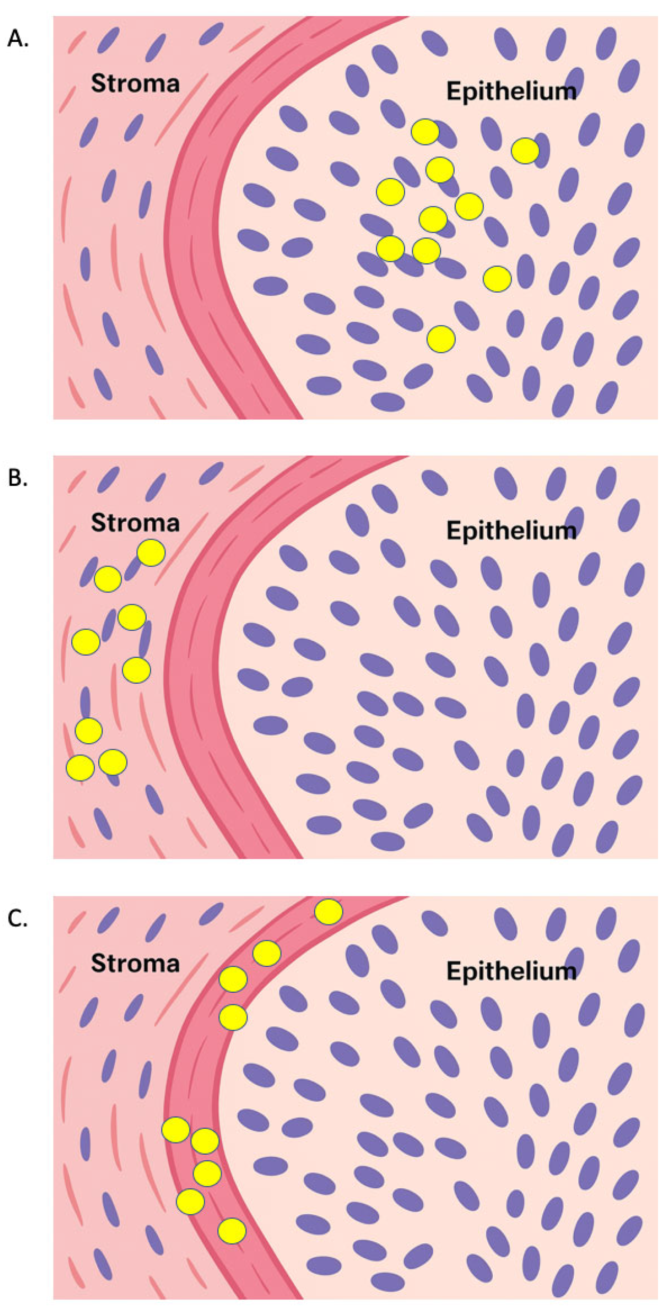
The schematic representation of these three compartments—epithelial, stromal, and interface—is shown in Figure 1, while Figure 2 illustrates their typical spatial organization and relative density in high-grade NMIBC.
In addition to isolated lymphocytes, some tumors may present lymphoid aggregates or actual tertiary lymphoid structures (TLS), considered sites of “trained” immune response [14]. In BUC, TIL reflect the host’s immune response and may offer prognostic information, but the evidence is heterogeneous: in ≥pT2 disease, greater TIL infiltration is associated with better survival [15], while in NMIBC/pT1 some studies reported a benefit of sTIL [10], whereas others failed to find such any prognostic value [8,11,16]. In MIBC, a recent integrated histological system combining morphology, sTIL, tumor budding, and growth patterns [17] improves risk stratification compared to the WHO criteria, and high levels of sTIL are associated with longer survival. According to the pan-cancer immunogenic analysis by Thorsson et al., the immune landscape places BUC predominantly in subtypes C1–C2 (pro-angiogenic/proliferative, with elevated Th2 and/or CD8) with less favorable outcomes than C3; however, within C1–C2, a more pronounced lymphocytic signature correlates with a better prognosis [18]. Overall, the prognostic value of TIL in BUC appears to depend on compartment (stromal vs. intratumoral), composition (e.g., CD8 fraction), and spatial architecture (e.g., epithelium–stroma gradients) rather than simple count, supporting the need for standardized metrics for valid clinical implementation. In NMIBC, TIL seems to reflect the biology of the tumor rather than providing a robust prognostic signal on its own: in high-grade pT1 tumors, increased stromal infiltration is associated with higher depth of invasion, but is not an independent predictor of survival (e.g., cancer-specific survival, CSS) [8,11]. In retrospective cohorts, increased TIL densities are related to greater thickness and more extensive invasion in TURBT chips, with no significant differences in recurrence or progression rates between TIL groups [16]. TIL tend to aggregate in the stroma of the papillary stalk and less commonly infiltrate the epithelium, often as individual cells rather that lymphoid aggregates, the median level of TIL in high-grade clinical T1 (cT1) being ~20% (range 5–60%) [8,19]. Some studies found no association with stage/grade, but reported correlations with tumor multiplicity [19,20]. Others described deeper CD3+/CD8+ stromal infiltration in T1 compared to Ta, and a lower proportion of intratumoral CD8+ in high-grade NMIBC compared to MIBC [21,22]. Overall, TIL density may indicate aggressiveness (e.g., associated with invasion), but prognostic value emerges mainly when considering composition and spatial metrics (e.g., epithelial-stromal CD8 gradients) rather than simple counts [11,14]. The prognostic role of TIL has been studied by several authors. High stromal TIL counts and increased expression of PD-L1 mRNA have been associated with better outcomes in pT1 by some [10,23]; on the other hand, several cohorts do not confirm a clear effect. In particular, in the pT1 series by Yaprak Bayrak et al. [16], the apparent ‘median PFS 0 months’ does not indicate immediate progression, but a distribution with few early events and many patients censored without progression; this data, together with the correction of the minimum time to recurrence in the >5% TIL group (4.5 months), highlights the limitations of median summaries when event rates are low and suggests functional heterogeneity of TIL. Supporting this heterogeneity, CD8+ and CD103+ (resident memory T) subgroups show favorable signals [24]; while Treg (regulatory T cell) FOXP3+ enrichment and checkpoint activation may reduce the protective effect, semi-quantitative assessment of sTIL might not detect transcriptional differences or may be limited by statistical power [7]. Consistently, in pT1 high-grade (HG)-NMIBC, stromal infiltrate correlates with depth of invasion but not with survival independently [8], while spatial and compositional metrics of the TME provide additional information, especially in the post-BCG setting [25]. Clinically, the relationship between TIL and prognosis should be interpreted in light of evasion mechanisms and the growing use of immunotherapy in high-risk NMIBC that does not respond to BCG [3,26]. In summary, in NMIBC, TILs are particularly significant when typed and mapped (e.g., CD8/CD103, epithelial-stromal gradients) and when integrated with morphological variables of aggressiveness (millimetric depth of invasion, tumor budding) for more robust stratification [27,28] (Table 1).
Beyond their prognostic relevance, TILs may also carry predictive value in determining the response to intravesical immunotherapy with Bacillus Calmette–Guérin (BCG), as discussed in the following section.

3. TIL as a Predictive Tool in BCG Treatment

When evaluating the prognostic value of TIL in relation to response to BCG, composition, functional relationships, and spatial metrics should be taken into account rather than simple density: pre-BCG studies that examined TAM, CD4+, CD8+, and Treg report results that are not always consistent [11,29,31,32,33]. In multiple cohorts, elevated CD68+ TAMs predict worse outcomes after BCG [30,31,32,34,35,36], while on the T-cell side, low Treg, high CD4+, and a higher CD4+/CD8+ ratio have been associated with better outcomes [29], although the absolute amount of CD8+ alone is not consistently predictive. Conversely, the CD8+ density gradient across the epithelium-stroma interface (and not density tout court) correlates with post-BCG recurrence-free survival (RFS) [11]. A converging line of evidence documents mechanisms of adaptive immune resistance induced or unmasked by BCG: in non-responders, pre-treatment co-localization of PD-L1 is observed in areas with high CD8+ density and low CD4+, and an increase in PD-L1 after BCG [37,38,39]. A recent comprehensive review has further emphasized that PD-L1 expression in NMIBC is frequently associated with BCG resistance and unfavorable outcomes, although results remain heterogeneous across different studies [40]. Beyond PD-L1, additional mechanisms of adaptive immune resistance have been described, including the BCG-induced IFN-γ response that can upregulate HLA-E and activate the natural killer group 2A/programmed cell death protein 1 (NKG2A/PD-1) axis, thereby promoting CD8+ exhaustion [41,42,43], while single-cell/spatial analyses imply a contribution of NK cells to IFN-γ in recurrent tumors [44] and a post-BCG expansion of NK CD56^high CD16+ and T γδ cells [45,46]. Consistently, a transcriptomic taxonomy defined three BRS (BCG response subtypes) with differential responses to BCG: BRS1 immuno-effector (most favorable), BRS2 intermediate, and BRS3 with high CD8+ activity but signs of exhaustion and poor clinical benefit [47]. Finally, a meta-analysis indicates that elevated CD4+, elevated GATA-3+, and a higher GATA-3+/T-bet+ ratio predict better response to BCG, while elevated Treg are associated with worse RFS/PFS [30,48,49,50] supporting a model in which the effector/regulator balance (including the T-cell/MDSC axis [51] and the spatial geography of the infiltrate provide the most informative signal for prognosis and predictivity of TILs in BCG-treated NMIBC.

4. Tumor-Associated Macrophages (TAM)

Macrophages display remarkable plasticity, polarizing toward either pro-inflammatory M1 or immunosuppressive M2 phenotypes [52,53,54]. In NMIBC, a predominance of M2-polarized TAMs correlates with poor BCG response and early recurrence, whereas M1-associated markers indicate an activated immune milieu [29,30,34,55]. Their dualistic nature positions TAMs as both potential biomarkers and therapeutic targets in modulating BCG efficacy [25,32,56,57]. Tumor-associated macrophages (TAMs) are major constituents of the bladder cancer immune microenvironment and play a pivotal role in shaping tumor behavior and therapeutic response. Although not lymphocytes, their functional interplay with T cells, B cells, and dendritic cells (DCs) makes them integral components of the tumor-infiltrating immune compartment in NMIBC.
TAMs display remarkable plasticity, with a polarization spectrum ranging from classically activated M1 macrophages (cytotoxic, pro-inflammatory, anti-tumor) to alternatively activated M2 macrophages (immunosuppressive, pro-angiogenic, and pro-invasive) [58]. The balance between these phenotypes profoundly influences tumor evolution and BCG responsiveness. In NMIBC, several studies have reported that a predominance of M2/CD163+ TAMs correlates with higher recurrence and progression rates, while enrichment in M1-related markers (e.g., iNOS, CD80) is linked to improved outcomes [29,30,32,34,36,55,59].
In patients receiving intravesical Bacillus Calmette–Guérin (BCG), TAM accumulation—particularly of the M2 subtype—has been repeatedly associated with unfavorable response and shorter recurrence-free survival [30,34,55]. For instance, Takayama et al. [34] showed that increased TAM infiltration predicted poor prognosis in carcinoma in situ after BCG, while Pichler et al. [29] identified a combined signature of high TAMs and regulatory T cells (Tregs) as markers of immune tolerance and BCG failure. Spatial analyses have confirmed that M2 polarization predominates within the lamina propria, supporting an immunosuppressive microenvironment [29,52]. Conversely, macrophage-rich interfaces characterized by M1-like activity and high CD11c density are linked to better post-BCG control [25,56].
Functional crosstalk between macrophages and lymphocytes further modulates NMIBC progression. TAMs can foster Treg recruitment and inhibit cytotoxic CD8+ responses through IL-6 and COX-2/PGE2 pathways [29,30,55,60], while Th1-polarized immunity and a high GATA-3+/T-bet+ ratio have been associated with improved BCG efficacy [48,50]. These observations emphasize that macrophage–lymphocyte interactions, rather than isolated cell counts, underlie the prognostic and predictive relevance of the immune microenvironment.
Standardized assessment of TAM subsets (CD68, CD163, CD206) and integration with digital spatial metrics could improve reproducibility and enable their inclusion in composite immunoscore models. Therapeutic reprogramming of TAMs toward an M1 phenotype—alone or in combination with BCG or checkpoint inhibitors—represents a promising avenue for enhancing local immune control in NMIBC.
Future work should aim to standardize TAM assessment by integrating immunohistochemical, digital, and transcriptomic markers to distinguish M1 and M2 phenotypes reproducibly. Exploring therapeutic macrophage reprogramming—either through cytokine modulation or targeted drugs—could enhance the efficacy of intravesical BCG and checkpoint inhibitors. Large multicenter studies are warranted to confirm the independent predictive value of TAM polarization in NMIBC.

5. T Cells

T lymphocytes represent the core effectors of adaptive antitumor immunity. Cytotoxic CD8+ cells mediate direct tumor cell killing through perforin–granzyme and Fas/FasL pathways, whereas CD4+ helper cells regulate both cytotoxic and B-cell responses. Conversely, regulatory T cells (Tregs) exert immunosuppressive effects by inhibiting effector function and promoting tolerance [61,62,63]. The balance among these subsets critically shapes the immune response to intravesical BCG and underlies the predictive significance of T-cell infiltration in NMIBC [29,30,48,50].
A decisive role is attributed to T lymphocytes, fundamental components of the immune system, which actively participate in both the onset and progression of neoplasms. The literature indicates that T lymphocytes are closely linked to bladder tumorigenesis, making them a prime target for therapeutic strategies, particularly immunomodulatory approaches [58,62]. Within T cells, CD4+ cells play a crucial orchestrating role, modulating the activity of other immune populations and supporting the activation of cytotoxic CD8+ cells, thereby directly impacting tumor growth control [62]. In the clinical setting, a higher density of CD4+ cells in the TME has been associated with worse overall survival in patients with NMIBC; although there is no statistically significant correlation with overall RFS, 10-year RFS is approximately 1.7 times shorter in patients with CD4+ infiltrate above the median [10]. However, other studies have not confirmed a strong association between CD4+ density and risk of recurrence, suggesting that factors such as location (stromal vs. epithelial), intratumoral heterogeneity, and the composite nature of the CD4+ compartment (which includes both pro-effector helper subsets and immunosuppressive regulatory T cells) may attenuate the prognostic signal in terms of RFS [61]. Overall, the global CD4+ count appears to be an incomplete indicator, and more refined approaches based on phenotyping and spatial analysis (distinction of subsets and their interactions) are needed to clarify the true prognostic value of CD4+ in NMIBC [13]. Within T cells, regulatory T cells (Tregs)—mostly FoxP3+—constitute the main immune brake: they maintain homeostasis and self-tolerance by suppressing the proliferation and differentiation of effector T cells and also modulating NK, B, macrophage, and DCs [63]. In NMIBC, the intratumoral proportion of Tregs correlates with adverse clinical-pathological variables (stage/high grade) and is associated with worse outcomes in several cohorts, including a series treated with BCG [30], while in another series, the median RFS was 20 months in patients with high percentages of FoxP3+ versus 113 months in cases with lower infiltrates [64]. Sexual dimorphism was also observed, with a higher number of FoxP3+ Tregs in low-grade tumors in women compared to men [61]. Despite this, the link between Tregs and outcome remains heterogeneous: some integrative analyses suggest a potential predictive value for recurrence/progression/OS [65], while others do not confirm independent associations with recurrence [33], highlighting the impact of compartmentalization, intratumoral heterogeneity, and quantification methods. In summary, in the context of NMIBC T cells, Tregs emerge as central nodes of TME immunosuppression and potential biomarkers/targets in BCG settings, but require standardized assessments and prospective studies for clinical validation [66,67]. Within T cells, CD8+ cells represent the main cytotoxic arm, but in NMIBC their prognostic value is context-dependent. In selected cohorts, defined stromal phenotypes (e.g., CD90+ stroma with high CD8a infiltration) are associated with 5-year survival >80% [68], and greater CD3+/CD8+ infiltration correlates with longer disease-free survival in NMIBC patients [20,52], while the CD3+/CD8+ ratio in the TME is prognostic for DFS in HR-NMIBC cases eligible for BCG [69]. Furthermore, transcriptomic signatures indicate that higher proportions of T CD8+ (together with activated memory CD4/Tfh) characterize low-risk groups with better outcomes [65], and the peripheral proportion of CD8+ is higher in responders to BCG [70]. In contrast, in a pT1 series, higher levels of CD3+ (but not CD8+) are associated with favorable outcomes, while intra-tumoral infiltration of CD3+/CD8+ correlates with grade G3 and worse prognosis, suggesting that the presence of CD8+ may reflect invasiveness and/or functional exhaustion [10]. In HG-pT1, stromal infiltrate is associated with invasion depth without independent prognostic value [8]. Overall, count, compartment (stroma vs. epithelium), and composite metrics (ratios and signatures) are more informative than the mere number of CD8+ cells, outlining a prognostic role for CD8+ cells that emerges when integrated into the geography and quality of the TME. In the specific context of NMIBC, Drachneris’ digital analysis showed that the CD8+ gradient at the epithelium-stroma interface is an independent predictor of RFS after BCG, unlike absolute counts [11]. The same group confirmed the prognostic value of spatial indices calculated not only for CD8, but also for Inducible T cell Co-stimulator (ICOS) and DCs’ CD11c, indicating that the arrangement of immune cells at the tumor interface is a key marker of effective immune response [25].
Future studies should aim to integrate phenotypic, functional, and spatial analyses of T-cell subsets, combining CD4+, CD8+, and Treg markers into standardized immunoscore models. Prospective, BCG-stratified cohorts and multiplex digital pathology are needed to validate these parameters as reliable predictors of clinical outcome. The application of single-cell and spatial transcriptomics will further clarify the dynamic interplay between effector and regulatory populations in NMIBC.

6. NK Cells

Natural killer (NK) cells are innate cytotoxic lymphocytes capable of eliminating tumor cells independently of antigen presentation, primarily through perforin–granzyme release and NKG2D-mediated recognition of stress-induced ligands [71,72,73]. Their activation state and abundance within the tumor microenvironment correlate with the efficacy of BCG-induced immune stimulation, linking NK-mediated cytotoxicity to improved recurrence-free survival [74,75,76].
NK cells are key players in early immune surveillance: they recognize and kill tumor cells via death receptor ligands, perforin/granzyme release, cytokine production, and antibody-dependent cellular cytotoxicity (ADCC) [72]. Consistent with this “innate” role, an effective adaptive response to BCG appears to rely on a well-functioning NK base; mouse models show that the benefit of BCG disappears in the absence of NK cells, and in patients, higher pre-treatment expression of NK receptor ligands (NCR) is associated with a better response [6,71,73,77]. Recent clinical evidence indicates that higher CD56+ infiltration in the urothelium of NMIBC prior to BCG predicts better outcomes, likely also through mechanisms of “trained immunity,” although methodological limitations (retrospective design, small cohorts) and confounding factors such as smoking, a known inhibitor of NK activity, remain [74,75,76]. NK infiltration appears to be greater in tumors ≤ 3 cm and in low-risk groups, but not all series confirm an independent prognostic value, suggesting that the stage of disease and the immunosuppressive heterogeneity of the TME influence the observable impact [52,65,78]. Phenotypically CD3/CD56+, NK cells orchestrate early immunity and shape adaptive immunity; they share several functional receptors with CD8+ T cells but act more upstream, making them natural targets for combination strategies in BC (and other solid tumors) [79,80,81,82,83,84]. Overall, the quantity and competence of pre-BCG NK cells emerge as candidate biomarkers of BCG sensitivity and potential therapeutic levers in NMIBC.
Future investigations should better characterize NK-cell subsets and their activation status within the NMIBC microenvironment, including the role of inhibitory checkpoints such as NKG2A. Combining histological quantification with circulating NK profiling and functional assays may help identify patients most likely to benefit from immunotherapy. Integration of NK metrics into immune-based classifiers could refine risk stratification and therapeutic decision-making.

7. B Cells

B cells contribute to tumor immunity both as antibody producers and as antigen-presenting cells that modulate T-cell activation. They can also promote immunoregulation through cytokine secretion and formation of tertiary lymphoid structures (TLS) [61,85,86,87]. The density and organization of B-cell infiltrates have been variably associated with BCG responsiveness, suggesting that the humoral compartment plays a context-dependent role in shaping local immune control [19,78,87,88].
In NMIBC, the role of B lymphocytes is less defined and more contradictory than that of T lymphocytes: although they are increased in the tumor compared to the adjacent normal mucosa and more abundant in high grades (typical markers: CD19, CD20, CD79a) [61,85,86,87,89], their prognostic association is not clear. Multiple data from heterogeneous cohorts of NMIBC patients, either treated with BCG or not, suggest that higher B cell density is associated with shorter RFS, and that high CD20+ infiltration correlates with worse disease-specific survival (DSS), with a tendency toward lower OS/DFS [14,25,87]. At the mechanistic level, intratumoral B cells can promote metastasis via the interleukin-8/androgene receptor/matrix metalloproteinase (IL-8/AR/MMP) axis [89]. However, other cohorts do not confirm significant relationships with recurrence (e.g., CD20+/CD68+ in solitary low-grade papillary tumors) or show effects only in univariate analysis for CD79a+ stromal infiltrate [61,78]. The contribution of B-cells is also modulated by local/systemic immunoregulatory factors, such as the cyclooxygenase 2/prostaglandin E2 (COX-2/PGE2) axis, which can shift the balance towards more immunosuppressive profiles [60]. Rather than providing a static or univocal prognostic signal, B cells likely exert context-dependent effects that mirror the complexity of the tumor microenvironment. Current research efforts are increasingly focused on the spatial and functional characterization of B–T cell interactions using multiplex immunohistochemistry, spatial transcriptomics, and digital image analysis. Future investigations integrating B-cell phenotyping and transcriptomic profiling, aiming to distinguish protective from tolerogenic profiles, could clarify whether humoral immunity acts as a protective or permissive force in NMIBC, ultimately elucidating the humoral component of the immune response in order to provide novel prognostic and therapeutic biomarkers in NMIBC, thus guiding the development of novel immunotherapy biomarkers and rational combination strategies.

8. Tertiary Lymphoid Structures (TLS)

Tertiary lymphoid structures (TLS) are organized aggregates of B and T cells that arise in chronically inflamed tissues and function as ectopic immune hubs for antigen presentation and local lymphocyte activation [90,91,92]. In NMIBC, the presence of mature TLS often reflects an immunologically “hot” microenvironment, potentially enhancing responsiveness to BCG and other immunotherapies [25,35,93,94]. Their predictive role remains under investigation but appears to mirror the broader orchestration of adaptive immunity within the tumor.
Tertiary lymphoid structures (TLS) are ectopic lymphoid aggregates, similar to the germinal centers of secondary lymphoid organs, which emerge in conditions of chronic inflammation and persistent antigen exposure and act as micro-niches for B cell activation/maturation within the TME. In terms of prognosis, the data are heterogeneous: in the ovary, colon, breast, and lung, the presence of TLS/intratumoral B-cell infiltrates has been associated with better outcomes, while in melanoma, prostate, kidney, and hepatocellular carcinoma, TLS have been found to be unfavorable, suggesting that the “presence” and “quality” (maturity, composition, and organization) of TLS are more relevant than simple count [61,90,91,92]. In urothelial bladder carcinoma, TLS has been documented more frequently in MIBC than in NMIBC (≈75% vs. ≈25%); They show organized B CD20+, CD21+ follicular DC networks, and a T CD3+/CD8+ outline, and their maturity may reflect the severity of the stromal inflammatory response and disease aggressiveness, albeit with the limitations of small cohorts [93]. In immunotherapy, different TLS architectural patterns have been observed in non-responders to checkpoint inhibitors, while solid data on their significance in the context of intravesical BCG therapy are still lacking [35,94]. In a BCG-treated NMIBC cohort, ‘binary’ TLS detection was not associated with RFS in univariate analysis (plausibly due to intratumoral heterogeneity and sampling issues), but some multivariate analyses suggested a contribution in the broader context of the TME; Furthermore, B (CD20+) density alone was not predictive, while spatial metrics of other subsets (CD8, ICOS, CD11c) entered the models, indicating that the informative value of B cells emerges mainly when inscribed in the TLS microarchitecture [25]. Overall, in BUC, TLS should therefore be considered as dynamic structures whose maturity, composition (B/T/DC), and spatial geography—rather than mere presence—can modulate prognosis and response to treatment. This calls for standardized assessments (maturity scores, cellular signatures, and spatial metrics) in future studies and clinical validation [10,35,94]. In summary, TLS emerge as dynamic elements of the bladder tumor microenvironment, potentially capable of modulating therapeutic response and clinical outcome. Their more detailed characterization, through spatial and functional approaches, could contribute in the future to better patient stratification and the identification of new immunotherapy targets.
Further studies should address the maturation spectrum and cellular architecture of TLS in NMIBC, linking structural patterns to treatment response. Advanced imaging and spatial omics could clarify how TLS organization reflects immune competence and tumor aggressiveness. A harmonized definition of TLS metrics will be critical for their adoption as routine biomarkers in pathology reports.

9. Methods of Assessment and Reproducibility

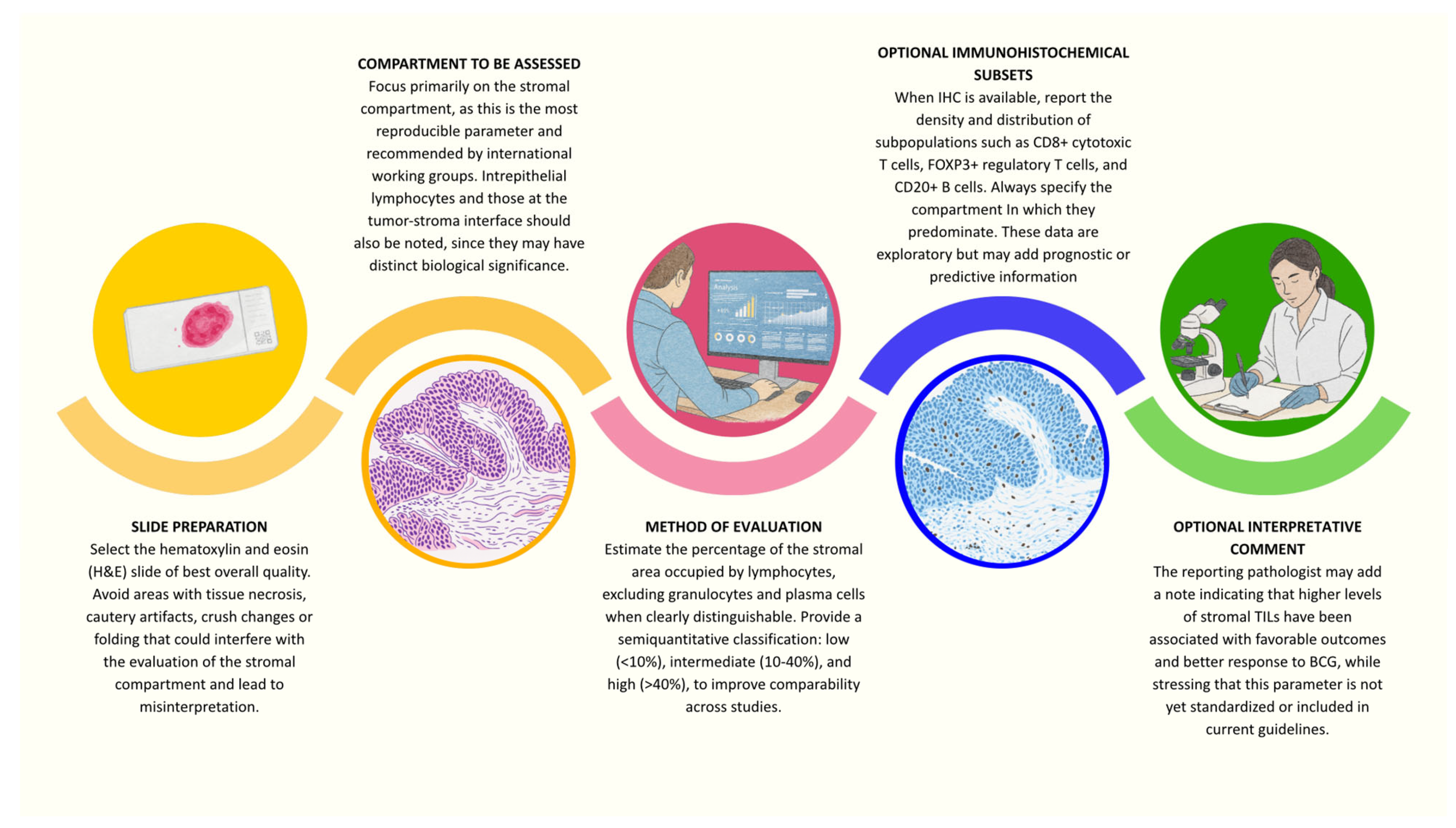
Current approaches for the evaluation of TILs in NMIBC are heterogeneous, ranging from traditional morphological estimates to advanced digital and molecular techniques. Standard hematoxylin–eosin evaluation remains the most widely used method because of its low cost and broad applicability. It is based on the estimation of the stromal lymphocyte percentage relative to the tumor area, following the recommendations of the International TILs Working Group (ITWG) [95], recommending the use of the percentage of stromal area occupied by lymphocytes as the main parameter. This approach has been applied in several multicenter studies [7,8,96], demonstrating its feasibility as a standardized tool. However, important limitations remain, including inter-observer variability, the need for arbitrary cut-offs, and difficulties in clearly distinguishing stromal versus intratumoral compartments in transurethral resection specimens. IHC allows for a more detailed characterization of TIL subsets (CD3, CD8, FOXP3, CD20) and enables compartment-specific analysis. Hülsen et al. [10] highlighted prognostic differences depending on stromal versus intratumoral location, while Bieri et al. [97] demonstrated the potential value of a modified Immunoscore in NMIBC. Moreover, Hassan et al. [98] reported a progressive increase in CD8+ cells along the NMIBC–MIBC continuum, underscoring the dynamic role of cytotoxic T cells in disease evolution. The introduction of digital pathology represents a qualitative leap, reducing subjectivity and enabling the quantitative analysis of complex spatial parameters. Paradigmatic examples are provided by the studies of Drachneris et al. [11,25], showing that spatial metrics—such as CD8+ density gradients across the epithelial–stromal interface or “interface density ratios” evaluating large-scale cell–cell interactions—outperform simple absolute counts in predicting prognosis and BCG response. Novel non-conventional strategies are also emerging. Chen et al. [99] developed a radiomic model capable of indirectly estimating TIL status from CT imaging, which correlated with both prognosis and BCG responsiveness, suggesting the feasibility of non-invasive immune stratification. At the molecular level, Damrauer et al. [100] identified transcriptomic signatures predictive of clinical outcomes, which may serve as the basis for genomic/transcriptional biomarker panels. Overall, there is a clear evolution from subjective morphological H&E-based estimates to quantitative, multiparametric, and digital assessments, complemented by radiomic and transcriptomic approaches. The main challenges remain inter-observer reproducibility, the establishment of standardized cut-offs, and the need for multicenter prospective validation before clinical implementation. In Figure 3, we provide a flowchart in the form of an infographic, which can be used for the evaluation of TIL in NMIBC in clinical practice.

10. Conclusions and Future Perspectives

Overall analysis of the available evidence shows that TIL in NMIBC represent a biologically plausible and clinically promising biomarker, but one that is not yet ready for routine inclusion in histopathological reports. Discrepancies between studies stem from methodological differences, the absence of shared criteria, and the limited number of prospective cohorts with adequate follow-up. The most robust data come from structured approaches (digital immunoscore) and spatial analyses, which seem to better capture the complexity of the tumor microenvironment than simple cell counts. With regard to T lymphocytes, the evidence supports their central role in both prognostic and predictive terms for response to BCG, especially when the assessment considers spatial distribution and functional signatures. In contrast, the contribution of B lymphocytes remains uncertain: rather than their absolute density, it may be useful to evaluate the presence and maturity of tertiary lymphoid structures and B–T interactions. Future prospects concern three main areas: (i) the standardization of H&E evaluation methods, possibly with the support of digital pathology and artificial intelligence; (ii) the development of integrated models combining TIL with other tissue and molecular biomarkers (e.g., transcriptomic subtypes, FGFR3 alterations, PD-L1), as already suggested in urothelial carcinoma in situ where PD-L1 and Ki-67 have been proposed as predictors of BCG response [101]; (iii) prospective validation in large multicenter cohorts, with the aim of incorporating these parameters into risk stratification systems and guiding personalized therapeutic decisions.
Beyond the identification and quantification of tumor-infiltrating lymphocytes, an emerging frontier in urothelial oncology is the therapeutic use of TILs themselves. Early-phase adoptive cell therapy (ACT) trials are exploring the feasibility of isolating, expanding, and reinfusing autologous TILs in bladder cancer, following the encouraging outcomes observed in other solid tumors, such as melanoma [102,103]. Preliminary data from UC suggest that tumors with a high CD8+ infiltrate and an inflamed immune phenotype may provide a suitable microenvironment for TIL expansion and reinfusion [104]. In this context, combination strategies using TIL-based ACT together with immune checkpoint inhibitors (e.g., anti-PD-1/PD-L1 or anti–NKG2A antibodies) or cytokine support (IL-2, IL-15) are under investigation to enhance lymphocyte persistence and cytotoxicity [105,106]. Although still experimental, these approaches may in the near future complement intravesical BCG or systemic immunotherapy, promoting a transition from immune monitoring to immune intervention in NMIBC.
In conclusion, TIL represent a rapidly evolving field of research in NMIBC: their clinical potential is high, but only through a coordinated, multicenter, and standardized approach will it be possible to translate this knowledge into tools that are truly useful for clinical practice.

Author Contributions

Conceptualization, R.M. and F.S.; methodology, R.M. and F.S.; validation, R.M. and F.S.; investigation, R.M., A.C. and F.S.; data curation, R.M., A.C. and F.S.; writing—original draft preparation, R.M., A.C. and F.S.; writing—review and editing, R.M. and F.S.; visualization, R.M. M.Z. (Magda Zanelli), M.Z. (Maurizio Zizzo), A.P., A.B.G. and F.S.; All authors have read and agreed to the published version of the manuscript.

Funding

This research received no external funding.

Institutional Review Board Statement

Not applicable.

Informed Consent Statement

Not applicable.

Conflicts of Interest

The authors declare no conflicts of interest.

Abbreviations

BCGBacillus Calmette–Guérin
CSSCancer-specific survival
HGHigh grade
H&EHematoxylin and eosin
NMIBCNon-muscle-invasive bladder cancer
OSOverall survival
RFSRecurrence-free survival
TAMTumor-associated macrophages
TILTumor-infiltrating lymphocytes

References

  1. IARC. Global Cancer Observatory: Cancer Today [Internet]. 2025. Available online: https://gco.iarc.fr/today/en/dataviz/bars?mode=cancer&group_populations=1&key=total (accessed on 31 August 2025).
  2. Smani, S.; DuBois, J.; Zhao, K.; Sutherland, R.; Rahman, S.N.; Humphrey, P.; Hesse, D.; Tan, W.S.; Martin, D.; Lokeshwar, S.D.; et al. Advancements in the Diagnosis, Treatment, and Risk Stratification of Non-Muscle Invasive Bladder Cancer. Curr. Oncol. Rep. 2025, 27, 236–246. [Google Scholar] [CrossRef]
  3. Holzbeierlein, J.M.; Bixler, B.R.; Buckley, D.I.; Chang, S.S.; Holmes, R.; James, A.C.; Kirkby, E.; McKiernan, J.M.; Schuckman, A.K. Diagnosis and Treatment of Non-Muscle Invasive Bladder Cancer: AUA/SUO Guideline: 2024 Amendment. J. Urol. 2024, 211, 533–538. [Google Scholar] [CrossRef]
  4. Cambier, S.; Sylvester, R.J.; Collette, L.; Gontero, P.; Brausi, M.A.; van Andel, G.; Kirkels, W.J.; Da Silva, F.C.; Oosterlinck, W.; Prescott, S.; et al. EORTC Nomograms and Risk Groups for Predicting Recurrence, Progression, and Disease-specific and Overall Survival in Non-Muscle-invasive Stage Ta-T1 Urothelial Bladder Cancer Patients Treated with 1–3 Years of Maintenance Bacillus Calmette-Guérin. Eur. Urol. 2016, 69, 60–69. [Google Scholar] [CrossRef]
  5. Babjuk, M.; Burger, M.; Capoun, O.; Cohen, D.; Compérat, E.M.; Dominguez Escrig, J.L.; Gontero, P.; Liedberg, F.; Masson-Lecomte, A.; Mostafid, A.H.; et al. European Association of Urology Guidelines on Non-muscle-invasive Bladder Cancer (Ta, T1, and Carcinoma in Situ). Eur. Urol. 2022, 81, 75–94. [Google Scholar] [CrossRef] [PubMed]
  6. Pettenati, C.; Ingersoll, M.A. Mechanisms of BCG immunotherapy and its outlook for bladder cancer. Nat. Rev. Urol. 2018, 15, 615–625. [Google Scholar] [CrossRef] [PubMed]
  7. Haas, M.; Engelmann, S.U.; Mayr, R.; Gossler, C.; Pickl, C.; Kälble, S.; Yang, Y.; Otto, W.; Hartmann, V.; Burger, M.; et al. A novel grading approach predicts worse outcomes in stage pT1 non-muscle-invasive bladder cancer. BJU Int. 2024, 134, 249–257. [Google Scholar] [CrossRef] [PubMed]
  8. Rouanne, M.; Betari, R.; Radulescu, C.; Goubar, A.; Signolle, N.; Neuzillet, Y.; Allory, Y.; Marabelle, A.; Adam, J.; Lebret, T. Stromal lymphocyte infiltration is associated with tumour invasion depth but is not prognostic in high-grade T1 bladder cancer. Eur. J. Cancer 2019, 108, 111–119. [Google Scholar] [CrossRef]
  9. Sanguedolce, F.; Falagario, U.G.; Zanelli, M.; Palicelli, A.; Zizzo, M.; Ascani, S.; Tortorella, S.; Mancini, V.; Cormio, A.; Carrieri, G.; et al. Clinicopathological Features and Survival Analysis in Molecular Subtypes of Muscle-Invasive Bladder Cancer. Int. J. Mol. Sci. 2023, 24, 6610. [Google Scholar] [CrossRef]
  10. Hülsen, S.; Lippolis, E.; Ferrazzi, F.; Otto, W.; Distel, L.; Fietkau, R.; Denzinger, S.; Breyer, J.; Burger, M.; Bertz, S.; et al. High Stroma T-Cell Infiltration is Associated with Better Survival in Stage pT1 Bladder Cancer. Int. J. Mol. Sci. 2020, 21, 8407. [Google Scholar] [CrossRef]
  11. Drachneris, J.; Rasmusson, A.; Morkunas, M.; Fabijonavicius, M.; Cekauskas, A.; Jankevicius, F.; Laurinavicius, A. CD8+ Cell Density Gradient across the Tumor Epithelium-Stromal Interface of Non-Muscle Invasive Papillary Urothelial Carcinoma Predicts Recurrence-Free Survival after BCG Immunotherapy. Cancers 2023, 15, 1205. [Google Scholar] [CrossRef]
  12. Lee, S.M. Prognosis in high-grade T1 bladder cancer: Host immune response and tumour infiltrating lymphocytes. Transl. Androl. Urol. 2019, 8, S335–S336. [Google Scholar] [CrossRef]
  13. De Carlo, C.; Valeri, M.; Corbitt, D.N.; Cieri, M.; Colombo, P. Non-muscle invasive bladder cancer biomarkers beyond morphology. Front. Oncol. 2022, 12, 947446. [Google Scholar] [CrossRef] [PubMed]
  14. Semeniuk-Wojtaś, A.; Modzelewska, M.; Poddębniak-Strama, K.; Kołaczyńska, S.; Lubas, A.; Górnicka, B.; Jakieła, A.; Stec, R. CD4, CD20 and PD-L1 as Markers of Recurrence in Non-Muscle-Invasive Bladder Cancer. Cancers 2023, 15, 5529. [Google Scholar] [CrossRef] [PubMed]
  15. Ledderose, S.; Rodler, S.; Eismann, L.; Ledderose, G.; Ledderose, C. Tumor-infiltrating lymphocytes predict survival in ≥ pT2 urothelial bladder cancer. Pathol. Res. Pract. 2022, 237, 154037. [Google Scholar] [CrossRef] [PubMed]
  16. Yaprak Bayrak, B.; Dogan, H.N.; Teke, K.; Yuvak, H.; Dillioglugil, O. The role of tumor-infiltrating lymphocytes and depth of invasion in T1 bladder cancer: A retrospective analysis. BMC Urol. 2025, 25, 135. [Google Scholar] [CrossRef]
  17. Eckstein, M.; Matek, C.; Wagner, P.; Erber, R.; Büttner-Herold, M.; Wild, P.J.; Taubert, H.; Wach, S.; Sikic, D.; Wullich, B.; et al. Proposal for a Novel Histological Scoring System as a Potential Grading Approach for Muscle-invasive Urothelial Bladder Cancer Correlating with Disease Aggressiveness and Patient Outcomes. Eur. Urol. Oncol. 2024, 7, 128–138. [Google Scholar] [CrossRef]
  18. Thorsson, V.; Gibbs, D.L.; Brown, S.D.; Wolf, D.; Bortone, D.S.; Ou Yang, T.H.; Chuah, E. The Immune Landscape of Cancer. Immunity 2018, 48, 812–830.e14. [Google Scholar] [CrossRef]
  19. Krpina, K.; Markić, D.; Rahelić, D.; Ahel, J.; Rubinić, N.; Španjol, J. 10-year survival of a patient with metastatic prostate cancer: Case report and literature review. Arch. Ital. Urol. Androl. 2015, 87, 252–253. [Google Scholar] [CrossRef]
  20. Liu, K.; Zhao, K.; Wang, L.; Sun, E. The prognostic values of tumor-infiltrating neutrophils, lymphocytes and neutrophil/lymphocyte rates in bladder urothelial cancer. Pathol. Res. Pract. 2018, 214, 1074–1080. [Google Scholar] [CrossRef]
  21. Roumiguié, M.; Kamat, A.M.; Bivalacqua, T.J.; Lerner, S.P.; Kassouf, W.; Böhle, A.; Brausi, M.; Buckley, R.; Persad, R.; Colombel, M.; et al. International Bladder Cancer Group Consensus Statement on Clinical Trial Design for Patients with Bacillus Calmette-Guérin-exposed High-risk Non-muscle-invasive Bladder Cancer. Eur Urol. 2022, 82, 34–46. [Google Scholar] [CrossRef]
  22. Viveiros, M.M.; Barreto, M.C.; Seca, A.M.L. Laurus azorica: Valorization through Its Phytochemical Study and Biological Activities. Separations 2022, 9, 211. [Google Scholar] [CrossRef]
  23. Breyer, J.; Wirtz, R.M.; Otto, W.; Erben, P.; Worst, T.S.; Stoehr, R.; Eckstein, M.; Denzinger, S.; Burger, M.; Hartmann, A. High PDL1 mRNA expression predicts better survival of stage pT1 non-muscle-invasive bladder cancer (NMIBC) patients. Cancer Immunol. Immunother. 2018, 67, 403–412. [Google Scholar] [CrossRef]
  24. Wang, B.; Wu, S.; Zeng, H.; Liu, Z.; Dong, W.; He, W.; Chen, X.; Dong, X.; Zheng, L.; Lin, T.; et al. CD103+ Tumor Infiltrating Lymphocytes Predict a Favorable Prognosis in Urothelial Cell Carcinoma of the Bladder. J. Urol. 2015, 194, 556–562. [Google Scholar] [CrossRef]
  25. Drachneris, J.; Morkunas, M.; Fabijonavicius, M.; Cekauskas, A.; Jankevicius, F.; Laurinavicius, A. Spatial Distribution of Macrophage and Lymphocyte Subtypes within Tumor Microenvironment to Predict Recurrence of Non-Muscle-Invasive Papillary Urothelial Carcinoma after BCG Immunotherapy. Int. J. Mol. Sci. 2024, 25, 4776. [Google Scholar] [CrossRef] [PubMed]
  26. Necchi, A.; Roumiguié, M.; Kamat, A.M.; Shore, N.D.; Boormans, J.L.; Esen, A.A.; Lebret, T.; Kandori, S.; Bajorin, D.F.; Krieger, L.E.M.; et al. Pembrolizumab monotherapy for high-risk non-muscle-invasive bladder cancer without carcinoma in situ and unresponsive to BCG (KEYNOTE-057): A single-arm, multicentre, phase 2 trial. Lancet Oncol. 2024, 25, 720–730. [Google Scholar] [CrossRef] [PubMed]
  27. Brimo, F.; Wu, C.; Zeizafoun, N.; Tanguay, S.; Aprikian, A.; Mansure, J.J.; Kassouf, W. Prognostic factors in T1 bladder urothelial carcinoma: The value of recording millimetric depth of invasion, diameter of invasive carcinoma, and muscularis mucosa invasion. Hum. Pathol. 2013, 44, 95–102. [Google Scholar] [CrossRef] [PubMed]
  28. Eckstein, M.; Kimmel, C.; Bruendl, J.; Weber, F.; Denzinger, S.; Gierth, M.; Burger, M.; Hartmann, A.; Otto, W.; Breyer, J. Tumor budding correlates with tumor invasiveness and predicts worse survival in pT1 non-muscle-invasive bladder cancer. Sci. Rep. 2021, 11, 17981. [Google Scholar] [CrossRef]
  29. Pichler, R.; Fritz, J.; Zavadil, C.; Schäfer, G.; Culig, Z.; Brunner, A. Tumor-infiltrating immune cell subpopulations influence the oncologic outcome after intravesical Bacillus Calmette-Guérin therapy in bladder cancer. Oncotarget 2016, 7, 39916–39930. [Google Scholar] [CrossRef]
  30. Miyake, M.; Tatsumi, Y.; Gotoh, D.; Ohnishi, S.; Owari, T.; Iida, K.; Ohnishi, K.; Hori, S.; Morizawa, Y.; Itami, Y.; et al. Regulatory T Cells and Tumor-Associated Macrophages in the Tumor Microenvironment in Non-Muscle Invasive Bladder Cancer Treated with Intravesical Bacille Calmette-Guérin: A Long-Term Follow-Up Study of a Japanese Cohort. Int. J. Mol. Sci. 2017, 18, 2186. [Google Scholar] [CrossRef]
  31. Ajili, F.; Kourda, N.; Darouiche, A.; Chebil, M.; Boubaker, S. Prognostic Value of Tumor-associated Macrophages Count in Human Non-muscle-invasive Bladder Cancer Treated by BCG Immunotherapy. Ultrastruct. Pathol. 2013, 37, 56–61. [Google Scholar] [CrossRef]
  32. Ayari, C.; LaRue, H.; Hovington, H.; Decobert, M.; Harel, F.; Bergeron, A.; Têtu, B.; Lacombe, L.; Fradet, Y. Bladder tumor infiltrating mature dendritic cells and macrophages as predictors of response to Bacillus Calmette-Guérin immunotherapy. Eur. Urol. 2009, 55, 1386–1395. [Google Scholar] [CrossRef]
  33. Eich, M.L.; Chaux, A.; Guner, G.; Taheri, D.; Mendoza Rodriguez, M.A.; Rodriguez Peña, M.D.C.; Baras, A.S.; Hahn, N.M.; Drake, C.; Sharma, R.; et al. Tumor immune microenvironment in non-muscle-invasive urothelial carcinoma of the bladder. Hum. Pathol. 2019, 89, 24–32. [Google Scholar] [CrossRef]
  34. Takayama, H.; Nishimura, K.; Tsujimura, A.; Nakai, Y.; Nakayama, M.; Aozasa, K.; Okuyama, A.; Nonomura, N. Increased infiltration of tumor associated macrophages is associated with poor prognosis of bladder carcinoma in situ after intravesical bacillus Calmette-Guerin instillation. J. Urol. 2009, 181, 1894–1900. [Google Scholar] [CrossRef] [PubMed]
  35. Vaghjiani, R.G.; Skitzki, J.J. Tertiary Lymphoid Structures as Mediators of Immunotherapy Response. Cancers 2022, 14, 3748. [Google Scholar] [CrossRef] [PubMed]
  36. Wang, L.-A.; Yang, B.; Tang, T.; Yang, Y.; Zhang, D.; Xiao, H.; Xu, J.; Wang, L.; Lin, L.; Jiang, J. Correlation of APE1 with VEGFA and CD163(+) macrophage infiltration in bladder cancer and their prognostic significance. Oncol. Lett. 2020, 20, 2881–2887. [Google Scholar] [CrossRef] [PubMed]
  37. Hashizume, A.; Umemoto, S.; Yokose, T.; Nakamura, Y.; Yoshihara, M.; Shoji, K.; Wada, S.; Miyagi, Y.; Kishida, T.; Sasada, T. Enhanced expression of PD-L1 in non-muscle-invasive bladder cancer after treatment with Bacillus Calmette-Guerin. Oncotarget 2018, 9, 34066–34078. [Google Scholar] [CrossRef]
  38. Kates, M.; Matoso, A.; Choi, W.; Baras, A.S.; Daniels, M.J.; Lombardo, K.; Brant, A.; Mikkilineni, N.; McConkey, D.J.; Kamat, A.M.; et al. Adaptive Immune Resistance to Intravesical BCG in Non-Muscle Invasive Bladder Cancer: Implications for Prospective BCG-Unresponsive Trials. Clin. Cancer Res. 2020, 26, 882–891. [Google Scholar] [CrossRef]
  39. Wang, J.; Li, J.; Cao, N.; Li, Z.; Han, J.; Li, L. Resveratrol, an activator of SIRT1, induces protective autophagy in non-small-cell lung cancer via inhibiting Akt/mTOR and activating p38-MAPK. OncoTargets Ther. 2018, 11, 7777–7786. [Google Scholar] [CrossRef]
  40. Sanguedolce, F.; Falagario, U.G.; Zanelli, M.; Palicelli, A.; Zizzo, M.; Busetto, G.M.; Cormio, A.; Carrieri, G.; Cormio, L. Integrating the PD-L1 Prognostic Biomarker in Non-Muscle Invasive Bladder Cancer in Clinical Practice—A Comprehensive Review on State-of-the-Art Advances and Critical Issues. J. Clin. Med. 2024, 13, 2182. [Google Scholar] [CrossRef]
  41. Cheng, X.; Zhao, Z.; Ventura, E.; Gran, B.; Shindler, K.S.; Rostami, A. The PD-1/PD-L pathway is up-regulated during IL-12-induced suppression of EAE mediated by IFN-gamma. J. Neuroimmunol. 2007, 185, 75–86. [Google Scholar] [CrossRef]
  42. Luo, Y.; Chen, X.; O’Donnell, M.A. Role of Th1 and Th2 cytokines in BCG-induced IFN-γ production: Cytokine promotion and simulation of BCG effect. Cytokine 2003, 21, 17–26. [Google Scholar] [CrossRef]
  43. Salomé, B.; Sfakianos, J.P.; Ranti, D.; Daza, J.; Bieber, C.; Charap, A.; Hammer, C.; Banchereau, R.; Farkas, A.M.; Ruan, D.F.; et al. NKG2A and HLA-E define an alternative immune checkpoint axis in bladder cancer. Cancer Cell 2022, 40, 1027–1043.e9. [Google Scholar] [CrossRef]
  44. Larsson, L.; Frisén, J.; Lundeberg, J. Spatially resolved transcriptomics adds a new dimension to genomics. Nat. Methods 2021, 18, 15–18. [Google Scholar] [CrossRef]
  45. Esteso, G.; Felgueres, M.J.; García-Jiménez, Á.F.; Reyburn-Valés, C.; Benguría, A.; Vázquez, E.; Reyburn, H.T.; Aguiló, N.; Martín, C.; Puentes, E.; et al. BCG-activation of leukocytes is sufficient for the generation of donor-independent innate anti-tumor NK and γδ T-cells that can be further expanded in vitro. Oncoimmunology 2023, 12, 2160094. [Google Scholar] [CrossRef]
  46. García-Cuesta, E.M.; Esteso, G.; Ashiru, O.; López-Cobo, S.; Álvarez-Maestro, M.; Linares, A.; Ho, M.M.; Martínez-Piñeiro, L.; Reyburn, H.T.; Valés-Gómez, M. Characterization of a human anti-tumoral NK cell population expanded after BCG treatment of leukocytes. Oncoimmunology 2017, 6, e1293212. [Google Scholar] [CrossRef] [PubMed]
  47. de Jong, F.C.; Laajala, T.D.; Hoedemaeker, R.F.; Jordan, K.R.; van der Made, A.C.J.; Boevé, E.R.; van der Schoot, D.K.E.; Nieuwkamer, B.; Janssen, E.A.M.; Mahmoudi, T.; et al. Non-muscle-invasive bladder cancer molecular subtypes predict differential response to intravesical Bacillus Calmette-Guérin. Sci. Transl. Med. 2023, 15, eabn4118. [Google Scholar] [CrossRef] [PubMed]
  48. Kardoust Parizi, M.; Shariat, S.F.; Margulis, V.; Mori, K.; Lotan, Y. Value of tumour-infiltrating immune cells in predicting response to intravesical BCG in patients with non-muscle-invasive bladder cancer: A systematic review and meta-analysis. BJU Int. 2021, 127, 617–625. [Google Scholar] [CrossRef] [PubMed]
  49. Martínez, R.; Tapia, G.; De Muga, S.; Hernández, A.; Cao, M.G.; Teixidó, C.; Urrea, V.; García, E.; Pedreño-López, S.; Ibarz, L.; et al. Combined assessment of peritumoral Th1/Th2 polarization and peripheral immunity as a new biomarker in the prediction of BCG response in patients with high-risk NMIBC. OncoImmunology 2019, 8, 1602460. [Google Scholar] [CrossRef]
  50. Nunez-Nateras, R.; Castle, E.P.; Protheroe, C.A.; Stanton, M.L.; Ocal, T.I.; Ferrigni, E.N.; Ochkur, S.I.; Jacobsen, E.A.; Hou, Y.-X.; Andrews, P.E.; et al. Predicting response to Bacillus Calmette-Guérin (BCG) in patients with carcinoma in situ of the bladder. Urol. Oncol. 2014, 32, 45.e23–45.e30. [Google Scholar] [CrossRef]
  51. Chevalier, M.F.; Trabanelli, S.; Racle, J.; Salomé, B.; Cesson, V.; Gharbi, D.; Bohner, P.; Domingos-Pereira, S.; Dartiguenave, F.; Fritschi, A.-S.; et al. ILC2-modulated T cell-to-MDSC balance is associated with bladder cancer recurrence. J. Clin. Investig. 2017, 127, 2916–2929. [Google Scholar] [CrossRef]
  52. Krpina, K.; Babarović, E.; Španjol, J.; Đorđević, G.; Maurer, T.; Jonjić, N. Correlation of tumor-associated macrophages and NK cells with bladder cancer size and T stage in patients with solitary low-grade urothelial carcinoma. Wien. Klin. Wochenschr. 2016, 128, 248–252. [Google Scholar] [CrossRef] [PubMed]
  53. Qian, B.Z.; Pollard, J.W. Macrophage diversity enhances tumor progression and metastasis. Cell 2010, 141, 39–51. [Google Scholar] [CrossRef] [PubMed]
  54. van der Horst, G.; Bos, L.; van der Pluijm, G. Epithelial plasticity, cancer stem cells, and the tumor-supportive stroma in bladder carcinoma. Mol. Cancer Res. 2012, 10, 995–1009. [Google Scholar] [CrossRef] [PubMed]
  55. Lima, L.; Oliveira, D.; Tavares, A.; Amaro, T.; Cruz, R.; Oliveira, M.J.; Ferreira, J.A.; Santos, L. The predominance of M2-polarized macrophages in the stroma of low-hypoxic bladder tumors is associated with BCG immunotherapy failure. Urol. Oncol. 2014, 32, 449–457. [Google Scholar] [CrossRef]
  56. Boström, M.M.; Irjala, H.; Mirtti, T.; Taimen, P.; Kauko, T.; Ålgars, A.; Jalkanen, S.; Boström, P.J. Tumor-Associated Macrophages Provide Significant Prognostic Information in Urothelial Bladder Cancer. PLoS ONE 2015, 10, e0133552. [Google Scholar] [CrossRef]
  57. Hori, S.; Miyake, M.; Tatsumi, Y.; Onishi, S.; Morizawa, Y.; Nakai, Y.; Tanaka, N.; Fujimoto, K. Topical and systemic immunoreaction triggered by intravesical chemotherapy in an N-butyl-N-(4-hydroxybutyl) nitorosamine induced bladder cancer mouse model. PLoS ONE 2017, 12, e0175494. [Google Scholar] [CrossRef]
  58. Pane, K.; Mirabelli, P.; Coppola, L.; Illiano, E.; Salvatore, M.; Franzese, M. New Roadmaps for Non-muscle-invasive Bladder Cancer with Unfavorable Prognosis. Front. Chem. 2020, 8, 600. [Google Scholar] [CrossRef]
  59. Hanada, T.; Nakagawa, M.; Emoto, A.; Nomura, T.; Nasu, N.; Nomura, Y. Prognostic value of tumor-associated macrophage count in human bladder cancer. Int. J. Urol. 2000, 7, 263–269. [Google Scholar] [CrossRef]
  60. Ibrahim, O.M.; Kalinski, P. Breaking Barriers: Modulation of Tumor Microenvironment to Enhance Bacillus Calmette-Guérin Immunotherapy of Bladder Cancer. Cells 2024, 13, 699. [Google Scholar] [CrossRef]
  61. Chenard, S.; Jackson, C.; Vidotto, T.; Chen, L.; Hardy, C.; Jamaspishvilli, T.; Berman, D.; Siemens, D.R.; Koti, M. Sexual Dimorphism in Outcomes of Non-muscle-invasive Bladder Cancer: A Role of CD163+ Macrophages, B cells, and PD-L1 Immune Checkpoint. Eur. Urol. Open Sci. 2021, 29, 50–58. [Google Scholar] [CrossRef]
  62. Rudensky, A.Y. Regulatory T cells and Foxp3. Immunol. Rev. 2011, 241, 260–268. [Google Scholar] [CrossRef]
  63. Sakaguchi, S.; Yamaguchi, T.; Nomura, T.; Ono, M. Regulatory T cells and immune tolerance. Cell 2008, 133, 775–787. [Google Scholar] [CrossRef]
  64. Murai, R.; Itoh, Y.; Kageyama, S.; Nakayama, M.; Ishigaki, H.; Teramoto, K.; Narita, M.; Yoshida, T.; Tomita, K.; Kobayashi, K.-I.; et al. Prediction of intravesical recurrence of non-muscle-invasive bladder cancer by evaluation of intratumoral Foxp3+ T cells in the primary transurethral resection of bladder tumor specimens. PLoS ONE 2018, 13, e0204745. [Google Scholar] [CrossRef] [PubMed]
  65. He, Y.; Wu, Y.; Liu, Z.; Li, B.; Jiang, N.; Xu, P.; Xu, A. Identification of Signature Genes Associated with Invasiveness and the Construction of a Prognostic Model That Predicts the Overall Survival of Bladder Cancer. Front. Genet. 2021, 12, 694777. [Google Scholar] [CrossRef] [PubMed]
  66. de Visser, K.E.; Joyce, J.A. The evolving tumor microenvironment: From cancer initiation to metastatic outgrowth. Cancer Cell 2023, 41, 374–403. [Google Scholar] [CrossRef] [PubMed]
  67. Skok, K.; Bräutigam, K. Tumor infiltrating lymphocytes (TILs)—Pathologia, quo vadis?—A global survey. Pathol. Res. Pract. 2025, 266, 155775. [Google Scholar] [CrossRef]
  68. Mezheyeuski, A.; Segersten, U.; Leiss, L.W.; Malmström, P.-U.; Hatina, J.; Östman, A.; Strell, C. Fibroblasts in urothelial bladder cancer define stroma phenotypes that are associated with clinical outcome. Sci. Rep. 2020, 10, 281. [Google Scholar] [CrossRef]
  69. Roumiguié, M.; Compérat, E.; Chaltiel, L.; Nouhaud, F.X.; Verhoest, G.; Masson-Lecomte, A.; Colin, P.; Audenet, F.; Houédé, N.; Larré, S.; et al. PD-L1 expression and pattern of immune cells in pre-treatment specimens are associated with disease-free survival for HR-NMIBC undergoing BCG treatment. World J. Urol. 2021, 39, 4055–4065. [Google Scholar] [CrossRef]
  70. Audenet, F.; Farkas, A.M.; Anastos, H.; Galsky, M.D.; Bhardwaj, N.; Sfakianos, J.P. Immune phenotype of peripheral blood mononuclear cells in patients with high-risk non-muscle invasive bladder cancer. World J. Urol. 2018, 36, 1741–1748. [Google Scholar] [CrossRef]
  71. Brandau, S.; Riemensberger, J.; Jacobsen, M.; Kemp, D.; Zhao, W.; Zhao, X.; Jocham, D.; Ratliff, T.L.; Böhle, A. NK cells are essential for effective BCG immunotherapy. Int. J. Cancer 2001, 92, 697–702. [Google Scholar] [CrossRef]
  72. Li, L.; Li, A.; Jin, H.; Li, M.; Jia, Q. Inhibitory receptors and checkpoints on NK cells: Implications for cancer immunotherapy. Pathol. Res. Pract. 2024, 253, 155003. [Google Scholar] [CrossRef] [PubMed]
  73. Ran, G.H.; Lin, Y.Q.; Tian, L.; Zhang, T.; Yan, D.M.; Yu, J.H.; Deng, Y.C. Natural killer cell homing and trafficking in tissues and tumors: From biology to application. Signal Transduct. Target Ther. 2022, 7, 205. [Google Scholar] [CrossRef] [PubMed]
  74. Jung, Y.S.; Park, J.H.; Park, D.I.; Sohn, C.I.; Lee, J.M.; Kim, T.I. Impact of Smoking on Human Natural Killer Cell Activity: A Large Cohort Study. J. Cancer Prev. 2020, 25, 13–20. [Google Scholar] [CrossRef] [PubMed]
  75. Lima, M.Z.; Bastos, D.A.; Mattedi, R.L.; Dzik, C.; Jardim, D.L.; Coelho, R.; Ribeiro-Filho, L.A.; Cordeiro, M.D.; Nahas, W.C.; Mello, E.S.; et al. Infiltrating Natural Killer cells influence the efficacy of BCG immunotherapy in non-muscle-invasive bladder cancer. Pathol. Res. Pract. 2025, 270, 155997. [Google Scholar] [CrossRef]
  76. Lu, L.M.; Zavitz, C.C.; Chen, B.; Kianpour, S.; Wan, Y.; Stämpfli, M.R. Cigarette smoke impairs NK cell-dependent tumor immune surveillance. J. Immunol. 2007, 178, 936–943. [Google Scholar] [CrossRef]
  77. Yutkin, V.; Pode, D.; Pikarsky, E.; Mandelboim, O. The expression level of ligands for natural killer cell receptors predicts response to bacillus Calmette-Guerin therapy: A pilot study. J. Urol. 2007, 178, 2660–2664. [Google Scholar] [CrossRef]
  78. Krpina, K.; Babarović, E.; Jonjić, N. Correlation of tumor-infiltrating lymphocytes with bladder cancer recurrence in patients with solitary low-grade urothelial carcinoma. Virchows Arch. 2015, 467, 443–448. [Google Scholar] [CrossRef]
  79. Abel, A.M.; Yang, C.; Thakar, M.S.; Malarkannan, S. Natural Killer Cells: Development, Maturation, and Clinical Utilization. Front. Immunol. 2018, 9, 1869. [Google Scholar] [CrossRef]
  80. Bhardwaj, N.; Farkas, A.M.; Gul, Z.; Sfakianos, J.P. Harnessing Natural Killer Cell Function for Genitourinary Cancers. Urol. Clin. N. Am. 2020, 47, 433–442. [Google Scholar] [CrossRef]
  81. Habif, G.; Crinier, A.; André, P.; Vivier, E.; Narni-Mancinelli, E. Targeting natural killer cells in solid tumors. Cell. Mol. Immunol. 2019, 16, 415–422. [Google Scholar] [CrossRef]
  82. Huntington, N.D.; Cursons, J.; Rautela, J. The cancer-natural killer cell immunity cycle. Nat. Rev. Cancer 2020, 20, 437–454. [Google Scholar] [CrossRef]
  83. Paul, S.; Lal, G. The Molecular Mechanism of Natural Killer Cells Function and Its Importance in Cancer Immunotherapy. Front. Immunol. 2017, 8, 1124. [Google Scholar] [CrossRef]
  84. Ranti, D.; Bieber, C.; Wang, Y.S.; Sfakianos, J.P.; Horowitz, A. Natural killer cells: Unlocking new treatments for bladder cancer. Trends Cancer 2022, 8, 698–710. [Google Scholar] [CrossRef]
  85. Chu, P.G.; Arber, D.A. CD79: A review. Appl. Immunohistochem. Mol. Morphol. 2001, 9, 97–106. [Google Scholar] [CrossRef]
  86. Sanz, I.; Wei, C.; Jenks, S.A.; Cashman, K.S.; Tipton, C.; Woodruff, M.C.; Hom, J.; Lee, F.E.-H. Challenges and Opportunities for Consistent Classification of Human B Cell and Plasma Cell Populations. Front. Immunol. 2019, 10, 2458. [Google Scholar] [CrossRef] [PubMed]
  87. Semeniuk-Wojtaś, A.; Poddębniak-Strama, K.; Modzelewska, M.; Baryła, M.; Dziąg-Dudek, E.; Syryło, T.; Górnicka, B.; Jakieła, A.; Stec, R. Tumour microenvironment as a predictive factor for immunotherapy in non-muscle-invasive bladder cancer. Cancer Immunol. Immunother. 2023, 72, 1971–1989. [Google Scholar] [CrossRef] [PubMed]
  88. Viveiros, N.; Flores, B.C.; Lobo, J.; Martins-Lima, C.; Cantante, M.; Lopes, P.; Deantonio, C.; Palu, C.; Sainson, R.C.; Henrique, R.; et al. Detailed bladder cancer immunoprofiling reveals new clues for immunotherapeutic strategies. Clin. Transl. Immunol. 2022, 11, e1402. [Google Scholar] [CrossRef] [PubMed]
  89. Ou, Z.; Wang, Y.; Liu, L.; Li, L.; Yeh, S.; Qi, L.; Chang, C. Tumor microenvironment B cells increase bladder cancer metastasis via modulation of the IL-8/androgen receptor (AR)/MMPs signals. Oncotarget 2015, 6, 26065–26078. [Google Scholar] [CrossRef]
  90. Dieu-Nosjean, M.-C.; Antoine, M.; Danel, C.; Heudes, D.; Wislez, M.; Poulot, V.; Rabbe, N.; Laurans, L.; Tartour, E.; de Chaisemartin, L.; et al. Long-term survival for patients with non-small-cell lung cancer with intratumoral lymphoid structures. J. Clin. Oncol. 2008, 26, 4410–4417. [Google Scholar] [CrossRef]
  91. Finkin, S.; Yuan, D.; Stein, I.; Taniguchi, K.; Weber, A.; Unger, K.; Browning, J.L.; Goossens, N.; Nakagawa, S.; Gunasekaran, G.; et al. Ectopic lymphoid structures function as microniches for tumor progenitor cells in hepatocellular carcinoma. Nat. Immunol. 2015, 16, 1235–1244. [Google Scholar] [CrossRef]
  92. Quinn, D.I.; Shore, N.D.; Egawa, S.; Gerritsen, W.R.; Fizazi, K. Immunotherapy for castration-resistant prostate cancer: Progress and new paradigms. Urol. Oncol. 2015, 33, 245–260. [Google Scholar] [CrossRef] [PubMed]
  93. Koti, M.; Xu, A.S.; Ren, K.Y.M.; Visram, K.; Ren, R.; Berman, D.M.; Siemens, D.R. Tertiary Lymphoid Structures Associate with Tumour Stage in Urothelial Bladder Cancer. Bladder Cancer 2017, 3, 259–267. [Google Scholar] [CrossRef] [PubMed]
  94. Van Dijk, N.; Gil-Jimenez, A.; Silina, K.; van Montfoort, M.L.; Einerhand, S.; Jonkman, L.; Voskuilen, C.S.; Peters, D.; Sanders, J.; Lubeck, Y.; et al. The Tumor Immune Landscape and Architecture of Tertiary Lymphoid Structures in Urothelial Cancer. Front. Immunol. 2021, 12, 793964. [Google Scholar] [CrossRef] [PubMed]
  95. Hendry, S.M.; Salgado, R.; Gevaert, T.; Russell, P.A.M.; John, T.; Thapa, B.M.; Christie, M.M.; van de Vijver, K.; Estrada, M.; Gonzalez-Ericsson, P.I.; et al. Assessing Tumor-Infiltrating Lymphocytes in Solid Tumors: A Practical Review for Pathologists and Proposal for a Standardized Method from the International Immuno-Oncology Biomarkers Working Group: Part 2: TILs in Melanoma, Gastrointestinal Tract Carcinomas, Non-Small Cell Lung Carcinoma and Mesothelioma, Endometrial and Ovarian Carcinomas, Squamous Cell Carcinoma of the Head and Neck, Genitourinary Carcinomas, and Primary Brain Tumors. Adv. Anat. Pathol. 2017, 24, 311–335. [Google Scholar]
  96. Bayrak, C.S.; Stein, D.; Jain, A.; Chaudhary, K.; Nadkarni, G.N.; Van Vleck, T.T.; Puel, A.; Boisson-Dupuis, S.; Okada, S.; Stenson, P.D.; et al. Identification of discriminative gene-level and protein-level features associated with pathogenic gain-of-function and loss-of-function variants. Am. J. Hum. Genet. 2021, 108, 2301–2318. [Google Scholar] [CrossRef]
  97. Bieri, U.; Scharl, M.; Sigg, S.; Szczerba, B.M.; Morsy, Y.; Rüschoff, J.H.; Schraml, P.H.; Krauthammer, M.; Hefermehl, L.J.; Eberli, D.; et al. Prospective observational study of the role of the microbiome in BCG responsiveness prediction (SILENT-EMPIRE): A study protocol. BMJ Open 2022, 12, e061421. [Google Scholar] [CrossRef]
  98. Hassan, W.A.; ElBanna, A.K.; Noufal, N.; El-Assmy, M.; Lotfy, H.; Ali, R.I. Significance of tumor-associated neutrophils, lymphocytes, and neutrophil-to-lymphocyte ratio in non-invasive and invasive bladder urothelial carcinoma. J. Pathol. Transl. Med. 2023, 57, 88–94. [Google Scholar] [CrossRef]
  99. Chen, L.; Xu, L.; Zhang, X.; Zhang, J.; Bai, X.; Peng, Q.; Guo, E.; Lu, X.; Yu, S.; Jin, Z.; et al. Diagnostic value of dual-layer spectral detector CT parameters for differentiating high- from low-grade bladder cancer. Insights Imaging 2025, 16, 6. [Google Scholar] [CrossRef]
  100. Damrauer, J.S.; Roell, K.R.; Smith, M.A.; Sun, X.; Kirk, E.L.; Hoadley, K.A.; Benefield, H.C.; Iyer, G.; Solit, D.B.; Milowsky, M.I.; et al. Identification of a Novel Inflamed Tumor Microenvironment Signature as a Predictive Biomarker of Bacillus Calmette-Guérin Immunotherapy in Non-Muscle-Invasive Bladder Cancer. Clin. Cancer Res. 2021, 27, 4599–4609. [Google Scholar] [CrossRef]
  101. Sanguedolce, F.; Cormio, A.; Zanelli, M.; Zizzo, M.; Palicelli, A.; Falagario, U.G.; Milanese, G.; Galosi, A.B.; Mazzucchelli, R.; Cormio, L.; et al. Diagnostic, Prognostic, and Predictive Tissue Biomarkers in Urothelial Carcinoma In Situ: A Narrative Review. Diagnostics 2025, 15, 2163. [Google Scholar] [CrossRef]
  102. Rohaan, M.W.; Wilgenhof, S.; Haanen, J. Adoptive cellular therapies: The current landscape. Virchows Arch. 2019, 474, 449–461. [Google Scholar] [CrossRef]
  103. Rosenberg, S.A.; Restifo, N.P.; Yang, J.C.; Morgan, R.A.; Dudley, M.E. Adoptive cell transfer: A clinical path to effective cancer immunotherapy. Nat. Rev. Cancer 2008, 8, 299–308. [Google Scholar] [CrossRef]
  104. Piombino, C.; Tonni, E.; Oltrecolli, M.; Pirola, M.; Pipitone, S.; Baldessari, C.; Dominici, M.; Sabbatini, R.; Vitale, M.G. Immunotherapy in urothelial cancer: Current status and future directions. Expert Rev. Anticancer Ther. 2023, 23, 1141–1155. [Google Scholar] [CrossRef]
  105. Kim, J.W.; Tomita, Y.; Trepel, J.; Apolo, A.B. Emerging immunotherapies for bladder cancer. Curr. Opin. Oncol. 2015, 27, 191–200. [Google Scholar] [CrossRef]
  106. Tran, E.; Robbins, P.F.; Rosenberg, S.A. ‘Final common pathway’ of human cancer immunotherapy: Targeting random somatic mutations. Nat. Immunol. 2017, 18, 255–262. [Google Scholar] [CrossRef]
Figure 1. Representative histological image of a high-grade non-muscle-invasive bladder carcinoma (HG-NMIBC) illustrating the spatial distribution of tumor-infiltrating lymphocytes (TILs). The epithelial (yellow asterisk) and stromal (blue asterisk) compartments are separated by an orange line marking the epithelial–stromal interface, corresponding to the dynamic zone of immune–tumor interaction described in the text.
Applsci 15 12032 g001
Figure 2. Schematic representation of the spatial compartments of tumor-infiltrating lymphocytes (TILs; yellow dots) in BUC. (A) iTILs: lymphocytes within tumor epithelium; (B) sTILs: lymphocytes in peritumoral stroma; (C) Interface: immune cells confined to the tumor–stroma boundary zone.
Applsci 15 12032 g002
Figure 3. Minimal checklist for reporting TILs in NMIBC.
Applsci 15 12032 g003
Table 1. Summary of key studies evaluating tumor-infiltrating lymphocytes (TILs) in non-muscle-invasive bladder cancer (NMIBC).
StudyCohortTIL Subsets/MethodologySignificant AssociationPrognostic Endpoint(s)
Hülsen S et al., 2020 [10]167 pT1 NMIBCStromal vs. intraepithelial TIL; CD3+, CD8+High stromal CD3+ infiltration—favorable outcome (OS, RFS)OS, RFS
Rouanne M et al., 2019 [8] 147 pT1 HG NMIBCStromal TIL density (%)Higher stromal TIL density—tumor invasion depthCSS
Drachneris J et al., 2023 [11]157 papillary NMIBC treated with BCGSpatial CD8+ gradient at epithelium–stroma interface (digital analysis)CD8+ gradient—RFSRFS
Yaprak Bayrak B et al., 2025 [16]154 pT1 NMIBCSemi-quantitative H&E TIL scoringHigher TILs ratios—increased tumor thickness and more extensive invasionRFS, PFS
Pichler R et al., 2016 [29]40 NMIBC treated with BCGTAM, B-cell, T-cell densities at different compartmentsHigher CD4+ and GATA3+ count—prolonged RFS. Higher Tregs and TAMs—BCG failureRFS
Miyake M et al., 2017 [30]71 NMIBC treated with BCGPeritumoral FOXP3+ Tregs and CD68+ TAMHigh counts of Treg and TAMs—shorter RFSRFS
Drachneris J et al., 2024 [25]165 papillary NMIBC treated with BCGEpithelial-stromal Interface Density Ratios for CD8+, ICOS+, CD11c+, CD20+, CD163+CD11c IDR > CD8 and ICOS IDR—longer RFSRFS
Disclaimer/Publisher’s Note: The statements, opinions and data contained in all publications are solely those of the individual author(s) and contributor(s) and not of MDPI and/or the editor(s). MDPI and/or the editor(s) disclaim responsibility for any injury to people or property resulting from any ideas, methods, instructions or products referred to in the content.