Article

An In Vitro Study Comparing Debonding of Orthodontic Ceramic and Metal Brackets Using Er:YAG Laser and Conventional Pliers

by
Aous Abdulmajeed
1,
Tiannie Phan
1,
Kinga Grzech-Leśniak
2,3,* and
Janina Golob Deeb
2
1
Department of General Practice, School of Dentistry, Virginia Commonwealth University, Richmond, VA 23298, USA
2
Department of Periodontics, School of Dentistry, Virginia Commonwealth University, Richmond, VA 23298, USA
3
Integrated Dentistry Department, Faculty of Medicine and Dentistry, Wroclaw Medical University, 50-367 Wroclaw, Poland
*
Author to whom correspondence should be addressed.
Appl. Sci. 2025, 15(21), 11844; https://doi.org/10.3390/app152111844
Submission received: 6 October 2025 / Revised: 31 October 2025 / Accepted: 31 October 2025 / Published: 6 November 2025

Abstract

Removing orthodontic brackets often presents clinical challenges, as it may cause patient discomfort, bracket fracture, or enamel damage resulting from strong adhesive bonds. Various techniques have been proposed to facilitate safer and more efficient debonding. Among them, laser-assisted methods have gained attention for their potential to minimize mechanical stress and improve patient comfort. The main objective of this study was to evaluate the effect of an erbium-doped yttrium–aluminum–garnet (Er:YAG) laser as an alternative to traditional mechanical methods for removing metal and ceramic orthodontic brackets. Materials and Methods: Thirty-six extracted premolars were prepared for bonding metal or ceramic brackets using a light-cure adhesive system. The control group consisted of six ceramic and six metal brackets removed with conventional orthodontic pliers. In the experimental groups, brackets were debonded using the Er:YAG laser (2940 nm, 0.6 mm spot size, 150 mJ; 15 Hz; (2.25 W) with an H14 handpiece. Irradiation time was recorded for each method, and teeth were rescanned to measure the surface area and volume of the crowns before and after bracket removal. Data were analyzed using one-way ANOVA and Tukey’s HSD test (p < 0.05). Scanning electron microscopy (SEM) was used for surface analysis. Results: A significant difference in debonding time (p = 0.001) was observed between the laser and traditional methods. The laser group took 52.5 s for metal and 56.25 s for ceramic brackets, compared to 1.05 s (metal) and 0.64 s (ceramic) in the traditional group. A significant difference in remaining cement volume was noted (p = 0.0002), but no differences were found between metal and ceramic brackets with laser removal. Conclusions: Er:YAG laser-assisted debonding is safe and minimally invasive but more time-consuming and costly than conventional methods, showing no improvement in clinical efficiency under current parameters.

1. Introduction

Bracket removal in orthodontic practice presents significant clinical challenges, including patient discomfort, bracket fracture, and enamel damage due to strong adhesive bonds. Ceramic brackets, because of their high elastic modulus and brittleness, are particularly difficult to debond without causing enamel microcracks and fractures [1]. Traditional mechanical techniques, such as the use of specialized pliers, often leave adhesive remnants on the enamel surface and risk enamel loss during clean-up, making the process both time-consuming and technique-sensitive. These limitations have prompted the exploration of alternative approaches that can improve safety, efficiency, and patient comfort [1,2,3]. Various mechanical and thermal methods, such as ultrasonic, electrothermal and laser-assisted debonding, have been proposed to overcome these limitations [2,3,4,5]. Among them, laser-assisted techniques have gained considerable attention for their potential to reduce mechanical stress and thermal damage while facilitating easier bracket removal. Erbium-based lasers, particularly the Er:YAG laser (wavelength 2940 nm), are well-suited for this purpose due to their strong absorption in water and resin monomers, allowing selective ablation of bonding materials without overheating the pulp or damaging enamel [6,7,8,9,10,11,12,13,14]. Recent studies have further confirmed the clinical safety and efficacy of erbium lasers for debonding orthodontic and restorative materials, highlighting their versatility and minimal invasiveness [15,16,17,18,19,20,21]. This characteristic makes the Er:YAG laser a promising tool for minimally invasive debonding of ceramic and metal brackets, as it can soften or vaporize the adhesive layer at the bracket–enamel interface, thereby reducing debonding force [22,23,24].
Previous studies have compared different laser types, including CO2, Er:YAG and, Er,Cr:YSGG, and reported variations in efficiency depending on laser energy, fluence, pulse duration, and bracket material [22,23,24,25,26,27]. The effectiveness depends on factors like laser type, energy setting, bracket material, resin type, and applied force [23,24,25,26,27,28]. For instance, Mundethu et al. achieved rapid removal of polycrystalline ceramic brackets using a 600 mJ Er:YAG laser, while other reports demonstrated efficient debonding in less than 10 s using higher power settings [29,30]. However, many earlier investigations primarily relied on qualitative assessments such as the Adhesive Remnant Index (ARI) and lacked high-resolution, quantitative analyses of enamel surface and volume changes. Recent advances in three-dimensional digital scanning, image segmentation, and volumetric modeling have now enabled accurate quantification of residual resin and enamel loss following bracket removal [31,32]. Establishing a quantitative relationship between laser preparation parameters (e.g., energy, fluence, pulse duration, and cooling ratio) and enamel surface alterations is crucial for optimizing clinical outcomes. Such process–structure–property correlations are well established in materials science and advanced manufacturing research, where quantitative modeling and parameter optimization help bridge preparation technology and design development. Luo et al. (2023) demonstrated multivariate correlations between processing parameters, microstructure, and mechanical properties in AlSi0Mg alloys using additive manufacturing frameworks [32], while similar relationships have been applied in surface engineering to predict energy–response behavior during laser–material interactions [33,34].
Therefore, the present study aims to evaluate the efficiency and safety of Er:YAG laser-assisted debonding with conventional plier-based removal for both metal and ceramic orthodontic brackets. The study integrates three-dimensional volumetric analysis with scanning electron microscopy (SEM) to quantitatively and qualitatively assess coronal volume changes and enamel surface morphology. By establishing measurable links between laser parameters and post-debonding enamel conditions, this research contributes a quantitative foundation for future development of optimized, clinically applicable laser-assisted debonding protocols.

2. Materials and Methods

Thirty-six extracted human premolars were randomly assigned to three groups (n = 12). All teeth were scanned using an intraoral scanner (TRIOS 4, 3Shape TRIOS A/S, Copenhagen, Denmark) for three-dimensional (3D) surface mapping following the protocol described by Vlasa et al. (2021) [35], using Autodesk Meshmixer (RRID:SCR_015736).
The extracted teeth were cleaned of any residual tissue using a hand scaler and stored in 10% neutrl buffered formalin (Sigma-Aldrich, Merck KGaA, Darmstadt, Germany). Enamel surfaces were polished with medium-grit pumice paste (Kerr, Orange, CA, USA) for 15 s, then rinsed and dried with compressed air. Each sample was etched with 37% phosphoric acid gel (Ultra-Etch™, Ultradent Products Inc., South Jordan, UT, USA) for 20 s, rinsed for 5–10 s, and dried with compressed air until a frosty white appearance was visible. A sixth-generation self-etch adhesive system (Clearfil™ SE Bond II, Kuraray Noritake, Tokyo, Japan) was applied according to the manufacturer’s instructions. Gentle airflow was used before light curing for 10 s using Valo LED curing light (Valo Curing Light, Ultradent Products Inc., South Jordan, UT, USA). A small amount of light-cure adhesive (3M™ Transbond™ XT, 3M ESPE, St. Paul, MN, USA) was applied to the bracket base, and positioned on enamel surfaces at the height of contour by direct application with digital manipulation. Excess adhesive was manually removed with an instrument and light-cured for 40 s (10 sec per bracket side) according to the protocol of Bishara and Trulove (1990) [2].
Two experimental groups were included:
-
Group C (n = 12): polycrystalline ceramic brackets (Transcend Series 6000, 3M Unitek, Monrovia, CA, USA);
-
Group M (n = 12): twin orthodontic metal bracket (Mini Master Series, American Orthodontics, Sheboygan, WI, USA).
The control group consisted of ceramic (XC, n = 6) and metal (XM, n = 6) brackets, which were removed using angulated orthodontic pliers (Hu Friedy, Chicago, IL, USA) (Figure 1).

2.1. Laser-Assisted Debonding

In the experimental groups, brackets were debonded using an Er:YAG laser (LightWalker, Fotona d.d., Ljubljana, Slovenia) operating at 2940 nm with an H14 handpiece and a 0.6 mm conical tip, at parameters: 150 mJ; 15 Hz; 2.25 W with operation mode 100 μm (MSP), fluence 88 J/cm2 and power density 884 W/cm2, 3/3 air/water, with a distance of 5 mm using a scanning irradiation. These parameters were selected based on Grzech-Leśniak et al. (2018) [36] and Downarowicz et al. (2020) [26] to ensure thermal safety and effective adhesive softening (Figure 1).
The irradiation time required for bracket removal was recorded for each specimen and each method. The Shapiro–Wilk (S-W) test was used to assess data normality. Since data followed a normal distribution, one-way ANOVA followed by Tukey’s HSD post hoc test (p < 0.05), at the 95% confidence level was performed using GraphPad Prism 9.0 (GraphPad Software, San Diego, CA, USA). In this study, Autodesk Meshmixer software was used to calculate coronal volume differences between pre- and post-debonding scans. Although this approach provided reliable 3D quantitative data, future investigations should consider incorporating automated image-processing and segmentation algorithms to improve the accuracy of enamel and resin surface differentiation.

2.2. Sample-Size Calculation and Study Endpoints

A priori sample size calculation was performed using G*Power software version 3.1.9.7 (Heinrich-Heine-University, Düsseldorf, Germany) (Faul et al., 2007) [36]. Assuming a medium effect size (f = 0.5), significance criterion α = 0.05, and power (1−β) = 0.80, the minimum total sample size estimated for one-way ANOVA was N = 36, confirming the adequacy of the chosen sample size.
The primary endpoint of this study was the debonding time required for bracket removal using laser-assisted and conventional plier methods. Secondary endpoints included the residual composite volume on enamel determined by 3D volumetric analysis and qualitative surface alterations evaluated by scanning electron microscopy (SEM).

2.3. 3D Volumetric Analysis

Each tooth was scanned before bracket bonding and after bracket removal using the TRIOS 4® intraoral scanner (3Shape A/S, Copenhagen, Denmark). The obtained STL files were imported into Autodesk Meshmixer® software version 3.5 (Autodesk Inc., San Rafael, CA, USA) for 3D mesh analysis. Coronal volumes were calculated before and after debonding to determine residual composite material, following the methodology of Deeb et al. (2021) [10] (Figure 2).

2.4. Surface Analysis

Surface characteristics following bracket removal were assesses using scanning electron microscopy, SEM (JEOL JSM-6610LV, JEOL Ltd., Tokyo, Japan). Samples were examined under 500× and 2000× magnifications to evaluate structural integrity and enamel surface changes. The procedure followed Dostalova et al. (2016) [25] and Grzech-Leśniak et al. (2018) [37].

3. Results

3.1. Debonding Time

Table 1 summarizes the mean debonding times for each group, comparing laser-assisted and conventional removal methods. Statistical analysis revealed a significant difference (p = 0.001) in debonding time between laser-assisted and traditional removal groups. The mean debonding time for laser-assisted groups was 52.5 ± 13.57 s for metal brackets and 56.25 ± 9.32 s for ceramic brackets, compared to 0.76 ± 0.28 s for metal and 0.64 ± 0.25 s for ceramic brackets when removed using the traditional method. However, no statistically significant differences were found between ceramic and metal brackets within the same removal method (p > 0.05).

3.2. Residual Composite

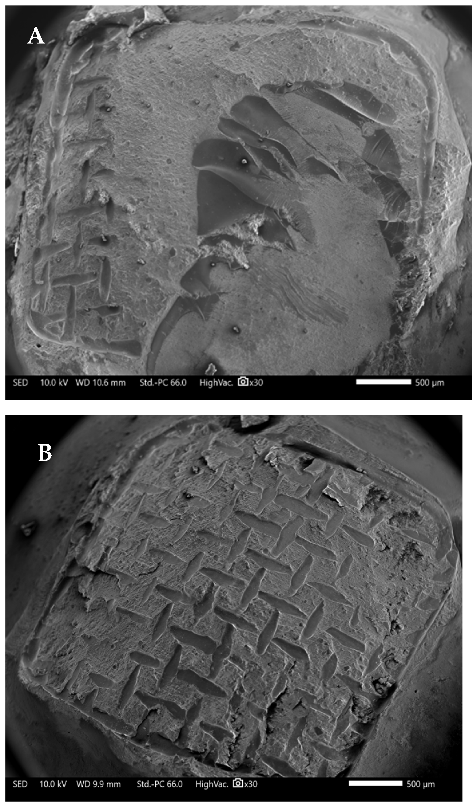
Statistical analysis revealed a significant difference (p = 0.0002) in tooth volume changes following bracket removal between the laser-assisted and traditional mechanical methods, regardless of bracket material. The mean volume change for laser-assisted groups was 1.99 ± 1.04 mm3 for metal brackets and 1.80 ± 1.29 mm3 for ceramic brackets, compared to 4.21 ± 1.22 mm3 when removed using the traditional method. No significant differences in volume were observed between ceramic and metal brackets within the laser-assisted groups (p > 0.05). The mean tooth volume changes after bracket removal, determined by 3D volumetric analysis, are presented in Table 2. The results of the volumetric analysis are visually summarized in Figure 3, which compares the representative 3D scans of tooth surfaces before and after bracket removal in the laser-assisted and mechanical groups. The images demonstrate reduced residual adhesive on enamel following laser-assisted debonding compared with the conventional technique, supporting the quantitative data presented in Table 2.

3.3. SEM Evaluation of Surface Morphology

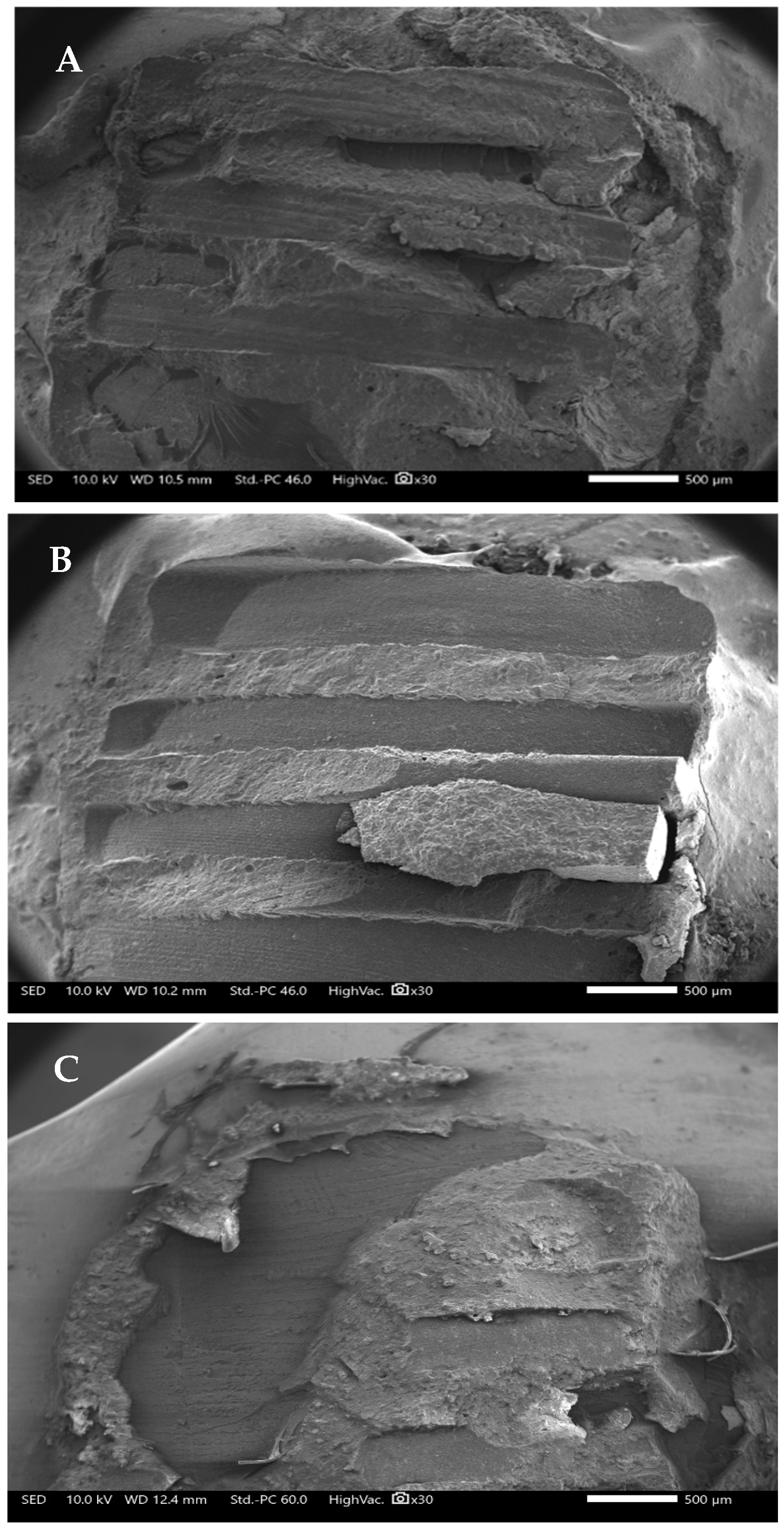
SEM analysis of the investigated groups also revealed that the laser-assisted debonding method resulted in less residual resin composite on the tooth surface compared with the traditional mechanical removal method (Figure 4). For metal brackets, laser irradiation was applied periphery (due to metal opacity), leading to localized composite ablation (p < 0.05 compared with control). In the ceramic bracket group laser irradiation penetrated through the bracket, producing more uniform adhesive softening (p > 0.05 compared with the metal laser group). SEM evaluation also confirmed no significant difference in surface morphology between ceramic and metal groups after laser use (p > 0.05), though both differed significantly from mechanically debonded samples (p < 0.01). Representative micrographs are presented in Figure 4, showing smoother enamel and reduced adhesive remnants after Er:YAG laser-assisted removal.

4. Discussion

Erbium lasers have been shown to be highly effective in retrieving fixed prosthetic ceramic appliances, achieving success rates exceeding 95% [13]. While Er:YAG lasers represent a viable method for orthodontic bracket removal, the present findings indicate that they do not provide a faster or more efficient alternative to conventional mechanical techniques, leading to the rejection of the null hypothesis. The laser-assisted method proved less time-efficient and posed a greater risk of enamel alteration, while its higher cost further limits economic feasibility.
However, it should be emphasized that the Er:YAG laser is not purchased exclusively for orthodontic applications. In clinical settings, this versatile device is routinely used for numerous procedures, including soft- and hard tissue surgery, peri-implant treatment, caries removal, and orthodontic corticotomy. Once available in a dental practice, the cost of performing laser-assisted bracket removal becomes comparable to that of conventional techniques, since no additional materials are required, only chairside time. Moreover, incorporating laser technology into orthodontic workflows may enhance patient perception and reflect a commitment to modern, high-technology care. Thus, the key consideration is not cost but the clinical justification and procedural efficiency, which motivated the present investigation.
Conventional orthodontic pliers allow rapid mechanical debonding; however, they leave significantly more adhesive residue on the enamel surface compared to laser-assisted removal. Volumetric analysis in this study confirmed that the bracket type did not significantly influence residual composite volume. Although laser-assisted debonding requires a longer procedure, it may ultimately reduce chairside time by minimizing the need for secondary adhesive removal. From a clinical standpoint, the Er:YAG laser represents an innovative and patient-friendly advancement in orthodontic debonding. Although laser-assisted bracket removal in this study required approximately 52–56 s per bracket compared with less than a second for mechanical pliers, this extra time can be justified by several advantages. The laser eliminates direct mechanical stress on enamel and brackets, greatly reducing the risk of cracks or fractures and improving patient comfort. It is a quiet, vibration-free, and minimally invasive method that many patients perceive as more pleasant and less anxiety-provoking than traditional plier removal. Furthermore, because the Er:YAG laser is already widely used for various dental procedures, its multifunctionality reinforces its clinical value. Despite the slightly longer procedure time, the advantages in safety, comfort, and tissue preservation highlight the laser’s potential as a modern, technology-driven modality in orthodontic care.
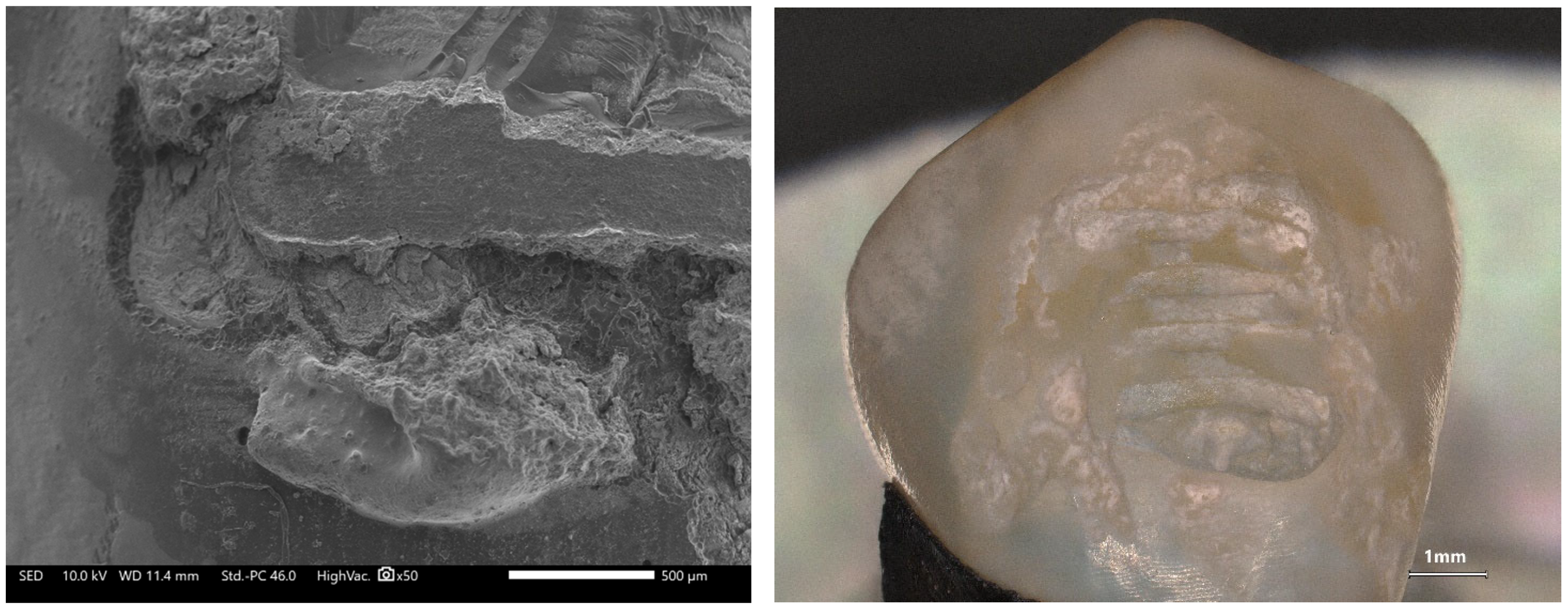
Laser irradiation likely contributes to composite ablation during bracket removal, facilitating detachment and reducing the need for post-debonding clean-up. Thermal softening of the bonding resin allows easier separation, and when heating is sufficiently rapid, thermal ablation may occur, vaporizing the adhesive before mechanical disruption [25,26,27,28,29,30,37]. Although SEM analysis (Figure 4) revealed less residual resin after laser-assisted debonding, subsequent micrographs (Figure 5) demonstrated localized enamel alterations such as microcracks and surface irregularities. This discrepancy reflects the trade-off between efficient adhesive ablation and the risk of thermal or mechanical enamel stress. Energy absorbed by the resin can cause rapid vaporization and interfacial expansion, promote bracket release but occasionally leading to superficial enamel disruption. Comparable findings were reported by Dostalova et al. (2016) [25] and Grzech-Leśniak et al. (2018) [37], who observed that lower laser power minimizes enamel alteration but leaves more residual resin, whereas higher power yields cleaner debonding at the cost of surface modification. Therefore, optimization of laser parameters remains crucial to balance resin-removal efficacy and enamel preservation.
Alternative approaches, such as the Adhesive Remnant Index (ARI), have traditionally been used to evaluate residual adhesive after debonding [37,38,39]. In the present study, however, a novel volumetric approach was employed to assess total crown volume analysis before and after bracket removal, providing a quantitative measure of residual resin. SEM evaluation further confirmed reduced resin on enamel surfaces following laser irradiation in both metal and ceramic bracket groups. These findings suggest that the prolonged irradiation time used in this study may have been excessive, likely due to the use of lower laser setting to minimize thermal effects on pulp and enamel. The laser parameters used in this experiment were lower than those used in a previous study [27,29,30], which utilized a similar power density to debond prefabricated zirconia crowns from primary anterior teeth using an Er,Cr:YSGG laser [40]. Although ceramic brackets provide greater strength, deformation resistance and bond strengths, they are more brittle and prone to breakage, which can increase enamel damage during debonding [41,42,43,44,45]. Other studies using longer irradiation times (140 s) for bracket removal with the Er:YAG laser, have reported less enamel damage and lower temperature increases compared to traditional removal method [25,26,37,43,45]. Our outcomes should also be contrasted with prior reports describing successful and safe laser-assisted debonding. Mundethu et al. (2014) [29] achieved rapid removal of polycrystalline ceramic brackets with Er:YAG, while Dostalova et al. (2016) [25] and Grzech-Leśniak et al. (2018) [37] reported minimal enamel alteration when parameters and cooling were optimized. Deeb et al. (2022) [13] demonstrated efficient erbium-laser retrieval of prefabricated ceramic crowns, and Downarowicz et al. (2020) [26] showed thermal effects within acceptable limits under controlled conditions. The discrepancies with our longer times and observed enamel changes likely reflect differences in: pulse energy and effective fluence at the interface, pulse duration (MSP vs. longer pulses) influencing peak power and micro-explosive effects, air/water cooling settings and tip-to-surface distance, which modulate heat dissipation, scan speed/irradiation time and energy delivery pattern, and bracket material (metal vs. ceramic) and adhesive composition affecting absorption and energy transfer. Our protocol (150 mJ, 15 Hz; moderate fluence with cautious cooling) prioritized thermal safety and may have required longer exposure to achieve adhesive softening, which in turn increased the risk of localized surface changes. Future work should systematically vary these parameters to identify a therapeutic window that maximizes resin removal while preserving enamel integrity.
SEM observations further revealed that the laser beam affected the resin surrounding the brackets, altering its morphology. Compared with mechanically debonded surfaces, laser-treated enamel exhibited more surface irregularities (Figure 5). These findings differ from reports describing no enamel cracks following expert-operated laser debonding [28,29,30,31,32,33,34,35,36,37,38,39,40,45,46].
Future research should include larger sample size and explore broader ranges of energy and frequency to refine selection and enhance clinical applicability. Previous investigations by Grzech-Leśniak et al. (2018) [37] and Downarowicz et al. (2020) [26] demonstrated that Er:YAG and Er,Cr:YSGG lasers can safely debond bracket without exceeding the critical pulpal temperature threshold (≤5.5 °C). Similarly, Deeb et al., (2021) [10], confirmed the safe and controlled use of erbium lasers for debonding prefabricated ceramic crowns from both natural teeth and implant abutments. Collectively, these findings reinforce the thermal safety of erbium lasers when appropriate clinical parameters are applied. In addition, future studies could benefit from using advanced image-processing and segmentation algorithms for more precise differentiation between enamel and residual adhesive surfaces. This would allow for higher reproducibility and objectivity in the assessment of post-debonding enamel alterations. While residual resin was assessed quantitatively using 3D volumetric analysis, the evaluation of enamel surface alterations on SEM micrographs was qualitative. The Adhesive Remnant Index (ARI), frequently used in previous research including our earlier study (Grzech-Leśniak et al., 2018) [37], represents a semi-quantitative visual scoring system. It was replaced here by volumetric evaluation to obtain numerical data. Future investigations should integrate ARI scoring with surface roughness or profilometric analyses to provide a more comprehensive assessment of post-debonding enamel alterations.

4.1. Limitation of the Study

The present study used specific bracket and adhesive systems, which may limit the generalizability of the findings. Although the sample size was determined by a priori power analysis and deemed statistically adequate, future studies using different manufacturers, materials, and bonding protocols are necessary to validate these results under varying clinical conditions. Another limitation was the lack of variation in laser parameters, as only a single energy and frequency setting was investigated. This design prevented a systematic evaluation of how different fluence levels, pulse durations, and cooling conditions might influence the efficiency of bracket removal and enamel surface integrity. Furthermore, because this was an in vitro study, the experimental conditions did not fully replicate intraoral factors such as temperature changes, saliva presence, and individual enamel variability. The SEM evaluation of enamel alterations was qualitative, without quantitative assessment of surface roughness or enamel loss. Although the laser parameters used were selected to ensure thermal safety, intrapulpal temperature changes were not monitored. Future research should address these limitations by incorporating larger sample sizes, testing a broader range of laser settings, and conducting in vivo studies to confirm the clinical applicability of the findings. Under the current parameters, the Er:YAG laser did not outperform conventional mechanical methods in terms of time efficiency; however, its favorable safety profile and potential for reducing enamel damage justify further optimization and clinical validation.

4.2. Recommendations for the Future Research

Future research should expand upon the current findings by exploring a broader range of laser parameters, including variations in energy output, frequency, pulse duration, and air/water cooling ratios, to identify optimal settings that maximize adhesive removal efficiency while minimizing enamel surface alterations. In vivo studies are needed to validate the clinical applicability of laser-assisted debonding, while considering patient comfort, procedural efficiency, and thermal safety. Quantitative surface analyses, such as profilometry, confocal microscopy, or 3D optical mapping, could complement SEM imaging to objectively assess enamel surface integrity. Future investigations should also include real-time intrapulpal temperature monitoring to ensure safe energy delivery. Moreover, comparative assessments across different bracket materials and adhesive systems would further clarify how optical properties influence laser absorption and debonding performance. Additionally, it should be considered that other variables, such as the type of adhesive system or pretreatment protocol, may significantly influence the bonding performance of orthodontic brackets. Previous studies have shown that the shear bond strength of universal adhesives depends on whether enamel is etched or left unetched (Beltrami et al., 2016) [46]. Similarly, deproteinizing agents such as hypochlorous acid have been reported to alter enamel surface properties and affect bonding efficiency (Polat & Çınar, 2024) [47]. These factors could partially explain the variability in residual adhesive and enamel surface morphology observed across different studies. Therefore, future research should systematically evaluate the impact of adhesive chemistry, pretreatment agents, and optical properties of bracket materials to optimize laser-assisted debonding protocols and improve clinical predictability. Finally, studies examining clinician ergonomics and patient-reported outcomes could provide valuable insights into the practical integration of Er:YAG laser-assisted debonding into routine orthodontic practice.

5. Conclusions

Within the limitations of this in vitro study, the Er:YAG laser demonstrated potential as a safe and minimally invasive alternative for orthodontic bracket debonding. Although laser-assisted removal was more time-consuming than conventional plier methods, it effectively reduced residual adhesive and minimized mechanical stress on enamel. Under the tested parameters, the laser did not improve clinical efficiency; however, optimization of energy settings, cooling conditions, and irradiation time may enhance its future feasibility. Continued development and refinement of laser protocols could support broader clinical implementation in orthodontics.

Author Contributions

Conceptualization, J.G.D. and K.G.-L.; Methodology, K.G.-L. and J.G.D.; Software, T.P.; Validation, J.G.D.; Formal analysis, K.G.-L.; Investigation, A.A., T.P. and J.G.D.; Resources, K.G.-L.; Data curation, T.P.; Writing—original draft, A.A., K.G.-L. and J.G.D.; Writing—review & editing, A.A., K.G.-L. and J.G.D.; Supervision, A.A. All authors have read and agreed to the published version of the manuscript.

Funding

This research received no external funding.

Institutional Review Board Statement

This study was considered exempt under the Code of Medical Ethics, Virginia Commonwealth University.

Informed Consent Statement

Not applicable.

Data Availability Statement

The original contributions presented in this study are included in the article. Further inquiries can be directed to the corresponding author.

Conflicts of Interest

The authors declare no conflict of interest.

References

  1. Azzeh, E.; Feldon, P.J. Laser debonding of ceramic brackets: A comprehensive review. Am. J. Orthod. Dentofac. Orthop. 2003, 123, 79–83. [Google Scholar] [CrossRef] [PubMed]
  2. Bishara, S.E.; Trulove, T.S. Comparisons of different debonding techniques for ceramic brackets: An in vitro study. Part I. Background and methods. Am. J. Orthod. Dentofac. Orthop. 1990, 98, 145–153. [Google Scholar] [CrossRef] [PubMed]
  3. Eliades, T.; Eliades, G.; Brantley, W.A. Orthodontic brackets. In Orthodontic Materials Scientific and Clinical Aspects; Brantley, W.A., Eliades, T., Eds.; Thieme: Stuttgart, Germany, 2001; pp. 143–172. [Google Scholar]
  4. Macri, R.T.; de Lima, F.A.; Bachmann, L.; Galo, R.; Romano, F.L.; Borsatto, M.C.; Matsumoto, M.A. CO2 laser as auxiliary in the debonding of ceramic brackets. Lasers Med. Sci. 2015, 30, 1835–1841. [Google Scholar] [CrossRef] [PubMed]
  5. Saito, A.; Namura, Y.; Isokawa, K.; Shimizu, N. CO2 laser debonding of a ceramic bracket bonded with orthodontic adhesive containing thermal expansion microcapsules. Lasers Med. Sci. 2015, 30, 869–874. [Google Scholar] [CrossRef]
  6. Tu, S.; Sun, C.; Zhao, N.; Xiong, Z. Safety and Efficacy of the Erbium Laser in Debonding Dental Accessories: A Narrative Review. Photobiomodulation Photomed. Laser Surg. 2024, 42, 327–338. [Google Scholar] [CrossRef]
  7. Grzech-Leśniak, K.; Bencharit, S.; Dalal, N.; Mroczka, K.; Deeb, J.G. In Vitro Examination of the Use of Er:YAG Laser to Retrieve Lithium Disilicate Crowns from Titanium Implant Abutments. J. Prosthodont. 2019, 28, 672–676. [Google Scholar] [CrossRef]
  8. Deeb, J.G.; Grzech-Leśniak, K.; Weaver, C.; Matys, J.; Bencharit, S. Retrieval of Glass Fiber Post Using Er:YAG Laser and Conventional Endodontic Ultrasonic Method: An In Vitro Study. J. Prosthodont. 2019, 28, 1024–1028. [Google Scholar] [CrossRef]
  9. Elkharashi, A.; Grzech-Leśniak, K.; Deeb, J.G.; Abdulmajeed, A.A.; Bencharit, S. Exploring the use of pulsed erbium lasers to retrieve a zirconia crown from a zirconia implant abutment. PLoS ONE 2020, 15, e0233536. [Google Scholar] [CrossRef]
  10. Deeb, J.G.; Skrjanc, L.; Kanduti, D.; Carrico, C.; Saturno, A.M.; Grzech-Leśniak, K. Evaluation of Er:YAG and Er,Cr:YSGG laser irradiation for the debonding of prefabricated zirconia crowns. Adv. Clin. Exp. Med. 2021, 30, 7–15. [Google Scholar] [CrossRef]
  11. Grzech-Leśniak, K.; Bencharit, S.; Skrjanc, L.; Kanduti, D.; Matys, J.; Deeb, J.G. Utilization of Er:YAG Laser in Retrieving and Reusing of Lithium Disilicate and Zirconia Monolithic Crowns in Natural Teeth: An In Vitro Study. Appl. Sci. 2020, 10, 4357. [Google Scholar] [CrossRef]
  12. Deeb, J.G.; McCall, C.; Carrico, C.K.; Dahlke, W.O.; Grzech-Leśniak, K. Retrieval of Prefabricated Zirconia Crowns with Er,Cr:YSGG Laser from Primary and Permanent Molars. Materials 2020, 13, 5569. [Google Scholar] [CrossRef]
  13. Deeb, J.G.; Grzech-Leśniak, K.; Brody, E.R.; Matys, J.; Bencharit, S. Erbium laser-assisted ceramic debonding: A scoping review. J. Prosthodont. 2022, 31, e100–e124. [Google Scholar] [CrossRef]
  14. Raafat, S.; Zohdy, M.; Eldemellawy, M.; Ghali, R.; Taha, D. Efficiency of dry versus wet Er,Cr:YSGG laser debonding of lithium disilicate veneers using different power outputs. J. Prosthet. Dent. 2025, 133, 193.e1–193.e8. [Google Scholar] [CrossRef]
  15. Khalil, A.S.; Tamish, N.M.; Elkalza, A.R. Assessment of chemical, ultrasonic, diode laser, and Er:YAG laser application on debonding of ceramic brackets. BMC Oral Health 2022, 22, 79. [Google Scholar] [CrossRef]
  16. Villalobos-Tinoco, J.; Andretti, F.; Conner, C.; Rojas-Rueda, S.; Fischer, N.G.; Pagan-Banchs, M.; Jurado, C.A. A Narrative Review and Clinical Study on Er:YAG Laser Debonding of Ceramic and Composite Veneers. Biomimetics 2025, 10, 295. [Google Scholar] [CrossRef]
  17. Florczak-Matyjek, A.; Nikodem, A.; Kensy, J.; Matys, J.; Grzech-Leśniak, K. The Efficacy of Erbium-Ion, Diode, and CO2 Lasers in Debonding Attachments Used During Overlay Orthodontic Treatment and the Risk of Hard Tooth Tissue Damage Compared to Traditional Methods—An In Vitro Study. Photonics 2025, 12, 621. [Google Scholar] [CrossRef]
  18. Grzech-Leśniak, K.; Matys, J. The Effect of Er:YAG Lasers on the Reduction of Aerosol Formation for Dental Workers. Materials 2021, 14, 2857. [Google Scholar] [CrossRef]
  19. Alves, L.V.G.L.; da Silva, M.B.F.; Borsatto, M.C.; Corona, S.A.M. Do erbium lasers promote changes in the tooth enamel during debonding of ceramic laminate veneers? A systematic review. Lasers Med. Sci. 2023, 38, 217. [Google Scholar] [CrossRef]
  20. Matys, J.; Grzech-Leśniak, K.; Flieger, R.; Dominiak, M. Assessment of an Impact of a Diode Laser Mode with Wavelength of 980 nm on a Temperature Rise Measured by Means of k-02 Thermocouple: Preliminary Results. Dent. Med. Probl. 2016, 53, 345–351. [Google Scholar] [CrossRef]
  21. Arnabat-Dominguez, J.; Vecchio, A.D.; Todea, C.; Grzech-Leśniak, K.; Vescovi, P.; Romeo, U.; Nammour, S. Laser dentistry in daily practice during the COVID-19 pandemic: Benefits, risks and recommendations for safe treatments. Adv. Clin. Exp. Med. 2021, 30, 119–125. [Google Scholar] [CrossRef]
  22. Ma, T.; Marangoni, R.D.; Flint, W. In vitro comparison of debonding force and intrapulpal temperature changes during ceramic orthodontic bracket removal using a carbon dioxide laser. Am. J. Orthod. Dentofac. Orthop. 1997, 111, 203–210. [Google Scholar] [CrossRef]
  23. Mimura, H.; Deguchi, T.; Obata, A.; Yamagishi, T.; Ito, M. Comparison of different bonding materials for laser debonding. Am. J. Orthod. Dentofac. Orthop. 1995, 108, 267–273. [Google Scholar] [CrossRef]
  24. Obata, A.; Tsumura, T.; Niwa, K.; Ashizawa, Y.; Deguchi, T.; Ito, M. Super pulse CO2 laser for bracket bonding and debonding. Eur. J. Orthod. 1999, 21, 193–198. [Google Scholar] [CrossRef]
  25. Dostalova, T.; Jelinkova, H.; Remes, M.; Šulc, J.; Němec, M. The Use of the Er:YAG Laser for Bracket Debonding and Its Effect on Enamel Damage. Photomed. Laser Surg. 2016, 34, 394–399. [Google Scholar] [CrossRef] [PubMed]
  26. Downarowicz, P.; Noszczyk, P.; Mikulewicz, M.; Nowak, R. Thermal effect of Er:YAG and Er,Cr:YSGG used for debonding ceramic and metal orthodontic brackets: An experimental analysis. Adv. Clin. Exp. Med. 2020, 29, 557–563. [Google Scholar] [CrossRef] [PubMed]
  27. Sabuncuoglu, F.A.; Ersahan, S.; Erturk, E. Debonding of ceramic brackets by Er:YAG laser. J. Istanb. Univ. Fac. Dent. 2016, 50, 24–30. [Google Scholar] [CrossRef] [PubMed]
  28. Crooks, M.; Hood, J.; Harkness, M. Thermal debonding of ceramic brackets: An in vitro study. Am. J. Orthod. Dentofac. Orthop. 1997, 111, 163–172. [Google Scholar] [CrossRef]
  29. Mundethu, A.R.; Gutknecht, N.; Franzen, R. Rapid debonding of polycrystalline ceramic orthodontic brackets with an Er:YAG laser: An in vitro study. Lasers Med. Sci. 2014, 29, 1551–1556. [Google Scholar] [CrossRef]
  30. Khalil, A.S.; Baowideen, F.A.; Alhujaili, A.S.; Alotaibi, N.F.; Almanjhi, W.A.; Yassin, H.Y.; Nasser, M.M.; Alzahrani, A.F.; Alrehaili, R.S. Efficacy of Lasers in Debonding Ceramic Brackets: Exploring the Rationale and Methods. Cureus 2024, 16, e61050. [Google Scholar] [CrossRef]
  31. Sohrabpoor, H.; Issa, A.; Al Hamaoy, A.; Ahad, I.U.; Chikarakara, E.; Bagga, K.; Brabazon, D. Chapter 24—Development of Laser Processing Technologies via Experimental Design. In Woodhead Publishing Series in Welding and Other Joining Technologies, Advances in Laser Materials Processing, 2nd ed.; Lawrence, J., Ed.; Woodhead Publishing: Cambridge, UK, 2018; pp. 707–729. ISBN 9780081012529. [Google Scholar] [CrossRef]
  32. Luo, Q.; Huang, N.; Fu, T.; Wang, J.; Bartles, D.L.; Simpson, T.W.; Beese, A.M. New insight into the multivariate relationships among process, structure, and properties in laser powder bed fusion AlSi10Mg. Addit. Manuf. 2023, 77, 103804. [Google Scholar] [CrossRef]
  33. Xian, G.; Qi, X.; Shi, J.; Tian, J.; Xiao, H. Toughened and self-healing carbon nanotube/epoxy resin composites modified with polycaprolactone filler for coatings, adhesives and FRP. J. Build. Eng. 2025, 111, 113207. [Google Scholar] [CrossRef]
  34. Zhang, S.-Q.; Dong, H.; Han, Y.; Xu, L.; Feng, Y.-K.; Li, P.-Y. Coupling Effect of Disconnected Pores and Grain Morphology on the Corrosion Tolerance of Laser-Clad 316L Coating. Coatings 2024, 14, 40. [Google Scholar] [CrossRef]
  35. Vlasa, A.; Bud, E.S.; Păcurar, M.; Lazăr, L.; Streiche, L.; Bucur, S.M.; Cocoș, D.I.; Bud, A. Effects of Composite Resin on the Enamel after Debonding: An In Vitro Study-Metal Brackets vs. Ceramic Brackets. Appl. Sci. 2021, 11, 7353. [Google Scholar] [CrossRef]
  36. Faul, F.; Erdfelder, E.; Lang, A.G.; Buchner, A. G*Power 3: A flexible statistical power analysis program for the social, behavioral, and biomedical sciences. Behav. Res. Methods 2007, 39, 175–191. [Google Scholar] [CrossRef]
  37. Grzech-Leśniak, K.; Matys, J.; Żmuda-Stawowiak, D.; Mroczka, K.; Dominiak, M.; Junior, A.B.; Gruber, R.; Romanos, G.E.; Sculean, A. Er:YAG Laser for Metal and Ceramic Bracket Debonding: An In Vitro Study on Intrapulpal Temperature, SEM, and EDS Analysis. Photomed. Laser Surg. 2018, 36, 595–600. [Google Scholar] [CrossRef]
  38. Abdulaziz, A.; El-Kholy, M.M.; Bushra, S.S.; Ali, S.M.; Shehab, K.A. Performance of two laser motion modes versus conventional orthodontic ceramic brackets debonding technique on enamel surface topography. Lasers Med. Sci. 2024, 39, 156. [Google Scholar] [CrossRef]
  39. Behnaz, M.; Dalaie, K.; Mirmohammadsadeghi, H.; Salehi, H.; Rakhshan, V.; Aslani, F. Shear bond strength and adhesive remnant index of orthodontic brackets bonded to enamel using adhesive systems mixed with TiO2 nanoparticles. Dental Press J. Orthod. 2018, 23, 43.e1–43.e7. [Google Scholar] [CrossRef]
  40. Deeb, J.G.; Crowell, A.; Richey, K.H.; Bencharit, S.; Carrico, C.K.; Williams, T.L.; Grzech-Leśniak, K. In Vitro Study of Laser-Assisted Prefabricated Ceramic Crown Debonding as Compared to Traditional Rotary Instrument Removal. Materials 2022, 15, 3617. [Google Scholar] [CrossRef]
  41. Jena, A.K.; Duggal, R.; Mehrotra, A.K. Physical properties and clinical characteristics of ceramic brackets: A comprehensive review. Trends Biomater. Artif. Organs 2007, 20, 101–115. [Google Scholar]
  42. Suliman, S.N.; Trojan, T.M.; Tantbirojn, D.; Versluis, A. Enamel loss following ceramic bracket debonding: A quantitative analysis in vitro. Angle Orthod. 2015, 85, 651–656. [Google Scholar] [CrossRef]
  43. Bora, N.; Mahanta, P.; Kalita, D.; Deka, S.; Konwar, R.; Phukan, C. Enamel Surface Damage following Debonding of Ceramic Brackets: A Hospital-Based Study. Sci. World J. 2021, 2021, 5561040. [Google Scholar] [CrossRef]
  44. Hoteit, M.; Nammour, S.; Zeinoun, T. Evaluation of Enamel Topography after Debonding Orthodontic Ceramic Brackets by Different Er,Cr:YSGG and Er:YAG Lasers Settings. Dent. J. 2020, 8, 6. [Google Scholar] [CrossRef]
  45. Ghazanfari, R.; Nokhbatolfoghahaei, H.; Alikhasi, M. Laser-Aided Ceramic Bracket Debonding: A Comprehensive Review. J. Lasers Med. Sci. 2016, 7, 2–11. [Google Scholar] [CrossRef]
  46. Beltrami, R.; Chiesa, M.; Scribante, A.; Allegretti, J.; Poggio, C. Comparison of shear bond strength of universal adhesives on etched and nonetched enamel. J. Appl. Biomater. Funct. Mater. 2016, 14, e78–e83. [Google Scholar] [CrossRef]
  47. Polat, S.; Çinar, Ç. The effect of the use of the deproteinization agent hypochlorous acid and two different pit and fissure sealant self-adhesive flowable composites upon its bonding with the enamel. J. Clin. Pediatr. Dent. 2024, 48, 144–151. [Google Scholar] [CrossRef]
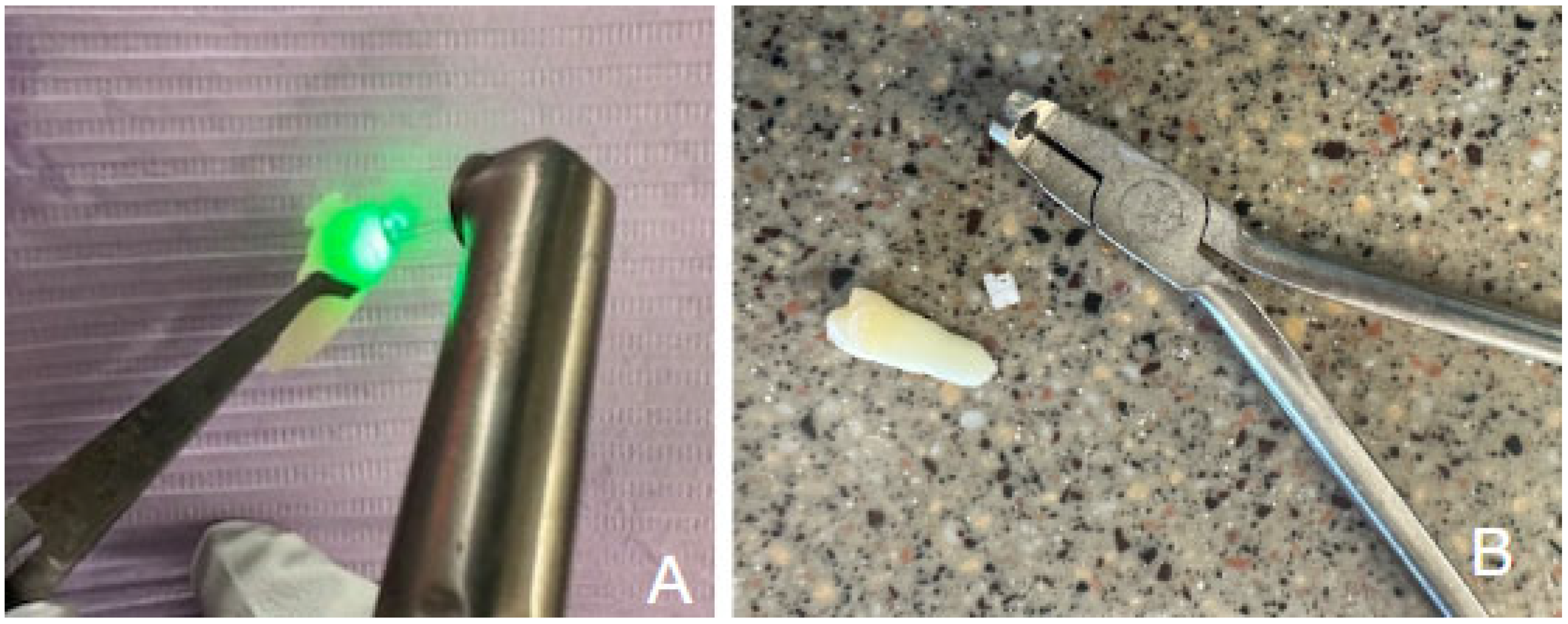
Figure 1. illustrates the experimental setup for bracket removal, showing the Er:YAG laser-assisted method (A) and the traditional mechanical plier method (B).
Applsci 15 11844 g001
Figure 2. To quantify the amount of residual resin based on coronal volume changes, the surfaces of the teeth were scanned prior to bracket placement and after bracket removal using laser for the Ceramic group (A) and Metal Group (B) and using mechanical removal for the Control group (C). Color maps illustrate surface height variations, with warmer colors indicating higher areas (residual resin) and cooler colors representing lower enamel surfaces.
Applsci 15 11844 g002
Figure 3. The SEM analysis validated the volumetric findings, indicating less residual resin remaining after bracket removal using the laser-assisted method (A) compared to the mechanical removal method (B).
Applsci 15 11844 g003
Figure 4. The surfaces of teeth were examined under SEM analysis after bracket removal with laser for the Ceramic group (A) and Metal Group (B), and mechanical removal for the Control group (C).
Applsci 15 11844 g004
Figure 5. SEM and imaging analysis confirmed that enamel damage occurred during laser-assisted bracket removal.
Applsci 15 11844 g005
Table 1. Mean values and standard deviations for bracket debonding in seconds using laser-assisted and traditional removal methods. Data were analyzed using one-way ANOVA and Tukey’s HSD test (p < 0.05). Different superscripts (a, b) indicate statistically significant differences (p < 0.001).
Group (n = 12)Mean Time
(Secs)
St.dev
Laser-assisted removal/metal bracket52.5 a13.57
Laser-assisted removal/ceramic bracket56.25 a9.32
Traditional removal/metal bracket0.76 b0.28
Traditional removal/ceramic bracket0.64 b0.25
Table 2. Mean values and standard deviations of tooth volume changes following bracket removal using laser-assisted and traditional removal methods. Data were analyzed using one-way ANOVA and Tukey’s HSD test (p < 0.05). Different superscripts (a, b) indicate statistically significant differences (p < 0.0002).
Group (n = 12)Volume Change (mm3)St.dev
Laser-assisted removal/metal bracket1.99 a1.04
Laser-assisted removal/ceramic1.80 a1.29
Traditional mechanical removal4.21 b1.22
Disclaimer/Publisher’s Note: The statements, opinions and data contained in all publications are solely those of the individual author(s) and contributor(s) and not of MDPI and/or the editor(s). MDPI and/or the editor(s) disclaim responsibility for any injury to people or property resulting from any ideas, methods, instructions or products referred to in the content.